WO2024259378A1 - T cell engager masking molecules - Google Patents
T cell engager masking molecules Download PDFInfo
- Publication number
- WO2024259378A1 WO2024259378A1 PCT/US2024/034204 US2024034204W WO2024259378A1 WO 2024259378 A1 WO2024259378 A1 WO 2024259378A1 US 2024034204 W US2024034204 W US 2024034204W WO 2024259378 A1 WO2024259378 A1 WO 2024259378A1
- Authority
- WO
- WIPO (PCT)
- Prior art keywords
- seq
- cdr
- depicted
- tce
- domain
- Prior art date
- Legal status (The legal status is an assumption and is not a legal conclusion. Google has not performed a legal analysis and makes no representation as to the accuracy of the status listed.)
- Pending
Links
Classifications
-
- C—CHEMISTRY; METALLURGY
- C07—ORGANIC CHEMISTRY
- C07K—PEPTIDES
- C07K16/00—Immunoglobulins [IG], e.g. monoclonal or polyclonal antibodies
- C07K16/18—Immunoglobulins [IG], e.g. monoclonal or polyclonal antibodies against material from animals or humans
- C07K16/28—Immunoglobulins [IG], e.g. monoclonal or polyclonal antibodies against material from animals or humans against receptors, cell surface antigens or cell surface determinants
- C07K16/2803—Immunoglobulins [IG], e.g. monoclonal or polyclonal antibodies against material from animals or humans against receptors, cell surface antigens or cell surface determinants against the immunoglobulin superfamily
-
- A—HUMAN NECESSITIES
- A61—MEDICAL OR VETERINARY SCIENCE; HYGIENE
- A61K—PREPARATIONS FOR MEDICAL, DENTAL OR TOILETRY PURPOSES
- A61K39/00—Medicinal preparations containing antigens or antibodies
- A61K39/395—Antibodies; Immunoglobulins; Immune serum, e.g. antilymphocytic serum
-
- A—HUMAN NECESSITIES
- A61—MEDICAL OR VETERINARY SCIENCE; HYGIENE
- A61P—SPECIFIC THERAPEUTIC ACTIVITY OF CHEMICAL COMPOUNDS OR MEDICINAL PREPARATIONS
- A61P35/00—Antineoplastic agents
- A61P35/02—Antineoplastic agents specific for leukemia
-
- C—CHEMISTRY; METALLURGY
- C07—ORGANIC CHEMISTRY
- C07K—PEPTIDES
- C07K14/00—Peptides having more than 20 amino acids; Gastrins; Somatostatins; Melanotropins; Derivatives thereof
- C07K14/435—Peptides having more than 20 amino acids; Gastrins; Somatostatins; Melanotropins; Derivatives thereof from animals; from humans
- C07K14/705—Receptors; Cell surface antigens; Cell surface determinants
- C07K14/70503—Immunoglobulin superfamily
- C07K14/7051—T-cell receptor (TcR)-CD3 complex
-
- C—CHEMISTRY; METALLURGY
- C07—ORGANIC CHEMISTRY
- C07K—PEPTIDES
- C07K16/00—Immunoglobulins [IG], e.g. monoclonal or polyclonal antibodies
- C07K16/18—Immunoglobulins [IG], e.g. monoclonal or polyclonal antibodies against material from animals or humans
- C07K16/28—Immunoglobulins [IG], e.g. monoclonal or polyclonal antibodies against material from animals or humans against receptors, cell surface antigens or cell surface determinants
- C07K16/2803—Immunoglobulins [IG], e.g. monoclonal or polyclonal antibodies against material from animals or humans against receptors, cell surface antigens or cell surface determinants against the immunoglobulin superfamily
- C07K16/2809—Immunoglobulins [IG], e.g. monoclonal or polyclonal antibodies against material from animals or humans against receptors, cell surface antigens or cell surface determinants against the immunoglobulin superfamily against the T-cell receptor (TcR)-CD3 complex
-
- C—CHEMISTRY; METALLURGY
- C07—ORGANIC CHEMISTRY
- C07K—PEPTIDES
- C07K16/00—Immunoglobulins [IG], e.g. monoclonal or polyclonal antibodies
- C07K16/18—Immunoglobulins [IG], e.g. monoclonal or polyclonal antibodies against material from animals or humans
- C07K16/28—Immunoglobulins [IG], e.g. monoclonal or polyclonal antibodies against material from animals or humans against receptors, cell surface antigens or cell surface determinants
- C07K16/2887—Immunoglobulins [IG], e.g. monoclonal or polyclonal antibodies against material from animals or humans against receptors, cell surface antigens or cell surface determinants against CD20
-
- C—CHEMISTRY; METALLURGY
- C07—ORGANIC CHEMISTRY
- C07K—PEPTIDES
- C07K7/00—Peptides having 5 to 20 amino acids in a fully defined sequence; Derivatives thereof
- C07K7/04—Linear peptides containing only normal peptide links
- C07K7/06—Linear peptides containing only normal peptide links having 5 to 11 amino acids
-
- A—HUMAN NECESSITIES
- A61—MEDICAL OR VETERINARY SCIENCE; HYGIENE
- A61K—PREPARATIONS FOR MEDICAL, DENTAL OR TOILETRY PURPOSES
- A61K39/00—Medicinal preparations containing antigens or antibodies
- A61K2039/505—Medicinal preparations containing antigens or antibodies comprising antibodies
-
- A—HUMAN NECESSITIES
- A61—MEDICAL OR VETERINARY SCIENCE; HYGIENE
- A61K—PREPARATIONS FOR MEDICAL, DENTAL OR TOILETRY PURPOSES
- A61K2300/00—Mixtures or combinations of active ingredients, wherein at least one active ingredient is fully defined in groups A61K31/00 - A61K41/00
-
- C—CHEMISTRY; METALLURGY
- C07—ORGANIC CHEMISTRY
- C07K—PEPTIDES
- C07K2317/00—Immunoglobulins specific features
- C07K2317/30—Immunoglobulins specific features characterized by aspects of specificity or valency
- C07K2317/31—Immunoglobulins specific features characterized by aspects of specificity or valency multispecific
-
- C—CHEMISTRY; METALLURGY
- C07—ORGANIC CHEMISTRY
- C07K—PEPTIDES
- C07K2317/00—Immunoglobulins specific features
- C07K2317/30—Immunoglobulins specific features characterized by aspects of specificity or valency
- C07K2317/34—Identification of a linear epitope shorter than 20 amino acid residues or of a conformational epitope defined by amino acid residues
-
- C—CHEMISTRY; METALLURGY
- C07—ORGANIC CHEMISTRY
- C07K—PEPTIDES
- C07K2317/00—Immunoglobulins specific features
- C07K2317/50—Immunoglobulins specific features characterized by immunoglobulin fragments
- C07K2317/56—Immunoglobulins specific features characterized by immunoglobulin fragments variable (Fv) region, i.e. VH and/or VL
- C07K2317/565—Complementarity determining region [CDR]
-
- C—CHEMISTRY; METALLURGY
- C07—ORGANIC CHEMISTRY
- C07K—PEPTIDES
- C07K2317/00—Immunoglobulins specific features
- C07K2317/90—Immunoglobulins specific features characterized by (pharmaco)kinetic aspects or by stability of the immunoglobulin
- C07K2317/92—Affinity (KD), association rate (Ka), dissociation rate (Kd) or EC50 value
Definitions
- This invention relates to aspects of biotechnology and medicine, in particular, to preventing and/or reducing the severity of cytokine release.
- BACKGROUND [2] The advent of redirecting T cell cytotoxicity in the field of immunotherapy has provided an abundantly increasing number of therapeutic approaches over the last years. Besides genetically modified T cells with chimeric antigen receptors (CAR), various bispecific T cell engagers (TCE) have successfully entered the market and are under development to treat various conditions including cancer.
- Exemplary bispecific molecules are recombinant protein constructs made from two flexibly linked antibody derived binding domains.
- TCEs are typically specific for a selected tumor-associated surface antigen on target cells; the second binding domain is specific for CD3, a subunit of the T cell receptor (TCR) complex on T cells.
- T cell redirection on-target activity like CAR T cells, is naturally associated with the risk of leading to a strong release of pro-inflammatory cytokines which may induce a cytokine release syndrome (CRS).
- CRS is considered a major dose-limiting clinical toxicity associated with T cell-directing immunotherapy that limits a patient’s ability to achieve efficacious TCE doses quickly and safely (Shimabukuro-Vornhagen, A. at al., (2016) Cytokine release syndrome. Journal for immunotherapy of cancer 6, 56).
- CRS CRS etiology
- clinical CRS is predominantly associated with first cycle TCE therapy;
- cytokine release tends to decrease with repeat TCE treatment cycles;
- TCE regimens that include step-dosing can lessen CRS incidence, but the success of this dosing strategy can be dependent on the specific target/indication (Jacobs, K. et al., (2017) Lead-in Dose Optimization to Mitigate Cytokine Release Syndrome in AML and MDS Patients Treated with Flotetuzumab, a CD123 x CD3 Dart® Molecule for T-Cell Redirected Therapy. Blood 130, 3856-3856).
- TCEs such as AMX-818, ANX007 and other clinical candidates
- approaches typically rely on protease-mediated mask cleavage to elicit TCE activity.
- one or more protease cleavage sites have to be introduced into the linker which covalently links the masking moiety and the drug molecule to be masked.
- These one or more cleavage sites are cleaved by proteases which may be frequently dysregulated in tumors but have a significant chance to be not dysregulated or overexpressed in the tumor which is actually targeted in each and every case.
- a covalently linked masking moiety requires significant molecule engineering of the specific drug molecule which may impact efficacy and/or stability properties of the original un-masked drug molecule. Also, engineering efforts have to be repeated for tailored masking for each drug molecule. [5] However, despite the advances in managing CRS and current engineering approaches, known mitigation efforts may reduce or eliminate entirely the efficacy of the immunotherapeutic treatment. Also, targeted approaches are typically not sufficient to prevent the strong and rapid onset of a cytokine storm that can occur after CAR T cell treatment or by molecules engineered with high avidity binding to the tumor-associated antigen and/or toward the, e.g., CD3 ⁇ of the TCR. Hence, there is a strong need for an improved versatile CRS mitigation strategy.
- TCE masking molecule comprising a binding peptide which is functionalized by a polymer for increased half-life and which is typically derived from the CD3 T-cell co-receptor, e.g., from the endogenous N-terminal sequence of CD3 ⁇ T cell co-receptor, when combined, e.g.
- this disclosure provides a T cell engager (TCE) masking molecule, the molecule comprising (i.) at least one binding peptide which binds to a T-cell engaging paratope of a T-cell engager (TCE) molecule, wherein the T-cell engaging paratope is an anti-CD3 paratope directed against an epitope located within CD3 ⁇ (SEQ ID NO: 256), CD3 ⁇ (SEQ ID NO: 257) or CD3 ⁇ and CD3 ⁇ , (ii.) at least one linker, wherein the linker is covalently linked to the C-terminus of the peptide; and (iii.) at least one half-life extending polymer of preferably at least 2 kDa, wherein the polymer
- the binding peptide binds to an anti-CD3 paratope of a TCE, wherein said anti-CD3 paratope binds to an epitope comprising at least one residue selected from CD3 ⁇ (SEQ ID NO:257): K73 and S83; and CD3 ⁇ (SEQ ID NO:256) K82 and C93, wherein the epitope preferably comprises the region of CD3 ⁇ defined by K73, N74, 175, G76, S77, D78, E79, D80, H81, L82, and S83, and wherein the epitope comprises the region of CD3 ⁇ defined by K82, E83, S84, T85, V86, Q87, V88, H89, Y90, R91, M92, and C93.
- a TCE masking molecule comprising: (i) a binding peptide that binds to an anti-CD3 epsilon (CD3 ⁇ ) paratope of a CD3 ⁇ binding domain of a TCE, wherein the anti-CD3 ⁇ paratope is directed against an epitope located within CD3 ⁇ , wherein CD3 ⁇ comprises the amino acid sequence of SEQ ID NO: 257; (ii) a linker covalently linked to the C-terminus of the binding peptide; and (iii) a half-life extending polymer covalently linked to the linker.
- the anti-CD3 ⁇ paratope is directed to a CD3 ⁇ epitope consisting of the amino acid sequence of SEQ ID NO: 258 or consisting of a shorter N-terminal sequence of SEQ ID NO: 258, preferably the amino acid sequence of SEQ ID NO: 259 or QDGNEE or QDGNEEM or QDGNEEMG.
- the binding peptide comprises 5, 6, 7, 8, 9, 10, 11, 12, 13, 14, 15, 16 ,17 or 18 amino acids, preferably 5, 6, 7, 8, 9 or 10 amino acids, more preferably 10 amino acids.
- the binding peptide binds to an anti-CD3 ⁇ paratope and comprises at the N-terminus at least the sequence of X1DGX2E (SEQ ID NO: 260), wherein X1 is selected from Q, pyroglutamic acid (pE) and S, and wherein X2 is selected from N, E, S, T, V, and I.
- the binding peptide binds to an anti-CD3 ⁇ paratope and comprises at the N-terminus at least the sequence of X1X2X3X4EX5 (SEQ ID NO: 392), wherein X1 is Q, pyroglutamic acid (pE), or S, and wherein X2 is D, H or N, and wherein X3 is G, F or Y, and wherein X4 is N, E, S, T, V, or I, and wherein X5 is E, L, P , or W.
- the binding peptide binds to an anti-CD3 ⁇ paratope and comprises at the N-terminus at least the sequence of X1DGX2EE (SEQ ID NO: 261), wherein X1 is Q, pE, or S, and wherein X2 is N, E, S, T, V, or I.
- the binding peptide binds to an anti-CD3 ⁇ paratope and comprises at the N-terminus at least the sequence of X1X2X3X4EX5X6X7 (SEQ ID NO: 393), wherein X1 is Q, pyroglutamic acid (pE), or S, and wherein X2 is D, H or N, and wherein X3 is G, F or Y, and wherein X4 is N, E, S, T, V, or I, and wherein X5 is E, L, P , or W, and wherein X6 is A, C, D, E, F, G, H, I, K, L , M , N,P, Q, R, S, T, V, W, or Y, and wherein X7 is A, C, D, E, F, G, H, I, K, L, M, N, P, Q, R, S, T, V, W, or Y, and wherein X7 is A, C, D
- the binding peptide binds to an anti-CD3 ⁇ paratope and comprises at the N-terminus at least the sequence of pEX1X2X3EX4LK (SEQ ID NO: 396), wherein X1 is D, H or N, and wherein X2 is G, F or Y, and wherein X3 is N, E, S, T, V, or I, and wherein X4 is E, L, P , or W.
- the anti-CD3 ⁇ binding peptide comprises a palindrome.
- the palindrome has a K at the position between the mirrored amino acids.
- the binding peptide binds to an anti-CD3 ⁇ paratope and comprises at the N-terminus at least the sequence of any one of SEQ ID NO: 263 to 284, 286 to 293, 295 to 303, 308 to 338, 385 to 388, 397 to 419 and 431 or any of 263 to 284, 286 to 293, 295 to 338, 385 to 388, 397 to 419 and 431.
- the binding peptide at its C-terminus further comprises the amino acids G and C or G and K.
- the binding peptide binds to an anti-CD3 ⁇ paratope and comprises the sequence of pEX1X2X3EX4X5X6GX7 (SEQ ID NO: 433), wherein X1 is D, H or N, and wherein X2 is G, F or Y, preferably G or F, and wherein X3 is N, H, Q, R, E, S, T, V, C, D, F, K L, M, W, Y or I, preferably N, E, H, Q or R, and wherein X4 is E, L, P , or W, and wherein X5 is A, C, D, E, F, G, H, I, K, L , M , N, P, Q, R, S, T, V, W, or or
- the binding peptide binds to an anti-CD3 ⁇ paratope and comprises the sequence of pEX1X2X3EELKGX4 (SEQ ID NO: 434), wherein X1 is D, H or N, and wherein X2 is G, F or Y, preferably G or F, and wherein X3 is N, H, Q, R, E, S, T, V, C, D, F, K L, M, W, Y or I, preferably N, E, H, Q or R, most preferably N or E, and wherein and wherein X4 is C or K.
- the binding peptide binds to an anti-CD3 ⁇ paratope and comprises the sequence of pEX1GX2EELKGX3 (SEQ ID NO: 435), wherein X1 is D, H or N, and wherein X2 is N, H, Q, R, E, S, T, V, C, D, F, K L, M, W, Y or I, preferably E, N, H, Q or R, most preferably N or E, and wherein and wherein X3 is C or K.
- the binding peptide binds to an anti-CD3 ⁇ paratope and comprises the sequence of pEDGX1EELKGX2 (SEQ ID NO: 436), wherein X1 is N, H, Q, R, E, S, T, V, C, D, F, K L, M, W, Y or I, preferably N, E, H, Q or R, most preferably N or E, and wherein and wherein X2 is C or K.
- the binding peptide binds to an anti-CD3 ⁇ paratope and comprises the sequence of SEQ ID NO: 297, 300, 303, 304, 305, 306, 307, 384 or 389.
- the linker is selected from the group consisting of (i.) disulfide linkers comprising disulfide (R-S-S-R’) wherein R is a half-life extending polymer and R’ is a binding peptide;, (ii.) free thiol containing linear or branched PEGs, OPSS derivatives, maleimides, nor-bornenes, acrylimides, acrylates, vinyl sulfones, and amines and (iii.) carboxyl linkers comprising amines, amides, and epsilon derivatized acetyl bromides.
- the linker is selected from the following moieties: is a half-life extending polymer and R’ is a CD3 ⁇ masking peptide.
- the linker is selected from a maleimide – thiosuccinimide linker having the formula when the C-terminal amino acid of the binding peptide is K, and an acetamie – thioether having the formula when the C-terminal amino acid of the binding peptide is C, and a disulfide having the formula R’-S-S-R when the C-terminal amino acid of the binding peptide is C, wherein R in the context of a linker herein is always a half-life extending polymer and R’ in the context of a liker herein is always a binding peptide.
- the half-life extending polymer is selected from mono-methoxy polyethylene glycol (mPEG), linear, 2-arm, 4-arm, 8-arm polyethylene glycol (PEG).; PLGA; peptide acrylate, polyglycerols, polyoxazolines, polyvinylpyrrolidone, polyacrylamides, poly(N- acryloylmorpholine), poly(N,N-dimethylacrylamide), ply(2-hydroxypropylmethacrylamide), polysarcosine, poly(2-hydroxyethylmethacrylamide), hyaluronic acids, sialic acid, poly[(organo)phosphazenes], and heparin.
- mPEG mono-methoxy polyethylene glycol
- PEG linear, 2-arm, 4-arm, 8-arm polyethylene glycol
- PLGA peptide acrylate, polyglycerols, polyoxazolines, polyvinylpyrrolidone, polyacrylamides, poly(N-
- the half-life extending polymer is a PEG selected from the following moieties: (i.) , wherein n equals an integer of about 20 to about 200, preferably 20 to 169, more preferably about 20, 30, 40, 50, 60, 70, 80, 90100, 103, 110 or 113, (ii.) , wherein n equals an integer of about 40 to about 400, preferably about 40 to about 208 or about 57 to about 338, more preferably about 50, 57, 60, 70, 80, 90, 100, 110, 120, 130, 140, 150, 160, 170, 180, 190, 200, 210, 220, 230 or about 225, and (iii.) , wherein n equals an integer of about 80 to about 700, preferably about 80 to about 675, more preferably about 80, 83, 90, 100, 110, 113, 120, 130, 140, 150, 160, 166, 170, 180, 190,
- the half-life extending polymer is a non-branched linear, or a branched 2-arm, 4-arm, or 8-arm PEG, preferably linear or 4-arm, with a molecular weight of about 2 kDa to about 60 kDa, preferably about 4 kDa to about 30 kDa or 10 kDa to about 30 kDa, or more preferably about 5 or about 20 kDa.
- the half-life extending polymer is a branched polymer, preferably a, 2-arm, 4-arm and/or 8-arm PEG with am molecular weight of about 2 to 60 kDa to which more than one binding peptide is linked via a linker each, preferably 2, 4 or 8 binding peptides to one half-life extending polymer.
- the binding peptide comprises the sequence of pEX1X2X3EELKGX4 (SEQ ID NO: 434), wherein X1 is D, H or N, and wherein X2 is G, F or Y, preferably G or F, and wherein X3 is N, H, Q, R, E, S, T, V, C, D, F, K L, M, W, Y or I, preferably N, E, H, Q or R, most preferably N or E, and (a.) wherein and wherein X4 is C; a linker chosen from maleimide – thiosuccinimide linker having the formula and a disulfide having the formula R’-S-S-R; and a half-life extending polymer that is a PEG of the following formula or having a molecular weight of about 5 to 20 kDa, preferably about 5 kDa or about 10 kDa or about 20 k
- the binding peptide comprises the sequence of pEX1GX2EELKGX3 (SEQ ID NO: 435), wherein X1 is D, H or N, and wherein X2 is N, H, Q, R, E, S, T, V, C, D, F, K L, M, W, Y or I, preferably E, N, H, Q or R, most preferably N or E, and (a.) wherein and wherein X3 is C; a linker chosen from maleimide – thiosuccinimide linker having the formula and a disulfide having the formula R’-S-S-R; and a half-life extending polymer that is a PEG of the following formula or having a molecular weight of about 5 to 20 kDa, preferably about 5 kDa or about 10 kDa or about 20 kDa; or (b.) wherein and wherein X3 is K; an
- the masking molecule comprises a T cell engager masking molecule comprising: (i.) a binding peptide binds to an anti-CD3 ⁇ paratope and comprises the amino acid sequence of any one of SEQ ID NO: 263 to284, 286 to 293, 295 to 303, 304 to 307, 308 to 338, 385 to 388, 397 to 419 and 431.; (ii.) a linker, wherein the linker is covalently linked to the C-terminus of the binding peptide; and (iii.) at least one half-life extending polymer of preferably at least 2 kDa, wherein the polymer is covalently linked to the linker.
- the masking molecule comprises a T cell engager masking molecule comprising: (a.) the amino acid sequence of SEQ ID 304, 306, 384 or 389, and a linker chosen from maleimide – thiosuccinimide linker having the formula and a disulfide having the formula R’-S-S-R; and a half-life extending polymer that is a PEG of the following formula or having a molecular weight of about 5 kDa to about 20 kDa, preferably about 5 kDa or about 10 kDa or about 20 kDa or (b) the amino acid sequence of SEQ ID 305 or 307; an acetamide – thioether having the formula and a half-life extending polymer that is a PEG of the following formula or having a molecular weight of about 5 kDa to about 20 kDa, preferably about 5 kDa or about 10 kDa
- the masking molecule comprises a T cell engager masking molecule comprising: (i.) a binding peptide having SEQ ID 304; (ii.) aleimide – thiosuccinimide linker having the formula or an disulfide R’-S-S-R covalently linked to the C-terminus of the binding peptide; and (iii.) a half-life extending polymer of the following formula or having a molecular weight of 5 kDa or 20Da.
- the masking molecule comprises a T cell engager masking molecule comprising: (i.) a binding peptide having SEQ ID 305; (ii.) an acetamides – thioether having the formula covalently linked to the C-terminus of the binding peptide; and (iii.) a half-life extending polymer of the following formula having a molecular weight of 5 kDa or 20Da.
- the masking molecule comprises a T cell engager masking molecule comprising: (i.) a binding peptide having SEQ ID 306; (ii.) a maleimide – thiosuccinimide linker having the formula or or an disulfide R’-S-S-R covalently linked to the C-terminus of the binding peptide; and (iii.) a half-life extending polymer of the following formula having a molecular weight of 5 kDa or 20Da.
- the masking molecule comprises a T cell engager masking molecule comprising: (i.) a binding peptide having SEQ ID 307; (ii.) a acetamides – thioether having the formula covalently linked to the C-terminus of the binding peptide; and (iii.) a half-life extending polymer of the following formula having a molecular weight of 5 kDa or 20Da.
- the TCE masking molecule comprises any of the combinations (a.) to (k.) of a binding peptide, a linker and a half-life extending polymer, wherein R’ stands for the binding peptide and R stands for the half-life extending polymer in following table 1: Table 1: Composition of exemplary TCE masking molecules [43] According to said aspect, it is also envisaged that the TCE masking molecule comprises any of the combinations (a.) to (k.) of a binding peptide, a linker and a half-life extending PEG polymer, wherein R’ stands for the binding peptide and R stands for the half-life extending PEG polymer: Table 2: Composition of exemplary TCE masking molecules with structural formula [44] According to said aspect, it is also envisaged that the TCE masking molecule has a half-life significantly shorter half-life than the TCE of about 1 to about 48 hours, preferably about 1.5 to about 24 hours, more
- the binding peptide binds to an anti-CD3 ⁇ paratope of a CD3 ⁇ binding domain of a TCE with an affinity in terms of a Kd value of typically less than 10 nM or less than 5 nM or less than 0.5 nM or about 0.1 nM to 5 nM.
- a Kd value typically less than 10 nM or less than 5 nM or less than 0.5 nM or about 0.1 nM to 5 nM.
- the amino acid sequence of the binding peptide is, e.g., pEX1GX2EELKGX3 (SEQ ID NO: 435), wherein X1 is D, H or N, and wherein X2 is N.
- the binding peptide binds to an anti-CD3 ⁇ paratope of a CD3 ⁇ binding domain of a TCE with an affinity of typically about 10 to 100 nM if the amino acid sequence of the binding peptide is pEX1GX2EELKGX3 (SEQ ID NO: 435), wherein X1 is D, H or N, and wherein X2 is E.
- the method comprising administering to the human subject an effective dose of a T cell engager masking molecule, wherein the TCE masking molecule comprises: (i.) a binding peptide that binds an anti-CD3 ⁇ paratope of a CD3 ⁇ binding domain of a TCE, wherein the anti-CD3 ⁇ paratope is directed against an epitope located within CD3 ⁇ , and wherein CD3 ⁇ comprises the amino acid sequence of SEQ ID NO: 257; (ii.) a linker, wherein the linker is covalently linked to the C-terminus of the binding peptide; and (iii.) at least one half-life extending polymer of preferably at least 2 kDa, wherein the polymer is covalently linked to the linker.
- the TCE masking molecule comprises: (i.) a binding peptide that binds an anti-CD3 ⁇ paratope of a CD3 ⁇ binding domain of a TCE, wherein the anti-CD3 ⁇ paratope is directed
- the T cell engager masking molecule comprises: a binding peptide which binds to an anti-CD3 ⁇ paratope and comprises the sequence of pEX1X2X3EELKGX4 (SEQ ID NO: 434), wherein X1 is D, H or N, and wherein X2 is G, F or Y, preferably G or F, and wherein X3 is N, H, Q, R, E, S, T, V, C, D, F, K L, M, W, Y or I, preferably N, E, H, Q or R, and (a.) wherein and wherein X4 is C; a linker chosen from maleimide – thiosuccinimide linker having the formula and a disulfide having the formula R’-S-S-R; and a half- life extending polymer that is a PEG of the following formula or having a molecular weight of about 5 to 20 kDa, preferably about 5 kD
- the T cell engager masking molecule comprises: a binding peptide which binds to an anti-CD3 ⁇ paratope and comprises the sequence of pEX1GX2EELKGX3 (SEQ ID NO: 435), wherein X1 is D, H or N, and wherein X2 is N, H, Q, R, E, S, T, V, C, D, F, K L, M, W, Y or I, preferably N, H, Q or R, and (a.) wherein and wherein X3 is C; a linker chosen from maleimide – thiosuccinimide linker having the formula and a disulfide having the formula R’-S-S-R; and a half-life extending polymer that is a PEG of the following formula having a molecular weight of about 5 to 20 kDa, preferably about 5 kDa or about 10 kDa or about 20 kDa; or (b.) wherein and
- the T cell engager masking molecule comprises: a binding peptide which binds to an anti-CD3 ⁇ paratope and comprises the sequence of pEDGX1EELKGX2 (SEQ ID NO: 436), wherein X1 is D, H or N, and wherein X1 is N, H, Q, R, E, S, T, V, C, D, F, K L, M, W, Y or I, preferably N, E, H, Q or R, most preferably N or E, and (a.) wherein and wherein X2 is C; a linker chosen from maleimide – thiosuccinimide linker having the formula and a disulfide having the formula R’-S-S-R; and a half-life extending polymer that is a PEG of the following formula or having a molecular weight of about 5 to 20 kDa, preferably about 5 kDa or about 10 kDa or about 20 kDa;
- the T cell engager masking molecule comprises: (a) the amino acid sequence of SEQ ID 304, 306, 384 or 389, and a linker chosen from maleimide – thiosuccinimide linker having the formula and a disulfide having the formula R’-S-S-R; and a half-life extending polymer that is a PEG of the following formula or having a molecular weight of about 5 kDa to about 20 kDa, preferably about 5 kDa or about 10 kDa or about 20 kDa; or (b) the amino acid sequence of SEQ ID 305 or 307; an acetamide – thioether having the formula and a half-life extending polymer that is a PEG of the following formula having a molecular weight of about 5 kDa to about 20 kDa, preferably about 5 kDa or about 10 kDa or about 20 kDa, wherein R’
- the method of reducing the severity of cytokine release in a human subject undergoing preferably treatment with a TCE comprises use or administration of the TCE masking molecule comprises any of the combinations (a.) to (k.) of a binding peptide, a linker and a half-life extending polymer, wherein R’ stands for the binding peptide and R stands for the half-life extending polymer:
- the method of reducing the severity of cytokine release in a human subject undergoing preferably treatment with a TCE comprises use or administration of any of the combinations (a.) to (k.) of a binding peptide, a linker and a half-life extending PEG polymer, wherein R’ stands for the binding peptide and R stands for the half-life extending PEG polymer:
- the immunotherapy comprises administering a T cell engager.
- the TCE masking molecule is administered before, during or after administration of a TCE.
- the molar ratio of masking molecule to TCE is in the range of 1000:1 to 10:1 or 250:1 to 10:1, preferably 100:1 to 25:1.
- the TCE is selected from (ii.) A TCE comprising at least three domains in an amino to carboxyl order, wherein: (a.) a first domain binds to a target cell surface antigen, which is preferably a tumor antigen; (b.) a second domain binds to an extracellular epitope of the human and/or the Macaca CD3 chain, preferably CD3 ⁇ ; and (c.) a third domain comprises two polypeptide monomers, each comprising a hinge, a CH2 and a CH3 domain, wherein said two polypeptide monomers are fused to each other via a peptide linker, wherein said third domain comprises in an amino to carboxyl order: hinge-CH2-CH3-linker-hinge-CH2-CH3; (ii.) A TCE comprising (a.) a first binding domain which binds to a first target cell surface antigen (e.g.
- TAA1 TAA1
- a second binding domain which binds to an extracellular epitope of the CD3 ⁇
- a spacer wherein said spacer is a single chain fragment crystallizable (scFc), human serum albumin (HSA), programmed death receptor 1 (PD1), or a hetero fragment crystallizable (hetero FC);
- a third binding domain which binds to a second target cell surface antigen (e.g.
- TAA2 TAA2
- a fourth binding domain which binds to an extracellular epitope of CD3 ⁇
- the first binding domain binds to the first target cell surface antigen and the third binding domain binds to the second target cell surface antigen simultaneously, wherein the first target cell surface antigen and the second target cell surface antigen are on the same target cell
- the TCE is a single polypeptide chain, wherein the first target cell surface antigen and the second target cell surface antigen are not identical, and wherein the first binding domain and the second binding domain form a first bispecific entity and the third and the fourth binding domain form a second bispecific entity, and wherein the spacer entity is positioned between the first and the second bispecific entities; ; and (iii.) an IgG-based full length bispecific antibody; or (iv.) a heterodimeric antibody.
- the TCE under [55] (i.) or (ii.) is a single chain molecule.
- a glycosylation site at Kabat position 314 of the CH2 domains in the third domain of the bispecific antigen-binding molecule is removed by a N314X substitution, wherein X is any amino acid excluding Q.
- each of said polypeptide monomers of the third domain has an amino acid sequence that is at least about 80, 85, 90, 95 or 100% identical to a sequence selected from the group consisting of: SEQ ID NOs: 437 to 444, or has an amino acid sequence selected from the group consisting of SEQ ID NOs: 437 to 444.
- the CH2 domain comprises an intra domain cysteine disulfide bridge.
- the tumor antigen is selected from the group consisting of CDH19, CDH3, MSLN, DLL3, FLT3, EGFRvIII, BCMA, PSMA, CD33, CD19, CD20, CLDN18.2, CLDN 6, MUC17, EpCAM, STEAP1 and CD70.
- the antibody construct comprises in an amino to carboxyl order: (a) the first domain; (b) a peptide linker having an amino acid sequence selected from the group consisting of SEQ ID NOs: 187-189; (c) the second domain; (d) a peptide linker having an amino acid sequence selected from the group consisting of SEQ ID NOs: 187, 188, 189, 195, 196, 197 and 198; (e) the first polypeptide monomer of the third domain having an amino acid sequence of any of SEQ ID NOs 437 to 444; (f) a peptide linker having an amino acid sequence selected from the group consisting of SEQ ID NOs: 191, 192, 193 and 194; and (g) the second polypeptide monomer of the third domain having an amino acid sequence of any of SEQ ID NOs 437 to 444.
- the first binding domain of the construct comprises a VH region comprising CDR-H1, CDR-H2 and CDR-H3 and a VL region comprising CDR- L1, CDR-L2 and CDR-L3 selected from the group consisting of: (a) CDR-H1 as depicted in SEQ ID NO: 4, CDR-H2 as depicted in SEQ ID NO: 5, CDR-H3 as depicted in SEQ ID NO: 6, CDR-L1 as depicted in SEQ ID NO: 1, CDR-L2 as depicted in SEQ ID NO: 2 and CDR-L3 as depicted in SEQ ID NO: 3, (b) CDR-H1 as depicted in SEQ ID NO: 29, CDR-H2 as depicted in SEQ ID NO: 30, CDR-H3 as depicted in SEQ ID NO: 31, CDR-L1 as depicted in SEQ ID NO: 34, CDR-L2 as depicted in
- the bispecific antigen-binding molecule has the SEQ ID NO: 17, 52, 63, 81, 93, 104, 114, 125, 136, 147, 162, 177, 213,226, 238, 248, 255 or 430, preferably 104 or 255.
- the bispecific antigen-binding molecule is a heterodimeric antibody comprising: a) a first monomer comprising a first heavy chain comprising: 1) a first variable heavy domain; 2) a first constant heavy chain comprising a first CH1 domain and a first Fc domain; 3) a scFv that binds human CD3 and comprises a scFv variable light domain, an scFv linker and a scFv variable heavy domain; wherein said scFv is covalently attached between the C-terminus of said CH1 domain and the Nterminus of said first Fc domain using domain linker(s); b) a second monomer comprising a second heavy chain comprising a second variable heavy domain and a second constant heavy chain comprising a second Fc domain; and c) a common light chain comprising a variable light domain and a constant light domain; wherein said first variable heavy domain and said variable light domain bind human STEAP
- the CD3 ⁇ binding domain of a TCE has the sequence of SEQ ID NO 26. 381, 382 or 383.
- a TCE masking molecule for use in reducing the severity of cytokine release in a human subject undergoing treatment with a TCE, wherein the TCE masking molecule comprises: (i.) a binding peptide that binds to an anti-CD3 ⁇ paratope of a CD3 ⁇ binding domain of a TCE, wherein the anti-CD3 ⁇ paratope is directed against an epitope located within CD3 ⁇ , and wherein CD3 ⁇ comprises the amino acid sequence of SEQ ID NO: 257; (ii) a linker covalently linked to the C-terminus of the binding peptide; and (iii) a half-life extending polymer covalently linked to the linker.
- a method of attenuating the in vivo exposure of a TCE comprising the steps of (a) providing a TCE masking molecule, comprising (i.) a binding peptide that binds to an anti-CD3 ⁇ paratope of a CD3 ⁇ binding domain of a TCE, wherein the anti-CD3 ⁇ paratope is directed against an epitope located within CD3 ⁇ , and wherein CD3 ⁇ comprises the amino acid sequence of SEQ ID NO: 257, (ii) a linker covalently linked to the C-terminus of the binding peptide; and (iii) a half-life extending polymer covalently linked to the linker, wherein the binding peptide binds to an anti-CD3 ⁇ paratope with a Kd which is at least 1.5-fold lower than the Kd for the amino acid sequence of SEQ ID NO: 262 binding to
- the TCE masking molecule is administered before, during or after the administration of the TCE.
- a method for reducing the severity of cytokine release in a human subject undergoing immunotherapy comprising administering to the human subject an effective dose of a T cell engager masking molecule before, concurrently with, or after immunotherapy, wherein the T cell engager masking molecule comprises: (i.) a binding peptide having SEQ ID selected from the group consisting of SEQ ID NOs 263 to 284, 286 to 293, 295 to 338, 385 to 388, 397 to 419 and 431; (ii.) a linker, wherein the linker is covalently linked to the C-terminus of the binding peptide; and (iii.) at least one half-life extending polymer of preferably at least 2 kDa, wherein the polymer is covalently linked to the linker.
- a method comprising administering to the subject in need of such treatment an effective dosage of the TCE masking molecule as disclosed herein or a pharmaceutically acceptable salt thereof, , in combination with a therapeutically effective amount of a T cell engager in a molar ration preferably in the range of 1000:1 to 10:1 or 250:1 to 10:1 (masking molecule : TCE).
- TCE masking molecule is administered before, during or after the administration of the TCE.
- a TCE masking molecule is envisaged for use in the attenuation of in vivo exposure of a TCE after administration of the TCE molecule, wherein the administration is preferably via the i.v.
- a TCE masking molecule comprising (i.) a binding peptide that binds to an anti-CD3 ⁇ paratope of a CD3 ⁇ binding domain of a TCE, wherein the anti-CD3 ⁇ paratope is directed against an epitope located within CD3 ⁇ , and wherein CD3 ⁇ comprises the amino acid sequence of SEQ ID NO: 257; (ii) a linker covalently linked to the C-terminus of the binding peptide; and (iii) a half-life extending polymer covalently linked to the linker, wherein the binding peptide binds to an anti-CD3 ⁇ paratope with a Kd which is at least 1.5-fold lower than the Kd for the amino acid sequence of SEQ ID NO: 262 binding to an anti-CD3 ⁇ paratope, preferably, at least 2-fold lower, more preferably 3-fold lower, and (b) administering the TCE masking molecule before, during
- FIG. 1 shows the general pharmacokinetic and pharmacodynamic relationship between TCE exposure and cytokine release.
- Left General PK profile for TCE administered by IV bolus and the relationship to cytokine release. The depiction here shows a TCE dosed to maintain minimal plasma concentrations above a pre-defined exposure target (EC90). This scenario often leads to patient overexposure that results in acute cytokine release at early times post-dose, which can induce runaway CRS.
- FIG. 2 shows exemplary in silico PK modeling highlighting the sensitivity of active, or ‘free’ TCE exposure to co-administration with masking molecule CD3 ⁇ PepPOL.
- (Left) In silico PK model schematic defining the relationship between plasma (Cp) and tissue (Cp) ‘free’ TCE concentrations, unbound CD3 ⁇ PepPOL (T1), and the binding kinetics driving TCE-(CD3 ⁇ PepPOL) complexation. Note, the elimination rate (kel,D) of TCE and TCE-(CD3 ⁇ PepPOL) is assumed to be identical. The elimination rate of CD3 ⁇ PepPOL, kel,T, is assumed >> than kel,D.
- FIG. 3 shows transient association of exemplary masking molecule CD3 ⁇ PepPOL with exemplary TCE molecule with a half-life extending scFc moiety.
- FIG. 4 shows exemplary plasma concentration over time in C57BL/6 mice of a binding peptide only vs. masking molecule with half-life extending polymer (SEQ ID NO: 297).
- Fig.5 shows PK impact of exemplary polymer on masking molecule in mice.
- Fig.6 FIG 6 shows PK impact of maleamide linker in exemplary masking molecules.
- Fig.7 Fig.7 shows TCE-mediated B-cell cytolytic effect.
- FIG.8 shows reduction in cytokine release with CD3 ⁇ PepPol co-administration at >1000X Peptide:TCE overage.
- FIG. 9 shows co-administration of muCD19 TCE + CD3 ⁇ -PepPOL, v1 co-administration reduces cytokine release as a function of CD3 ⁇ -PepPOL, v1 dose.
- Fig. 10 shows co-administration of muCD19 TCE + CD3 ⁇ -PepPol, v1 reduces cytokine release with minimal impact on PD response in the huCD3 ⁇ KI model.
- Fig. 11 Fig.
- FIG. 11 shows Co-administration of CD3 ⁇ -PepPOL, v1 reduces cytokine release with retention of anti-tumor activity in preclinical MC38-muCD19 tumor model.
- A Serum cytokine INF-y;
- B serum cytokine release TNF-a,
- C Target cell depletion in spleen and
- D in blood ;
- E Tumor growth inhibition. Note: Statistical analysis performed using 1 way ANOVA for panels (A) and (B) and Repeated Measures 2way ANOVA for panel (C); GraphPad Prism v 9.5.1.
- Fig.12 shows Co-administration of CD20 TCE + CD3 ⁇ -PepPOL, v1 uncouples cytokine release from PD response (CD20+ B cell lysis) in cynomolgus monkeys.
- Fig. 13 Figure 13 shows clinical chemistry parameters upon Co-administration of CD20 TCE + high affinity CD3 ⁇ -PepPOL (pGlu, 20kDa).
- Fig. 14 Figure 14 shows Co-administration of CD20 TCE + high affinity CD3 ⁇ -PepPOL (pGlu, 20kDa) reduces cytokine release without impacting PD effect in cynomolgus monkeys.
- Fig. 12 shows Co-administration of CD20 TCE + CD3 ⁇ -PepPOL, v1 uncouples cytokine release from PD response (CD20+ B cell lysis) in cynomolgus monkeys.
- Figure 15 shows inhibition of cytotoxicity of heterodimeric STEAP1xCD3 TCE (comprising CD3e binder SEQ ID NO 381), CLDN6xCD3 TCE (comprising CD3e binder I2E, SEQ ID NO 383) and DLL3xCD3 TCE (comprising CD33 binder I2C, SEQ ID NO 26) by masking CD3e.
- CD3e pepPEG with high affinity against CD3e binder showed higher inhibition activity against TCE whereas peptides with intermediate affinity showed lower inhibition activity. All the peptides with high affinity showed similar potency with respect to each other.
- Fig. 16 shows inhibition of cytokine release shows representative TCE masking molecules’ inhibition of cytokine release induced by heterodimeric STEAP1xCD3 ⁇ TCE
- Fig. 17 shows inhibition of cytokine release shows representative TCE masking molecules’ inhibition of cytokine release induced by DLL3xCD3 ⁇ TCE.
- Fig. 16 shows inhibition of cytokine release shows representative TCE masking molecules’ inhibition of cytokine release induced by DLL3xCD3 ⁇ TCE.
- cytokine release shows representative TCE masking molecules’ inhibition of cytokine release induced by CLDN6xCD3 ⁇ TCE.
- Fig.19 shows affinity of representative binding peptides and one negative example on FLT3xCD3 TCE and STEAP1xCD3 TCE.
- Fig.20 shows inhibition of cytotoxicity on CDH3xMSLN dual targeting TCE
- Fig.21 shows inhibition of cytokine release on CDH3xMSN dual targeting TCE DETAILED DESCRIPTION [95]
- pro-inflammatory cytokine release such as in CRS is a phenomenon rooted in sub-optimal TCE pharmacokinetics in the context of high TCE therapeutic potency.
- the general principle of underlying pharmacokinetic and pharmacodynamic relationship between TCE exposure -non controlled and controlled, e.g., by a masking molecule- and cytokine release is depicted in Figure 1.
- the masking molecule of the invention typically comprises a peptide which binds to the T-cell engaging paratope of the TCE, a linker and a half-life extending polymer.
- CD3 binding domains of TCEs comprise those which bind to CD3 ⁇ , CD3 ⁇ , or both.
- CD3 ⁇ The masking molecule may be abbreviated as CD3 PepPOL in the following.
- CD3 ⁇ PepPOL refers to a masking molecule which binds to a CD3 ⁇ binding paratope of a TCE.
- Some of the benefits of the present disclosure are (i.) to lessen the frequency of CRS incidence at any TCE dose level, (ii.) to aid clinical dosing schema by allowing for a higher initial dose of the TCE, and (iii.) to reduce the number of dose fractionation steps of the TCE required to achieve an efficacious dose thereby reducing therapeutic cycle time.
- the masking molecules CD3 PepPOL if combined, e.g. administered before, during or after administration of a TCE, can achieve a more favorable TCE exposure profile than TCE administration without masking molecule, e.g. IV bolus or s.c. administration of TCE monotherapy.
- CD3 PepPOL binds the anti-CD3 T cell engager moiety antagonistically with moderate to high affinity to prevent T cell engagement.
- TCR T cell receptor
- CD3 PepPOL is typically cleared from the plasma quicker than the TCE
- competitive blockade of TCE on T cells is transient, with the duration defined by CD3 PepPOL clearance (CL) and volume of distribution (V), as well as the amount of molar excess CD3 PepPOL administered.
- CL CD3 PepPOL clearance
- V volume of distribution
- V volume of distribution
- Cmax attenuated maximum concentration
- tmax delayed time of maximal exposure
- binding peptides of the present disclosure are preferred which have a higher affinity in terms of a lower Kd value, preferably equilibrium Kd value, for the CD3 ⁇ paratope of the TCE to be masked than the physiological CD3 ⁇ epitope (e.g. a sequence comprising SEQ ID NO: 262, QDGNEEMG).
- Kd values of preferred binding peptides are at least 1.5 lower than, e.g., reference epitope QDGNEEMG (SEQ ID NO: 262), preferably at least 2-fold lower, more preferably 3- fold lower or even about 5-fold, about 10-fold or about 100-fold lower.
- binding affinity in terms of a Kd of 35.8 nM for a FLT3xCD3 ⁇ TCE (SEQ ID NO 93), see Example 1.
- Particularly preferred are binding peptides of, for example, any of SEQ ID NO: 300, 304 to 307, 384 and 389.
- a binding peptide with high affinity will typically allow a lower molar ratio of masing molecule : TCE and thus, a more economic employment of the masking molecule.
- Binding peptides are preferred which provide affinity gains, in particular by the substitution of N- terminal amino acid Q by pE.
- Affinity can further be fine-tuned, in particular, by the amino acid in position 4 in N to C orientation of a binding peptide according to the present invention.
- the amino acid N is typically associated with higher affinity and the amino acid E with slightly attenuated, i.e. intermediate affinity. Intermediate affinity can be of advantage to facilitate a relatively short time of masking the TCE.
- An important problem solved by the present invention is to mitigate cytokine release triggered by the TCE therapy. As explained herein and without wanting to be bound by theory, said cytokine release may be associated with initial high systemic exposure of TCE, e.g. in the blood plasma. Accordingly, it is sufficient to mask the TCE transiently to mitigate cytokine release on the onset of the TCE therapy.
- the masking time is typically driven by the affinity to the CD3 ⁇ paratope and the stability of the TCE masking molecule.
- a binding peptide with a half-life of less than about 24 hours, preferably about 12, 10, 8, 6, 4 or 2 hours is advantageous to mitigate initial high systemic exposure of TCE but do not reduce TCE exposure longer than required for initial cytokine release mitigation in order to ensure TCE on target activity.
- TCEs typically comprising a half-life extending domain such as an Fc-based domain typically have a half-life of more than 24 hours, preferably 2 days or more.
- Half-live of the binding peptide alone would be only a few minutes as shown in the Examples, which would be too short to effectively mitigate the initial TCE-mediated cytokine release.
- Half-life is typically understood herein as half-life in serum.
- all polymers disclosed herein are suitable to be linked to a binding peptide as disclosed herein via a linker as disclosed herein in order to increase the half-life of the binding peptide.
- Peptides of proven clinical safety and regulatory approval are preferred.
- PEG and PLGA are preferred.
- the branching of the polymer can provide additional benefits for TCEs having more than one CD3 ⁇ domain.
- a dual targeting CDH3xMSLN molecule with two CD3 ⁇ domains such as SEQ ID NO 255 may be more efficiently masked by a a masking molecule comprising a 4-arm PEG than a linear PEG.
- a binding peptide which binds to an anti-CD3 ⁇ paratope of a CD3 ⁇ binding domain of a TCE with an affinity indicated as Kd of about 0.05 to about 10 nM, typically 0.1 pM to 10 nM or 500 pM to 5 nM is considered of “high” affinity
- a binding peptide which binds to an anti- CD3 ⁇ paratope of a CD3 ⁇ binding domain of a TCE with an affinity of about 10 to 150, preferably 10 to 100 mM is considered of “high” affinity.
- the exact value may vary with the respective TCE.
- Binding peptides are preferred which comprise (i.) a pE at the N-terminus to preferably provide the affinity gain with respect to the physiologic epitope comprising QDGNEEM to which the anti-CD3 ⁇ paratope of a CD3 ⁇ binder of a TCE binds, (ii.) an E or N is position 4 in N to C orientation to preferably fine tune high or intermediate affinity, (iii.) an E in position 5 in E to N orientation and (iv.) a C or K at the N-terminus to preferably provide for efficient linker connection.
- a 10mer binding peptide such as pEX1X2X3EX4X5X6GX7 (SEQ ID NO: 433)
- X1 is D, H or N, preferably D
- X2 is G, F or Y, preferably G
- X3 is N, H, Q, R, E, S, T, V, C, D, F, K L, M, W, Y or I, preferably N, or E
- X4 is E, L, P , or W, preferably E
- X5 can be any proteogenic amino acid, i.e.
- X5 can be any proteogenic amino acid, i.e. A, C, D, E, F, G, H, I, K, L, M , N,P, Q, R, S, T, V, W, or Y, and wherein X7 is C or K, preferably pEX1X2X3EEX5X6GX7, more preferably pEDGX3EEX5X6GX7.
- binding peptide in the context of the present disclosure refers to a chain of about 5 to about 20 amino acids, preferably about 6 to about 12 amino acids, e.g.10 amino acids, which binds to the anti-CD3 ⁇ paratope of the CD3 ⁇ binding domain of a TCE.
- the amino acid chain may comprise proteogenic and non-proteogenic amino acids.
- a preferred non-proteogenic amino acid is pyroglutamic acid (pE) also known as 5-oxoproline.
- pE pyroglutamic acid
- the term “CD3” refers to “cluster of differentiation 3” which is a T cell co-receptor that is involved in activating, e.g., cytotoxic CD8+ T cells.
- a co-receptor is generally known as a cell surface receptor that binds a signaling molecule in addition to a primary receptor in order to facilitate ligand recognition and initiate biological processes.
- TCE masking molecule refers to a molecule comprising an anti-CD3 paratope binding peptide, preferably an anti-CD3 ⁇ paratope binding peptide, a suitable linker and a suitable polymer which extends the half-life of the TCE masking molecule with respect to the binding peptide alone.
- the TCE masking molecule may interchangeably also be referred to herein as CD3 peptide polymer (CD3 pepPOL) or more specifically as CD3 pepPEG.
- antibody product refers to “secreted protein” or “secreted recombinant protein” and means a protein (e.g., a recombinant protein) that originally contained at least one secretion signal sequence when it is translated within a mammalian cell, and through, at least in part, enzymatic cleavage of the secretion signal sequence in the mammalian cell, is secreted at least partially into the extracellular space (e.g., a liquid culture medium).
- the extracellular space e.g., a liquid culture medium.
- bispecific antibody refers to full-length bispecific antibodies such as IgG-based antibodies. In contrast, bispecific antibody fragments are not full-length antibodies but parts thereof with a designated function, wherein both are colloquially referred to herein as bispecific antigen-binding molecules.
- T cell engager refers to a bispecific antigen-binding molecule which comprises at least one binding domain which binds to an antigen or target (e.g. the target cell surface antigen, preferably a tumor associated antigen (TAA)), and the second binding domain binds to another antigen or target, in the present context CD3, preferably CD3 ⁇ . If not indicated otherwise, any reference in this disclosure to CD3 is meant to refer to CD3 ⁇ .
- CD3 ⁇ may also be written as CD3e and is intended to refer to the same subject matter.
- a TCE is understood to be an “antigen-binding molecule” which refers to a molecule in which the structure and/or function is/are based on the structure and/or function of an antibody, e.g., of a full-length or whole immunoglobulin molecule and/or is/are drawn from the variable heavy chain (VH) and/or variable light chain (VL) domains of an antibody or fragment thereof.
- VH variable heavy chain
- VL variable light chain
- An antigen-binding molecule is hence binds to its specific target or antigen.
- the binding domain of an antigen-binding molecule according to the invention comprises the minimum structural requirements of an antibody which allow for the target binding. This minimum requirement may e.g.
- an antibody be defined by the presence of at least the three light chain CDRs (i.e. CDR1, CDR2 and CDR3 of the VL region) and/or the three heavy chain CDRs (i.e. CDR1, CDR2 and CDR3 of the VH region), preferably of all six CDRs.
- An alternative approach to define the minimal structure requirements of an antibody is the definition of the epitope of the antibody within the structure of the specific target, respectively, the protein domain of the target protein composing the epitope region (epitope cluster) or by reference to an specific antibody competing with the epitope of the defined antibody.
- the antibodies on which the constructs according to the invention are based include for example monoclonal, recombinant, chimeric, deimmunized, humanized and human antibodies.
- the binding domain of an antigen-binding molecule according to the invention may e.g. comprise the above referred groups of CDRs.
- those CDRs are comprised in the framework of an antibody light chain variable region (VL) and an antibody heavy chain variable region (VH); however, it does not have to comprise both.
- Fd fragments for example, have two VH regions and often retain some antigen- binding function of the intact antigen-binding domain.
- antibody fragments, antibody variants or binding domains include (1) a Fab fragment, a monovalent fragment having the VL, VH, CL and CH1 domains; (2) a F(ab')2 fragment, a bivalent fragment having two Fab fragments linked by a disulfide bridge at the hinge region; (3) an Fd fragment having the two VH and CH1 domains; (4) an Fv fragment having the VL and VH domains of a single arm of an antibody, (5) a dAb fragment (Ward et al., (1989) Nature 341 :544-546), which has a VH domain; (6) an isolated complementarity determining region (CDR), and (7) a single chain Fv (scFv) , the latter being preferred (for example, derived from an scFV-library).
- a Fab fragment a monovalent fragment having the VL, VH, CL and CH1 domains
- F(ab')2 fragment a bivalent fragment having two Fab fragments linked by
- antigen-binding molecules examples are e.g. described in WO 00/006605, WO 2005/040220, WO 2008/119567, WO 2010/037838, WO 2013/026837, WO 2013/026833, US 2014/0308285, US 2014/0302037, WO 2014/144722, WO 2014/151910, and WO 2015/048272.
- binding domain or “domain which binds” are fragments of full- length antibodies, such as VH, VHH, VL, (s)dAb, Fv, Fd, Fab, Fab’, F(ab')2 or “r IgG” (“half antibody”).
- Antigen-binding molecules according to the invention may also comprise modified fragments of antibodies, also called antibody variants, such as scFv, di-scFv or bi(s)-scFv, scFv-Fc, scFv-zipper, scFab, Fab2, Fab3, diabodies, single chain diabodies, tandem diabodies (Tandab’s), tandem di-scFv, tandem tri- scFv, “multibodies” such as triabodies or tetrabodies, and single domain antibodies such as nanobodies or single variable domain antibodies comprising merely one variable domain, which might be VHH, VH or VL, that specifically bind an antigen or epitope independently of other V regions or domains.
- antibody variants such as scFv, di-scFv or bi(s)-scFv, scFv-Fc, scFv-zipper, scFab, Fab2, Fab3, diabodies, single
- single-chain Fv single polypeptide chain antibody fragments that comprise the variable regions from both the heavy and light chains, but lack the constant regions.
- a single-chain antibody further comprises a polypeptide linker between the VH and VL domains which enables it to form the desired structure which would allow for antigen binding.
- Single chain antibodies are discussed in detail by Pluckthun in The Pharmacology of Monoclonal Antibodies, vol. 113, Rosenburg and Moore eds. Springer-Verlag, New York, pp. 269-315 (1994).
- Various methods of generating single chain antibodies are known, including those described in U.S. Pat. Nos.
- single-chain antibodies can also be bispecific, multispecific, human, and/or humanized and/or synthetic.
- the definition of the term “antigen-binding molecule” includes monovalent, bivalent and polyvalent / multivalent constructs and, thus, bispecific constructs, specifically binding to only two antigenic structure, as well as polyspecific / multispecific constructs, which specifically bind more than two antigenic structures, e.g. three, four or more, through distinct binding domains.
- the definition of the term “antigen-binding molecule” includes molecules consisting of only one polypeptide chain as well as molecules consisting of more than one polypeptide chain, which chains can be either identical (homodimers, homotrimers or homo oligomers) or different (heterodimer, heterotrimer or heterooligomer).
- polypeptide as used herein describes a group of molecules, which usually consist of more than 30 amino acids. Polypeptides may further form multimers such as dimers, trimers and higher oligomers, i.e., consisting of more than one polypeptide molecule. Polypeptide molecules forming such dimers, trimers etc.
- heteromultimer is an antibody molecule, which, in its naturally occurring form, consists of two identical light polypeptide chains and two identical heavy polypeptide chains.
- the terms “peptide”, “polypeptide” and “protein” also refer to naturally modified peptides / polypeptides / proteins wherein the modification is effected e.g. by post-translational modifications like glycosylation, acetylation, phosphorylation and the like.
- a “peptide”, “polypeptide” or “protein” when referred to herein may also be chemically modified such as pegylated.
- bispecific refers to an antigen-binding molecule which is “at least bispecific”, i.e., it comprises at least a first binding domain and a second binding domain, wherein the first binding domain binds to one antigen or target (e.g. the target cell surface antigen), and the second binding domain binds to another antigen or target.
- one of the two binding domains of a bispecific antigen-binding molecule binds to CD3, preferably CD3 ⁇ , more preferably and extracellular epitope of CD3 ⁇ .
- bispecific antigen-binding molecule is used herein interchangeably with T cell engager (TCE) or TCE molecule.
- TCE T cell engager
- a TCE having two CD3 ⁇ binding domains in addition to at least one target binding domain is also considered bispecific.
- a TCE which comprises at least two target binding domains in addition to at least one CD3 ⁇ binding domain is understood to be dual targeting in addition to being bispecific.
- antigen-binding molecules according to the invention comprise specificities for at least two different antigens or targets.
- the first domain does preferably not bind to an extracellular epitope of CD3 ⁇ of one or more of the species as described herein.
- target cell surface antigen refers to an antigenic structure expressed by a cell and which is present at the cell surface such that it is accessible for an antigen-binding molecule as described herein. It may be a protein, preferably the extracellular portion of a protein, or a carbohydrate structure, preferably a carbohydrate structure of a protein, such as a glycoprotein. It is preferably a tumor antigen.
- bispecific antigen-binding molecule of the invention also encompasses multispecific antigen-binding molecules such as trispecific antigen-binding molecules, the latter ones including three binding domains, or constructs having more than three (e.g. four, five...) specificities.
- antigen-binding molecules according to the invention are (at least) bispecific, they do not occur naturally and they are markedly different from naturally occurring products.
- a “bispecific” antigen-binding molecule or immunoglobulin is hence an artificial hybrid antibody or immunoglobulin having at least two distinct binding sides with different specificities.
- Bispecific antigen-binding molecules can be produced by a variety of methods including fusion of hybridomas or linking of Fab' fragments. See, e.g., Songsivilai & Lachmann, Clin. Exp. Immunol.79:315-321 (1990).
- the at least two binding domains and the variable domains (VH / VL) of the antigen-binding molecule of the present invention may or may not comprise peptide linkers (spacer peptides).
- the term “peptide linker” comprises in accordance with the present invention an amino acid sequence by which the amino acid sequences of one (variable and/or binding) domain and another (variable and/or binding) domain of the antigen-binding molecule of the invention are linked with each other.
- the peptide linkers can also be used to fuse the third domain to the other domains of the antigen-binding molecule of the invention.
- An essential technical feature of such peptide linker is that it does not comprise any polymerization activity.
- the peptide linkers can also be used to attach other domains or modules or regions (such as half-life extending domains) to the antigen-binding molecule of the invention.
- the antigen-binding molecules of the present invention are preferably “in vitro generated antigen- binding molecules”.
- variable region e.g., at least one CDR
- a non-immune cell selection e.g., an in vitro phage display, protein chip or any other method in which candidate sequences can be tested for their ability to bind to an antigen.
- a “recombinant antibody” is an antibody made through the use of recombinant DNA technology or genetic engineering.
- mAb monoclonal antibody
- monoclonal antigen-binding molecule refers to an antibody obtained from a population of substantially homogeneous antibodies, i.e., the individual antibodies comprising the population are identical except for possible naturally occurring mutations and/or post-translation modifications (e.g., isomerizations, amidations) that may be present in minor amounts.
- Monoclonal antibodies are highly specific, being directed against a single antigenic side or determinant on the antigen, in contrast to conventional (polyclonal) antibody preparations which typically include different antibodies directed against different determinants (or epitopes).
- the monoclonal antibodies are advantageous in that they are synthesized by the hybridoma culture, hence uncontaminated by other immunoglobulins.
- the modifier “monoclonal” indicates the character of the antibody as being obtained from a substantially homogeneous population of antibodies, and is not to be construed as requiring production of the antibody by any particular method.
- any technique providing antibodies produced by continuous cell line cultures can be used.
- monoclonal antibodies to be used may be made by the hybridoma method first described by Koehler et al., Nature, 256: 495 (1975), or may be made by recombinant DNA methods (see, e.g., U.S.
- Hybridomas can then be screened using standard methods, such as enzyme-linked immunosorbent assay (ELISA) and surface plasmon resonance (BIACORETM) analysis, to identify one or more hybridomas that produce an antibody that specifically binds with a specified antigen.
- ELISA enzyme-linked immunosorbent assay
- BIACORETM surface plasmon resonance
- any form of the relevant antigen may be used as the immunogen, e.g., recombinant antigen, naturally occurring forms, any variants or fragments thereof, as well as an antigenic peptide thereof.
- Surface plasmon resonance as employed in the BIAcore system can be used to increase the efficiency of phage antibodies which bind to an epitope of a target cell surface antigen, (Schier, Human Antibodies Hybridomas 7 (1996), 97-105; Malmborg, J. Immunol. Methods 183 (1995), 7-13).
- Another exemplary method of making monoclonal antibodies includes screening protein expression libraries, e.g., phage display or ribosome display libraries.
- the relevant antigen can be used to immunize a non-human animal, e.g., a rodent (such as a mouse, hamster, rabbit or rat).
- a rodent such as a mouse, hamster, rabbit or rat.
- the non-human animal includes at least a part of a human immunoglobulin gene.
- a monoclonal antibody can also be obtained from a non-human animal, and then modified, e.g., humanized, deimmunized, rendered chimeric etc., using recombinant DNA techniques known in the art.
- modified antigen-binding molecules include humanized variants of non-human antibodies, "affinity matured” antibodies (see, e.g. Hawkins et al. J. Mol. Biol. 254, 889-896 (1992) and Lowman et al., Biochemistry 30, 10832- 10837 (1991)) and antibody mutants with altered effector function(s) (see, e.g., US Patent 5,648,260, Kontermann and Dübel (2010), loc. cit. and Little (2009), loc. cit.).
- affinity maturation is the process by which B cells produce antibodies with increased affinity for antigen during the course of an immune response. With repeated exposures to the same antigen, a host will produce antibodies of successively greater affinities.
- the in vitro affinity maturation is based on the principles of mutation and selection.
- the in vitro affinity maturation has successfully been used to optimize antibodies, antigen-binding molecules, and antibody fragments. Random mutations inside the CDRs are introduced using radiation, chemical mutagens or error- prone PCR. In addition, the genetic diversity can be increased by chain shuffling. Two or three rounds of mutation and selection using display methods like phage display usually results in antibody fragments with affinities in the low nanomolar range.
- a preferred type of an amino acid substitutional variation of the antigen-binding molecules involves substituting one or more hypervariable region residues of a parent antibody (e. g. a humanized or human antibody).
- the resulting variant(s) selected for further development will have improved biological properties relative to the parent antibody from which they are generated.
- a convenient way for generating such substitutional variants involves affinity maturation using phage display. Briefly, several hypervariable region sides (e. g.6-7 sides) are mutated to generate all possible amino acid substitutions at each side.
- the antibody variants thus generated are displayed in a monovalent fashion from filamentous phage particles as fusions to the gene III product of M13 packaged within each particle.
- the phage- displayed variants are then screened for their biological activity (e. g. binding affinity) as herein disclosed.
- alanine scanning mutagenesis can be performed to identify hypervariable region residues contributing significantly to antigen binding.
- Such contact residues and neighboring residues are candidates for substitution according to the techniques elaborated herein.
- the monoclonal antibodies and antigen-binding molecules of the present invention specifically include “chimeric” antibodies (immunoglobulins) in which a portion of the heavy and/or light chain is identical with or homologous to corresponding sequences in antibodies derived from a particular species or belonging to a particular antibody class or subclass, while the remainder of the chain(s) is/are identical with or homologous to corresponding sequences in antibodies derived from another species or belonging to another antibody class or subclass, as well as fragments of such antibodies, so long as they exhibit the desired biological activity (U.S. Patent No.4,816,567; Morrison et al., Proc. Natl. Acad. Sci. USA, 81: 6851-6855 (1984)).
- chimeric antibodies immunoglobulins
- Chimeric antibodies of interest herein include “primitized” antibodies comprising variable domain antigen-binding sequences derived from a non-human primate (e.g., Old World Monkey, Ape etc.) and human constant region sequences.
- a non-human primate e.g., Old World Monkey, Ape etc.
- human constant region sequences e.g., human constant region sequences.
- a variety of approaches for making chimeric antibodies have been described. See e.g., Morrison et al., Proc. Natl. Acad. Sci U.S.A.81:6851 , 1985; Takeda et al., Nature 314:452, 1985, Cabilly et al., U.S. Patent No.4,816,567; Boss et al., U.S.
- An antibody, antigen-binding molecule, antibody fragment or antibody variant may also be modified by specific deletion of human T cell epitopes (a method called “deimmunization”) by the methods disclosed for example in WO 98/52976 or WO 00/34317. Briefly, the heavy and light chain variable domains of an antibody can be analyzed for peptides that bind to MHC class II; these peptides represent potential T cell epitopes (as defined in WO 98/52976 and WO 00/34317).
- peptide threading For detection of potential T cell epitopes, a computer modeling approach termed “peptide threading” can be applied, and in addition a database of human MHC class Il binding peptides can be searched for motifs present in the VH and VL sequences, as described in WO 98/52976 and WO 00/34317. These motifs bind to any of the 18 major MHC class Il DR allotypes, and thus constitute potential T cell epitopes.
- Potential T cell epitopes detected can be eliminated by substituting small numbers of amino acid residues in the variable domains, or preferably, by single amino acid substitutions. Typically, conservative substitutions are made. Often, but not exclusively, an amino acid common to a position in human germline antibody sequences may be used.
- Consensus human framework regions can also be used, for example as described in US Patent No.6,300,064.
- “Humanized” antibodies, antigen-binding molecules, variants or fragments thereof are antibodies or immunoglobulins of mostly human sequences, which contain (a) minimal sequence(s) derived from non-human immunoglobulin.
- humanized antibodies are human immunoglobulins (recipient antibody) in which residues from a hypervariable region (also CDR) of the recipient are replaced by residues from a hypervariable region of a non-human (e.g., rodent) species (donor antibody) such as mouse, rat, hamster or rabbit having the desired specificity, affinity, and capacity.
- donor antibody e.g., rodent
- Fv framework region (FR) residues of the human immunoglobulin are replaced by corresponding non-human residues.
- “humanized antibodies” as used herein may also comprise residues which are found neither in the recipient antibody nor the donor antibody. These modifications are made to further refine and optimize antibody performance.
- the humanized antibody may also comprise at least a portion of an immunoglobulin constant region (Fc), typically that of a human immunoglobulin.
- Fc immunoglobulin constant region
- Humanized antibodies or fragments thereof can be generated by replacing sequences of the Fv variable domain that are not directly involved in antigen binding with equivalent sequences from human Fv variable domains. Exemplary methods for generating humanized antibodies or fragments thereof are provided by Morrison (1985) Science 229:1202-1207; by Oi et al.
- Those methods include isolating, manipulating, and expressing the nucleic acid sequences that encode all or part of immunoglobulin Fv variable domains from at least one of a heavy or light chain.
- nucleic acids may be obtained from a hybridoma producing an antibody against a predetermined target, as described above, as well as from other sources.
- the recombinant DNA encoding the humanized antibody molecule can then be cloned into an appropriate expression vector.
- Humanized antibodies may also be produced using transgenic animals such as mice that express human heavy and light chain genes, but are incapable of expressing the endogenous mouse immunoglobulin heavy and light chain genes. Winter describes an exemplary CDR grafting method that may be used to prepare the humanized antibodies described herein (U.S. Patent No.5,225,539). All of the CDRs of a particular human antibody may be replaced with at least a portion of a non-human CDR, or only some of the CDRs may be replaced with non-human CDRs. It is only necessary to replace the number of CDRs required for binding of the humanized antibody to a predetermined antigen.
- a humanized antibody can be optimized by the introduction of conservative substitutions, consensus sequence substitutions, germline substitutions and/or back mutations.
- Such altered immunoglobulin molecules can be made by any of several techniques known in the art, (e.g., Teng et al., Proc. Natl. Acad. Sci. U.S.A., 80: 7308-7312, 1983; Kozbor et al., Immunology Today, 4: 7279, 1983; Olsson et al., Meth. Enzymol., 92: 3-16, 1982, and EP 239400).
- human antibody includes antibodies, antigen-binding molecules and binding domains having antibody regions such as variable and constant regions or domains which correspond substantially to human germline immunoglobulin sequences known in the art, including, for example, those described by Kabat et al. (1991) (loc. cit.).
- the human antibodies, antigen-binding molecules or binding domains of the invention may include amino acid residues not encoded by human germline immunoglobulin sequences (e.g., mutations introduced by random or side-specific mutagenesis in vitro or by somatic mutation in vivo), for example in the CDRs, and in particular, in CDR3.
- human antibodies, antigen-binding molecules or binding domains can have at least one, two, three, four, five, or more positions replaced with an amino acid residue that is not encoded by the human germline immunoglobulin sequence.
- the antigen-binding molecules of the invention are “isolated” or “substantially pure” antigen-binding molecules. “Isolated” or “substantially pure”, when used to describe the antigen-binding molecules disclosed herein, means an antigen-binding molecule that has been identified, separated and/or recovered from a component of its production environment. Preferably, the antigen-binding molecule is free or substantially free of association with all other components from its production environment. Contaminant components of its production environment, such as that resulting from recombinant transfected cells, are materials that would typically interfere with diagnostic or therapeutic uses for the polypeptide, and may include enzymes, hormones, and other proteinaceous or non- proteinaceous solutes.
- the antigen-binding molecules may e.g constitute at least about 5%, or at least about 50% by weight of the total protein in a given sample. It is understood that the isolated protein may constitute from 5% to 99.9% by weight of the total protein content, depending on the circumstances.
- the polypeptide may be made at a significantly higher concentration through the use of an inducible promoter or high expression promoter, such that it is made at increased concentration levels.
- the definition includes the production of an antigen-binding molecule in a wide variety of organisms and/or host cells that are known in the art.
- the antigen-binding molecule will be purified (1) to a degree sufficient to obtain at least 15 residues of N-terminal or internal amino acid sequence by use of a spinning cup sequenator, or (2) to homogeneity by SDS-PAGE under non-reducing or reducing conditions using Coomassie blue or, preferably, silver stain. Ordinarily, however, an isolated antigen-binding molecule will be prepared by at least one purification step.
- binding domain characterizes in connection with the present invention a domain which (specifically) binds to / interacts with / recognizes a given target epitope or a given target side on the target molecules (antigens), e.g. CD33 and CD3, respectively.
- the structure and function of the first binding domain (recognizing e.g. CD33), and preferably also the structure and/or function of the second binding domain (recognizing e.g. CD3), is/are based on the structure and/or function of an antibody, e.g. of a full- length or whole immunoglobulin molecule and/or is/are drawn from the variable heavy chain (VH) and/or variable light chain (VL) domains of an antibody or fragment thereof.
- the first binding domain is characterized by the presence of three light chain CDRs (i.e. CDR1, CDR2 and CDR3 of the VL region) and/or three heavy chain CDRs (i.e. CDR1, CDR2 and CDR3 of the VH region).
- the second binding domain preferably also comprises the minimum structural requirements of an antibody which allow for the target binding. More preferably, the second binding domain comprises at least three light chain CDRs (i.e. CDR1, CDR2 and CDR3 of the VL region) and/or three heavy chain CDRs (i.e. CDR1, CDR2 and CDR3 of the VH region). It is envisaged that the first and/or second binding domain is produced by or obtainable by phage-display or library screening methods rather than by grafting CDR sequences from a pre-existing (monoclonal) antibody into a scaffold. [136] According to the present invention, binding domains are in the form of one or more polypeptides.
- polypeptides may include proteinaceous parts and non-proteinaceous parts (e.g. chemical linkers or chemical cross-linking agents such as glutaraldehyde). Proteins (including fragments thereof, preferably biologically active fragments, and peptides, usually having less than 30 amino acids) comprise two or more amino acids coupled to each other via a covalent peptide bond (resulting in a chain of amino acids).
- polypeptide as used herein describes a group of molecules, which usually consist of more than 30 amino acids. Polypeptides may further form multimers such as dimers, trimers and higher oligomers, i.e., consisting of more than one polypeptide molecule. Polypeptide molecules forming such dimers, trimers etc.
- heteromultimer is an antibody molecule, which, in its naturally occurring form, consists of two identical light polypeptide chains and two identical heavy polypeptide chains.
- the terms “peptide”, “polypeptide” and “protein” also refer to naturally modified peptides / polypeptides / proteins wherein the modification is effected e.g. by post-translational modifications like glycosylation, acetylation, phosphorylation and the like.
- a “peptide”, “polypeptide” or “protein” when referred to herein may also be chemically modified such as pegylated.
- the binding domain which binds to the target cell surface antigen and/or the binding domain which binds to CD3 ⁇ is/are human binding domains.
- Antibodies and antigen-binding molecules comprising at least one human binding domain avoid some of the problems associated with antibodies or antigen-binding molecules that possess non-human such as rodent (e.g. murine, rat, hamster or rabbit) variable and/or constant regions. The presence of such rodent derived proteins can lead to the rapid clearance of the antibodies or antigen-binding molecules or can lead to the generation of an immune response against the antibody or antigen-binding molecule by a patient.
- rodent e.g. murine, rat, hamster or rabbit
- human or fully human antibodies / antigen-binding molecules can be generated through the introduction of human antibody function into a rodent so that the rodent produces fully human antibodies.
- the ability to clone and reconstruct megabase-sized human loci in YACs and to introduce them into the mouse germline provides a powerful approach to elucidating the functional components of very large or crudely mapped loci as well as generating useful models of human disease.
- the use of such technology for substitution of mouse loci with their human equivalents could provide unique insights into the expression and regulation of human gene products during development, their communication with other systems, and their involvement in disease induction and progression.
- antigen-specific human mAbs with the desired specificity could be readily produced and selected.
- This general strategy was demonstrated in connection with the generation of the first XenoMouse mouse strains (see Green et al. Nature Genetics 7:13-21 (1994)).
- the XenoMouse strains were engineered with yeast artificial chromosomes (YACs) containing 245 kb and 190 kb-sized germline configuration fragments of the human heavy chain locus and kappa light chain locus, respectively, which contained core variable and constant region sequences.
- YACs yeast artificial chromosomes
- the human Ig containing YACs proved to be compatible with the mouse system for both rearrangement and expression of antibodies and were capable of substituting for the inactivated mouse Ig genes.
- minilocus an exogenous Ig locus is mimicked through the inclusion of pieces (individual genes) from the Ig locus.
- VH genes, one or more DH genes, one or more JH genes, a mu constant region, and a second constant region are formed into a construct for insertion into an animal. This approach is described in U.S. Pat.
- Kirin has also demonstrated the generation of human antibodies from mice in which, through microcell fusion, large pieces of chromosomes, or entire chromosomes, have been introduced. See European Patent Application Nos.773288 and 843961. Xenerex Biosciences is developing a technology for the potential generation of human antibodies. In this technology, SCID mice are reconstituted with human lymphatic cells, e.g., B and/or T cells. Mice are then immunized with an antigen and can generate an immune response against the antigen. See U.S. Pat. Nos.5,476,996; 5,698,767; and 5,958,765.
- HAMA Human anti-mouse antibody
- HACA human anti-chimeric antibody
- binding domain interacts or specifically interacts with a given epitope or a given target side on the target molecules (antigens), here: target cell surface antigen and CD3 ⁇ , respectively.
- epitope refers to a side on an antigen to which a binding domain, such as an antibody or immunoglobulin, or a derivative, fragment or variant of an antibody or an immunoglobulin, binds.
- epitope is antigenic and thus the term epitope is sometimes also referred to herein as “antigenic structure” or “antigenic determinant”.
- the binding domain is an “antigen interaction side”. Said binding/interaction is also understood to define a “specific recognition”.
- “Epitopes” can be formed both by contiguous amino acids or non-contiguous amino acids juxtaposed by tertiary folding of a protein.
- a “linear epitope” is an epitope where an amino acid primary sequence comprises the recognized epitope.
- a linear epitope typically includes at least 3 or at least 4, and more usually, at least 5 or at least 6 or at least 7, for example, about 8 to about 10 or 5 or 27 amino acids in a unique sequence.
- a “conformational epitope”, in contrast to a linear epitope, is an epitope wherein the primary sequence of the amino acids comprising the epitope is not the sole defining component of the epitope recognized (e.g., an epitope wherein the primary sequence of amino acids is not necessarily recognized by the binding domain).
- a conformational epitope comprises an increased number of amino acids relative to a linear epitope.
- the binding domain recognizes a three-dimensional structure of the antigen, preferably a peptide or protein or fragment thereof (in the context of the present invention, the antigenic structure for one of the binding domains is comprised within the target cell surface antigen protein).
- a protein molecule folds to form a three- dimensional structure
- certain amino acids and/or the polypeptide backbone forming the conformational epitope become juxtaposed enabling the antibody to recognize the epitope.
- Methods of determining the conformation of epitopes include, but are not limited to, x-ray crystallography, two-dimensional nuclear magnetic resonance (2D-NMR) spectroscopy and site-directed spin labelling and electron paramagnetic resonance (EPR) spectroscopy.
- 2D-NMR two-dimensional nuclear magnetic resonance
- EPR electron paramagnetic resonance
- a method for epitope mapping is described in the following: When a region (a contiguous amino acid stretch) in the human target cell surface antigen protein is exchanged / replaced with its corresponding region of a non-human and non-primate target cell surface antigen (e.g., mouse target cell surface antigen, but others like chicken, rat, hamster, rabbit etc. might also be conceivable), a decrease in the binding of the binding domain is expected to occur, unless the binding domain is cross-reactive for the non-human, non-primate target cell surface antigen used.
- a non-human and non-primate target cell surface antigen e.g., mouse target cell surface antigen, but others like chicken, rat, hamster, rabbit etc. might also be conceivable
- Said decrease is preferably at least 10%, 20%, 30%, 40%, or 50%; more preferably at least 60%, 70%, or 80%, and most preferably 90%, 95% or even 100% in comparison to the binding to the respective region in the human target cell surface antigen protein, whereby binding to the respective region in the human target cell surface antigen protein is set to be 100%.
- the aforementioned human target cell surface antigen / non-human target cell surface antigen chimeras are expressed in CHO cells. It is also envisaged that the human target cell surface antigen / non-human target cell surface antigen chimeras are fused with a transmembrane domain and/or cytoplasmic domain of a different membrane-bound protein such as EpCAM.
- truncated versions of the human target cell surface antigen extracellular domain can be generated in order to determine a specific region that is recognized by a binding domain.
- the different extracellular target cell surface antigen domains / sub-domains or regions are stepwise deleted, starting from the N-terminus.
- the truncated target cell surface antigen versions may be expressed in CHO cells. It is also envisaged that the truncated target cell surface antigen versions may be fused with a transmembrane domain and/or cytoplasmic domain of a different membrane-bound protein such as EpCAM.
- the truncated target cell surface antigen versions may encompass a signal peptide domain at their N-terminus, for example a signal peptide derived from mouse IgG heavy chain signal peptide. It is furthermore envisaged that the truncated target cell surface antigen versions may encompass a v5 domain at their N-terminus (following the signal peptide) which allows verifying their correct expression on the cell surface. A decrease or a loss of binding is expected to occur with those truncated target cell surface antigen versions which do not encompass any more the target cell surface antigen region that is recognized by the binding domain.
- the decrease of binding is preferably at least 10%, 20%, 30%, 40%, 50%; more preferably at least 60%, 70%, 80%, and most preferably 90%, 95% or even 100%, whereby binding to the entire human target cell surface antigen protein (or its extracellular region or domain) is set to be 100.
- a further method to determine the contribution of a specific residue of a target cell surface antigen to the recognition by an antigen-binding molecule or binding domain is alanine scanning (see e.g. Morrison KL & Weiss GA. Cur Opin Chem Biol. 2001 Jun;5(3):302-7), where each residue to be analyzed is replaced by alanine, e.g. via site-directed mutagenesis.
- Alanine is used because of its non-bulky, chemically inert, methyl functional group that nevertheless mimics the secondary structure references that many of the other amino acids possess. Sometimes bulky amino acids such as valine or leucine can be used in cases where conservation of the size of mutated residues is desired. Alanine scanning is a mature technology which has been used for a long period of time. [153] The interaction between the binding domain and the epitope or the region comprising the epitope implies that a binding domain exhibits appreciable affinity for the epitope / the region comprising the epitope on a particular protein or antigen (here: target cell surface antigen and CD3, respectively) and, generally, does not exhibit significant reactivity with proteins or antigens other than the target cell surface antigen or CD3.
- a particular protein or antigen here: target cell surface antigen and CD3, respectively
- “Appreciable affinity” includes binding with an affinity of about 10 -6 M (KD) or stronger. Preferably, binding is considered specific when the binding affinity is about 10 -12 to 10 -8 M, 10 -12 to 10- 9 M, 10 -12 to 10 -10 M, 10 -11 to 10 -8 M, preferably of about 10 -11 to 10 -9 M. Whether a binding domain specifically reacts with or binds to a target can be tested readily by, inter alia, comparing the reaction of said binding domain with a target protein or antigen with the reaction of said binding domain with proteins or antigens other than the target cell surface antigen or CD3.
- a binding domain of the invention does not essentially or substantially bind to proteins or antigens other than the target cell surface antigen or CD3 (i.e., the first binding domain is preferably not capable of binding to proteins other than the target cell surface antigen and the second binding domain is not capable of binding to proteins other than CD3). It is an envisaged characteristic of the antigen-binding molecules according to the present invention to have superior affinity characteristics in comparison to other HLE formats. Such a superior affinity, in consequence, suggests a prolonged half-life in vivo. The longer half-life of the antigen-binding molecules according to the present invention may reduce the duration and frequency of administration which typically contributes to improved patient compliance.
- the term “paratope” is understood as an antigen-binding site, i.e. the part of an antibody or an antibody-derived molecule such as a TCE which recognizes and binds to an antigen.
- the paratope is generally understood as a small region within the Fv.
- Each paratope comprises six complementarity- determining regions (CDRs), three derived from each of the light and heavy chains.
- CDRs complementarity- determining regions
- a CD3 paratope or more preferred a CD3 ⁇ paratope facilitates the binding of a TCE molecule to the CD3 receptor on a T cell.
- the CD3 T-cell co-receptor is the antigen, more preferably the CD3 ⁇ chain thereof, and a paratope of the TCE is the part of the CD3 binding domain which binds and makes contact with the epitope of said antigen.
- the term “anti-CD3 ⁇ paratope” is understood as the CD3 ⁇ -binding site within the CD3 ⁇ binding domain of a TCE which binds to the corresponding epitope on CD3 ⁇ .
- the anti-CD3 ⁇ paratope is the site to which the binding peptide of the TCE masking molecule binds in order to preferably transiently mask the TCE.
- binding domain of the present invention does not bind a protein or antigen other than the target cell surface antigen or CD3, i.e., does not show reactivity of more than 30%, preferably not more than 20%, more preferably not more than 10%, particularly preferably not more than 9%, 8%, 7%, 6% or 5% with proteins or antigens other than the target cell surface antigen or CD3, whereby binding to the target cell surface antigen or CD3, respectively, is set to be 100%.
- Binding is believed to be effected by specific motifs in the amino acid sequence of the binding domain and the antigen.
- binding is achieved as a result of their primary, secondary and/or tertiary structure as well as the result of secondary modifications of said structures.
- the specific interaction of the antigen-interaction-side with its specific antigen may result in a simple binding of said side to the antigen.
- the specific interaction of the antigen-interaction-side with its specific antigen may alternatively or additionally result in the initiation of a signal, e.g. due to the induction of a change of the conformation of the antigen, an oligomerization of the antigen, etc.
- variable refers to the portions of the antibody or immunoglobulin domains that exhibit variability in their sequence and that are involved in determining the specificity and binding affinity of a particular antibody (i.e., the “variable domain(s)”).
- VH variable heavy chain
- VL variable light chain
- Variability is not evenly distributed throughout the variable domains of antibodies; it is concentrated in sub-domains of each of the heavy and light chain variable regions. These sub-domains are called “hypervariable regions” or “complementarity determining regions” (CDRs).
- variable domains The more conserved (i.e., non-hypervariable) portions of the variable domains are called the “framework” regions (FRM or FR) and provide a scaffold for the six CDRs in three dimensional space to form an antigen-binding surface.
- the variable domains of naturally occurring heavy and light chains each comprise four FRM regions (FR1, FR2, FR3, and FR4), largely adopting a ⁇ -sheet configuration, connected by three hypervariable regions, which form loops connecting, and in some cases forming part of, the ⁇ -sheet structure.
- the hypervariable regions in each chain are held together in close proximity by the FRM and, with the hypervariable regions from the other chain, contribute to the formation of the antigen-binding side (see Kabat et al., loc.
- CDR refers to the complementarity determining region of which three make up the binding character of a light chain variable region (CDR-L1, CDR-L2 and CDR- L3) and three make up the binding character of a heavy chain variable region (CDR-H1, CDR-H2 and CDR-H3).
- CDRs contain most of the residues responsible for specific interactions of the antibody with the antigen and hence contribute to the functional activity of an antibody molecule: they are the main determinants of antigen specificity.
- the exact definitional CDR boundaries and lengths are subject to different classification and numbering systems.
- CDRs may therefore be referred to by Kabat, Chothia, contact or any other boundary definitions, including the numbering system described herein. Despite differing boundaries, each of these systems has some degree of overlap in what constitutes the so called “hypervariable regions” within the variable sequences. CDR definitions according to these systems may therefore differ in length and boundary areas with respect to the adjacent framework region. See for example Kabat (an approach based on cross-species sequence variability), Chothia (an approach based on crystallographic studies of antigen- antibody complexes), and/or MacCallum (Kabat et al., loc. cit.; Chothia et al., J. MoI.
- CDRs form a loop structure that can be classified as a canonical structure.
- canonical structure refers to the main chain conformation that is adopted by the antigen binding (CDR) loops. From comparative structural studies, it has been found that five of the six antigen binding loops have only a limited repertoire of available conformations. Each canonical structure can be characterized by the torsion angles of the polypeptide backbone. Correspondent loops between antibodies may, therefore, have very similar three dimensional structures, despite high amino acid sequence variability in most parts of the loops (Chothia and Lesk, J. MoI.
- the term “canonical structure” may also include considerations as to the linear sequence of the antibody, for example, as catalogued by Kabat (Kabat et al., loc. cit.).
- Kabat numbering scheme system
- the Kabat numbering scheme is a widely adopted standard for numbering the amino acid residues of an antibody variable domain in a consistent manner and is the preferred scheme applied in the present invention as also mentioned elsewhere herein. Additional structural considerations can also be used to determine the canonical structure of an antibody. For example, those differences not fully reflected by Kabat numbering can be described by the numbering system of Chothia et al. and/or revealed by other techniques, for example, crystallography and two- or three-dimensional computational modeling.
- a given antibody sequence may be placed into a canonical class which allows for, among other things, identifying appropriate chassis sequences (e.g., based on a desire to include a variety of canonical structures in a library).
- Kabat numbering of antibody amino acid sequences and structural considerations as described by Chothia et al., loc. cit. and their implications for construing canonical aspects of antibody structure are described in the literature.
- the subunit structures and three-dimensional configurations of different classes of immunoglobulins are well known in the art. For a review of the antibody structure, see Antibodies: A Laboratory Manual, Cold Spring Harbor Laboratory, eds. Harlow et al., 1988.
- the CDR3 of the light chain and, particularly, the CDR3 of the heavy chain may constitute the most important determinants in antigen binding within the light and heavy chain variable regions.
- the heavy chain CDR3 appears to constitute the major area of contact between the antigen and the antibody.
- CDR3 is typically the greatest source of molecular diversity within the antibody-binding side.
- H3 for example, can be as short as two amino acid residues or greater than 26 amino acids.
- each light (L) chain is linked to a heavy (H) chain by one covalent disulfide bond, while the two H chains are linked to each other by one or more disulfide bonds depending on the H chain isotype.
- the CH domain most proximal to VH is usually designated as CH1.
- the constant (“C”) domains are not directly involved in antigen binding, but exhibit various effector functions, such as antibody-dependent, cell-mediated cytotoxicity and complement activation.
- the Fc region of an antibody is comprised within the heavy chain constant domains and is for example able to interact with cell surface located Fc receptors.
- the sequence of antibody genes after assembly and somatic mutation is highly varied, and these varied genes are estimated to encode 10 10 different antibody molecules (Immunoglobulin Genes, 2 nd ed., eds. Jonio et al., Academic Press, San Diego, CA, 1995). Accordingly, the immune system provides a repertoire of immunoglobulins.
- the term “repertoire” refers to at least one nucleotide sequence derived wholly or partially from at least one sequence encoding at least one immunoglobulin.
- the sequence(s) may be generated by rearrangement in vivo of the V, D, and J segments of heavy chains, and the V and J segments of light chains.
- sequence(s) can be generated from a cell in response to which rearrangement occurs, e.g., in vitro stimulation.
- part or all of the sequence(s) may be obtained by DNA splicing, nucleotide synthesis, mutagenesis, and other methods, see, e.g., U.S. Patent 5,565,332.
- a repertoire may include only one sequence or may include a plurality of sequences, including ones in a genetically diverse collection.
- the term "Fc portion" or "Fc monomer” means in connection with this invention a polypeptide comprising at least one domain having the function of a CH2 domain and at least one domain having the function of a CH3 domain of an immunoglobulin molecule.
- an Fc monomer can be a polypeptide comprising at least a fragment of the constant region of an immunoglobulin excluding the first constant region immunoglobulin domain of the heavy chain (CH1), but maintaining at least a functional part of one CH2 domain and a functional part of one CH3 domain, wherein the CH2 domain is amino terminal to the CH3 domain.
- an Fc monomer can be a polypeptide constant region comprising a portion of the Ig-Fc hinge region, a CH2 region and a CH3 region, wherein the hinge region is amino terminal to the CH2 domain.
- an Fc monomer can be a polypeptide region comprising a portion of a CH2 region and a CH3 region.
- Such Fc polypeptide molecules can be obtained by pepsin digestion of an immunoglobulin molecule, for example and not limitation.
- the polypeptide sequence of an Fc monomer is substantially similar to an Fc polypeptide sequence of: an IgG1 Fc region, an IgG2 Fc region, an IgG3 Fc region, an IgG4 Fc region, an IgM Fc region, an IgA Fc region, an IgD Fc region and an IgE Fc region.
- an Fc polypeptide sequence of: an IgG1 Fc region, an IgG2 Fc region, an IgG3 Fc region, an IgG4 Fc region, an IgM Fc region, an IgA Fc region, an IgD Fc region and an IgE Fc region See, e.g., Padlan, Molecular Immunology, 31(3), 169-217 (1993)).
- Fc monomer refers to the last two heavy chain constant region immunoglobulin domains of IgA, IgD, and IgG, and the last three heavy chain constant region immunoglobulin domains of IgE and IgM. As mentioned, the Fc monomer can also include the flexible hinge N-terminal to these domains. For IgA and IgM, the Fc monomer may include the J chain. For IgG, the Fc portion comprises immunoglobulin domains CH2 and CH3 and the hinge between the first two domains and CH2.
- CH2 and CH3 domain can be defined e.g. to comprise residues D231 (of the hinge domain – corresponding to D234 in Table 4 below)) to P476, respectively L476 (for IgG 4 ) of the carboxyl-terminus of the CH3 domain, wherein the numbering is according to Kabat.
- the two Fc portions or Fc monomers, which are fused to each other via a peptide linker define the third domain of the antigen-binding molecule of the invention, which may also be defined as scFc domain.
- a scFc domain as disclosed herein, respectively the Fc monomers fused to each other are comprised only in the third domain of the antigen-binding molecule.
- An IgG hinge region can be identified by analogy using the Kabat numbering as set forth in Table 4. In line with the above, it is envisaged that a hinge domain/region may comprise the amino acid residues corresponding to the IgG 1 sequence stretch of D234 to P243 according to the Kabat numbering.
- a hinge domain/region of may comprise or consist of the IgG1 hinge sequence DKTHTCPPCP (SEQ ID NO: 182) (corresponding to the stretch D234 to P243 as shown in Table 4 below – variations of said sequence are also envisaged provided that the hinge region still promotes dimerization ).
- the glycosylation site at Kabat position 314 of the CH2 domains in the third domain of the antigen-binding molecule is removed by a N314X substitution, wherein X is any amino acid excluding Q. Said substitution is preferably a N314G substitution.
- said CH2 domain additionally comprises the following substitutions (position according to Kabat) V321C and R309C (these substitutions introduce the intra domain cysteine disulfide bridge at Kabat positions 309 and 321).
- the third domain of the antigen-binding molecule comprises or consists in an amino to carboxyl order: DKTHTCPPCP (SEQ ID NO: 182) (i.e. hinge) –CH2-CH3-linker- DKTHTCPPCP (SEQ ID NO: 182) (i.e. hinge) –CH2-CH3.
- the peptide linker of the aforementioned antigen-binding molecule is in a preferred embodiment characterized by the amino acid sequence Gly-Gly-Gly-Gly-Ser, i.e. Gly4Ser (SEQ ID NO: 187), or polymers thereof, i.e. (Gly4Ser)x, where x is an integer of 5 or greater (e.g. 5, 6, 7, 8 etc. or greater), 6 being preferred ((Gly4Ser)6).
- Said construct may further comprise the aforementioned substitutions N314X, preferably N314G, and/or the further substitutions V321C and R309C.
- the second domain binds to an extracellular epitope of the human and/or the Macaca CD3 ⁇ chain.
- Table 5 Kabat numbering of the amino acid residues of the hinge region
- the hinge domain/region comprises or consists of the IgG2 subtype hinge sequence ERKCCVECPPCP (SEQ ID NO: 183), the IgG3 subtype hinge sequence ELKTPLDTTHTCPRCP (SEQ ID NO: 184) or ELKTPLGDTTHTCPRCP (SEQ ID NO: 185), and/or the IgG4 subtype hinge sequence ESKYGPPCPSCP (SEQ ID NO: 186).
- the IgG1 subtype hinge sequence may be the following one EPKSCDKTHTCPPCP (as shown in Table 4 and SEQ ID NO: 183). These core hinge regions are thus also envisaged in the context of the present invention.
- the location and sequence of the IgG CH2 and IgG CD3 domain can be identified by analogy using the Kabat numbering as set forth in Table 5: Table 6: Kabat numbering of the amino acid residues of the IgG CH2 and CH3 region I gG subtype CH2 aa CH2 Kabat CH3 aa CH3 Kabat t ranslation translation [170] In one embodiment of the invention the emphasized bold amino acid residues in the CH3 domain of the first or both Fc monomers are deleted.
- the peptide linker by whom the polypeptide monomers (“Fc portion” or “Fc monomer”) of the third domain are fused to each other, preferably comprises at least 25 amino acid residues (25, 26, 27, 28, 29, 30 etc.). More preferably, this peptide linker comprises at least 30 amino acid residues (30, 31, 32, 33, 34, 35 etc.). It is also preferred that the linker comprises up to 40 amino acid residues, more preferably up to 35 amino acid residues, most preferably exactly 30 amino acid residues.
- a preferred embodiment of such peptide linker is characterized by the amino acid sequence Gly-Gly-Gly-Gly-Ser, i.e.
- Gly4Ser (SEQ ID NO: 187), or polymers thereof, i.e. (Gly4Ser)x, where x is an integer of 5 or greater (e.g.6, 7 or 8). Preferably the integer is 6 or 7, more preferably the integer is 6. [172] In the event that a linker is used to fuse the first domain to the second domain, or the first or second domain to the third domain, this linker is preferably of a length and sequence sufficient to ensure that each of the first and second domains can, independently from one another, retain their differential binding specificities.
- peptide linkers which connect the at least two binding domains (or two variable domains) in the antigen-binding molecule of the invention
- those peptide linkers are preferred which comprise only a few number of amino acid residues, e.g. 12 amino acid residues or less.
- peptide linkers of 12, 11, 10, 9, 8, 7, 6 or 5 amino acid residues are preferred.
- An envisaged peptide linker with less than 5 amino acids comprises 4, 3, 2 or one amino acid(s), wherein Gly-rich linkers are preferred.
- a preferred embodiment of the peptide linker for a fusion the first and the second domain is depicted in SEQ ID NO:1.
- a preferred linker embodiment of the peptide linker for a fusion the second and the third domain is a (Gly) 4 -linker, respectively G 4 -linker.
- a particularly preferred “single” amino acid in the context of one of the above described “peptide linker” is Gly. Accordingly, said peptide linker may consist of the single amino acid Gly.
- a peptide linker is characterized by the amino acid sequence Gly-Gly-Gly- Gly-Ser, i.e. Gly 4 Ser (SEQ ID NO: 187), or polymers thereof, i.e. (Gly 4 Ser)x, where x is an integer of 1 or greater (e.g. 2 or 3).
- Preferred linkers are depicted in SEQ ID Nos: 1 to 12.
- the characteristics of said peptide linker, which comprise the absence of the promotion of secondary structures, are known in the art and are described e.g. in Dall’Acqua et al. (Biochem. (1998) 37, 9266-9273), Cheadle et al. (Mol Immunol (1992) 29, 21-30) and Raag and Whitlow (FASEB (1995) 9(1), 73-80).
- Peptide linkers which furthermore do not promote any secondary structures are preferred.
- the linkage of said domains to each other can be provided, e.g., by genetic engineering, as described in the examples.
- the first and second domain form an antigen-binding molecule in a format selected from the group consisting of (scFv)2, scFv-single domain mAb, diabody and oligomers of any of the those formats [175]
- the first and the second domain of the antigen-binding molecule of the invention is a “bispecific single chain antigen-binding molecule”, more preferably a bispecific “single chain Fv” (scFv).
- VL and VH are coded for by separate genes, they can be joined, using recombinant methods, by a synthetic linker – as described hereinbefore – that enables them to be made as a single protein chain in which the VL and VH regions pair to form a monovalent molecule; see e.g., Huston et al. (1988) Proc. Natl. Acad. Sci USA 85:5879-5883).
- synthetic linker – as described hereinbefore – that enables them to be made as a single protein chain in which the VL and VH regions pair to form a monovalent molecule; see e.g., Huston et al. (1988) Proc. Natl. Acad. Sci USA 85:5879-5883).
- These antibody fragments are obtained using conventional techniques known to those with skill in the art, and the fragments are evaluated for function in the same manner as are whole or full-length antibodies.
- a single-chain variable fragment is hence a fusion protein of the variable region of the heavy chain (VH) and of the light chain (VL) of immunoglobulins, usually connected with a short linker peptide of about ten to about 25 amino acids, preferably about 15 to 20 amino acids.
- the linker is usually rich in glycine for flexibility, as well as serine or threonine for solubility, and can either connect the N-terminus of the VH with the C-terminus of the VL, or vice versa. This protein retains the specificity of the original immunoglobulin, despite removal of the constant regions and introduction of the linker.
- Bispecific single chain antigen-binding molecules are known in the art and are described in WO 99/54440, Mack, J. Immunol. (1997), 158, 3965-3970, Mack, PNAS, (1995), 92, 7021-7025, Kufer, Cancer Immunol. Immunother., (1997), 45, 193-197, Löffler, Blood, (2000), 95, 6, 2098-2103, Brühl, Immunol., (2001), 166, 2420-2426, Kipriyanov, J. Mol. Biol., (1999), 293, 41-56. Techniques described for the production of single chain antibodies (see, inter alia, US Patent 4,946,778, Kontermann and Dübel (2010), loc. Cit. and Little (2009), loc.
- Bivalent (also called divalent) or bispecific single-chain variable fragments (bi-scFvs or di-scFvs having the format (scFv) 2 can be engineered by linking two scFv molecules (e.g. with linkers as described hereinbefore). If these two scFv molecules have the same binding specificity, the resulting (scFv) 2 molecule will preferably be called bivalent (i.e. it has two valences for the same target epitope). If the two scFv molecules have different binding specificities, the resulting (scFv) 2 molecule will preferably be called bispecific.
- the linking can be done by producing a single peptide chain with two VH regions and two VL regions, yielding tandem scFvs (see e.g. Kufer P. et al., (2004) Trends in Biotechnology 22(5):238-244).
- Another possibility is the creation of scFv molecules with linker peptides that are too short for the two variable regions to fold together (e.g. about five amino acids), forcing the scFvs to dimerize. This type is known as diabodies (see e.g. Hollinger, Philipp et al., (July 1993) Proceedings of the National Academy of Sciences of the United States of America 90 (14): 6444-8).
- Either the first, the second or the first and the second domain may comprise a single domain antibody, respectively the variable domain or at least the CDRs of a single domain antibody.
- Single domain antibodies comprise merely one (monomeric) antibody variable domain which is able to bind selectively to a specific antigen, independently of other V regions or domains.
- the first single domain antibodies were engineered from havy chain antibodies found in camelids, and these are called VHH fragments.
- Cartilaginous fishes also have heavy chain antibodies (IgNAR) from which single domain antibodies called VNAR fragments can be obtained.
- An alternative approach is to split the dimeric variable domains from common immunoglobulins e.g.
- a (single domain mAb)2 is hence a monoclonal antigen-binding molecule composed of (at least) two single domain monoclonal antibodies, which are individually selected from the group comprising VH, VL, VHH and VNAR.
- the linker is preferably in the form of a peptide linker.
- an “scFv-single domain mAb” is a monoclonal antigen-binding molecule composed of at least one single domain antibody as described above and one scFv molecule as described above.
- the linker is preferably in the form of a peptide linker.
- T cells or T lymphocytes are a type of lymphocyte (itself a type of white blood cell) that play a central role in cell-mediated immunity. There are several subsets of T cells, each with a distinct function. T cells can be distinguished from other lymphocytes, such as B cells and NK cells, by the presence of a T cell receptor (TCR) on the cell surface.
- TCR T cell receptor
- the TCR is responsible for recognizing antigens bound to major histocompatibility complex (MHC) molecules and is composed of two different protein chains. In 95% of the T cells, the TCR consists of an alpha ( ⁇ ) and beta ( ⁇ ) chain. When the TCR engages with antigenic peptide and MHC (peptide / MHC complex), the T lymphocyte is activated through a series of biochemical events mediated by associated enzymes, co-receptors, specialized adaptor molecules, and activated or released transcription factors.
- the CD3 receptor complex is a protein complex and is composed of four chains. In mammals, the complex contains a CD3 ⁇ (gamma) chain, a CD3 ⁇ (delta) chain, and two CD3 ⁇ (epsilon) chains.
- TCR T cell receptor
- ⁇ zeta chain
- the CD3 ⁇ (gamma), CD3 ⁇ (delta), and CD3 ⁇ (epsilon) chains are highly related cell-surface proteins of the immunoglobulin superfamily containing a single extracellular immunoglobulin domain.
- the intracellular tails of the CD3 molecules contain a single conserved motif known as an immunoreceptor tyrosine-based activation motif or ITAM for short, which is essential for the signaling capacity of the TCR.
- ITAM immunoreceptor tyrosine-based activation motif
- the CD3 epsilon molecule is a polypeptide which in humans is encoded by the CD3E gene which resides on chromosome 11.
- the most preferred epitope of CD3 epsilon is comprised within amino acid residues 1-27 of the human CD3 epsilon extracellular domain.
- antigen-binding molecules according to the present invention typically and advantageously show less unspecific T cell activation, which is not desired in specific immunotherapy. This translates to a reduced risk of side effects.
- the redirected lysis of target cells via the recruitment of T cells by a multispecific, at least bispecific, antigen-binding molecule involves cytolytic synapse formation and delivery of perforin and granzymes.
- the engaged T cells are capable of serial target cell lysis, and are not affected by immune escape mechanisms interfering with peptide antigen processing and presentation, or clonal T cell differentiation; see, for example, WO 2007/042261.
- Cytotoxicity mediated by antigen-binding molecules of the invention can be measured in various ways. Effector cells can be e.g. stimulated enriched (human) CD8 positive T cells or unstimulated (human) peripheral blood mononuclear cells (PBMC). If the target cells are of macaque origin or express or are transfected with macaque target cell surface antigen which is bound by the first domain, the effector cells should also be of macaque origin such as a macaque T cell line, e.g. 4119LnPx.
- the target cells should express (at least the extracellular domain of) the target cell surface antigen, e.g. human or macaque target cell surface antigen.
- Target cells can be a cell line (such as CHO) which is stably or transiently transfected with target cell surface antigen, e.g. human or macaque target cell surface antigen.
- the target cells can be a target cell surface antigen positive natural expresser cell line.
- E:T effector to target cell (E:T) ratio is usually about 10:1, but can also vary.
- Cytotoxic activity of target cell surface antigenxCD3 bispecific antigen-binding molecules can be measured in a 51 Cr-release assay (incubation time of about 18 hours) or in a in a FACS-based cytotoxicity assay (incubation time of about 48 hours). Modifications of the assay incubation time (cytotoxic reaction) are also possible.
- Other methods of measuring cytotoxicity are well-known to the skilled person and comprise MTT or MTS assays, ATP-based assays including bioluminescent assays, the sulforhodamine B (SRB) assay, WST assay, clonogenic assay and the ECIS technology.
- the cytotoxic activity mediated by target cell surface antigenxCD3 bispecific antigen-binding molecules of the present invention is preferably measured in a cell-based cytotoxicity assay. It may also be measured in a 51 Cr-release assay. It is represented by the EC50 value, which corresponds to the half maximal effective concentration (concentration of the antigen-binding molecule which induces a cytotoxic response halfway between the baseline and maximum).
- the EC50 value of the target cell surface antigenxCD3 bispecific antigen-binding molecules is ⁇ 5000 pM or ⁇ 4000 pM, more preferably ⁇ 3000 pM or ⁇ 2000 pM, even more preferably ⁇ 1000 pM or ⁇ 500 pM, even more preferably ⁇ 400 pM or ⁇ 300 pM, even more preferably ⁇ 200 pM, even more preferably ⁇ 100 pM, even more preferably ⁇ 50 pM, even more preferably ⁇ 20 pM or ⁇ 10 pM, and most preferably ⁇ 5 pM.
- the above given EC50 values can be measured in different assays.
- an EC50 value can be expected to be lower when stimulated / enriched CD8 + T cells are used as effector cells, compared with unstimulated PBMC. It can furthermore be expected that the EC50 values are lower when the target cells express a high number of the target cell surface antigen compared with a low target expression rat.
- the EC50 value of the target cell surface antigenxCD3 bispecific antigen-binding molecule is preferably ⁇ 1000 pM, more preferably ⁇ 500 pM, even more preferably ⁇ 250 pM, even more preferably ⁇ 100 pM, even more preferably ⁇ 50 pM, even more preferably ⁇ 10 pM, and most preferably ⁇ 5 pM.
- the EC50 value of the target cell surface antigenxCD3 bispecific antigen-binding molecule is preferably ⁇ 5000 pM or ⁇ 4000 pM (in particular when the target cells are target cell surface antigen positive human cell lines), more preferably ⁇ 2000 pM (in particular when the target cells are target cell surface antigen transfected cells such as CHO cells), more preferably ⁇ 1000 pM or ⁇ 500 pM, even more preferably ⁇ 200 pM, even more preferably ⁇ 150 pM, even more preferably ⁇ 100 pM, and most preferably ⁇ 50 pM, or lower.
- the EC 50 value of the target cell surface antigenxCD3 bispecific antigen-binding molecule is preferably ⁇ 2000 pM or ⁇ 1500 pM, more preferably ⁇ 1000 pM or ⁇ 500 pM, even more preferably ⁇ 300 pM or ⁇ 250 pM, even more preferably ⁇ 100 pM, and most preferably ⁇ 50 pM.
- the target cell surface antigenxCD3 bispecific antigen-binding molecules of the present invention do not induce / mediate lysis or do not essentially induce / mediate lysis of target cell surface antigen negative cells such as CHO cells.
- the term “do not induce lysis”, “do not essentially induce lysis”, “do not mediate lysis” or “do not essentially mediate lysis” means that an antigen-binding molecule of the present invention does not induce or mediate lysis of more than 30%, preferably not more than 20%, more preferably not more than 10%, particularly preferably not more than 9%, 8%, 7%, 6% or 5% of target cell surface antigen negative cells, whereby lysis of a target cell surface antigen positive human cell line is set to be 100%. This usually applies for concentrations of the antigen-binding molecule of up to 500 nM. The skilled person knows how to measure cell lysis without further ado.
- Potency gap The difference in cytotoxic activity between the monomeric and the dimeric isoform of individual target cell surface antigenxCD3 bispecific antigen-binding molecules is referred to as “potency gap”. This potency gap can e.g. be calculated as ratio between EC50 values of the molecule’s monomeric and dimeric form. Potency gaps of the target cell surface antigenxCD3 bispecific antigen-binding molecules of the present invention are preferably ⁇ 5, more preferably ⁇ 4, even more preferably ⁇ 3, even more preferably ⁇ 2 and most preferably ⁇ 1.
- the first and/or the second (or any further) binding domain(s) of the antigen-binding molecule of the invention is/are preferably cross-species specific for members of the mammalian order of primates.
- Cross-species specific CD3 binding domains are, for example, described in WO 2008/119567.
- the first and/or second binding domain in addition to binding to human target cell surface antigen and human CD3, respectively, will also bind to target cell surface antigen / CD3 of primates including (but not limited to) new world primates (such as Callithrix jacchus, Saguinus Oedipus or Saimiri sciureus), old world primates (such baboons and macaques), gibbons, and non-human homininae.
- new world primates such as Callithrix jacchus, Saguinus Oedipus or Saimiri sciureus
- old world primates such baboons and macaques
- gibbons such as gibbons, and non-human homininae.
- the first domain binds to human target cell surface antigen and further binds to macaque target cell surface antigen, such as target cell surface antigen of Macaca fascicularis, and more preferably, to macaque target cell surface antigen expressed on the surface macaque cells.
- the affinity of the first binding domain for macaque target cell surface antigen is preferably ⁇ 15 nM, more preferably ⁇ 10 nM, even more preferably ⁇ 5 nM, even more preferably ⁇ 1 nM, even more preferably ⁇ 0.5 nM, even more preferably ⁇ 0.1 nM, and most preferably ⁇ 0.05 nM or even ⁇ 0.01 nM.
- the affinity gap of the antigen-binding molecules according to the invention for binding macaque target cell surface antigen versus human target cell surface antigen is ⁇ 100, preferably ⁇ 20, more preferably ⁇ 15, further preferably ⁇ 10, even more preferably ⁇ 8, more preferably ⁇ 6 and most preferably ⁇ 2.
- Preferred ranges for the affinity gap of the antigen-binding molecules according to the invention for binding macaque target cell surface antigen versus human target cell surface antigen are between 0.1 and 20, more preferably between 0.2 and 10, even more preferably between 0.3 and 6, even more preferably between 0.5 and 3 or between 0.5 and 2.5, and most preferably between 0.5 and 2 or between 0.6 and 2.
- the second (binding) domain of the antigen-binding molecule binds to human CD3 epsilon and/or to Macaca CD3 epsilon. In a preferred embodiment, the second domain further binds to Callithrix jacchus, Saguinus Oedipus or Saimiri sciureus CD3 epsilon.
- the second domain which binds to an extracellular epitope of the human and/or the Macaca CD3 comprises a VL region comprising CDR-L1, CDR-L2 and CDR-L3 selected from: (a) CDR-L1 as depicted in SEQ ID NO: 27 of WO 2008/119567, CDR-L2 as depicted in SEQ ID NO: 28 of WO 2008/119567 and CDR-L3 as depicted in SEQ ID NO: 29 of WO 2008/119567; (b) CDR-L1 as depicted in SEQ ID NO: 117 of WO 2008/119567, CDR-L2 as depicted in SEQ ID NO: 118 of WO 2008/11
- the second domain which binds to an extracellular epitope of the human and/or the Macaca CD3 epsilon chain comprises a VH region comprising CDR-H 1, CDR-H2 and CDR-H3 selected from: (a) CDR-H1 as depicted in SEQ ID NO: 12 of WO 2008/119567, CDR-H2 as depicted in SEQ ID NO: 13 of WO 2008/119567 and CDR-H3 as depicted in SEQ ID NO: 14 of WO 2008/119567; (b) CDR-H1 as depicted in SEQ ID NO: 30 of WO 2008/119567, CDR-H2 as depicted in SEQ ID NO: 31 of WO 2008/119567 and CDR-H3 as depicted in SEQ ID NO: 32 of WO 2008/119567; I CDR-H1 as depicted in SEQ ID NO: 48 of WO 2008/119567, C
- the above described three groups of VL CDRs are combined with the above described ten groups of VH CDRs within the second binding domain to form (30) groups, each comprising CDR-L 1-3 and CDR-H 1-3.
- the second domain which binds to CD3 comprises a VL region selected from the group consisting of a VL region as depicted in SEQ ID NO: 25, 346, or 354; or as depicted o SEQ ID NO 17, 21, 35, 39, 53, 57, 71, 75, 89, 93, 107, 111, 125, 129, 143, 147, 161, 165, 179 or 183 of WO 2008/119567 or as depicted in SEQ ID NO: 200.
- the second domain which binds to CD3 comprises a VH region selected from the group consisting of a VH region as depicted in SEQ ID NO: 25, 346, or 354; or as depicted o SEQ ID NO 15, 19, 33, 37, 51, 55, 69, 73, 87, 91, 105, 109, 123, 127, 141, 145, 159, 163, 177 or 181 of WO 2008/119567 or as depicted in SEQ ID NO: 201.
- the antigen-binding molecule is characterized by a second domain which binds to CD3 ⁇ comprising a VL region and a VH region selected from the group consisting of: (a) a VL region as depicted in SEQ ID NO: 17 or 21 of WO 2008/119567 and a VH region as depicted in SEQ ID NO: 15 or 19 of WO 2008/119567; (b) a VL region as depicted in SEQ ID NO: 35 or 39 of WO 2008/119567 and a VH region as depicted in SEQ ID NO: 33 or 37 of WO 2008/119567; I a VL region as depicted in SEQ ID NO: 53 or 57 of WO 2008/119567 and a VH region as depicted in SEQ ID NO: 51 or 55 of WO 2008/119567; (d) a VL region as depicted in SEQ ID NO: 71 or 75 of WO 2008/119567 and a VH region as depicted
- the first and/or the second domain have the following format:
- the pairs of VH regions and VL regions are in the format of a single chain antibody (scFv).
- the VH and VL regions are arranged in the order VH-VL or VL-VH. It is preferred that the VH-region is positioned N-terminally of a linker sequence, and the VL-region is positioned C- terminally of the linker sequence.
- a preferred embodiment of the above described TCE is characterized by a domain which binds to CD3 ⁇ comprising an amino acid sequence selected from the group consisting of SEQ ID Nos: 23, 25, 41, 43, 59, 61, 77, 79, 95, 97, 113, 115, 131, 133, 149, 151, 167, 169, 185 or 187 of WO 2008/119567 or comprising a sequence having 90, 95, 99 or 100% sequence identity with SEQ ID NO: 26. 381, 382 or 383.
- Covalent modifications of the antigen-binding molecules are also included within the scope of this invention, and are generally, but not always, done post-translationally.
- Cysteinyl residues also are derivatized by reaction with bromotrifluoroacetone, ⁇ -bromo- ⁇ -(5- imidozoyl)propionic acid, chloroacetyl phosphate, N-alkylmaleimides, 3-nitro-2-pyridyl disulfide, methyl 2-pyridyl disulfide, p-chloromercuribenzoate, 2-chloromercuri-4-nitrophenol, or chloro-7-nitrobenzo-2- oxa-1,3-diazole.
- Histidyl residues are derivatized by reaction with diethylpyrocarbonate at pH 5.5-7.0 because this agent is relatively specific for the histidyl side chain.
- Para-bromophenacyl bromide also is useful; the reaction is preferably performed in 0.1 M sodium cacodylate at pH 6.0. Lysinyl and amino terminal residues are reacted with succinic or other carboxylic acid anhydrides. Derivatization with these agents has the effect of reversing the charge of the lysinyl residues.
- Suitable reagents for derivatizing alpha- amino-containing residues include imidoesters such as methyl picolinimidate; pyridoxal phosphate; pyridoxal; chloroborohydride; trinitrobenzenesulfonic acid; O-methylisourea; 2,4-pentanedione; and transaminase-catalyzed reaction with glyoxylate.
- imidoesters such as methyl picolinimidate; pyridoxal phosphate; pyridoxal; chloroborohydride; trinitrobenzenesulfonic acid; O-methylisourea; 2,4-pentanedione; and transaminase-catalyzed reaction with glyoxylate.
- Arginyl residues are modified by reaction with one or several conventional reagents, among them phenylglyoxal, 2,3-butanedione, 1,2-cyclohexanedione, and n
- arginine residues requires that the reaction be performed in alkaline conditions because of the high pKa of the guanidine functional group. Furthermore, these reagents may react with the groups of lysine as well as the arginine epsilon-amino group. [206]
- the specific modification of tyrosyl residues may be made, with particular interest in introducing spectral labels into tyrosyl residues by reaction with aromatic diazonium compounds or tetranitromethane. Most commonly, N-acetylimidizole and tetranitromethane are used to form O-acetyl tyrosyl species and 3-nitro derivatives, respectively.
- Tyrosyl residues are iodinated using 125 I or 131 I to prepare labeled proteins for use in radioimmunoassay, the chloramine T method described above being suitable.
- crosslinking agents include, e.g., 1,1-bis(diazoacetyl)-2-phenylethane, glutaraldehyde, N-hydroxysuccinimide esters, for example, esters with 4-azidosalicylic acid, homobifunctional imidoesters, including disuccinimidyl esters such as 3,3’-dithiobis(succinimidylpropionate), and bifunctional maleimides such as bis-N- maleimido-1,8-octane.
- 1,1-bis(diazoacetyl)-2-phenylethane glutaraldehyde
- N-hydroxysuccinimide esters for example, esters with 4-azidosalicylic acid
- homobifunctional imidoesters including disuccinimidyl esters such as 3,3’-dithiobis(succinimidylpropionate)
- bifunctional maleimides such as bis-N- maleimido-1,
- Derivatizing agents such as methyl-3-[(p-azidophenyl)dithio]propioimidate yield photoactivatable intermediates that are capable of forming crosslinks in the presence of light.
- reactive water-insoluble matrices such as cyanogen bromide-activated carbohydrates and the reactive substrates as described in U.S. Pat. Nos.3,969,287; 3,691,016; 4,195,128; 4,247,642; 4,229,537; and 4,330,440 are employed for protein immobilization.
- Glutaminyl and asparaginyl residues are frequently deamidated to the corresponding glutamyl and aspartyl residues, respectively.
- residues are deamidated under mildly acidic conditions. Either form of these residues falls within the scope of this invention.
- Other modifications include hydroxylation of proline and lysine, phosphorylation of hydroxyl groups of seryl or threonyl residues, methylation of the ⁇ -amino groups of lysine, arginine, and histidine side chains (T. E. Creighton, Proteins: Structure and Molecular Properties, W. H. Freeman & Co., San Francisco, 1983, pp.79-86), acetylation of the N-terminal amine, and amidation of any C-terminal carboxyl group.
- Another type of covalent modification of the antigen-binding molecules included within the scope of this invention comprises altering the glycosylation pattern of the protein.
- glycosylation patterns can depend on both the sequence of the protein (e.g., the presence or absence of particular glycosylation amino acid residues, discussed below), or the host cell or organism in which the protein is produced. Particular expression systems are discussed below.
- Glycosylation of polypeptides is typically either N-linked or O-linked. N-linked refers to the attachment of the carbohydrate moiety to the side chain of an asparagine residue.
- the tri-peptide sequences asparagine-X-serine and asparagine-X-threonine, where X is any amino acid except proline, are the recognition sequences for enzymatic attachment of the carbohydrate moiety to the asparagine side chain.
- O-linked glycosylation refers to the attachment of one of the sugars N-acetylgalactosamine, galactose, or xylose, to a hydroxyamino acid, most commonly serine or threonine, although 5-hydroxyproline or 5- hydroxylysine may also be used.
- glycosylation sites to the antigen-binding molecule is conveniently accomplished by altering the amino acid sequence such that it contains one or more of the above-described tri-peptide sequences (for N-linked glycosylation sites). The alteration may also be made by the addition of, or substitution by, one or more serine or threonine residues to the starting sequence (for O-linked glycosylation sites).
- the amino acid sequence of an antigen-binding molecule is preferably altered through changes at the DNA level, particularly by mutating the DNA encoding the polypeptide at preselected bases such that codons are generated that will translate into the desired amino acids.
- Another means of increasing the number of carbohydrate moieties on the antigen-binding molecule is by chemical or enzymatic coupling of glycosides to the protein. These procedures are advantageous in that they do not require production of the protein in a host cell that has glycosylation capabilities for N- and O-linked glycosylation.
- the sugar(s) may be attached to (a) arginine and histidine, (b) free carboxyl groups, (c) free sulfhydryl groups such as those of cysteine, (d) free hydroxyl groups such as those of serine, threonine, or hydroxyproline, I aromatic residues such as those of phenylalanine, tyrosine, or tryptophan, or (f) the amide group of glutamine.
- Removal of carbohydrate moieties present on the starting antigen-binding molecule may be accomplished chemically or enzymatically.
- Chemical deglycosylation requires exposure of the protein to the compound trifluoromethanesulfonic acid, or an equivalent compound. This treatment results in the cleavage of most or all sugars except the linking sugar (N-acetylglucosamine or N-acetylgalactosamine), while leaving the polypeptide intact.
- Chemical deglycosylation is described by Hakimuddin et al., 1987, Arch. Biochem. Biophys. 259:52 and by Edge et al., 1981, Anal. Biochem. 118:131.
- Enzymatic cleavage of carbohydrate moieties on polypeptides can be achieved by the use of a variety of endo- and exo- glycosidases as described by Thotakura et al., 1987, Meth. Enzymol.138:350. Glycosylation at potential glycosylation sites may be prevented by the use of the compound tunicamycin as described by Duskin et al., 1982, J. Biol. Chem.257:3105. Tunicamycin blocks the formation of protein-N-glycoside linkages. [216] Other modifications of the antigen-binding molecule are also contemplated herein.
- another type of covalent modification of the antigen-binding molecule comprises linking the antigen- binding molecule to various non-proteinaceous polymers, including, but not limited to, various polyols such as polyethylene glycol, polypropylene glycol, polyoxyalkylenes, or copolymers of polyethylene glycol and polypropylene glycol, in the manner set forth in U.S. Patent Nos.4,640,835; 4,496,689; 4,301,144; 4,670,417; 4,791,192 or 4,179,337.
- amino acid substitutions may be made in various positions within the antigen-binding molecule, e.g. in order to facilitate the addition of polymers such as PEG.
- the covalent modification of the antigen-binding molecules of the invention comprises the addition of one or more labels.
- the labelling group may be coupled to the antigen-binding molecule via spacer arms of various lengths to reduce potential steric hindrance.
- spacer arms of various lengths to reduce potential steric hindrance.
- labelling proteins are known in the art and can be used in performing the present invention.
- label or “labelling group” refers to any detectable label.
- labels fall into a variety of classes, depending on the assay in which they are to be detected – the following examples include, but are not limited to: a) isotopic labels, which may be radioactive or heavy isotopes, such as radioisotopes or radionuclides (e.g., 3 H, 14 C, 15 N, 35 S, 89 Zr, 90 Y, 99 Tc, 111 In, 125 I, 131 I) b) magnetic labels (e.g., magnetic particles) c) redox active moieties d) optical dyes (including, but not limited to, chromophores, phosphors and fluorophores) such as fluorescent groups (e.g., FITC, rhodamine, lanthanide phosphors), chemiluminescent groups, and fluorophores which can be either “small molecule” fluores or proteinaceous fluores e) enzymatic groups (e.g.
- isotopic labels which may be radioactive or heavy iso
- fluorescent label any molecule that may be detected via its inherent fluorescent properties.
- Suitable fluorescent labels include, but are not limited to, fluorescein, rhodamine, tetramethylrhodamine, eosin, erythrosin, coumarin, methyl-coumarins, pyrene, Malacite green, stilbene, Lucifer Yellow, Cascade BlueJ, Texas Red, IAEDANS, EDANS, BODIPY FL, LC Red 640, Cy 5, Cy 5.5, LC Red 705, Oregon green, the Alexa-Fluor dyes (Alexa Fluor 350, Alexa Fluor 430, Alexa Fluor 488, Alexa Fluor 546, Alexa Fluor 568, Alexa Fluor 594, Alexa Fluor 633, Alexa Fluor 660, Alexa Fluor 680), Cascade Blue, Cascade Yellow and R-phycoerythrin (PE) (Molecular Probes, Eugene, OR), FITC, Rhodamine, and Texas Red (Pierce, Rockford, IL), Cy5, Cy5.5, Cy7 (
- Suitable optical dyes including fluorophores, are described in Molecular Probes Handbook by Richard P. Haugland.
- Suitable proteinaceous fluorescent labels also include, but are not limited to, green fluorescent protein, including a Renilla, Ptilosarcus, or Aequorea species of GFP (Chalfie et al., 1994, Science 263:802-805), EGFP (Clontech Laboratories, Inc., Genbank Accession Number U55762), blue fluorescent protein (BFP, Quantum Biotechnologies, Inc. 1801 de Maisonneuve Blvd. West, 8 th Floor, Montreal, Quebec, Canada H3H 1J9; Stauber, 1998, Biotechniques 24:462-471; Heim et al., 1996, Curr.
- Biol.6:178- 182) enhanced yellow fluorescent protein (EYFP, Clontech Laboratories, Inc.), luciferase (Ichiki et al., 1993, J. Immunol. 150:5408-5417), ⁇ galactosidase (Nolan et al., 1988, Proc. Natl. Acad. Sci. U.S.A. 85:2603-2607) and Renilla (WO92/15673, WO95/07463, WO98/14605, WO98/26277, WO99/49019, U.S.
- EYFP enhanced yellow fluorescent protein
- Clontech Laboratories, Inc. luciferase
- ⁇ galactosidase Nolan et al., 1988, Proc. Natl. Acad. Sci. U.S.A. 85:2603-2607
- Renilla WO92/15673, WO95/07463, WO98/14605, WO98/26277, WO99/49019
- the antigen-binding molecule of the invention may also comprise additional domains, which are e.g. helpful in the isolation of the molecule or relate to an adapted pharmacokinetic profile of the molecule. Domains helpful for the isolation of an antigen-binding molecule may be selected from peptide motives or secondarily introduced moieties, which can be captured in an isolation method, e.g. an isolation column.
- Non-limiting embodiments of such additional domains comprise peptide motives known as Myc-tag, HAT-tag, HA-tag, TAP-tag, GST-tag, chitin binding domain (CBD-tag), maltose binding protein (MBP- tag), Flag-tag, Strep-tag and variants thereof (e.g. StrepII-tag) and His-tag.
- All herein disclosed antigen- binding molecules characterized by the identified CDRs may comprise a His-tag domain, which is generally known as a repeat of consecutive His residues in the amino acid sequence of a molecule, preferably of five, and more preferably of six His residues (hexa-histidine).
- the His-tag may be located e.g.
- HHHHHH hexa-histidine tag
- SEQ ID NO:199 a hexa-histidine tag
- a conjugate system of PLGA-PEG-PLGA may be combined with a poly-histidine tag for sustained release application and improved pharmacokinetic profile.
- Amino acid sequence modifications of the antigen-binding molecules described herein are also contemplated. For example, it may be desirable to improve the binding affinity and/or other biological properties of the antigen-binding molecule.
- Amino acid sequence variants of the antigen-binding molecules are prepared by introducing appropriate nucleotide changes into the antigen-binding molecules nucleic acid, or by peptide synthesis. All of the below described amino acd sequence modifications should result in an antigen-binding molecule which still retains the desired biological activity (binding to the target cell surface antigen and to CD3) of the unmodified parental molecule.
- amino acid typically refers to an amino acid having its art recognized definition such as an amino acid selected from the group consisting of: alanine (Ala or A); arginine (Arg or R); asparagine (Asn or N); aspartic acid (Asp or D); cysteine (Cys or C); glutamine (Gin or Q); glutamic acid (Giu or E); glycine (Giy or G); histidine (His or H); isoleucine (He or I): leucine (Leu or L); lysine (Lys or K); methionine (Met or M); phenylalanine (Phe or F); pro line (Pro or P); serine (Ser or S); threonine (Thr or T); tryptophan (Trp or W); tyrosine (Tyr or Y); and valine (VaI or V), although modified, synthetic, or rare amino acids may
- amino acids can be grouped as having a nonpolar side chain (e.g., Ala, Cys, He, Leu, Met, Phe, Pro, VaI); a negatively charged side chain (e.g., Asp, Giu); a positively charged sidechain (e.g., Arg, His, Lys); or an uncharged polar side chain (e.g., Asn, Cys, Gin, Giy, His, Met, Phe, Ser, Thr, Trp, and Tyr).
- a nonpolar side chain e.g., Ala, Cys, He, Leu, Met, Phe, Pro, VaI
- a negatively charged side chain e.g., Asp, Giu
- a positively charged sidechain e.g., Arg, His, Lys
- an uncharged polar side chain e.g., Asn, Cys, Gin, Giy, His, Met, Phe, Ser, Thr, Trp, and Tyr.
- amino acid changes also may alter post- translational processes of the antigen-binding molecules, such as changing the number or position of glycosylation sites.
- 1, 2, 3, 4, 5, or 6 amino acids may be inserted, substituted or deleted in each of the CDRs (of course, dependent on their length), while 1, 2, 3, 4, 5, 6, 7, 8, 9, 10, 11, 12, 13, 14, 15, 16, 17, 18, 19, 20, or 25 amino acids may be inserted, substituted or deleted in each of the FRs.
- amino acid sequence insertions into the antigen-binding molecule include amino- and/or carboxyl-terminal fusions ranging in length from 1, 2, 3, 4, 5, 6, 7, 8, 9 or 10 residues to polypeptides containing a hundred or more residues, as well as intra-sequence insertions of single or multiple amino acid residues.
- amino acid sequence insertions into the antigen-binding molecule include amino- and/or carboxyl-terminal fusions ranging in length from 1, 2, 3, 4, 5, 6, 7, 8, 9 or 10 residues to polypeptides containing a hundred or more residues, as well as intra-sequence insertions of single or multiple amino acid residues.
- Corresponding modifications may also performed within the third domain of the antigen-binding molecule of the invention.
- An insertional variant of the antigen-binding molecule of the invention includes the fusion to the N-terminus or to the C-terminus of the antigen-binding molecule of an enzyme or the fusion to a polypeptide
- the sites of greatest interest for substitutional mutagenesis include (but are not limited to) the CDRs of the heavy and/or light chain, in particular the hypervariable regions, but FR alterations in the heavy and/or light chain are also contemplated.
- the substitutions are preferably conservative substitutions as described herein.
- 1, 2, 3, 4, 5, 6, 7, 8, 9, or 10 amino acids may be substituted in a CDR, while 1, 2, 3, 4, 5, 6, 7, 8, 9, 10, 11, 12, 13, 14, 15, 16, 17, 18, 19, 20, or 25 amino acids may be substituted in the framework regions (FRs), depending on the length of the CDR or FR.
- FRs framework regions
- a CDR sequence encompasses 15 amino acids it is envisaged that one, two, three, four, five or six of these amino acids are substituted.
- a useful method for identification of certain residues or regions of the antigen-binding molecules that are preferred locations for mutagenesis is called “alanine scanning mutagenesis” as described by Cunningham and Wells in Science, 244: 1081-1085 (1989).
- a residue or group of target residues within the antigen-binding molecule is/are identified (e.g.
- alanine scanning or random mutagenesis may be conducted at a target codon or region, and the expressed antigen-binding molecule variants are screened for the optimal combination of desired activity.
- Techniques for making substitution mutations at predetermined sites in the DNA having a known sequence are well known, for example, M13 primer mutagenesis and PCR mutagenesis. Screening of the mutants is done using assays of antigen binding activities, such as the target cell surface antigen or CD3 binding.
- the then-obtained “substituted” sequence is at least 60% or 65%, more preferably 70% or 75%, even more preferably 80% or 85%, and particularly preferably 90% or 95% identical to the “original” CDR sequence. This means that it is dependent of the length of the CDR to which degree it is identical to the “substituted” sequence.
- a CDR having 5 amino acids is preferably 80% identical to its substituted sequence in order to have at least one amino acid substituted.
- the CDRs of the antigen-binding molecule may have different degrees of identity to their substituted sequences, e.g., CDRL1 may have 80%, while CDRL3 may have 90%.
- Preferred substitutions (or replacements) are conservative substitutions.
- any substitution is envisaged as long as the antigen-binding molecule retains its capability to bind to the target cell surface antigen via the first domain and to CD3, respectively CD3 epsilon, via the second domain and/or its CDRs have an identity to the then substituted sequence (at least 60% or 65%, more preferably 70% or 75%, even more preferably 80% or 85%, and particularly preferably 90% or 95% identical to the “original” CDR sequence).
- Conservative substitutions are shown in Table 3 under the heading of “preferred substitutions”.
- substitutions result in a change in biological activity, then more substantial changes, denominated “exemplary substitutions” in Table 3, or as further described below in reference to amino acid classes, may be introduced and the products screened for a desired characteristic.
- Table 7 Amino acid substitutions [231] Substantial modifications in the biological properties of the antigen-binding molecule are accomplished by selecting substitutions that differ significantly in their effect on maintaining (a) the structure of the polypeptide backbone in the area of the substitution, for example, as a sheet or helical conformation, (b) the charge or hydrophobicity of the molecule at the target site, or (c) the bulk of the side chain.
- Naturally occurring residues are divided into groups based on common side-chain properties: (1) hydrophobic: norleucine, met, ala, val, leu, ile; (2) neutral hydrophilic: cys, ser, thr, asn, gln; (3) acidic: asp, glu; (4) basic: his, lys, arg; (5) residues that influence chain orientation: gly, pro; and (6) aromatic : trp, tyr, phe. [232] Non-conservative substitutions will entail exchanging a member of one of these classes for another class.
- cysteine residues not involved in maintaining the proper conformation of the antigen-binding molecule may be substituted, generally with serine, to improve the oxidative stability of the molecule and prevent aberrant crosslinking.
- cysteine bond(s) may be added to the antibody to improve its stability (particularly where the antibody is an antibody fragment such as an Fv fragment).
- sequence identity and/or similarity is determined by using standard techniques known in the art, including, but not limited to, the local sequence identity algorithm of Smith and Waterman, 1981, Adv. Appl. Math. 2:482, the sequence identity alignment algorithm of Needleman and Wunsch, 1970, J. Mol. Biol. 48:443, the search for similarity method of Pearson and Lipman, 1988, Proc. Nat.
- PILEUP creates a multiple sequence alignment from a group of related sequences using progressive, pairwise alignments. It can also plot a tree showing the clustering relationships used to create the alignment. PILEUP uses a simplification of the progressive alignment method of Feng & Doolittle, 1987, J. Mol. Evol. 35:351-360; the method is similar to that described by Higgins and Sharp, 1989, CABIOS 5:151-153.
- Useful PILEUP parameters including a default gap weight of 3.00, a default gap length weight of 0.10, and weighted end gaps.
- Another example of a useful algorithm is the BLAST algorithm, described in: Altschul et al., 1990, J. Mol. Biol.215:403-410; Altschul et al., 1997, Nucleic Acids Res.25:3389-3402; and Karin et al., 1993, Proc. Natl. Acad. Sci. U.S.A. 90:5873-5787.
- a particularly useful BLAST program is the WU-BLAST-2 program which was obtained from Altschul et al., 1996, Methods in Enzymology 266:460-480. WU- BLAST-2 uses several search parameters, most of which are set to the default values.
- the HSP S and HSP S2 parameters are dynamic values and are established by the program itself depending upon the composition of the particular sequence and composition of the particular database against which the sequence of interest is being searched; however, the values may be adjusted to increase sensitivity. [236] An additional useful algorithm is gapped BLAST as reported by Altschul et al., 1993, Nucl. Acids Res. 25:3389-3402.
- Gapped BLAST uses BLOSUM-62 substitution scores; threshold T parameter set to 9; the two-hit method to trigger ungapped extensions, charges gap lengths of k a cost of 10+k; Xu set to 16, and Xg set to 40 for database search stage and to 67 for the output stage of the algorithms. Gapped alignments are triggered by a score corresponding to about 22 bits.
- amino acid homology, similarity, or identity between individual variant CDRs or VH / VL sequences are at least 60% to the sequences depicted herein, and more typically with preferably increasing homologies or identities of at least 65% or 70%, more preferably at least 75% or 80%, even more preferably at least 85%, 90%, 91%, 92%, 93%, 94%, 95%, 96%, 97%, 98%, 99%, and almost 100%.
- “percent (%) nucleic acid sequence identity” with respect to the nucleic acid sequence of the binding proteins identified herein is defined as the percentage of nucleotide residues in a candidate sequence that are identical with the nucleotide residues in the coding sequence of the antigen-binding molecule.
- a specific method utilizes the BLASTN module of WU-BLAST-2 set to the default parameters, with overlap span and overlap fraction set to 1 and 0.125, respectively.
- nucleic acid sequence homology, similarity, or identity between the nucleotide sequences encoding individual variant CDRs or VH / VL sequences and the nucleotide sequences depicted herein are at least 60%, and more typically with preferably increasing homologies or identities of at least 65%, 70%, 75%, 80%, 81%, 82%, 83%, 84%, 85%, 86%, 87%, 88%, 89%, 90%, 91%, 92%, 93%, 94%, 95%, 96%, 97%, 98%, or 99%, and almost 100%.
- a “variant CDR” or a “variant VH / VL region” is one with the specified homology, similarity, or identity to the parent CDR / VH / VL of the invention, and shares biological function, including, but not limited to, at least 60%, 65%, 70%, 75%, 80%, 81%, 82%, 83%, 84%, 85%, 86%, 87%, 88%, 89%, 90%, 91%, 92%, 93%, 94%, 95%, 96%, 97%, 98%, or 99% of the specificity and/or activity of the parent CDR or VH / VL.
- the percentage of identity to human germline of the antigen-binding molecules according to the invention is ⁇ 70% or ⁇ 75%, more preferably ⁇ 80% or ⁇ 85%, even more preferably ⁇ 90%, and most preferably ⁇ 91%, ⁇ 92%, ⁇ 93%, ⁇ 94%, ⁇ 95% or even ⁇ 96%.
- Identity to human antibody germline gene products is thought to be an important feature to reduce the risk of therapeutic proteins to elicit an immune response against the drug in the patient during treatment.
- Hwang & Foote (“Immunogenicity of engineered antibodies”; Methods 36 (2005) 3-10) demonstrate that the reduction of non-human portions of drug antigen-binding molecules leads to a decrease of risk to induce anti-drug antibodies in the patients during treatment.
- humanization of the V- regions of antibodies makes the protein less immunogenic (average 5.1 % of patients) than antibodies carrying unaltered non-human V regions (average 23.59 % of patients).
- a higher degree of identity to human sequences is hence desirable for V-region based protein therapeutics in the form of antigen-binding molecules.
- the V-regions of VL can be aligned with the amino acid sequences of human germline V segments and J segments (http://vbase.mrc-cpe.cam.ac.uk/) using Vector NTI software and the amino acid sequence calculated by dividing the identical amino acid residues by the total number of amino acid residues of the VL in percent.
- the same can be for the VH segments (http://vbase.mrc-cpe.cam.ac.uk/) with the exception that the VH CDR3 may be excluded due to its high diversity and a lack of existing human germline VH CDR3 alignment partners.
- Recombinant techniques can then be used to increase sequence identity to human antibody germline genes.
- the bispecific antigen-binding molecules exhibit high monomer yields under standard research scale conditions, e.g., in a standard two-step purification process.
- the monomer yield of the antigen-binding molecules according to the invention is ⁇ 0.25 mg/L supernatant, more preferably ⁇ 0.5 mg/L, even more preferably ⁇ 1 mg/L, and most preferably ⁇ 3 mg/L supernatant.
- the yield of the dimeric antigen-binding molecule isoforms and hence the monomer percentage (i.e., monomer : (monomer+dimer)) of the antigen-binding molecules can be determined.
- the productivity of monomeric and dimeric antigen-binding molecules and the calculated monomer percentage can e.g. be obtained in the SEC purification step of culture supernatant from standardized research-scale production in roller bottles.
- the monomer percentage of the antigen-binding molecules is ⁇ 80%, more preferably ⁇ 85%, even more preferably ⁇ 90%, and most preferably ⁇ 95%.
- the antigen-binding molecules have a preferred plasma stability (ratio of EC50 with plasma to EC50 w/o plasma) of ⁇ 5 or ⁇ 4, more preferably ⁇ 3.5 or ⁇ 3, even more preferably ⁇ 2.5 or ⁇ 2, and most preferably ⁇ 1.5 or ⁇ 1.
- the plasma stability of an antigen-binding molecule can be tested by incubation of the construct in human plasma at 37°C for 24 hours followed by EC50 determination in a 51 chromium release cytotoxicity assay.
- the effector cells in the cytotoxicity assay can be stimulated enriched human CD8 positive T cells.
- Target cells can e.g. be CHO cells transfected with the human target cell surface antigen.
- the effector to target cell (E:T) ratio can be chosen as 10:1.
- the human plasma pool used for this purpose is derived from the blood of healthy donors collected by EDTA coated syringes. Cellular components are removed by centrifugation and the upper plasma phase is collected and subsequently pooled.
- antigen-binding molecules are diluted immediately prior to the cytotoxicity assay in RPMI-1640 medium.
- the plasma stability is calculated as ratio of EC50 (after plasma incubation) to EC50 (control).
- the monomer to dimer conversion of antigen-binding molecules of the invention is low.
- the conversion can be measured under different conditions and analyzed by high performance size exclusion chromatography. For example, incubation of the monomeric isoforms of the antigen-binding molecules can be carried out for 7 days at 37°C and concentrations of e.g. 100 ⁇ g/ml or 250 ⁇ g/ml in an incubator.
- the antigen-binding molecules of the invention show a dimer percentage that is ⁇ 5%, more preferably ⁇ 4%, even more preferably ⁇ 3%, even more preferably ⁇ 2.5%, even more preferably ⁇ 2%, even more preferably ⁇ 1.5%, and most preferably ⁇ 1% or ⁇ 0.5% or even 0%.
- the bispecific antigen-binding molecules present with very low dimer conversion after a number of freeze/thaw cycles.
- the antigen-binding molecule monomer is adjusted to a concentration of 250 ⁇ g/ml e.g.
- the dimer percentages of the bispecific antigen-binding molecules are ⁇ 5%, more preferably ⁇ 4%, even more preferably ⁇ 3%, even more preferably ⁇ 2.5%, even more preferably ⁇ 2%, even more preferably ⁇ 1.5%, and most preferably ⁇ 1% or even ⁇ 0.5%, for example after three freeze/thaw cycles.
- the bispecific antigen-binding molecules preferably show a favorable thermostability with aggregation temperatures ⁇ 45°C or ⁇ 50°C, more preferably ⁇ 52°C or ⁇ 54°C, even more preferably ⁇ 56°C or ⁇ 57°C, and most preferably ⁇ 58°C or ⁇ 59°C.
- the thermostability parameter can be determined in terms of antibody aggregation temperature as follows: Antibody solution at a concentration 250 ⁇ g/ml is transferred into a single use cuvette and placed in a Dynamic Light Scattering (DLS) device. The sample is heated from 40°C to 70°C at a heating rate of 0.5°C/min with constant acquisition of the measured radius.
- DLS Dynamic Light Scattering
- temperature melting curves can be determined by Differential Scanning Calorimetry (DSC) to determine intrinsic biophysical protein stabilities of the antigen-binding molecules. These experiments are performed using a MicroCal LLC (Northampton, MA, U.S.A) VP-DSC device. The energy uptake of a sample containing an antigen-binding molecule is recorded from 20°C to 90°C compared to a sample containing only the formulation buffer. The antigen-binding molecules are adjusted to a final concentration of 250 ⁇ g/ml e.g. in SEC running buffer.
- DSC Differential Scanning Calorimetry
- the overall sample temperature is increased stepwise.
- T energy uptake of the sample and the formulation buffer reference is recorded.
- the difference in energy uptake Cp (kcal/mole/°C) of the sample minus the reference is plotted against the respective temperature.
- the melting temperature is defined as the temperature at the first maximum of energy uptake.
- the target cell surface antigenxCD3 bispecific antigen-binding molecules of the invention are also envisaged to have a turbidity (as measured by OD340 after concentration of purified monomeric antigen- binding molecule to 2.5 mg/ml and over night incubation) of ⁇ 0.2, preferably of ⁇ 0.15, more preferably of ⁇ 0.12, even more preferably of ⁇ 0.1, and most preferably of ⁇ 0.08.
- a turbidity as measured by OD340 after concentration of purified monomeric antigen- binding molecule to 2.5 mg/ml and over night incubation
- ⁇ turbidity as measured by OD340 after concentration of purified monomeric antigen- binding molecule to 2.5 mg/ml and over night incubation
- ⁇ 0.2 preferably of ⁇ 0.15, more preferably of ⁇ 0.12, even more preferably of ⁇ 0.1, and most preferably of ⁇ 0.08.
- the bispecific antigen-binding molecules exhibit therapeutic efficacy or anti-tumor activity. This can
- the tumor growth inhibition T/C [%] is ⁇ 70 or ⁇ 60, more preferably ⁇ 50 or ⁇ 40, even more preferably ⁇ 30 or ⁇ 20 and most preferably ⁇ 10 or ⁇ 5 or even ⁇ 2.5.
- the antigen-binding molecule is a single chain antigen-binding molecule.
- said third domain comprises in an amino to carboxyl order: hinge-CH2-CH3-linker-hinge-CH2-CH3.
- the CH2 domain of one or preferably each (both) polypeptide monomers of the third domain comprises an intra domain cysteine disulfide bridge.
- cysteine disulfide bridge refers to a functional group with the general structure R–S– S–R.
- the linkage is also called an SS-bond or a disulfide bridge and is derived by the coupling of two thiol groups of cysteine residues. It is particularly preferred for the antigen-binding molecule of the invention that the cysteines forming the cysteine disulfide bridge in the mature antigen-binding molecule are introduced into the amino acid sequence of the CH2 domain corresponding to 309 and 321 (Kabat numbering). [253] In one embodiment of the invention a glycosylation site in Kabat position 314 of the CH2 domain is removed. It is preferred that this removal of the glycosylation site is achieved by a N314X substitution, wherein X is any amino acid excluding Q. Said substitution is preferably a N314G substitution.
- said CH2 domain additionally comprises the following substitutions (position according to Kabat) V321C and R309C (these substitutions introduce the intra domain cysteine disulfide bridge at Kabat positions 309 and 321).
- substitutions position according to Kabat
- V321C and R309C substitutions introduce the intra domain cysteine disulfide bridge at Kabat positions 309 and 321.
- the CH2 domains in the third domain of the antigen-binding molecule of the invention comprise the intra domain cysteine disulfide bridge at Kabat positions 309 and 321 and/or the glycosylation site at Kabat position 314 is removed by a N314X substitution as above, preferably by a N314G substitution.
- the CH2 domains in the third domain of the antigen-binding molecule of the invention comprise the intra domain cysteine disulfide bridge at Kabat positions 309 and 321 and the glycosylation site at Kabat position 314 is removed by a N314G substitution.
- the invention provides an antigen-binding molecule, wherein: (182) the first domain comprises two antibody variable domains and the second domain comprises two antibody variable domains; (ii) the first domain comprises one antibody variable domain and the second domain comprises two antibody variable domains; (iii) the first domain comprises two antibody variable domains and the second domain comprises one antibody variable domain; or (iv) the first domain comprises one antibody variable domain and the second domain comprises one antibody variable domain.
- the first and the second domain may be binding domains comprising each two antibody variable domains such as a VH and a VL domain. Examples for such binding domains comprising two antibody variable domains where described herein above and comprise e.g.
- first and second domain are fused to the third domain via a peptide linker.
- Preferred peptide linker have been described herein above and are characterized by the amino acid sequence Gly-Gly-Gly-Gly-Ser, i.e. Gly4Ser (SEQ ID NO: 187), or polymers thereof, i.e. (Gly4Ser)x, where x is an integer of 1 or greater (e.g. 2 or 3).
- Gly4Ser amino acid sequence Gly-Gly-Gly-Gly-Gly-Ser
- x is an integer of 1 or greater (e.g. 2 or 3).
- a particularly preferred linker for the fusion of the first and second domain to the third domain is depicted in SEQ ID Nos: 1.
- the antigen-binding molecule of the invention is characterized to comprise in an amino to carboxyl order: (a) the first domain; (b) a peptide linker having an amino acid sequence selected from the group consisting of SEQ ID Nos: 187-189; I the second domain; (d) a peptide linker having an amino acid sequence selected from the group consisting of SEQ ID NO: 187, 188, 189, 195, 196, 197 and 198; I the first polypeptide monomer of the third domain; (f) a peptide linker having an amino acid sequence selected from the group consisting of SEQ ID Nos: 191, 192, 193 and 194; and (g) the second polypeptide monomer of the third domain.
- the target cell surface antigen bound by the first domain is a tumor antigen, an antigen specific for an immunological disorder or a viral antigen.
- tumor antigen“ as used herein may be understood as those antigens that are presented on tumor cells. These antigens can be presented on the cell surface with an extracellular part, which is often combined with a transmembrane and cytoplasmic part of the molecule. These antigens can sometimes be presented only by tumor cells and never by the normal ones. Tumor antigens can be exclusively expressed on tumor cells or might represent a tumor specific mutation compared to normal cells. In this case, they are called tumor-specific antigens.
- tumor-associated antigens More common are antigens that are presented by tumor cells and normal cells, and they are called tumor-associated antigens. These tumor-associated antigens can be overexpressed compared to normal cells or are accessible for antibody binding in tumor cells due to the less compact structure of the tumor tissue compared to normal tissue.
- tumor antigens as used herein are CDH19, MSLN, DLL3, FLT3, EGFRvIII, CD33, CD19, MUC17, CLDN18.2, CDH3, CD70, BCMA and PSMA.
- Further target cell surface antigens specific for an immunological disorder in the context comprise, for example, TL1A and TNF-alpha.
- a bispecific antigen-binding molecule which is preferably a full length antibody.
- an antibody is a hetero IgG antibody.
- the tumor antigen preferably tumor antigen, is selected from the group consisting of CDH19, MSLN, DLL3, FLT3, EGFRvIII, CD33, CD19, MUC17, CLDN18.2, CDH3, CD70, CLDN6, STEAP 1, BCMA and PSMA.
- the antigen-binding molecule comprises in an amino to carboxyl order: (a) the first domain having 80, 90, 95, 99 or 100% sequence identity with an amino acid sequence selected from the group consisting of SEQ ID Nos: 7, 8, 17, 27, 28, 37, 38, 39, 40, 41, 48, 49, 50, 51,52, 59, 60, 61, 62, 63, 64, 71, 72, 73, 74, 75.
- the TCE molecule which is understood herein to be a bispecific antigen-binding molecule, is characterized by having an amino acid sequence selected from the group consisting of and being directed to the respective target cell surface antigen: (a) SEQ ID Nos: 27, 28, 37 to 41; CD33 (b) SEQ ID Nos: each of 48 to 52; EGFRvIII (c) SEQ ID Nos: each of 59 to 64; MSLN (d) SEQ ID Nos: each of 71 to 82 CDH19 (e) SEQ ID Nos: each of 100 to 104 DLL3 (f) SEQ ID Nos: 7, 8, 17, 113 and 114 CD19 (g) SEQ ID Nos: each of 89 to 93 FLT3 (h) SEQ ID Nos: each of 121 to 125 CDH3 (i) SEQ ID Nos: each of 132 to 136 BCMA (j) SEQ ID Nos: each of 143 to 151, 158 to 166 and 173 to 18
- the disclosure further provides a polynucleotide / nucleic acid molecule encoding an antigen- binding molecule of the invention.
- a polynucleotide is a biopolymer composed of 13 or more nucleotide monomers covalently bonded in a chain.
- DNA such as cDNA
- RNA such as mRNA
- Nucleotides are organic molecules that serve as the monomers or subunits of nucleic acid molecules like DNA or RNA.
- the nucleic acid molecule or polynucleotide can be double stranded and single stranded, linear and circular.
- the genetic code is the set of rules by which information encoded within genetic material (nucleic acids) is translated into proteins. Biological decoding in living cells is accomplished by the ribosome which links amino acids in an order specified by mRNA, using tRNA molecules to carry amino acids and to read the mRNA three nucleotides at a time.
- the code defines how sequences of these nucleotide triplets, called codons, specify which amino acid will be added next during protein synthesis. With some exceptions, a three-nucleotide codon in a nucleic acid sequence specifies a single amino acid. Because the vast majority of genes are encoded with exactly the same code, this particular code is often referred to as the canonical or standard genetic code. While the genetic code determines the protein sequence for a given coding region, other genomic regions can influence when and where these proteins are produced. [267] Furthermore, the disclosure provides a vector comprising a polynucleotide / nucleic acid molecule.
- a vector is a nucleic acid molecule used as a vehicle to transfer (foreign) genetic material into a cell.
- the term “vector” encompasses – but is not restricted to – plasmids, viruses, cosmids and artificial chromosomes.
- engineered vectors comprise an origin of replication, a multicloning site and a selectable marker.
- the vector itself is generally a nucleotide sequence, commonly a DNA sequence that comprises an insert (transgene) and a larger sequence that serves as the “backbone” of the vector.
- Modern vectors may encompass additional features besides the transgene insert and a backbone: promoter, genetic marker, antibiotic resistance, reporter gene, targeting sequence, protein purification tag.
- control sequences refers to DNA sequences necessary for the expression of an operably linked coding sequence in a particular host organism.
- the control sequences that are suitable for prokaryotes, for example, include a promoter, optionally an operator sequence, and a ribosome binding side.
- Eukaryotic cells are known to utilize promoters, polyadenylation signals, and enhancers.
- a nucleic acid is “operably linked” when it is placed into a functional relationship with another nucleic acid sequence.
- DNA for a presequence or secretory leader is operably linked to DNA for a polypeptide if it is expressed as a preprotein that participates in the secretion of the polypeptide; a promoter or enhancer is operably linked to a coding sequence if it affects the transcription of the sequence; or a ribosome binding side is operably linked to a coding sequence if it is positioned so as to facilitate translation.
- “operably linked” means that the DNA sequences being linked are contiguous, and, in the case of a secretory leader, contiguous and in reading phase. However, enhancers do not have to be contiguous. Linking is accomplished by ligation at convenient restriction sites.
- Transfection is the process of deliberately introducing nucleic acid molecules or polynucleotides (including vectors) into target cells. The term is mostly used for non-viral methods in eukaryotic cells. Transduction is often used to describe virus-mediated transfer of nucleic acid molecules or polynucleotides. Transfection of animal cells typically involves opening transient pores or “holes” in the cell membrane, to allow the uptake of material.
- Transfection can be carried out using calcium phosphate, by electroporation, by cell squeezing or by mixing a cationic lipid with the material to produce liposomes, which fuse with the cell membrane and deposit their cargo inside.
- transformation is used to describe non-viral transfer of nucleic acid molecules or polynucleotides (including vectors) into bacteria, and also into non-animal eukaryotic cells, including plant cells. Transformation is hence the genetic alteration of a bacterial or non-animal eukaryotic cell resulting from the direct uptake through the cell membrane(s) from its surroundings and subsequent incorporation of exogenous genetic material (nucleic acid molecules). Transformation can be effected by artificial means.
- the disclosure provides a host cell transformed or transfected with the polynucleotide / nucleic acid molecule or with the vector.
- the terms “host cell” or “recipient cell” are intended to include any individual cell or cell culture that can be or has/have been recipients of vectors, exogenous nucleic acid molecules, and polynucleotides encoding the antigen-binding molecule disclosed herein; and/or recipients of the antigen-binding molecule itself.
- the introduction of the respective material into the cell is carried out by way of transformation, transfection and the like.
- host cell is also intended to include progeny or potential progeny of a single cell. Because certain modifications may occur in succeeding generations due to either natural, accidental, or deliberate mutation or due to environmental influences, such progeny may not, in fact, be completely identical (in morphology or in genomic or total DNA complement) to the parent cell, but is still included within the scope of the term as used herein.
- Suitable host cells include prokaryotic or eukaryotic cells, and also include but are not limited to bacteria, yeast cells, fungi cells, plant cells, and animal cells such as insect cells and mammalian cells, e.g., murine, rat, macaque or human.
- the antigen-binding molecule can be produced in bacteria.
- the antigen-binding molecule is isolated from the E. coli cell paste in a soluble fraction and can be purified through, e.g., affinity chromatography and/or size exclusion. Final purification can be carried out similar to the process for purifying antibody expressed e.g., in CHO cells.
- eukaryotic microbes such as filamentous fungi or yeast are suitable cloning or expression hosts for the antigen-binding molecule. Saccharomyces cerevisiae, or common baker’s yeast, is the most commonly used among lower eukaryotic host microorganisms.
- K. lactis a number of other genera, species, and strains are commonly available and useful herein, such as Schizosaccharomyces pombe, Kluyveromyces hosts such as K. lactis, K. fragilis (ATCC 12424), K. bulgaricus (ATCC 16045), K. wickeramii (ATCC 24178), K. waltii (ATCC 56500), K. drosophilarum (ATCC 36906), K. thermotolerans, and K.
- Schizosaccharomyces pombe Kluyveromyces hosts such as K. lactis, K. fragilis (ATCC 12424), K. bulgaricus (ATCC 16045), K. wickeramii (ATCC 24178), K. waltii (ATCC 56500), K. drosophilarum (ATCC 36906), K. thermotolerans, and K.
- Suitable host cells for the expression of glycosylated antigen-binding molecule are derived from multicellular organisms. Examples of invertebrate cells include plant and insect cells.
- baculoviral strains and variants and corresponding permissive insect host cells from hosts such as Spodoptera frugiperda (caterpillar), Aedes aegypti (mosquito), Aedes albopictus (mosquito), Drosophila melanogaster (fruit fly), and Bombyx mori have been identified.
- a variety of viral strains for transfection are publicly available, e.g., the L-1 variant of Autographa californica NPV and the Bm-5 strain of Bombyx mori NPV, and such viruses may be used as the virus herein, particularly for transfection of Spodoptera frugiperda cells.
- Plant cell cultures of cotton, corn, potato, soybean, petunia, tomato, Arabidopsis and tobacco can also be used as hosts.
- Cloning and expression vectors useful in the production of proteins in plant cell culture are known to those of skill in the art. See e.g. Hiatt et al., Nature (1989) 342: 76-78, Owen et al. (1992) Bio/Technology 10: 790-794, Artsaenko et al. (1995) The Plant J 8: 745-750, and Fecker et al. (1996) Plant Mol Biol 32: 979-986. [277]
- interest has been greatest in vertebrate cells, and propagation of vertebrate cells in culture (tissue culture) has become a routine procedure.
- Examples of useful mammalian host cell lines are monkey kidney CV1 line transformed by SV40 (COS-7, ATCC CRL 1651); human embryonic kidney line (293 or 293 cells subcloned for growth in suspension culture, Graham et al. , J. Gen Virol.36 : 59 (1977)); baby hamster kidney cells (BHK, ATCC CCL 10); Chinese hamster ovary cells/-DHFR (CHO, Urlaub et al., Proc. Natl. Acad. Sci. USA 77: 4216 (1980)); mouse sertoli cells (TM4, Mather, Biol.
- the disclosure provides a process for the production of an antigen-binding molecule, said process comprising culturing a host cell under conditions allowing the expression of the antigen-binding molecule and recovering the produced antigen-binding molecule from the culture.
- culturing refers to the in vitro maintenance, differentiation, growth, proliferation and/or propagation of cells under suitable conditions in a medium.
- the term “expression” includes any step involved in the production of an antigen-binding molecule including, but not limited to, transcription, post-transcriptional modification, translation, post-translational modification, and secretion.
- the antigen-binding molecule can be produced intracellularly, in the periplasmic space, or directly secreted into the medium. If the antigen-binding molecule is produced intracellularly, as a first step, the particulate debris, either host cells or lysed fragments, are removed, for example, by centrifugation or ultrafiltration.
- a protease inhibitor such as PMSF may be included in any of the foregoing steps to inhibit proteolysis and antibiotics may be included to prevent the growth of adventitious contaminants.
- the antigen-binding molecule prepared from the host cells can be recovered or purified using, for example, hydroxylapatite chromatography, gel electrophoresis, dialysis, and affinity chromatography.
- the matrix to which the affinity ligand is attached is most often agarose, but other matrices are available.
- Mechanically stable matrices such as controlled pore glass or poly (styrenedivinyl) benzene allow for faster flow rates and shorter processing times than can be achieved with agarose.
- the homogeneity of the antigen-binding molecule is ⁇ 80%, more preferably ⁇ 81%, ⁇ 82%, ⁇ 83%, ⁇ 84%, or ⁇ 85%, further preferably ⁇ 86%, ⁇ 87%, ⁇ 88%, ⁇ 89%, or ⁇ 90%, still further preferably, ⁇ 91%, ⁇ 92%, ⁇ 93%, ⁇ 94%, or ⁇ 95% and most preferably ⁇ 96%, ⁇ 97%, ⁇ 98% or ⁇ 99%.
- the term “pharmaceutical composition” relates to a composition which is suitable for administration to a patient, preferably a human patient.
- the particularly preferred pharmaceutical composition comprises one or a plurality of the antigen-binding molecule(s) on, preferably in a therapeutically effective amount.
- the pharmaceutical composition further comprises suitable formulations of one or more (pharmaceutically effective) carriers, stabilizers, excipients, diluents, solubilizers, surfactants, emulsifiers, preservatives and/or adjuvants. Acceptable constituents of the composition are preferably nontoxic to recipients at the dosages and concentrations employed.
- Pharmaceutical compositions include, but are not limited to, liquid, frozen, and lyophilized compositions. [285]
- the compositions may comprise a pharmaceutically acceptable carrier.
- “pharmaceutically acceptable carrier” means any and all aqueous and non-aqueous solutions, sterile solutions, solvents, buffers, e.g. phosphate buffered saline (PBS) solutions, water, suspensions, emulsions, such as oil/water emulsions, various types of wetting agents, liposomes, dispersion media and coatings, which are compatible with pharmaceutical administration, in particular with parenteral administration.
- PBS phosphate buffered saline
- compositions comprising the antigen-binding molecule and further one or more excipients such as those illustratively described in this section and elsewhere herein.
- Excipients can be used in this regard for a wide variety of purposes, such as adjusting physical, chemical, or biological properties of formulations, such as adjustment of viscosity, and or processes to improve effectiveness and or to stabilize such formulations and processes against degradation and spoilage due to, for instance, stresses that occur during manufacturing, shipping, storage, pre-use preparation, administration, and thereafter.
- the pharmaceutical composition may contain formulation materials for the purpose of modifying, maintaining or preserving, e.g., the pH, osmolarity, viscosity, clarity, color, isotonicity, odor, sterility, stability, rate of dissolution or release, adsorption or penetration of the composition (see, REMINGTON’S PHARMACEUTICAL SCIENCES, 18” Edition, (A.R. Genrmo, ed.), 1990, Mack Publishing Company).
- formulation materials for the purpose of modifying, maintaining or preserving, e.g., the pH, osmolarity, viscosity, clarity, color, isotonicity, odor, sterility, stability, rate of dissolution or release, adsorption or penetration of the composition (see, REMINGTON’S PHARMACEUTICAL SCIENCES, 18” Edition, (A.R. Genrmo, ed.), 1990, Mack Publishing Company).
- suitable formulation materials may include, but are not limited to: ⁇ amino acids such as glycine, alanine, glutamine, asparagine, threonine, proline, 2-phenylalanine, including charged amino acids, preferably lysine, lysine acetate, arginine, glutamate and/or histidine ⁇ antimicrobials such as antibacterial and antifungal agents ⁇ antioxidants such as ascorbic acid, methionine, sodium sulfite or sodium hydrogen-sulfite; ⁇ buffers, buffer systems and buffering agents which are used to maintain the composition at physiological pH or at a slightly lower pH; examples of buffers are borate, bicarbonate, Tris-HCl, citrates, phosphates or other organic acids, succinate, phosphate, and histidine; for example Tris buffer of about pH 7.0-8.5; ⁇ non-aqueous solvents such as propylene glycol, polyethylene glycol, vegetable oils such as olive oil, and injectable organic solvents such as
- compositions can have different effects, for example, and amino acid can act as a buffer, a stabilizer and/or an antioxidant; mannitol can act as a bulking agent and/or a tonicity enhancing agent; sodium chloride can act as delivery vehicle and/or tonicity enhancing agent; etc.
- amino acid can act as a buffer, a stabilizer and/or an antioxidant
- mannitol can act as a bulking agent and/or a tonicity enhancing agent
- sodium chloride can act as delivery vehicle and/or tonicity enhancing agent
- the composition might comprise, in addition to the polypeptide defined herein, further biologically active agents, depending on the intended use of the composition.
- agents might be drugs acting on the gastro-intestinal system, drugs acting as cytostatica, drugs preventing hyperurikemia, drugs inhibiting immunoreactions (e.g.
- compositions may influence the physical state, stability, rate of in vivo release and rate of in vivo clearance of the antigen-binding molecule.
- the primary vehicle or carrier in a pharmaceutical composition may be either aqueous or non-aqueous in nature.
- a suitable vehicle or carrier may be water for injection, physiological saline solution or artificial cerebrospinal fluid, possibly supplemented with other materials common in compositions for parenteral administration.
- Neutral buffered saline or saline mixed with serum albumin are further exemplary vehicles.
- the antigen-binding molecule of the compositions may be prepared for storage by mixing the selected composition having the desired degree of purity with optional formulation agents (REMINGTON’S PHARMACEUTICAL SCIENCES, supra) in the form of a lyophilized cake or an aqueous solution.
- the antigen-binding molecule may be formulated as a lyophilizate using appropriate excipients such as sucrose.
- the therapeutic compositions for use herein may be provided in the form of a pyrogen-free, parenterally acceptable aqueous solution comprising the desired antigen-binding molecule of the invention in a pharmaceutically acceptable vehicle.
- a particularly suitable vehicle for parenteral injection is sterile distilled water in which the antigen-binding molecule is formulated as a sterile, isotonic solution, properly preserved.
- the preparation can involve the formulation of the desired molecule with an agent, such as injectable microspheres, bio-erodible particles, polymeric compounds (such as polylactic acid or polyglycolic acid), beads or liposomes, that may provide controlled or sustained release of the product which can be delivered via depot injection.
- hyaluronic acid may also be used, having the effect of promoting sustained duration in the circulation.
- implantable drug delivery devices may be used to introduce the desired antigen-binding molecule.
- the antigen-binding molecule may also be entrapped in microcapsules prepared, for example, by coacervation techniques or by interfacial polymerization (for example, hydroxymethylcellulose or gelatin- microcapsules and poly (methylmethacylate) microcapsules, respectively), in colloidal drug delivery systems (for example, liposomes, albumin microspheres, microemulsions, nanoparticles and nanocapsules), or in macroemulsions.
- compositions used for in vivo administration are typically provided as sterile preparations. Sterilization can be accomplished by filtration through sterile filtration membranes. When the composition is lyophilized, sterilization using this method may be conducted either prior to or following lyophilisation and reconstitution.
- Compositions for parenteral administration can be stored in lyophilized form or in a solution. Parenteral compositions generally are placed into a container having a sterile access port, for example, an intravenous solution bag or vial having a stopper pierceable by a hypodermic injection needle.
- Another aspect includes self-buffering antigen-binding molecule formulations, which can be used as pharmaceutical compositions, as described in international patent application WO 06138181A2 (PCT/US2006/022599).
- PCT/US2006/022599 A variety of expositions are available on protein stabilization and formulation materials and methods useful in this regard, such as Arakawa et al., “Solvent interactions in pharmaceutical formulations,” Pharm Res. 8(3): 285-91 (1991); Kendrick et al., “Physical stabilization of proteins in aqueous solution” in: RATIONAL DESIGN OF STABLE PROTEIN FORMULATIONS: THEORY AND PRACTICE, Carpenter and Manning, eds.
- Salts may be used in accordance with certain embodiments to, for example, adjust the ionic strength and/or the isotonicity of a formulation and/or to improve the solubility and/or physical stability of a protein or other ingredient of a composition.
- ions can stabilize the native state of proteins by binding to charged residues on the protein’s surface and by shielding charged and polar groups in the protein and reducing the strength of their electrostatic interactions, attractive, and repulsive interactions. Ions also can stabilize the denatured state of a protein by binding to, in particular, the denatured peptide linkages (--CONH) of the protein. Furthermore, ionic interaction with charged and polar groups in a protein also can reduce intermolecular electrostatic interactions and, thereby, prevent or reduce protein aggregation and insolubility. [296] Ionic species differ significantly in their effects on proteins. A number of categorical rankings of ions and their effects on proteins have been developed that can be used in formulating pharmaceutical compositions.
- Hofmeister series which ranks ionic and polar non-ionic solutes by their effect on the conformational stability of proteins in solution.
- Stabilizing solutes are referred to as “kosmotropic”.
- Destabilizing solutes are referred to as “chaotropic”.
- Kosmotropes commonly are used at high concentrations (e.g., >1 molar ammonium sulfate) to precipitate proteins from solution (“salting- out”).
- Chaotropes commonly are used to denture and/or to solubilize proteins (“salting-in”). The relative effectiveness of ions to “salt-in” and “salt-out” defines their position in the Hofmeister series.
- Free amino acids can be used in the antigen-binding molecule formulations in accordance with various embodiments as bulking agents, stabilizers, and antioxidants, as well as other standard uses. Lysine, proline, serine, and alanine can be used for stabilizing proteins in a formulation. Glycine is useful in lyophilisation to ensure correct cake structure and properties. Arginine may be useful to inhibit protein aggregation, in both liquid and lyophilized formulations. Methionine is useful as an antioxidant.
- Polyols include sugars, e.g., mannitol, sucrose, and sorbitol and polyhydric alcohols such as, for instance, glycerol and propylene glycol, and, for purposes of discussion herein, polyethylene glycol (PEG) and related substances.
- Polyols are kosmotropic. They are useful stabilizing agents in both liquid and lyophilized formulations to protect proteins from physical and chemical degradation processes. Polyols also are useful for adjusting the tonicity of formulations.
- mannitol commonly used to ensure structural stability of the cake in lyophilized formulations. It ensures structural stability to the cake.
- a lyoprotectant e.g., sucrose.
- Sorbitol and sucrose are among preferred agents for adjusting tonicity and as stabilizers to protect against freeze-thaw stresses during transport or the preparation of bulks during the manufacturing process.
- Reducing sugars (which contain free aldehyde or ketone groups), such as glucose and lactose, can glycate surface lysine and arginine residues. Therefore, they generally are not among preferred polyols for useherein.
- sugars that form such reactive species such as sucrose, which is hydrolyzed to fructose and glucose under acidic conditions, and consequently engenders glycation, also is not among preferred polyols in this regard.
- Embodiments of the antigen-binding molecule formulations further comprise surfactants.
- Protein molecules may be susceptible to adsorption on surfaces and to denaturation and consequent aggregation at air-liquid, solid-liquid, and liquid-liquid interfaces. These effects generally scale inversely with protein concentration. These deleterious interactions generally scale inversely with protein concentration and typically are exacerbated by physical agitation, such as that generated during the shipping and handling of a product.
- Surfactants routinely are used to prevent, minimize, or reduce surface adsorption.
- Useful surfactants in this regard include polysorbate 20, polysorbate 80, other fatty acid esters of sorbitan polyethoxylates, and poloxamer 188. Surfactants also are commonly used to control protein conformational stability. The use of surfactants in this regard is protein-specific since, any given surfactant typically will stabilize some proteins and destabilize others.
- Polysorbates are susceptible to oxidative degradation and often, as supplied, contain sufficient quantities of peroxides to cause oxidation of protein residue side-chains, especially methionine. Consequently, polysorbates should be used carefully, and when used, should be employed at their lowest effective concentration. In this regard, polysorbates exemplify the general rule that excipients should be used in their lowest effective concentrations.
- Embodiments of the antigen-binding molecule of the invention formulations further comprise one or more antioxidants.
- antioxidants can be used as well to prevent oxidative degradation of proteins.
- useful antioxidants in this regard are reducing agents, oxygen/free-radical scavengers, and chelating agents.
- Antioxidants for use in therapeutic protein formulations in accordance with the invention preferably are water-soluble and maintain their activity throughout the shelf life of a product.
- EDTA is a preferred antioxidant in accordance with the invention in this regard. Antioxidants can damage proteins.
- Formulations in accordance with the invention may include metal ions that are protein co-factors and that are necessary to form protein coordination complexes, such as zinc necessary to form certain insulin suspensions. Metal ions also can inhibit some processes that degrade proteins. However, metal ions also catalyze physical and chemical processes that degrade proteins. Magnesium ions (10-120 mM) can be used to inhibit isomerization of aspartic acid to isoaspartic acid.
- Embodiments of the antigen-binding molecule of the invention formulations further comprise one or more preservatives. Preservatives are necessary when developing multi-dose parenteral formulations that involve more than one extraction from the same container.
- preservatives Their primary function is to inhibit microbial growth and ensure product sterility throughout the shelf-life or term of use of the drug product.
- Commonly used preservatives include benzyl alcohol, phenol and m-cresol.
- preservatives have a long history of use with small-molecule parenterals, the development of protein formulations that includes preservatives can be challenging. Preservatives almost always have a destabilizing effect (aggregation) on proteins, and this has become a major factor in limiting their use in multi-dose protein formulations.
- most protein drugs have been formulated for single-use only. However, when multi- dose formulations are possible, they have the added advantage of enabling patient convenience, and increased marketability.
- hGH human growth hormone
- Freeze-dried products can be lyophilized without the preservative and reconstituted with a preservative containing diluent at the time of use. This shortens the time for which a preservative is in contact with the protein, significantly minimizing the associated stability risks. With liquid formulations, preservative effectiveness and stability should be maintained over the entire product shelf-life (about 18 to 24 months). An important point to note is that preservative effectiveness should be demonstrated in the final formulation containing the active drug and all excipient components. [305]
- the antigen-binding molecules disclosed herein may also be formulated as liposomes.
- a “liposome” is a small vesicle composed of various types of lipids, phospholipids and/or surfactant which is useful for delivery of a drug to a mammal.
- the components of the liposome are commonly arranged in a bilayer formation, similar to the lipid arrangement of biological membranes.
- Liposomes containing the antigen-binding molecule are prepared by methods known in the art, such as described in Epstein et al., Proc. Natl. Acad. Sci. USA, 82: 3688 (1985); Hwang et al. , Proc. Natl Acad. Sci. USA, 77: 4030 (1980); US Pat. Nos. 4,485,045 and 4,544,545; and W097/38731.
- Liposomes with enhanced circulation time are disclosed in US Patent No.5,013, 556.
- Particularly useful liposomes can be generated by the reverse phase evaporation method with a lipid composition comprising phosphatidylcholine, cholesterol and PEG- derivatized phosphatidylethanolamine (PEG-PE). Liposomes are extruded through filters of defined pore size to yield liposomes with the desired diameter.
- Fab’ fragments of the antigen-binding molecule of the present invention can be conjugated to the liposomes as described in Martin et al. J. Biol. Chem.257: 286- 288 (1982) via a disulfide interchange reaction.
- a chemotherapeutic agent is optionally contained within the liposome.
- the pharmaceutical composition may be stored in sterile vials as a solution, suspension, gel, emulsion, solid, crystal, or as a dehydrated or lyophilized powder. Such formulations may be stored either in a ready-to-use form or in a form (e.g., lyophilized) that is reconstituted prior to administration.
- the biological activity of the pharmaceutical composition defined herein can be determined for instance by cytotoxicity assays, as described in the following examples, in WO 99/54440 or by Schlereth et al. (Cancer Immunol. Immunother. 20 (2005), 1-12).
- “Efficacy” or “in vivo efficacy” as used herein refers to the response to therapy by the pharmaceutical composition of the invention, using e.g. standardized NCI response criteria.
- the success or in vivo efficacy of the therapy using a pharmaceutical composition of the invention refers to the effectiveness of the composition for its intended purpose, i.e. the ability of the composition to cause its desired effect, i.e. depletion of pathologic cells, e.g. tumor cells.
- the in vivo efficacy may be monitored by established standard methods for the respective disease entities including, but not limited to white blood cell counts, differentials, Fluorescence Activated Cell Sorting, bone marrow aspiration.
- various disease specific clinical chemistry parameters and other established standard methods may be used.
- positron-emission tomography scanning white blood cell counts, differentials, Fluorescence Activated Cell Sorting, bone marrow aspiration, lymph node biopsies/histologies, and various lymphoma specific clinical chemistry parameters (e.g. lactate dehydrogenase) and other established standard methods may be used.
- Another major challenge in the development of drugs such as the pharmaceutical composition of the invention is the predictable modulation of pharmacokinetic properties.
- a pharmacokinetic profile of the drug candidate i.e. a profile of the pharmacokinetic parameters that affect the ability of a particular drug to treat a given condition, can be established.
- Pharmacokinetic parameters of the drug influencing the ability of a drug for treating a certain disease entity include, but are not limited to: half- life, volume of distribution, hepatic first-pass metabolism and the degree of blood serum binding.
- the efficacy of a given drug agent can be influenced by each of the parameters mentioned above. It is an envisaged characteristic of the antigen-binding molecules of the present invention provided with the specific FC modality that they comprise, for example, differences in pharmacokinetic behavior.
- a half-life extended targeting antigen-binding molecule according to the present invention preferably shows a surprisingly increased residence time in vivo in comparison to “canonical” non-HLE versions of said antigen-binding molecule.
- “Half-life” means the time where 50% of an administered drug are eliminated through biological processes, e.g. metabolism, excretion, etc.
- hepatic first-pass metabolism is meant the propensity of a drug to be metabolized upon first contact with the liver, i.e. during its first pass through the liver.
- “Volume of distribution” means the degree of retention of a drug throughout the various compartments of the body, like e.g. intracellular and extracellular spaces, tissues and organs, etc. and the distribution of the drug within these compartments.
- “Degree of blood serum binding” means the propensity of a drug to interact with and bind to blood serum proteins, such as albumin, leading to a reduction or loss of biological activity of the drug.
- Pharmacokinetic parameters also include bioavailability, lag time (Tlag), Tmax, absorption rates, more onset and/or Cmax for a given amount of drug administered.
- Bioavailability means the amount of a drug in the blood compartment.
- Lag time means the time delay between the administration of the drug and its detection and measurability in blood or plasma.
- Tmax is the time after which maximal blood concentration of the drug is reached, and “Cmax” is the blood concentration maximally obtained with a given drug. The time to reach a blood or tissue concentration of the drug which is required for its biological effect is influenced by all parameters.
- the pharmaceutical composition is stable for at least four weeks at about -20°C.
- the quality of an antigen-binding molecule of the invention vs. the quality of corresponding state of the art antigen-binding molecules may be tested using different systems.
- HMWS per size exclusion For the assessment of the quality of a pharmaceutical composition comprising an antigen-binding molecule of the invention may be analyzed e.g. by analyzing the content of soluble aggregates in a solution (HMWS per size exclusion). It is preferred that stability for at least four weeks at about -20°C is characterized by a content of less than about 5% HMWS, more preferably of less than 2.5% HMWS, even more preferably by less than 1.5% HMWS.
- Other examples for the assessment of the stability of an antigen-binding molecule in form of a pharmaceutical composition are provided in the appended examples 4-12.
- antigen-binding molecules are tested with respect to different stress conditions in different pharmaceutical formulations and the results compared with other half-life extending (HLE) formats of bispecific T cell engaging antigen-binding molecule known from the art.
- HLE half-life extending
- antigen-binding molecules provided with the specific FC modality according to the present invention are typically more stable over a broad range of stress conditions such as temperature and light stress, both compared to antigen-binding molecules provided with different HLE formats and without any HLE format (e.g. “canonical” antigen-binding molecules).
- Said temperature stability may relate both to decreased (below room temperature including freezing) and increased (above room temperature including temperatures up to or above body temperature) temperature.
- One embodiment provides the antigen-binding molecule of the invention or the antigen-binding molecule produced according to the process of the invention for use in the prevention, treatment or amelioration of a proliferative disease, a tumorous disease, a viral disease or an immunological disorder.
- the formulations described herein are useful as pharmaceutical compositions in the treatment, amelioration and/or prevention of the pathological medical condition as described herein in a patient in need thereof.
- treatment refers to both therapeutic treatment and prophylactic or preventative measures.
- Treatment includes the application or administration of the formulation to the body, an isolated tissue, or cell from a patient who has a disease/disorder, a symptom of a disease/disorder, or a predisposition toward a disease/disorder, with the purpose to cure, heal, alleviate, relieve, alter, remedy, ameliorate, improve, or affect the disease, the symptom of the disease, or the predisposition toward the disease.
- amelioration refers to any improvement of the disease state of a patient having a tumor or cancer or a metastatic cancer as specified herein below, by the administration of an antigen-binding molecule according to the invention to a subject in need thereof.
- prevention means the avoidance of the occurrence or re- occurrence of a patient having a tumor or cancer or a metastatic cancer as specified herein below, by the administration of an antigen-binding molecule according to the invention to a subject in need thereof.
- disease refers to any condition that would benefit from treatment with the antigen- binding molecule or the pharmaceutic composition described herein. This includes chronic and acute disorders or diseases including those pathological conditions that predispose the mammal to the disease in question.
- Neoplasm is an abnormal growth of tissue, usually but not always forming a mass. When also forming a mass, it is commonly referred to as a “tumor”. Neoplasms or tumors or can be benign, potentially malignant (pre-cancerous), or malignant. Malignant neoplasms are commonly called cancer. They usually invade and destroy the surrounding tissue and may form metastases, i.e., they spread to other parts, tissues or organs of the body. Hence, the term “metatstatic cancer” encompasses metastases to other tissues or organs than the one of the original tumor. Lymphomas and leukemias are lymphoid neoplasms.
- the term “viral disease” describes diseases, which are the result of a viral infection of a subject.
- the term “immunological disorder” as used herein describes in line with the common definition of this term immunological disorders such as autoimmune diseases, hypersensitivities, immune deficiencies.
- the invention provides a method for the treatment or amelioration of a proliferative disease, a tumorous disease, a viral disease or an immunological disorder, comprising the step of administering to a subject in need thereof the antigen-binding molecule of the invention, or produced according to the process of the invention.
- the terms “subject in need” or those “in need of treatment” includes those already with the disorder, as well as those in which the disorder is to be prevented.
- the subject in need or “patient” includes human and other mammalian subjects that receive either prophylactic or therapeutic treatment.
- the molecules of the invention will generally be designed for specific routes and methods of administration, for specific dosages and frequencies of administration, for specific treatments of specific diseases, with ranges of bio-availability and persistence, among other things.
- the materials of the composition are preferably formulated in concentrations that are acceptable for the site of administration.
- Formulations and compositions thus may be designed in accordance with the invention for delivery by any suitable route of administration.
- the routes of administration include, but are not limited to ⁇ topical routes (such as epicutaneous, inhalational, nasal, ophthalmic, auricular / aural, vaginal, mucosal); ⁇ enteral routes (such as oral, gastrointestinal, sublingual, sublabial, buccal, rectal); and ⁇ parenteral routes (such as intravenous, intraarterial, intraosseous, intramuscular, intracerebral, intracerebroventricular, epidural, intrathecal, subcutaneous, intraperitoneal, extra-amniotic, intraarticular, intracardiac, intradermal, intralesional, intrauterine, intravesical, intravitreal, transdermal, intranasal, transmucosal, intrasynovial, intraluminal).
- ⁇ topical routes such as epicutaneous, inhalational, nasal, ophthalmic, auricular / aural, vaginal, mucosal
- enteral routes such as oral, gastrointestinal, sub
- compositions and the antigen-binding molecule of this invention are particularly useful for parenteral administration, e.g., subcutaneous or intravenous delivery, for example by injection such as bolus injection, or by infusion such as continuous infusion.
- Pharmaceutical compositions may be administered using a medical device. Examples of medical devices for administering pharmaceutical compositions are described in U.S. Patent Nos. 4,475,196; 4,439,196; 4,447,224; 4,447, 233; 4,486,194; 4,487,603; 4,596,556; 4,790,824; 4,941,880; 5,064,413; 5,312,335; 5,312,335; 5,383,851; and 5,399,163.
- the present invention provides for an uninterrupted administration of the suitable composition.
- uninterrupted or substantially uninterrupted, i.e. continuous administration may be realized by a small pump system worn by the patient for metering the influx of therapeutic agent into the body of the patient.
- the pharmaceutical composition comprising the antigen- binding molecule of the invention can be administered by using said pump systems.
- pump systems are generally known in the art, and commonly rely on periodic exchange of cartridges containing the therapeutic agent to be infused. When exchanging the cartridge in such a pump system, a temporary interruption of the otherwise uninterrupted flow of therapeutic agent into the body of the patient may ensue.
- the continuous or uninterrupted administration of the antigen-binding molecules of the invention may be intravenous or subcutaneous by way of a fluid delivery device or small pump system including a fluid driving mechanism for driving fluid out of a reservoir and an actuating mechanism for actuating the driving mechanism.
- Pump systems for subcutaneous administration may include a needle or a cannula for penetrating the skin of a patient and delivering the suitable composition into the patient’s body.
- Said pump systems may be directly fixed or attached to the skin of the patient independently of a vein, artery or blood vessel, thereby allowing a direct contact between the pump system and the skin of the patient.
- the pump system can be attached to the skin of the patient for 24 hours up to several days.
- the pump system may be of small size with a reservoir for small volumes.
- the volume of the reservoir for the suitable pharmaceutical composition to be administered can be between 0.1 and 50 ml.
- the continuous administration may also be transdermal by way of a patch worn on the skin and replaced at intervals.
- patch systems for drug delivery suitable for this purpose is aware of patch systems for drug delivery suitable for this purpose.
- transdermal administration is especially amenable to uninterrupted administration, as exchange of a first exhausted patch can advantageously be accomplished simultaneously with the placement of a new, second patch, for example on the surface of the skin immediately adjacent to the first exhausted patch and immediately prior to removal of the first exhausted patch. Issues of flow interruption or power cell failure do not arise.
- the lyophilized material is first reconstituted in an appropriate liquid prior to administration.
- the lyophilized material may be reconstituted in, e.g., bacteriostatic water for injection (BWFI), physiological saline, phosphate buffered saline (PBS), or the same formulation the protein had been in prior to lyophilisation.
- compositions of the present invention can be administered to the subject at a suitable dose which can be determined e.g. by dose escalating studies by administration of increasing doses of the antigen-binding molecule of the invention exhibiting cross-species specificity described herein to non- chimpanzee primates, for instance macaques.
- the antigen-binding molecule of the invention exhibiting cross-species specificity described herein can be advantageously used in identical form in preclinical testing in non-chimpanzee primates and as drug in humans.
- the term “effective dose” or “effective dosage” is defined as an amount sufficient to achieve or at least partially achieve the desired effect.
- terapéuticaally effective dose is defined as an amount sufficient to cure or at least partially arrest the disease and its complications in a patient already suffering from the disease. Amounts or doses effective for this use will depend on the condition to be treated (the indication), the delivered antigen-binding molecule, the therapeutic context and objectives, the severity of the disease, prior therapy, the patient’s clinical history and response to the therapeutic agent, the route of administration, the size (body weight, body surface or organ size) and/or condition (the age and general health) of the patient, and the general state of the patient’s own immune system.
- a therapeutic effective amount of an antigen-binding molecule of the invention preferably results in a decrease in severity of disease symptoms, an increase in frequency or duration of disease symptom- free periods or a prevention of impairment or disability due to the disease affliction.
- a therapeutically effective amount of the antigen-binding molecule of the invention e.g. an anti-target cell antigen/anti-CD3 antigen-binding molecule, preferably inhibits cell growth or tumor growth by at least about 20%, at least about 40%, at least about 50%, at least about 60%, at least about 70%, at least about 80%, or at least about 90% relative to untreated patients.
- the ability of a compound to inhibit tumor growth may be evaluated in an animal model predictive of efficacy.
- the pharmaceutical composition can be administered as a sole therapeutic or in combination with additional therapies such as anti-cancer therapies as needed, e.g. other proteinaceous and non- proteinaceous drugs. These drugs may be administered simultaneously with the composition comprising the antigen-binding molecule of the invention as defined herein or separately before or after administration of said antigen-binding molecule in timely defined intervals and doses.
- the term “effective and non-toxic dose” as used herein refers to a tolerable dose of an inventive antigen-binding molecule which is high enough to cause depletion of pathologic cells, tumor elimination, tumor shrinkage or stabilization of disease without or essentially without major toxic effects.
- toxicity refers to the toxic effects of a drug manifested in adverse events or severe adverse events. These side events might refer to a lack of tolerability of the drug in general and/or a lack of local tolerance after administration. Toxicity could also include teratogenic or carcinogenic effects caused by the drug.
- safety in vivo safety or “tolerability” as used herein defines the administration of a drug without inducing severe adverse events directly after administration (local tolerance) and during a longer period of application of the drug. “Safety”, “in vivo safety” or “tolerability” can be evaluated e.g. at regular intervals during the treatment and follow-up period. Measurements include clinical evaluation, e.g. organ manifestations, and screening of laboratory abnormalities. Clinical evaluation may be carried out and deviations to normal findings recorded/coded according to NCI-CTC and/or MedDRA standards.
- Organ manifestations may include criteria such as allergy/immunology, blood/bone marrow, cardiac arrhythmia, coagulation and the like, as set forth e.g. in the Common Terminology Criteria for adverse events v3.0 (CTCAE).
- Laboratory parameters which may be tested include for instance hematology, clinical chemistry, coagulation profile and urine analysis and examination of other body fluids such as serum, plasma, lymphoid or spinal fluid, liquor and the like.
- Safety can thus be assessed e.g. by physical examination, imaging techniques (i.e. ultrasound, x-ray, CT scans, Magnetic Resonance Imaging (MRI), other measures with technical devices (i.e. electrocardiogram), vital signs, by measuring laboratory parameters and recording adverse events.
- imaging techniques i.e. ultrasound, x-ray, CT scans, Magnetic Resonance Imaging (MRI), other measures with technical devices (i.e. electrocardiogram), vital signs, by measuring laboratory parameters and recording adverse events.
- MRI Magnetic Resonance Imaging
- the invention provides a kit comprising an antigen-binding molecule of the invention or produced according to the process of the invention, a pharmaceutical composition of the invention, a polynucleotide of the invention, a vector of the invention and/or a host cell of the invention.
- kit means two or more components – one of which corresponding to the antigen-binding molecule, the pharmaceutical composition, the vector or the host cell of the invention – packaged together in a container, recipient or otherwise.
- a kit can hence be described as a set of products and/or utensils that are sufficient to achieve a certain goal, which can be marketed as a single unit.
- the kit may comprise one or more recipients (such as vials, ampoules, containers, syringes, bottles, bags) of any appropriate shape, size and material (preferably waterproof, e.g. plastic or glass) containing the antigen-binding molecule or the pharmaceutical composition of the present invention in an appropriate dosage for administration (see above).
- the kit may additionally contain directions for use (e.g. in the form of a leaflet or instruction manual), means for administering the antigen-binding molecule of the present invention such as a syringe, pump, infuser or the like, means for reconstituting the antigen-binding molecule of the invention and/or means for diluting the antigen-binding molecule of the invention.
- directions for use e.g. in the form of a leaflet or instruction manual
- means for administering the antigen-binding molecule of the present invention such as a syringe, pump, infuser or the like
- means for reconstituting the antigen-binding molecule of the invention and/or means for diluting the antigen-binding molecule of the invention.
- the invention also provides kits for a single-dose administration unit.
- the kit of the invention may also contain a first recipient comprising a dried / lyophilized antigen-binding molecule and a second recipient comprising an aque
- kits containing single-chambered and multi-chambered pre-filled syringes are provided.
- the singular forms “a”, “an”, and “the”, include plural references unless the context clearly indicates otherwise.
- reference to “a reagent” includes one or more of such different reagents and reference to “the method” includes reference to equivalent steps and methods known to those of ordinary skill in the art that could be modified or substituted for the methods described herein.
- the term “at least” preceding a series of elements is to be understood to refer to every element in the series.
- Example 1 Binding peptide modification, binding peptide to polymer conjugation and characterization
- Peptide – Carrier conjugation Disulfide conjugation reaction: Reagents: Reaction Buffer: PBS 100 mM Phosphate, 250 mM NaCl, pH 6.5, filtered (Endotoxin Free) Cell Culture Water 15 mL Falcon Tube Amicon filtration centrifuge tubes Procedure: Dissolve OPSS or Maleimide containing PEG to 4 mM in Reaction Buffer. Dissolve binding peptide to 5 mM in Cell Culture Water or Reaction Buffer.
- Reactions were carried out with between 62.5 uM and 125 uM PEG in a total volume of 7200 ul.
- Add PEG and Reaction Buffer to 10 mL Falcon Tube. With gentle vortexing, add 8 equivalents of peptide, slowly. Rotate mixture for 3 hours at room temperature. After 3 hours, add reaction mixture to appropriately sized MW amicon filtration unit (i.e. 3kDa filter for conjugates derived from 4-arm 2- and 5-kDa PEG, 10-kDa filters for conjugates derived from 4-arm 10- and 20-kDa PEG).
- Add reaction buffer to 15 mL and concentrate reaction to between 1 and 2 mL. Repeat addition of buffer and concentration for a total of 4 concentration steps.
- Conjugates are stored at -20 °C until the concentration could be measured via mass spec quantitation assay. Reagents have been stored between 3-25 mg/mL in reaction buffer.
- Thioether conjugation reaction (Mal, AcBr): 1. Reduce OPSS- or thiol-functionalized PEG with TCEP 2. Remove TCEP (if necessary) and buffer exchange to reaction buffer (50 mM sodium phosphate, 2mM EDTA, pH7.5, or similar). 3. Add BrAc-functionalized peptide in excess. Incubate at RT O/N. 4. Purify by RP-HPLC.
- Azide / Alkyne cycloaddition Exemplary preparation of 73136 pE-DGNEELKGK(mPEG)-OH, linear, 20kDa, acetamide
- the binding peptide solution was first prepared by dissolving pE-DGNEELKGK(bromoacetyl)-OH in Reaction Buffer 1 (50 mM Phosphate, 2 mM EDTA, pH 7.5) at 5 mg/mL.
- Reaction Buffer 1 50 mM Phosphate, 2 mM EDTA, pH 7.5
- the peptide solution was added to the tube containing the PEG to the final ratio of 2 equivalent of the bromoacetyl-peptide to the thiol.
- the mixture was pulse-vortexed gently until the PEG was completely dissolved.
- the pH of the solution was adjusted to 8 by adding a sufficient amount of 1 M HEPES, pH 8.5.
- the reaction mixture was incubated overnight at an ambient temperature on a rotary incubator. The completed reaction was stored at -20°C until purification.
- peptide solution was first prepared by dissolving pE-DGEEELKGC-NH2 at 5 mg/mL in Reaction Buffer 2 (100mM Sodium phosphate 250mM NaCl, pH 6.5) in a 50-mL conical tube.
- the PEG solution was prepared by dissolving 5 kDa 4-Arm PEG-MAL (PSB-453, Creative PEGWorks, Chapel Hill, NC) in the Reaction Buffer 2 at 50 mg/mL.
- the PEG stock solution was kept on ice during the operation.
- the PEG solution was added to the peptide solution dropwise at an ambient temperature while gently vortexing intermittently, to the final ratio of 2 equivalent thiol-peptide to maleimide.
- the completed reaction was stored at -20°C until purification.
- General procedure of purification of TCE masking molecules The binding peptide-PEG conjugates were purified by reverse-phase high-performance liquid chromatography using Agilent HPLC system 1290 Infinity II, IFC equipped with Phenomenex Gemini 5um NX-C18110A, 250x30mm LC column.
- the compounds were eluted at the flow rate of 30 mL/min in the gradient of 0-60 %B over 45 min, with the mobile phase A: 20 mM ammonium bicarbonate, 5% acetonitrile in water, and B: 20 mM ammonium bicarbonate, 75% acetonitrile in water.
- Fractions containing the desired product were pooled, lyophilized, solubilized in deionized water, and filter-sterilized using 0.22 ⁇ m PDVF filter (Steriflip® Vacuum Tube Top Filter, Millipore, Burlington. MA). The solution was re-lyophilized to remove any residual ammonium bicarbonate.
- the product was re-solubilized in sterile deionized water and the peptide content was determined by chemiluminescence nitrogen detection using Antek MultiTek (PAC, Houston, TX) according to the manufacturer’s instructions.
- the final product was formulated as pre-quantified dry powder by aliquoting the final solution of known peptide content into sterile vials and lyophilizing.
- -Binding assays SPR solution assay of competition binding with TCE Equilibrium binding affinity measurement by KinExA 1.
- Preparation of NHS-activated sepharose fast-flow beads 2. Fast flow sepharose suspension in 100% IPA, washed twice with milliQ H20, mild centrifugation to pool beads in between wash steps. 3.
- Constant Binding Partner was FLT3xCD3 TCE (SEQ ID NO: 93), MW 105720, 3.2 mg/mL 31.21 uM was diluted to 333 pM and the pGlu-DGNEEMGKGMEENGDQC peptide or pGlu-DGNEEMGKGMEENGDQC-PEG (Pep-PEG v2) were titrated from 1 uM or 400 nM down to 2.3 pM concentrations relative to effective peptide molarity. Association kinetics by KinExA Results In vitro binding affinity and kinetics Table 5. Tabulated in vitro binding affinity and kinetic parameters for CD3 ⁇ Peptide and CD3 ⁇ PepPOL variant forms.
- Example 2 Mouse pharmacokinetic study to evaluate in vivo PK To evaluate the extension of peptide half-life through PEG-peptide conjugation, the dimeric and palindromic cysteine containing peptide, QDGNEEMGKGMEENGDQC, containing lysine engineered in and envisaged as a tryptic cleavable handle for analytical mass spectrometric quantitation, was chosen for initial evaluation in mice.
- the 4-arm 20kDa PepPOL conjugate was dosed by IV administration to BL/6 mice at 0.82 or 8.2 mg/kg dose levels.
- Two non-conjugated parent peptide forms i.e., QDGNEEMGKGMEENGDQ and QDGNEEMGKGMEENGDQC, were also dosed at 1 mg/kg as control arms to benchmark the half-life extension.
- the PK profiles for each peptide were evaluated by quantifying plasma concentrations of total intact peptide material over time (see methods).
- the terminal elimination half-life of the PepPOL conjugate was approximated to be 88 minutes based on curve fitting to the terminal slope in log-linear plotted form.
- the unconjugated peptides exhibited a terminal elimination half-life of between 3-10 minutes, consistent with their small size and presumably higher degree of renal clearance (see Fig.4).
- Example 3 2 nd mouse PK study A second PK study was conducted in BL/6 mice to evaluate the plasma pharmacokinetics of the masking peptide in 8-arm (octavalent), 40kDa PEG form. Both CD3 ⁇ PepPOL, v1 (40kDa) and CD3 ⁇ PepPOL, v1 (20kDa) (see Table 6 for sequences) were administered to mice by IV bolus at 5 mg/kg dose level and the results are shown in Fig. XX.
- the plasma PK of the CD3 ⁇ PepPOL, v1 (20kDa) dosed as a control arm in the study and the that of the larger CD3 ⁇ PepPOL, v1 (40kDa) form were similar, which indicates that the rate determining clearance mechanism may be driven by loss of the peptide and not intact CD3 ⁇ PepPOL.
- the terminal elimination half-lives were estimated to be between 90 min and 120 min for the 20kDa or 40kDa CD3 ⁇ PepPOL, v1 forms, respectively.
- Example 4 TCE masking molecule in PK mouse study
- a 4-arm 20kDa disulfide linked version ((pE- DGNEEMGKGMEENGDQC)4 – PEG(20kDa)) (CD3 ⁇ PepPOL, v2) was synthesized and evaluated in a single dose mouse PK study (C57BL/6).
- the PK profile of CD3 ⁇ PepPOL, v2 form is compared to the original CD3 ⁇ PepPOL, v1 to illustrate their similar behavior.
- Example 5 PK evaluation of binding peptideMaleimide trials To evaluate the impact of linker chemistry on the plasma PK in rodents, the monomeric p- GluDGNEELGKC peptide was coupled through maleimide conjugation to 4-arm PEG carriers of 2-, 5-, 10-, and 20-kDa sizes, and their single dose plasma PK was evaluated in mice. The data are plotted in Fig 6, and PK parameters tabulated in Table 3.
- Example 6 Study muCD19 TCE + CD3 ⁇ PepPOL, v1 co-administration in huCD3 ⁇ TCR KI mouse model. 100ug/kg IV dose murine anti-muCD19 TCE investigated >2300X. An initial pilot study in huCD3 ⁇ TCR KI mice was conducted to establish the desired PD effect with CD3 ⁇ PepPol v1.
- CD3 ⁇ PepPol, v1 was co-administered at ⁇ 1000X molar overage (peptide : TCE) with 100 ug/kg muCD19 TCE.
- the molar overage is given as ⁇ 1000X because the PK revealed that the TCE was underdosed, resulting in a higher than desired ratio. Nonetheless, the impact on cytokine reduction can be seen for both 20kDa and 40kDa PepPols across the panel, while 20kDa PepPOL co-admin retained a greater degree of PD effect with respect to CD19+ B cell depletion.
- Example 7 Study muCD19 TCE + CD3 ⁇ PepPOL, v1 co-administration in huCD3 ⁇ TCR KI mouse model. 500ug/kg IV dose murine anti-muCD19 TCE +25X, 100X, and 500X molar excess CD3 ⁇ PepPOL, v1.
- CD3 ⁇ PepPOL, v1 dose response was characterized in huCD3 ⁇ TCR KI micewith co- administration of 500 ug/kg muCD19 TCE delivered via an IV bolus injection.
- Pharmacodynamic endpoints included: a) CD19+ B cell immunophenotyping of both splenic and peripheral B cell populations by flow cytometry; and b) serum cytokines at 0-96hr.
- Example 9 Cynomolgus Study CD20 TCE (10ug/kg) + CD3 ⁇ PepPOL, v1 (+250X (0.160 mg/kg), +750X (0.480 mg/kg) co-administration in cynomolgus monkeys.
- This PK/PD study aimed to characterize the ability of CD3 ⁇ PepPOL, v1 to uncouple cytokine release from CD20 TCE PD response (CD20+ B cell lysis) across a 3-fold CD3 ⁇ PepPOL, v1 dose range in combination with 10 ug/kg CD20 TCE dose.
- PD response was conserved across all groups with near complete depletion of circulating CD20+ B lymphocyte populations by 24hrs post dose.
- a CD3 ⁇ PepPOL, v1 dose response was observed for MCP-1, IL-2, and IL-6 cytokines, with near complete loss of detectable cytokines at all time points at the top CD3 ⁇ PepPOL, v1 dose of 0.480 mg/kg (750X molar in peptide).
- Example 10 Cynomolgus Study CD20 TCE (0.010 mg/kg) + CD3 ⁇ PepPOL, v2 (+25X (0.016 mg/kg), +250X (0.16 mg/kg) co-administration in cynomolgus monkeys PK/PD study 157130 was an exploratory dose-ranging study conducted in cynomolgus monkeys aimed to establish activity of a ‘high affinity’ CD3 ⁇ PepPOL, v2 when co-administered with CD20 TCE.
- CD20 TCE was dosed at 10ug/kg alone, or in combination with CD3 ⁇ PepPOL, v2 at 0.016 mg/kg (25X molar excess CD3 ⁇ peptide), or 0.160 mg/kg (250X molar excess CD3 ⁇ peptide).
- Cytokines Serum cytokines MCP-1, IL-6, TNF ⁇ , IFN ⁇ , and IL-2 were assessed by multiplex sandwich immunoassay at multiple time points from 0 – 168 hours post dose.
- TNF- ⁇ and INF- ⁇ were not detected above the assay LLOQ at all time points in both animals dosed with CD20 TCE in combination with 0.160 mg/kg CD3 ⁇ PepPOL, v2. No detectable IL-2 response was observed in any group.
- T lymphocyte activation CD3 ⁇ PepPOL, v2 co-administration had a pronounced effect on T cell activation, as assessed through both early (CD69+) and late (CD25+) markers monitored by flow cytometry.
- CD20 TCE cytolytic activity Following Intravenous (IV) administration of 0.010 mg/kg CD20 TCE alone, or in combination with CD3 ⁇ -PepPOL v2, a complete elimination of circulating CD20+ B lymphocytes was observed. No apparent difference in CD20 TCE cytolytic activity was discernable between CD20 TCE control group dosed at 0.010 mg/kg by IV administration vs. CD3 ⁇ -PepPOL v2 dosed groups suggesting that CD3 ⁇ - PepPOL v2 successfully uncouples cytokine/inflammatory responses from the CD20 TCE PD effect.
- Example 11 In silico mutational screen of binding peptide binding to CD3 ⁇ domains I2C (SEQ ID NO 26), I2E (SEQ ID NO 382) and CD3 binder of heterodimeric antibody (SEQ ID NO 381) Computational methods were utilized to identify tolerable mutation combinations for CD3 ⁇ peptide binding to I2C.
- the binding free energy difference ( ⁇ G) between the wild-type and mutant complexes is then calculated using Rosetta’s Talaris energy function.
- Generalized additive model (GAM) is used to reweigh Rosetta’s predicted ⁇ G to experimentally known values.
- a ⁇ G ⁇ 1 indicates a destabilizing mutation
- a 1 ⁇ ⁇ G ⁇ -1 indicates a neutral mutation
- a ⁇ G ⁇ -1 indicates a stabilizing mutation.
- Table 1 CD3 ⁇ peptide sequences with tolerable mutations for I2C binding
- I2E a closely related variant of I2C with three residues different in the CDRs.
- a high-resolution crystal structure (2.25 ⁇ ) of I2E scFv is available.
- predicted complex structure of I2E with CD3 ⁇ peptide was generated by superimposing I2E crystal structure onto I2C/CD3 ⁇ peptide co-crystal structure.
- mutational saturation was performed with residues 2, 6, 7, and 8 using the Flex ddG protocol. From the prediction results, two additional mutations E and F were found to be allowable at residue 2 compared to I2C.
- Example 12 In vitro cytotoxicity and cytokine release inhibition by binding peptides
- TCE T cell engager
- TDCC T cell dependent cellular cytotoxicity
- PBMCs Peripheral blood mononuclear cells
- effector cells were co- cultured with tumor target cells at an effector cell-to-target cell ratio of 10:1 in the presence of mixture of TCE and CD3e pepPEG for 48 hours at 37°C.
- Cytotoxicity of TCE was assessed by Steady-Glo (Promega) for luciferase-tagged target cells and CellTiter-Glo (Promega) for non-luciferase-tagged target cells after plates were washed to eliminate T cells.
- Specific cytotoxicity curves and EC50 values were derived using Prism 7.04 (GraphPad).
- Example 13 A high affinity SPR ‘capture’ surface was first constructed to facilitate the SPR analysis of pre-equilibrated peptide analyte + TCE solutions.
- CM5 chip was functionalized with PDEA (2- (2pyridinyldithio)ethaneamine) under standard EDC/NHS coupling conditions where the PDEA is dissolved to 20 mM 2-(2-pyridinyldithio)ethaneamine and 1 M NaCl in 0.1 M sodium acetate pH 4.3.
- 6 peptide including pEDGNEELKGC (ID 14974, SEQ ID NO 304) were tested: Respective peptide was dissolved to between 50 and 100-ug/mL for injection over the activated CM5 surface in PBST, pH 6.5, and coupling times were optimized to yield the desired surface density ( ⁇ 300RU).
- Example 14 SPR assay of immobilized peptides binding with TCE
- a cytiva CM5 chip was first functionalized with PDEA (2- under standard ((1-ethyl-3-(3-dimethylaminopropyl)carbodiimide)/(N-hydroxysuccinimide) EDC/NHS coupling conditions where the PDEA is dissolved to 20 mM and 1 M NaCl in 0.1 M sodium acetate pH 4.3. Binding peptides were dissolved to 50- or 100-ug/mL respectively in PBST, pH 6.5, and coupling times were optimized to yield the desired surface density.
- Example 15 TCE masking molecule inhibition of cytotoxicity and cytokine release of representative CDH3xMSLN dual targeting TCE with two CD3e binders (SEQ ID NO 255)
- TCE T cell engager
- TDCC T cell dependent cellular cytotoxicity
- TCE tumor necrosis factor-associated gamma-associated gamma-associated gamma-associated ⁇ -associated ⁇ -associated ⁇ -associated ⁇ -associated ⁇ -associated ⁇ -associated ⁇ -associated ⁇ -associated ⁇ -associated ⁇ -associated ⁇ -associated ⁇ -associated ⁇ -associated ⁇ -associated ⁇ -associated ⁇ -associated ⁇ -associated ⁇ -associated ⁇ -associated ⁇ -associated ⁇ -associated ⁇ ⁇ ⁇ ⁇ ⁇ ⁇ ⁇ ⁇ ⁇ ⁇ ⁇ -associated ⁇ -associated ⁇ -associated ⁇ -associated ⁇ -associated ⁇ -associated ⁇ -associated ⁇ -associated ⁇ -associated ⁇ -associated ⁇ -associated ⁇ -associated ⁇ -associated ⁇ -associated ⁇ -associated ⁇ -associated ⁇ -associated ⁇ -associated ⁇ -associated ⁇ -associated ⁇ -associated ⁇ -associated ⁇ -associated ⁇ -associated ⁇ -associated ⁇ -associated ⁇ -associated ⁇ -associated ⁇ -associated ⁇ -associated ⁇
Landscapes
- Health & Medical Sciences (AREA)
- Chemical & Material Sciences (AREA)
- Immunology (AREA)
- Organic Chemistry (AREA)
- Life Sciences & Earth Sciences (AREA)
- Medicinal Chemistry (AREA)
- General Health & Medical Sciences (AREA)
- Biochemistry (AREA)
- Genetics & Genomics (AREA)
- Molecular Biology (AREA)
- Proteomics, Peptides & Aminoacids (AREA)
- Biophysics (AREA)
- Veterinary Medicine (AREA)
- Public Health (AREA)
- Animal Behavior & Ethology (AREA)
- Pharmacology & Pharmacy (AREA)
- Nuclear Medicine, Radiotherapy & Molecular Imaging (AREA)
- General Chemical & Material Sciences (AREA)
- Chemical Kinetics & Catalysis (AREA)
- Oncology (AREA)
- Hematology (AREA)
- Cell Biology (AREA)
- Toxicology (AREA)
- Zoology (AREA)
- Gastroenterology & Hepatology (AREA)
- Engineering & Computer Science (AREA)
- Bioinformatics & Cheminformatics (AREA)
- Microbiology (AREA)
- Mycology (AREA)
- Epidemiology (AREA)
- Peptides Or Proteins (AREA)
- Medicinal Preparation (AREA)
Abstract
Description


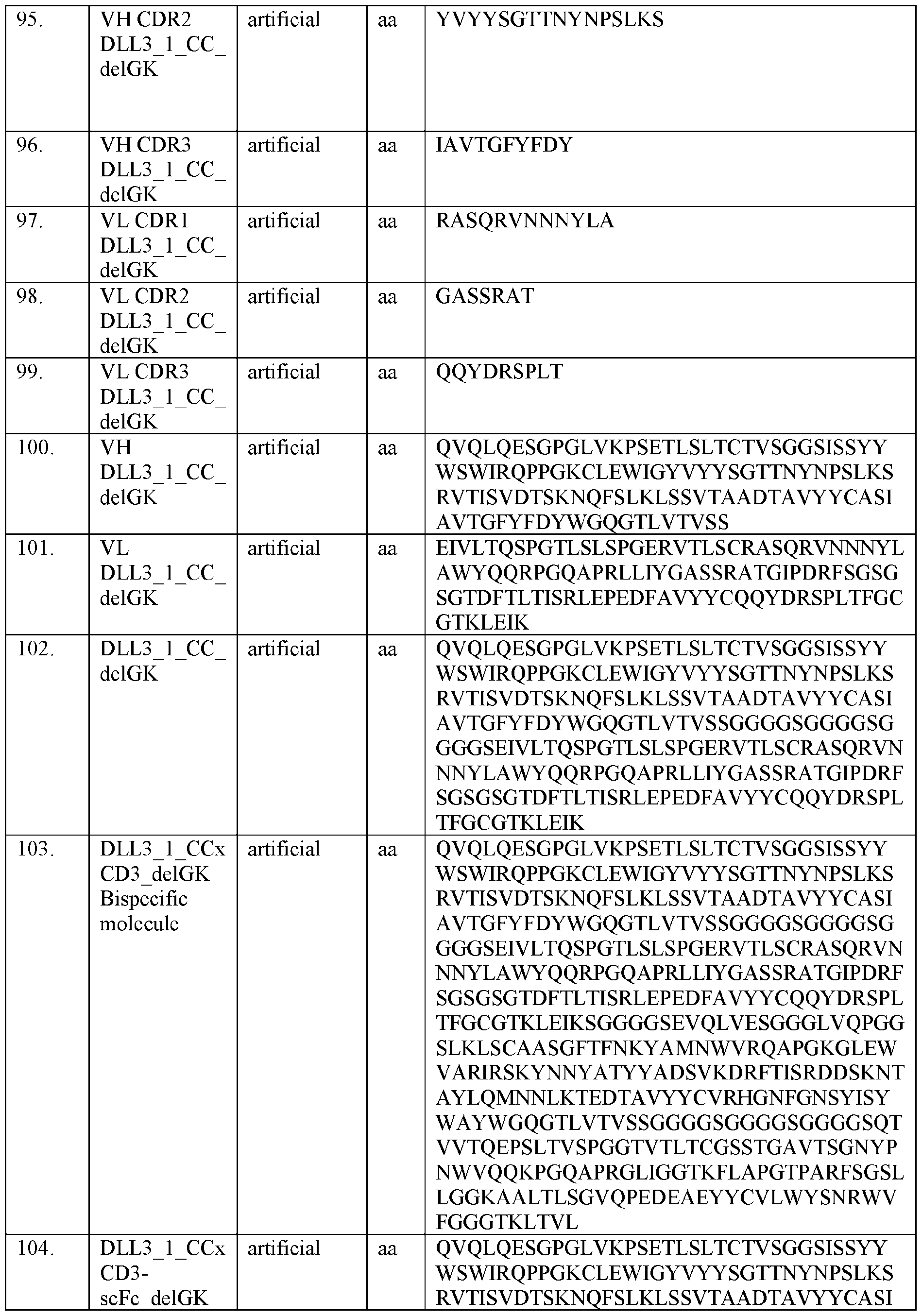


Claims


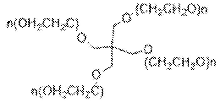


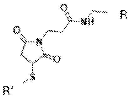


Priority Applications (4)
| Application Number | Priority Date | Filing Date | Title |
|---|---|---|---|
| AU2024302269A AU2024302269A1 (en) | 2023-06-14 | 2024-06-14 | T cell engager masking molecules |
| CN202480038215.4A CN121358496A (en) | 2023-06-14 | 2024-06-14 | T-cell junction masking molecules |
| MX2025014635A MX2025014635A (en) | 2023-06-14 | 2024-06-14 | T CELL MASKING HOST MOLECULES |
| IL324231A IL324231A (en) | 2023-06-14 | 2025-10-26 | Molecules that mask T cell activators |
Applications Claiming Priority (4)
| Application Number | Priority Date | Filing Date | Title |
|---|---|---|---|
| US202363508265P | 2023-06-14 | 2023-06-14 | |
| US63/508,265 | 2023-06-14 | ||
| US202463647855P | 2024-05-15 | 2024-05-15 | |
| US63/647,855 | 2024-05-15 |
Publications (2)
| Publication Number | Publication Date |
|---|---|
| WO2024259378A1 true WO2024259378A1 (en) | 2024-12-19 |
| WO2024259378A9 WO2024259378A9 (en) | 2025-02-13 |
Family
ID=91853576
Family Applications (1)
| Application Number | Title | Priority Date | Filing Date |
|---|---|---|---|
| PCT/US2024/034204 Pending WO2024259378A1 (en) | 2023-06-14 | 2024-06-14 | T cell engager masking molecules |
Country Status (7)
| Country | Link |
|---|---|
| CN (1) | CN121358496A (en) |
| AU (1) | AU2024302269A1 (en) |
| IL (1) | IL324231A (en) |
| MX (1) | MX2025014635A (en) |
| TW (1) | TW202510911A (en) |
| UY (1) | UY40797A (en) |
| WO (1) | WO2024259378A1 (en) |
Citations (146)
| Publication number | Priority date | Publication date | Assignee | Title |
|---|---|---|---|---|
| US556A (en) | 1838-01-09 | Machine foe | ||
| US5013A (en) | 1847-03-13 | Improvement in apparatus for the manufacture of malleable iron | ||
| US3180193A (en) | 1963-02-25 | 1965-04-27 | Benedict David | Machines for cutting lengths of strip material |
| US3691016A (en) | 1970-04-17 | 1972-09-12 | Monsanto Co | Process for the preparation of insoluble enzymes |
| US3969287A (en) | 1972-12-08 | 1976-07-13 | Boehringer Mannheim Gmbh | Carrier-bound protein prepared by reacting the protein with an acylating or alkylating compound having a carrier-bonding group and reacting the product with a carrier |
| US4179337A (en) | 1973-07-20 | 1979-12-18 | Davis Frank F | Non-immunogenic polypeptides |
| US4195128A (en) | 1976-05-03 | 1980-03-25 | Bayer Aktiengesellschaft | Polymeric carrier bound ligands |
| US4229537A (en) | 1978-02-09 | 1980-10-21 | New York University | Preparation of trichloro-s-triazine activated supports for coupling ligands |
| US4247642A (en) | 1977-02-17 | 1981-01-27 | Sumitomo Chemical Company, Limited | Enzyme immobilization with pullulan gel |
| US4301144A (en) | 1979-07-11 | 1981-11-17 | Ajinomoto Company, Incorporated | Blood substitute containing modified hemoglobin |
| US4330440A (en) | 1977-02-08 | 1982-05-18 | Development Finance Corporation Of New Zealand | Activated matrix and method of activation |
| US4439196A (en) | 1982-03-18 | 1984-03-27 | Merck & Co., Inc. | Osmotic drug delivery system |
| US4447233A (en) | 1981-04-10 | 1984-05-08 | Parker-Hannifin Corporation | Medication infusion pump |
| US4447224A (en) | 1982-09-20 | 1984-05-08 | Infusaid Corporation | Variable flow implantable infusion apparatus |
| US4475196A (en) | 1981-03-06 | 1984-10-02 | Zor Clair G | Instrument for locating faults in aircraft passenger reading light and attendant call control system |
| US4485045A (en) | 1981-07-06 | 1984-11-27 | Research Corporation | Synthetic phosphatidyl cholines useful in forming liposomes |
| US4486194A (en) | 1983-06-08 | 1984-12-04 | James Ferrara | Therapeutic device for administering medicaments through the skin |
| US4487603A (en) | 1982-11-26 | 1984-12-11 | Cordis Corporation | Implantable microinfusion pump system |
| US4496689A (en) | 1983-12-27 | 1985-01-29 | Miles Laboratories, Inc. | Covalently attached complex of alpha-1-proteinase inhibitor with a water soluble polymer |
| US4544545A (en) | 1983-06-20 | 1985-10-01 | Trustees University Of Massachusetts | Liposomes containing modified cholesterol for organ targeting |
| EP0171496A2 (en) | 1984-08-15 | 1986-02-19 | Research Development Corporation of Japan | Process for the production of a chimera monoclonal antibody |
| EP0173494A2 (en) | 1984-08-27 | 1986-03-05 | The Board Of Trustees Of The Leland Stanford Junior University | Chimeric receptors by DNA splicing and expression |
| EP0183070A2 (en) | 1984-10-30 | 1986-06-04 | Phillips Petroleum Company | Transformation of yeasts of the genus pichia |
| US4596556A (en) | 1985-03-25 | 1986-06-24 | Bioject, Inc. | Hypodermic injection apparatus |
| GB2177096A (en) | 1984-09-03 | 1987-01-14 | Celltech Ltd | Production of chimeric antibodies |
| US4640835A (en) | 1981-10-30 | 1987-02-03 | Nippon Chemiphar Company, Ltd. | Plasminogen activator derivatives |
| US4670417A (en) | 1985-06-19 | 1987-06-02 | Ajinomoto Co., Inc. | Hemoglobin combined with a poly(alkylene oxide) |
| WO1987005330A1 (en) | 1986-03-07 | 1987-09-11 | Michel Louis Eugene Bergh | Method for enhancing glycoprotein stability |
| US4694778A (en) | 1984-05-04 | 1987-09-22 | Anicon, Inc. | Chemical vapor deposition wafer boat |
| EP0239400A2 (en) | 1986-03-27 | 1987-09-30 | Medical Research Council | Recombinant antibodies and methods for their production |
| EP0244234A2 (en) | 1986-04-30 | 1987-11-04 | Alko Group Ltd. | Transformation of trichoderma |
| WO1988001649A1 (en) | 1986-09-02 | 1988-03-10 | Genex Corporation | Single polypeptide chain binding molecules |
| US4751180A (en) | 1985-03-28 | 1988-06-14 | Chiron Corporation | Expression using fused genes providing for protein product |
| WO1988009344A1 (en) | 1987-05-21 | 1988-12-01 | Creative Biomolecules, Inc. | Targeted multifunctional proteins |
| US4791192A (en) | 1986-06-26 | 1988-12-13 | Takeda Chemical Industries, Ltd. | Chemically modified protein with polyethyleneglycol |
| US4790824A (en) | 1987-06-19 | 1988-12-13 | Bioject, Inc. | Non-invasive hypodermic injection device |
| US4816397A (en) | 1983-03-25 | 1989-03-28 | Celltech, Limited | Multichain polypeptides or proteins and processes for their production |
| US4816567A (en) | 1983-04-08 | 1989-03-28 | Genentech, Inc. | Recombinant immunoglobin preparations |
| US4935233A (en) | 1985-12-02 | 1990-06-19 | G. D. Searle And Company | Covalently linked polypeptide cell modulators |
| US4941880A (en) | 1987-06-19 | 1990-07-17 | Bioject, Inc. | Pre-filled ampule and non-invasive hypodermic injection device assembly |
| US4946778A (en) | 1987-09-21 | 1990-08-07 | Genex Corporation | Single polypeptide chain binding molecules |
| EP0402226A1 (en) | 1989-06-06 | 1990-12-12 | Institut National De La Recherche Agronomique | Transformation vectors for yeast yarrowia |
| US5064413A (en) | 1989-11-09 | 1991-11-12 | Bioject, Inc. | Needleless hypodermic injection device |
| WO1992003918A1 (en) | 1990-08-29 | 1992-03-19 | Genpharm International, Inc. | Transgenic non-human animals capable of producing heterologous antibodies |
| WO1992015673A1 (en) | 1991-03-11 | 1992-09-17 | The University Of Georgia Research Foundation, Inc. | Cloning and expression of renilla luciferase |
| WO1992022647A1 (en) | 1991-06-12 | 1992-12-23 | Genpharm International, Inc. | Early detection of transgenic emryros |
| WO1992022645A1 (en) | 1991-06-14 | 1992-12-23 | Genpharm International, Inc. | Transgenic immunodeficient non-human animals |
| WO1993012227A1 (en) | 1991-12-17 | 1993-06-24 | Genpharm International, Inc. | Transgenic non-human animals capable of producing heterologous antibodies |
| US5223409A (en) | 1988-09-02 | 1993-06-29 | Protein Engineering Corp. | Directed evolution of novel binding proteins |
| US5225539A (en) | 1986-03-27 | 1993-07-06 | Medical Research Council | Recombinant altered antibodies and methods of making altered antibodies |
| US5260203A (en) | 1986-09-02 | 1993-11-09 | Enzon, Inc. | Single polypeptide chain binding molecules |
| WO1994000569A1 (en) | 1992-06-18 | 1994-01-06 | Genpharm International, Inc. | Methods for producing transgenic non-human animals harboring a yeast artificial chromosome |
| WO1994002602A1 (en) | 1992-07-24 | 1994-02-03 | Cell Genesys, Inc. | Generation of xenogeneic antibodies |
| US5292658A (en) | 1989-12-29 | 1994-03-08 | University Of Georgia Research Foundation, Inc. Boyd Graduate Studies Research Center | Cloning and expressions of Renilla luciferase |
| US5312335A (en) | 1989-11-09 | 1994-05-17 | Bioject Inc. | Needleless hypodermic injection device |
| US5313198A (en) | 1987-12-09 | 1994-05-17 | Omron Tateisi Electronics Co. | Data communication apparatus |
| WO1994025585A1 (en) | 1993-04-26 | 1994-11-10 | Genpharm International, Inc. | Transgenic non-human animals capable of producing heterologous antibodies |
| US5383851A (en) | 1992-07-24 | 1995-01-24 | Bioject Inc. | Needleless hypodermic injection device |
| WO1995007463A1 (en) | 1993-09-10 | 1995-03-16 | The Trustees Of Columbia University In The City Of New York | Uses of green fluorescent protein |
| US5476996A (en) | 1988-06-14 | 1995-12-19 | Lidak Pharmaceuticals | Human immune system in non-human animal |
| WO1996014436A1 (en) | 1994-11-04 | 1996-05-17 | Genpharm International, Inc. | Method for making recombinant yeast artificial chromosomes |
| EP0463151B1 (en) | 1990-01-12 | 1996-06-12 | Cell Genesys, Inc. | Generation of xenogeneic antibodies |
| US5545806A (en) | 1990-08-29 | 1996-08-13 | Genpharm International, Inc. | Ransgenic non-human animals for producing heterologous antibodies |
| US5545807A (en) | 1988-10-12 | 1996-08-13 | The Babraham Institute | Production of antibodies from transgenic animals |
| US5565332A (en) | 1991-09-23 | 1996-10-15 | Medical Research Council | Production of chimeric antibodies - a combinatorial approach |
| WO1996033735A1 (en) | 1995-04-27 | 1996-10-31 | Abgenix, Inc. | Human antibodies derived from immunized xenomice |
| WO1996034096A1 (en) | 1995-04-28 | 1996-10-31 | Abgenix, Inc. | Human antibodies derived from immunized xenomice |
| US5585089A (en) | 1988-12-28 | 1996-12-17 | Protein Design Labs, Inc. | Humanized immunoglobulins |
| US5591669A (en) | 1988-12-05 | 1997-01-07 | Genpharm International, Inc. | Transgenic mice depleted in a mature lymphocytic cell-type |
| US5612205A (en) | 1990-08-29 | 1997-03-18 | Genpharm International, Incorporated | Homologous recombination in mammalian cells |
| WO1997013852A1 (en) | 1995-10-10 | 1997-04-17 | Genpharm International, Inc. | Transgenic non-human animals capable of producing heterologous antibodies |
| US5625825A (en) | 1993-10-21 | 1997-04-29 | Lsi Logic Corporation | Random number generating apparatus for an interface unit of a carrier sense with multiple access and collision detect (CSMA/CD) ethernet data network |
| US5625126A (en) | 1990-08-29 | 1997-04-29 | Genpharm International, Inc. | Transgenic non-human animals for producing heterologous antibodies |
| EP0773288A2 (en) | 1995-08-29 | 1997-05-14 | Kirin Beer Kabushiki Kaisha | Chimeric animal and method for producing the same |
| US5633425A (en) | 1990-08-29 | 1997-05-27 | Genpharm International, Inc. | Transgenic non-human animals capable of producing heterologous antibodies |
| US5648260A (en) | 1987-03-18 | 1997-07-15 | Scotgen Biopharmaceuticals Incorporated | DNA encoding antibodies with altered effector functions |
| US5661016A (en) | 1990-08-29 | 1997-08-26 | Genpharm International Inc. | Transgenic non-human animals capable of producing heterologous antibodies of various isotypes |
| WO1997038731A1 (en) | 1996-04-18 | 1997-10-23 | The Regents Of The University Of California | Immunoliposomes that optimize internalization into target cells |
| US5683888A (en) | 1989-07-22 | 1997-11-04 | University Of Wales College Of Medicine | Modified bioluminescent proteins and their use |
| WO1998014605A1 (en) | 1996-10-04 | 1998-04-09 | Loma Linda University | Renilla luciferase and green fluorescent protein fusion genes |
| US5741668A (en) | 1994-02-04 | 1998-04-21 | Rutgers, The State University Of New Jersey | Expression of a gene for a modified green-fluorescent protein |
| WO1998024884A1 (en) | 1996-12-02 | 1998-06-11 | Genpharm International | Transgenic non-human animals capable of producing heterologous antibodies |
| WO1998024893A2 (en) | 1996-12-03 | 1998-06-11 | Abgenix, Inc. | TRANSGENIC MAMMALS HAVING HUMAN IG LOCI INCLUDING PLURAL VH AND Vλ REGIONS AND ANTIBODIES PRODUCED THEREFROM |
| WO1998026277A2 (en) | 1996-12-12 | 1998-06-18 | Prolume, Ltd. | Apparatus and method for detecting and identifying infectious agents |
| US5777079A (en) | 1994-11-10 | 1998-07-07 | The Regents Of The University Of California | Modified green fluorescent proteins |
| US5789650A (en) | 1990-08-29 | 1998-08-04 | Genpharm International, Inc. | Transgenic non-human animals for producing heterologous antibodies |
| US5789215A (en) | 1991-08-20 | 1998-08-04 | Genpharm International | Gene targeting in animal cells using isogenic DNA constructs |
| US5804387A (en) | 1996-02-01 | 1998-09-08 | The Board Of Trustees Of The Leland Stanford Junior University | FACS-optimized mutants of the green fluorescent protein (GFP) |
| US5814318A (en) | 1990-08-29 | 1998-09-29 | Genpharm International Inc. | Transgenic non-human animals for producing heterologous antibodies |
| WO1998052976A1 (en) | 1997-05-21 | 1998-11-26 | Biovation Limited | Method for the production of non-immunogenic proteins |
| US5859205A (en) | 1989-12-21 | 1999-01-12 | Celltech Limited | Humanised antibodies |
| US5874299A (en) | 1990-08-29 | 1999-02-23 | Genpharm International, Inc. | Transgenic non-human animals capable of producing heterologous antibodies |
| US5874304A (en) | 1996-01-18 | 1999-02-23 | University Of Florida Research Foundation, Inc. | Humanized green fluorescent protein genes and methods |
| US5876995A (en) | 1996-02-06 | 1999-03-02 | Bryan; Bruce | Bioluminescent novelty items |
| US5877397A (en) | 1990-08-29 | 1999-03-02 | Genpharm International Inc. | Transgenic non-human animals capable of producing heterologous antibodies of various isotypes |
| US5925558A (en) | 1996-07-16 | 1999-07-20 | The Regents Of The University Of California | Assays for protein kinases using fluorescent protein substrates |
| US5958765A (en) | 1995-06-07 | 1999-09-28 | Idec Pharmaceuticals Corporation | Neutralizing high affinity human monoclonal antibodies specific to RSV F-protein and methods for their manufacture and therapeutic use thereof |
| WO1999049019A2 (en) | 1998-03-27 | 1999-09-30 | Prolume, Ltd. | Luciferases, fluorescent proteins, nucleic acids encoding the luciferases and fluorescent proteins and the use thereof in diagnostics |
| WO1999054440A1 (en) | 1998-04-21 | 1999-10-28 | Micromet Gesellschaft Für Biomedizinische Forschung Mbh | CD19xCD3 SPECIFIC POLYPEPTIDES AND USES THEREOF |
| US5981175A (en) | 1993-01-07 | 1999-11-09 | Genpharm Internation, Inc. | Methods for producing recombinant mammalian cells harboring a yeast artificial chromosome |
| WO2000006605A2 (en) | 1998-07-28 | 2000-02-10 | Micromet Ag | Heterominibodies |
| US6075181A (en) | 1990-01-12 | 2000-06-13 | Abgenix, Inc. | Human antibodies derived from immunized xenomice |
| WO2000034317A2 (en) | 1998-12-08 | 2000-06-15 | Biovation Limited | Method for reducing immunogenicity of proteins |
| US6150584A (en) | 1990-01-12 | 2000-11-21 | Abgenix, Inc. | Human antibodies derived from immunized xenomice |
| US6162963A (en) | 1990-01-12 | 2000-12-19 | Abgenix, Inc. | Generation of Xenogenetic antibodies |
| WO2000076310A1 (en) | 1999-06-10 | 2000-12-21 | Abgenix, Inc. | Transgenic animals for producing specific isotypes of human antibodies via non-cognate switch regions |
| US6255458B1 (en) | 1990-08-29 | 2001-07-03 | Genpharm International | High affinity human antibodies and human antibodies against digoxin |
| US6300064B1 (en) | 1995-08-18 | 2001-10-09 | Morphosys Ag | Protein/(poly)peptide libraries |
| US6407213B1 (en) | 1991-06-14 | 2002-06-18 | Genentech, Inc. | Method for making humanized antibodies |
| WO2003047336A2 (en) | 2001-11-30 | 2003-06-12 | Abgenix, Inc. | TRANSGENIC ANIMALS BEARING HUMAN Igμ LIGHT CHAIN GENES |
| WO2005040220A1 (en) | 2003-10-16 | 2005-05-06 | Micromet Ag | Multispecific deimmunized cd3-binders |
| WO2006105993A2 (en) * | 2005-04-05 | 2006-10-12 | Istituto Di Ricerche Di Biologia Molecolare P Angeletti Spa | Method for shielding functional sites or epitopes on proteins |
| WO2006138181A2 (en) | 2005-06-14 | 2006-12-28 | Amgen Inc. | Self-buffering protein formulations |
| WO2007042261A2 (en) | 2005-10-11 | 2007-04-19 | Micromet Ag | Compositions comprising cross-species-specific antibodies and uses thereof |
| WO2008119567A2 (en) | 2007-04-03 | 2008-10-09 | Micromet Ag | Cross-species-specific cd3-epsilon binding domain |
| US7466008B2 (en) | 2007-03-13 | 2008-12-16 | Taiwan Semiconductor Manufacturing Company, Ltd. | BiCMOS performance enhancement by mechanical uniaxial strain and methods of manufacture |
| US7574748B2 (en) | 2006-03-07 | 2009-08-18 | Nike, Inc. | Glove with support system |
| US7575962B2 (en) | 2006-08-11 | 2009-08-18 | Samsung Electronics Co., Ltd. | Fin structure and method of manufacturing fin transistor adopting the fin structure |
| US7610515B2 (en) | 2005-10-27 | 2009-10-27 | Hitachi, Ltd. | Disk array device and failure response verification method thereof |
| WO2010037838A2 (en) | 2008-10-01 | 2010-04-08 | Micromet Ag | Cross-species-specific single domain bispecific single chain antibody |
| US7853408B2 (en) | 2005-08-17 | 2010-12-14 | Biosigma S.A. | Method for the design of oligonucleotides for molecular biology techniques |
| US7904068B2 (en) | 2003-06-06 | 2011-03-08 | At&T Intellectual Property I, L.P. | System and method for providing integrated voice and data services utilizing wired cordless access with unlicensed spectrum and wired access with licensed spectrum |
| US7919297B2 (en) | 2006-02-21 | 2011-04-05 | Cornell Research Foundation, Inc. | Mutants of Aspergillus niger PhyA phytase and Aspergillus fumigatus phytase |
| US7990860B2 (en) | 2006-06-16 | 2011-08-02 | Harris Corporation | Method and system for rule-based sequencing for QoS |
| US8155301B2 (en) | 2006-10-30 | 2012-04-10 | Huawei Technologies Co., Ltd. | System and method for dialing prompt |
| US8165699B2 (en) | 2005-03-14 | 2012-04-24 | Omron Corporation | Programmable controller system |
| US8161739B2 (en) | 2007-11-29 | 2012-04-24 | Schaeffler Technologies AG & Co. KG | Force transmission device in particular for power transmission between a drive engine and an output |
| US8209741B2 (en) | 2007-09-17 | 2012-06-26 | Microsoft Corporation | Human performance in human interactive proofs using partial credit |
| US8234145B2 (en) | 2005-07-12 | 2012-07-31 | International Business Machines Corporation | Automatic computation of validation metrics for global logistics processes |
| US8376279B2 (en) | 2008-01-23 | 2013-02-19 | Aurora Flight Sciences Corporation | Inflatable folding wings for a very high altitude aircraft |
| WO2013026833A1 (en) | 2011-08-23 | 2013-02-28 | Roche Glycart Ag | Bispecific t cell activating antigen binding molecules |
| WO2013026837A1 (en) | 2011-08-23 | 2013-02-28 | Roche Glycart Ag | Bispecific t cell activating antigen binding molecules |
| US8430938B1 (en) | 2006-07-13 | 2013-04-30 | The United States Of America As Represented By The Secretary Of The Navy | Control algorithm for autothermal reformer |
| US8463191B2 (en) | 2009-04-02 | 2013-06-11 | Qualcomm Incorporated | Beamforming options with partial channel knowledge |
| US8462837B2 (en) | 1998-10-30 | 2013-06-11 | Broadcom Corporation | Constellation-multiplexed transmitter and receiver |
| US8464584B2 (en) | 2007-10-19 | 2013-06-18 | Food Equipment Technologies Company, Inc. | Beverage dispenser with level measuring apparatus and display |
| US8486859B2 (en) | 2002-05-15 | 2013-07-16 | Bioenergy, Inc. | Use of ribose to enhance plant growth |
| US8486853B2 (en) | 2009-03-04 | 2013-07-16 | Nissan Motor Co., Ltd. | Exhaust gas purifying catalyst and method for manufacturing the same |
| US8759620B2 (en) | 2001-08-31 | 2014-06-24 | Syngenta Participations Ag | Transgenic plants expressing modified CRY3A |
| WO2014144722A2 (en) | 2013-03-15 | 2014-09-18 | Amgen Inc. | BISPECIFIC-Fc MOLECULES |
| WO2014151910A1 (en) | 2013-03-15 | 2014-09-25 | Amgen Inc. | Heterodimeric bispecific antibodies |
| US20140308285A1 (en) | 2013-03-15 | 2014-10-16 | Amgen Inc. | Heterodimeric bispecific antibodies |
| WO2015048272A1 (en) | 2013-09-25 | 2015-04-02 | Amgen Inc. | V-c-fc-v-c antibody |
| US9676298B2 (en) | 2010-12-30 | 2017-06-13 | C. Rob. Hammerstein Gmbh & Co. Kg | Longitudinal adjustment device for a motor vehicle seat, comprising two pairs of rails |
| US11284898B2 (en) | 2014-09-18 | 2022-03-29 | Cilag Gmbh International | Surgical instrument including a deployable knife |
| WO2022272033A2 (en) * | 2021-06-25 | 2022-12-29 | Harpoon Therapeutics, Inc. | Extended-release immune cell engaging proteins and methods of treatment |
-
2024
- 2024-06-14 UY UY0001040797A patent/UY40797A/en unknown
- 2024-06-14 CN CN202480038215.4A patent/CN121358496A/en active Pending
- 2024-06-14 MX MX2025014635A patent/MX2025014635A/en unknown
- 2024-06-14 AU AU2024302269A patent/AU2024302269A1/en active Pending
- 2024-06-14 WO PCT/US2024/034204 patent/WO2024259378A1/en active Pending
- 2024-06-14 TW TW113122224A patent/TW202510911A/en unknown
-
2025
- 2025-10-26 IL IL324231A patent/IL324231A/en unknown
Patent Citations (165)
| Publication number | Priority date | Publication date | Assignee | Title |
|---|---|---|---|---|
| US556A (en) | 1838-01-09 | Machine foe | ||
| US5013A (en) | 1847-03-13 | Improvement in apparatus for the manufacture of malleable iron | ||
| US3180193A (en) | 1963-02-25 | 1965-04-27 | Benedict David | Machines for cutting lengths of strip material |
| US3691016A (en) | 1970-04-17 | 1972-09-12 | Monsanto Co | Process for the preparation of insoluble enzymes |
| US3969287A (en) | 1972-12-08 | 1976-07-13 | Boehringer Mannheim Gmbh | Carrier-bound protein prepared by reacting the protein with an acylating or alkylating compound having a carrier-bonding group and reacting the product with a carrier |
| US4179337A (en) | 1973-07-20 | 1979-12-18 | Davis Frank F | Non-immunogenic polypeptides |
| US4195128A (en) | 1976-05-03 | 1980-03-25 | Bayer Aktiengesellschaft | Polymeric carrier bound ligands |
| US4330440A (en) | 1977-02-08 | 1982-05-18 | Development Finance Corporation Of New Zealand | Activated matrix and method of activation |
| US4247642A (en) | 1977-02-17 | 1981-01-27 | Sumitomo Chemical Company, Limited | Enzyme immobilization with pullulan gel |
| US4229537A (en) | 1978-02-09 | 1980-10-21 | New York University | Preparation of trichloro-s-triazine activated supports for coupling ligands |
| US4301144A (en) | 1979-07-11 | 1981-11-17 | Ajinomoto Company, Incorporated | Blood substitute containing modified hemoglobin |
| US4475196A (en) | 1981-03-06 | 1984-10-02 | Zor Clair G | Instrument for locating faults in aircraft passenger reading light and attendant call control system |
| US4447233A (en) | 1981-04-10 | 1984-05-08 | Parker-Hannifin Corporation | Medication infusion pump |
| US4485045A (en) | 1981-07-06 | 1984-11-27 | Research Corporation | Synthetic phosphatidyl cholines useful in forming liposomes |
| US4640835A (en) | 1981-10-30 | 1987-02-03 | Nippon Chemiphar Company, Ltd. | Plasminogen activator derivatives |
| US4439196A (en) | 1982-03-18 | 1984-03-27 | Merck & Co., Inc. | Osmotic drug delivery system |
| US4447224A (en) | 1982-09-20 | 1984-05-08 | Infusaid Corporation | Variable flow implantable infusion apparatus |
| US4487603A (en) | 1982-11-26 | 1984-12-11 | Cordis Corporation | Implantable microinfusion pump system |
| US4816397A (en) | 1983-03-25 | 1989-03-28 | Celltech, Limited | Multichain polypeptides or proteins and processes for their production |
| US4816567A (en) | 1983-04-08 | 1989-03-28 | Genentech, Inc. | Recombinant immunoglobin preparations |
| US4486194A (en) | 1983-06-08 | 1984-12-04 | James Ferrara | Therapeutic device for administering medicaments through the skin |
| US4544545A (en) | 1983-06-20 | 1985-10-01 | Trustees University Of Massachusetts | Liposomes containing modified cholesterol for organ targeting |
| US4496689A (en) | 1983-12-27 | 1985-01-29 | Miles Laboratories, Inc. | Covalently attached complex of alpha-1-proteinase inhibitor with a water soluble polymer |
| US4694778A (en) | 1984-05-04 | 1987-09-22 | Anicon, Inc. | Chemical vapor deposition wafer boat |
| EP0171496A2 (en) | 1984-08-15 | 1986-02-19 | Research Development Corporation of Japan | Process for the production of a chimera monoclonal antibody |
| EP0173494A2 (en) | 1984-08-27 | 1986-03-05 | The Board Of Trustees Of The Leland Stanford Junior University | Chimeric receptors by DNA splicing and expression |
| GB2177096A (en) | 1984-09-03 | 1987-01-14 | Celltech Ltd | Production of chimeric antibodies |
| EP0183070A2 (en) | 1984-10-30 | 1986-06-04 | Phillips Petroleum Company | Transformation of yeasts of the genus pichia |
| US4596556A (en) | 1985-03-25 | 1986-06-24 | Bioject, Inc. | Hypodermic injection apparatus |
| US4751180A (en) | 1985-03-28 | 1988-06-14 | Chiron Corporation | Expression using fused genes providing for protein product |
| US4670417A (en) | 1985-06-19 | 1987-06-02 | Ajinomoto Co., Inc. | Hemoglobin combined with a poly(alkylene oxide) |
| US4935233A (en) | 1985-12-02 | 1990-06-19 | G. D. Searle And Company | Covalently linked polypeptide cell modulators |
| WO1987005330A1 (en) | 1986-03-07 | 1987-09-11 | Michel Louis Eugene Bergh | Method for enhancing glycoprotein stability |
| EP0239400A2 (en) | 1986-03-27 | 1987-09-30 | Medical Research Council | Recombinant antibodies and methods for their production |
| US5225539A (en) | 1986-03-27 | 1993-07-06 | Medical Research Council | Recombinant altered antibodies and methods of making altered antibodies |
| EP0244234A2 (en) | 1986-04-30 | 1987-11-04 | Alko Group Ltd. | Transformation of trichoderma |
| US4791192A (en) | 1986-06-26 | 1988-12-13 | Takeda Chemical Industries, Ltd. | Chemically modified protein with polyethyleneglycol |
| US5260203A (en) | 1986-09-02 | 1993-11-09 | Enzon, Inc. | Single polypeptide chain binding molecules |
| WO1988001649A1 (en) | 1986-09-02 | 1988-03-10 | Genex Corporation | Single polypeptide chain binding molecules |
| US5648260A (en) | 1987-03-18 | 1997-07-15 | Scotgen Biopharmaceuticals Incorporated | DNA encoding antibodies with altered effector functions |
| WO1988009344A1 (en) | 1987-05-21 | 1988-12-01 | Creative Biomolecules, Inc. | Targeted multifunctional proteins |
| US4941880A (en) | 1987-06-19 | 1990-07-17 | Bioject, Inc. | Pre-filled ampule and non-invasive hypodermic injection device assembly |
| US4790824A (en) | 1987-06-19 | 1988-12-13 | Bioject, Inc. | Non-invasive hypodermic injection device |
| US4946778A (en) | 1987-09-21 | 1990-08-07 | Genex Corporation | Single polypeptide chain binding molecules |
| US5313198A (en) | 1987-12-09 | 1994-05-17 | Omron Tateisi Electronics Co. | Data communication apparatus |
| US5698767A (en) | 1988-06-14 | 1997-12-16 | Lidak Pharmaceuticals | Human immune system in non-human animal |
| US5476996A (en) | 1988-06-14 | 1995-12-19 | Lidak Pharmaceuticals | Human immune system in non-human animal |
| US5223409A (en) | 1988-09-02 | 1993-06-29 | Protein Engineering Corp. | Directed evolution of novel binding proteins |
| US5545807A (en) | 1988-10-12 | 1996-08-13 | The Babraham Institute | Production of antibodies from transgenic animals |
| US6023010A (en) | 1988-12-05 | 2000-02-08 | Genpharm International | Transgenic non-human animals depleted in a mature lymphocytic cell-type |
| US5591669A (en) | 1988-12-05 | 1997-01-07 | Genpharm International, Inc. | Transgenic mice depleted in a mature lymphocytic cell-type |
| US5693762A (en) | 1988-12-28 | 1997-12-02 | Protein Design Labs, Inc. | Humanized immunoglobulins |
| US5585089A (en) | 1988-12-28 | 1996-12-17 | Protein Design Labs, Inc. | Humanized immunoglobulins |
| US5693761A (en) | 1988-12-28 | 1997-12-02 | Protein Design Labs, Inc. | Polynucleotides encoding improved humanized immunoglobulins |
| EP0402226A1 (en) | 1989-06-06 | 1990-12-12 | Institut National De La Recherche Agronomique | Transformation vectors for yeast yarrowia |
| US5683888A (en) | 1989-07-22 | 1997-11-04 | University Of Wales College Of Medicine | Modified bioluminescent proteins and their use |
| US5064413A (en) | 1989-11-09 | 1991-11-12 | Bioject, Inc. | Needleless hypodermic injection device |
| US5312335A (en) | 1989-11-09 | 1994-05-17 | Bioject Inc. | Needleless hypodermic injection device |
| US5859205A (en) | 1989-12-21 | 1999-01-12 | Celltech Limited | Humanised antibodies |
| US5292658A (en) | 1989-12-29 | 1994-03-08 | University Of Georgia Research Foundation, Inc. Boyd Graduate Studies Research Center | Cloning and expressions of Renilla luciferase |
| US5418155A (en) | 1989-12-29 | 1995-05-23 | University Of Georgia Research Foundation, Inc. | Isolated Renilla luciferase and method of use thereof |
| US5939598A (en) | 1990-01-12 | 1999-08-17 | Abgenix, Inc. | Method of making transgenic mice lacking endogenous heavy chains |
| US6075181A (en) | 1990-01-12 | 2000-06-13 | Abgenix, Inc. | Human antibodies derived from immunized xenomice |
| JP3068506B2 (en) | 1990-01-12 | 2000-07-24 | アブジェニックス インコーポレイテッド | Generation of heterologous antibodies |
| EP0463151B1 (en) | 1990-01-12 | 1996-06-12 | Cell Genesys, Inc. | Generation of xenogeneic antibodies |
| JP3068180B2 (en) | 1990-01-12 | 2000-07-24 | アブジェニックス インコーポレイテッド | Generation of heterologous antibodies |
| JP3068507B2 (en) | 1990-01-12 | 2000-07-24 | アブジェニックス インコーポレイテッド | Generation of heterologous antibodies |
| US6114598A (en) | 1990-01-12 | 2000-09-05 | Abgenix, Inc. | Generation of xenogeneic antibodies |
| US6150584A (en) | 1990-01-12 | 2000-11-21 | Abgenix, Inc. | Human antibodies derived from immunized xenomice |
| US6162963A (en) | 1990-01-12 | 2000-12-19 | Abgenix, Inc. | Generation of Xenogenetic antibodies |
| EP0546073B1 (en) | 1990-08-29 | 1997-09-10 | GenPharm International, Inc. | production and use of transgenic non-human animals capable of producing heterologous antibodies |
| US5721367A (en) | 1990-08-29 | 1998-02-24 | Pharming B.V. | Homologous recombination in mammalian cells |
| US5612205A (en) | 1990-08-29 | 1997-03-18 | Genpharm International, Incorporated | Homologous recombination in mammalian cells |
| US5874299A (en) | 1990-08-29 | 1999-02-23 | Genpharm International, Inc. | Transgenic non-human animals capable of producing heterologous antibodies |
| US5814318A (en) | 1990-08-29 | 1998-09-29 | Genpharm International Inc. | Transgenic non-human animals for producing heterologous antibodies |
| US5625126A (en) | 1990-08-29 | 1997-04-29 | Genpharm International, Inc. | Transgenic non-human animals for producing heterologous antibodies |
| US6255458B1 (en) | 1990-08-29 | 2001-07-03 | Genpharm International | High affinity human antibodies and human antibodies against digoxin |
| US5633425A (en) | 1990-08-29 | 1997-05-27 | Genpharm International, Inc. | Transgenic non-human animals capable of producing heterologous antibodies |
| US5789650A (en) | 1990-08-29 | 1998-08-04 | Genpharm International, Inc. | Transgenic non-human animals for producing heterologous antibodies |
| WO1992003918A1 (en) | 1990-08-29 | 1992-03-19 | Genpharm International, Inc. | Transgenic non-human animals capable of producing heterologous antibodies |
| US5661016A (en) | 1990-08-29 | 1997-08-26 | Genpharm International Inc. | Transgenic non-human animals capable of producing heterologous antibodies of various isotypes |
| US5770429A (en) | 1990-08-29 | 1998-06-23 | Genpharm International, Inc. | Transgenic non-human animals capable of producing heterologous antibodies |
| US5877397A (en) | 1990-08-29 | 1999-03-02 | Genpharm International Inc. | Transgenic non-human animals capable of producing heterologous antibodies of various isotypes |
| US5545806A (en) | 1990-08-29 | 1996-08-13 | Genpharm International, Inc. | Ransgenic non-human animals for producing heterologous antibodies |
| WO1992015673A1 (en) | 1991-03-11 | 1992-09-17 | The University Of Georgia Research Foundation, Inc. | Cloning and expression of renilla luciferase |
| WO1992022647A1 (en) | 1991-06-12 | 1992-12-23 | Genpharm International, Inc. | Early detection of transgenic emryros |
| WO1992022670A1 (en) | 1991-06-12 | 1992-12-23 | Genpharm International, Inc. | Early detection of transgenic embryos |
| WO1992022645A1 (en) | 1991-06-14 | 1992-12-23 | Genpharm International, Inc. | Transgenic immunodeficient non-human animals |
| US6407213B1 (en) | 1991-06-14 | 2002-06-18 | Genentech, Inc. | Method for making humanized antibodies |
| US5789215A (en) | 1991-08-20 | 1998-08-04 | Genpharm International | Gene targeting in animal cells using isogenic DNA constructs |
| US5565332A (en) | 1991-09-23 | 1996-10-15 | Medical Research Council | Production of chimeric antibodies - a combinatorial approach |
| WO1993012227A1 (en) | 1991-12-17 | 1993-06-24 | Genpharm International, Inc. | Transgenic non-human animals capable of producing heterologous antibodies |
| WO1994000569A1 (en) | 1992-06-18 | 1994-01-06 | Genpharm International, Inc. | Methods for producing transgenic non-human animals harboring a yeast artificial chromosome |
| WO1994002602A1 (en) | 1992-07-24 | 1994-02-03 | Cell Genesys, Inc. | Generation of xenogeneic antibodies |
| US5383851A (en) | 1992-07-24 | 1995-01-24 | Bioject Inc. | Needleless hypodermic injection device |
| US5399163A (en) | 1992-07-24 | 1995-03-21 | Bioject Inc. | Needleless hypodermic injection methods and device |
| US5981175A (en) | 1993-01-07 | 1999-11-09 | Genpharm Internation, Inc. | Methods for producing recombinant mammalian cells harboring a yeast artificial chromosome |
| WO1994025585A1 (en) | 1993-04-26 | 1994-11-10 | Genpharm International, Inc. | Transgenic non-human animals capable of producing heterologous antibodies |
| WO1995007463A1 (en) | 1993-09-10 | 1995-03-16 | The Trustees Of Columbia University In The City Of New York | Uses of green fluorescent protein |
| US5625825A (en) | 1993-10-21 | 1997-04-29 | Lsi Logic Corporation | Random number generating apparatus for an interface unit of a carrier sense with multiple access and collision detect (CSMA/CD) ethernet data network |
| US5741668A (en) | 1994-02-04 | 1998-04-21 | Rutgers, The State University Of New Jersey | Expression of a gene for a modified green-fluorescent protein |
| WO1996014436A1 (en) | 1994-11-04 | 1996-05-17 | Genpharm International, Inc. | Method for making recombinant yeast artificial chromosomes |
| US5643763A (en) | 1994-11-04 | 1997-07-01 | Genpharm International, Inc. | Method for making recombinant yeast artificial chromosomes by minimizing diploid doubling during mating |
| US5777079A (en) | 1994-11-10 | 1998-07-07 | The Regents Of The University Of California | Modified green fluorescent proteins |
| WO1996033735A1 (en) | 1995-04-27 | 1996-10-31 | Abgenix, Inc. | Human antibodies derived from immunized xenomice |
| WO1996034096A1 (en) | 1995-04-28 | 1996-10-31 | Abgenix, Inc. | Human antibodies derived from immunized xenomice |
| US5958765A (en) | 1995-06-07 | 1999-09-28 | Idec Pharmaceuticals Corporation | Neutralizing high affinity human monoclonal antibodies specific to RSV F-protein and methods for their manufacture and therapeutic use thereof |
| US6300064B1 (en) | 1995-08-18 | 2001-10-09 | Morphosys Ag | Protein/(poly)peptide libraries |
| EP0843961A1 (en) | 1995-08-29 | 1998-05-27 | Kirin Beer Kabushiki Kaisha | Chimeric animal and method for constructing the same |
| EP0773288A2 (en) | 1995-08-29 | 1997-05-14 | Kirin Beer Kabushiki Kaisha | Chimeric animal and method for producing the same |
| WO1997013852A1 (en) | 1995-10-10 | 1997-04-17 | Genpharm International, Inc. | Transgenic non-human animals capable of producing heterologous antibodies |
| US5874304A (en) | 1996-01-18 | 1999-02-23 | University Of Florida Research Foundation, Inc. | Humanized green fluorescent protein genes and methods |
| US5804387A (en) | 1996-02-01 | 1998-09-08 | The Board Of Trustees Of The Leland Stanford Junior University | FACS-optimized mutants of the green fluorescent protein (GFP) |
| US5876995A (en) | 1996-02-06 | 1999-03-02 | Bryan; Bruce | Bioluminescent novelty items |
| WO1997038731A1 (en) | 1996-04-18 | 1997-10-23 | The Regents Of The University Of California | Immunoliposomes that optimize internalization into target cells |
| US5925558A (en) | 1996-07-16 | 1999-07-20 | The Regents Of The University Of California | Assays for protein kinases using fluorescent protein substrates |
| WO1998014605A1 (en) | 1996-10-04 | 1998-04-09 | Loma Linda University | Renilla luciferase and green fluorescent protein fusion genes |
| WO1998024884A1 (en) | 1996-12-02 | 1998-06-11 | Genpharm International | Transgenic non-human animals capable of producing heterologous antibodies |
| US20030070185A1 (en) | 1996-12-03 | 2003-04-10 | Aya Jakobovits | Transgenic mammals having human Ig loci including plural Vh and Vk regions and antibodies produced therefrom |
| WO1998024893A2 (en) | 1996-12-03 | 1998-06-11 | Abgenix, Inc. | TRANSGENIC MAMMALS HAVING HUMAN IG LOCI INCLUDING PLURAL VH AND Vλ REGIONS AND ANTIBODIES PRODUCED THEREFROM |
| WO1998026277A2 (en) | 1996-12-12 | 1998-06-18 | Prolume, Ltd. | Apparatus and method for detecting and identifying infectious agents |
| WO1998052976A1 (en) | 1997-05-21 | 1998-11-26 | Biovation Limited | Method for the production of non-immunogenic proteins |
| WO1999049019A2 (en) | 1998-03-27 | 1999-09-30 | Prolume, Ltd. | Luciferases, fluorescent proteins, nucleic acids encoding the luciferases and fluorescent proteins and the use thereof in diagnostics |
| WO1999054440A1 (en) | 1998-04-21 | 1999-10-28 | Micromet Gesellschaft Für Biomedizinische Forschung Mbh | CD19xCD3 SPECIFIC POLYPEPTIDES AND USES THEREOF |
| WO2000006605A2 (en) | 1998-07-28 | 2000-02-10 | Micromet Ag | Heterominibodies |
| US8462837B2 (en) | 1998-10-30 | 2013-06-11 | Broadcom Corporation | Constellation-multiplexed transmitter and receiver |
| WO2000034317A2 (en) | 1998-12-08 | 2000-06-15 | Biovation Limited | Method for reducing immunogenicity of proteins |
| WO2000076310A1 (en) | 1999-06-10 | 2000-12-21 | Abgenix, Inc. | Transgenic animals for producing specific isotypes of human antibodies via non-cognate switch regions |
| US8759620B2 (en) | 2001-08-31 | 2014-06-24 | Syngenta Participations Ag | Transgenic plants expressing modified CRY3A |
| WO2003047336A2 (en) | 2001-11-30 | 2003-06-12 | Abgenix, Inc. | TRANSGENIC ANIMALS BEARING HUMAN Igμ LIGHT CHAIN GENES |
| US8486859B2 (en) | 2002-05-15 | 2013-07-16 | Bioenergy, Inc. | Use of ribose to enhance plant growth |
| US7904068B2 (en) | 2003-06-06 | 2011-03-08 | At&T Intellectual Property I, L.P. | System and method for providing integrated voice and data services utilizing wired cordless access with unlicensed spectrum and wired access with licensed spectrum |
| WO2005040220A1 (en) | 2003-10-16 | 2005-05-06 | Micromet Ag | Multispecific deimmunized cd3-binders |
| US8165699B2 (en) | 2005-03-14 | 2012-04-24 | Omron Corporation | Programmable controller system |
| WO2006105993A2 (en) * | 2005-04-05 | 2006-10-12 | Istituto Di Ricerche Di Biologia Molecolare P Angeletti Spa | Method for shielding functional sites or epitopes on proteins |
| WO2006138181A2 (en) | 2005-06-14 | 2006-12-28 | Amgen Inc. | Self-buffering protein formulations |
| US8234145B2 (en) | 2005-07-12 | 2012-07-31 | International Business Machines Corporation | Automatic computation of validation metrics for global logistics processes |
| US7853408B2 (en) | 2005-08-17 | 2010-12-14 | Biosigma S.A. | Method for the design of oligonucleotides for molecular biology techniques |
| WO2007042261A2 (en) | 2005-10-11 | 2007-04-19 | Micromet Ag | Compositions comprising cross-species-specific antibodies and uses thereof |
| US7610515B2 (en) | 2005-10-27 | 2009-10-27 | Hitachi, Ltd. | Disk array device and failure response verification method thereof |
| US7919297B2 (en) | 2006-02-21 | 2011-04-05 | Cornell Research Foundation, Inc. | Mutants of Aspergillus niger PhyA phytase and Aspergillus fumigatus phytase |
| US7574748B2 (en) | 2006-03-07 | 2009-08-18 | Nike, Inc. | Glove with support system |
| US7990860B2 (en) | 2006-06-16 | 2011-08-02 | Harris Corporation | Method and system for rule-based sequencing for QoS |
| US8430938B1 (en) | 2006-07-13 | 2013-04-30 | The United States Of America As Represented By The Secretary Of The Navy | Control algorithm for autothermal reformer |
| US7575962B2 (en) | 2006-08-11 | 2009-08-18 | Samsung Electronics Co., Ltd. | Fin structure and method of manufacturing fin transistor adopting the fin structure |
| US8155301B2 (en) | 2006-10-30 | 2012-04-10 | Huawei Technologies Co., Ltd. | System and method for dialing prompt |
| US7466008B2 (en) | 2007-03-13 | 2008-12-16 | Taiwan Semiconductor Manufacturing Company, Ltd. | BiCMOS performance enhancement by mechanical uniaxial strain and methods of manufacture |
| WO2008119567A2 (en) | 2007-04-03 | 2008-10-09 | Micromet Ag | Cross-species-specific cd3-epsilon binding domain |
| US8209741B2 (en) | 2007-09-17 | 2012-06-26 | Microsoft Corporation | Human performance in human interactive proofs using partial credit |
| US8464584B2 (en) | 2007-10-19 | 2013-06-18 | Food Equipment Technologies Company, Inc. | Beverage dispenser with level measuring apparatus and display |
| US8161739B2 (en) | 2007-11-29 | 2012-04-24 | Schaeffler Technologies AG & Co. KG | Force transmission device in particular for power transmission between a drive engine and an output |
| US8376279B2 (en) | 2008-01-23 | 2013-02-19 | Aurora Flight Sciences Corporation | Inflatable folding wings for a very high altitude aircraft |
| WO2010037838A2 (en) | 2008-10-01 | 2010-04-08 | Micromet Ag | Cross-species-specific single domain bispecific single chain antibody |
| US8486853B2 (en) | 2009-03-04 | 2013-07-16 | Nissan Motor Co., Ltd. | Exhaust gas purifying catalyst and method for manufacturing the same |
| US8463191B2 (en) | 2009-04-02 | 2013-06-11 | Qualcomm Incorporated | Beamforming options with partial channel knowledge |
| US9676298B2 (en) | 2010-12-30 | 2017-06-13 | C. Rob. Hammerstein Gmbh & Co. Kg | Longitudinal adjustment device for a motor vehicle seat, comprising two pairs of rails |
| WO2013026837A1 (en) | 2011-08-23 | 2013-02-28 | Roche Glycart Ag | Bispecific t cell activating antigen binding molecules |
| WO2013026833A1 (en) | 2011-08-23 | 2013-02-28 | Roche Glycart Ag | Bispecific t cell activating antigen binding molecules |
| WO2014144722A2 (en) | 2013-03-15 | 2014-09-18 | Amgen Inc. | BISPECIFIC-Fc MOLECULES |
| WO2014151910A1 (en) | 2013-03-15 | 2014-09-25 | Amgen Inc. | Heterodimeric bispecific antibodies |
| US20140302037A1 (en) | 2013-03-15 | 2014-10-09 | Amgen Inc. | BISPECIFIC-Fc MOLECULES |
| US20140308285A1 (en) | 2013-03-15 | 2014-10-16 | Amgen Inc. | Heterodimeric bispecific antibodies |
| WO2015048272A1 (en) | 2013-09-25 | 2015-04-02 | Amgen Inc. | V-c-fc-v-c antibody |
| US11284898B2 (en) | 2014-09-18 | 2022-03-29 | Cilag Gmbh International | Surgical instrument including a deployable knife |
| WO2022272033A2 (en) * | 2021-06-25 | 2022-12-29 | Harpoon Therapeutics, Inc. | Extended-release immune cell engaging proteins and methods of treatment |
Non-Patent Citations (96)
| Title |
|---|
| "Antibody Engineering Lab Manual", SPRINGER-VERLAG, article "Protein Sequence and Structure Analysis of Antibody Variable Domains" |
| "Macromolecule Sequencing and Synthesis, Selected Methods and Applications", 1988, ALAN R. LISS, INC, article "Current Methods in Sequence Comparison and Analysis", pages: 127 - 149 |
| "Preclinical safety evaluation of biotechnology-derived pharmaceuticals S6", ICH HARMONISED TRIPARTITE GUIDELINE; ICH STEERING COMMITTEE MEETING, 16 July 1997 (1997-07-16) |
| "REMINGTON'S PHARMACEUTICAL SCIENCES", 1990, MACK PUBLISHING COMPANY |
| ALTSCHUL ET AL., J. MOL. BIOL., vol. 215, 1990, pages 403 - 410 |
| ALTSCHUL ET AL., METHODS IN ENZYMOLOGY, vol. 266, 1996, pages 460 - 480 |
| ALTSCHUL ET AL., NUCL. ACIDS RES., vol. 25, 1993, pages 3389 - 3402 |
| ALTSCHUL ET AL., NUCLEIC ACIDS RES., vol. 25, 1997, pages 3389 - 3402 |
| APLINWRISTON, CRC CRIT. REV. BIOCHEM., 1981, pages 259 - 306 |
| ARAKAWA ET AL.: "Solvent interactions in pharmaceutical formulations", PHARM RES., vol. 8, no. 3, 1991, pages 285 - 91, XP009052919, DOI: 10.1023/A:1015825027737 |
| ARTSAENKO ET AL., THE PLANT J, vol. 8, 1995, pages 745 - 750 |
| BARLOW ET AL., J. PHYS. CHEM, 2018 |
| BRIIHL, IMMUNOL., vol. 166, 2001, pages 2420 - 2426 |
| CARTER ET AL., BIO/TECHNOLOGY, vol. 10, 1992, pages 163 - 167 |
| CHALFIE ET AL., SCIENCE, vol. 263, 1994, pages 802 - 805 |
| CHEADLE ET AL., MOL IMMUNOL, vol. 29, 1992, pages 21 - 30 |
| CHEN T TIMOTHY: "Conditionally active T cell engagers for the treatment of solid tumors: rationale and clinical development", EXPERT OPINION ON BIOLOGICAL THERAPY, vol. 22, no. 8, 21 July 2022 (2022-07-21), pages 955 - 963, XP093119758, ISSN: 1471-2598, DOI: 10.1080/14712598.2022.2098674 * |
| CHEN, X. ET AL., A MODELING FRAMEWORK TO CHARACTERIZE CYTOKINE RELEASE UPON T-CELL-ENGAGING BISPECIFIC ANTIBODY TREATMENT: METHODOLOGY AND OPPORTUNITIES., vol. 12, 2019, pages 600 - 608 |
| CHESON BDHORNING SJCOIFFIER BSHIPP MAFISHER RICONNORS JMLISTER TAVOSE JGRILLO-LOPEZ AHAGENBEEK A: "Report of an international workshop to standardize response criteria for non-Hodgkin's lymphomas. NCI Sponsored International Working Group", J CLIN ONCOL., vol. 17, no. 4, April 1999 (1999-04-01), pages 1244 |
| CHOTHIA ET AL., J. MOL. BIOL, vol. 196, 1987, pages 901 - 917 |
| CHOTHIA ET AL., NATURE, vol. 342, 1989, pages 54454 - 546 |
| CHOTHIALESK, J. MOL. BIOL., vol. 196, 1987, pages 901 |
| CLACKSON ET AL., NATURE, vol. 352, 1991, pages 624 - 628 |
| COOK, G.P. ET AL., IMMUNOL. TODAY, vol. 16, no. 5, 1995, pages 237 - 242 |
| CUNNINGHAMWELLS, SCIENCE, vol. 244, 1989, pages 1081 - 1085 |
| DALL'ACQUA ET AL., BIOCHEM., vol. 37, 1998, pages 9266 - 9273 |
| DEVEREUX ET AL., NUCL. ACID RES., vol. 12, 1984, pages 387 - 395 |
| EDGE ET AL., ANAL. BIOCHEM., vol. 118, 1981, pages 131 |
| EPSTEIN ET AL., PROC. NATL. ACAD. SCI. USA, vol. 82, 1985, pages 3688 |
| FECKER, PLANT MOL BIOL, vol. 32, 1996, pages 979 - 986 |
| FENGDOOLITTLE, J. MOL. EVOL., vol. 35, 1987, pages 351 - 360 |
| G LECLERCY ET AL.: "Novel strategies for the mitigation of cytokine release syndrome induced by T cell engaging therapies with a focus on the use of kinase inhibitors", ONCOIMMUNOLOGY, vol. 11, no. 1, 2022 |
| GABIZON ET AL., J. NATIONAL CANCER INST., vol. 81, no. 19, 1989, pages 1484 |
| GRAHAM ET AL., J. GEN VIROL., vol. 36, 1977, pages 59 |
| GREEN ET AL., NATURE GENETICS, vol. 113, 1994, pages 269 - 315 |
| GREENJAKOBOVITS, J. EXP. MED., vol. 188, 1998, pages 483 - 495 |
| HAKIMUDDIN ET AL., ARCH. BIOCHEM. BIOPHYS., vol. 259, 1987, pages 52 |
| HEIM ET AL., CURR. BIOL., vol. 6, 1996, pages 178 - 182 |
| HIGGINSSHARP, CABIOS, vol. 5, 1989, pages 151 - 153 |
| HOLLINGER, PHILIPP ET AL., PROCEEDINGS OF THE NATIONAL ACADEMY OF SCIENCES OF THE UNITED STATES OF AMERICA, vol. 90, no. 14, July 1993 (1993-07-01), pages 6444 - 8 |
| HUSTON ET AL., PROC. NATL. ACAD. SCI USA, vol. 85, 1988, pages 5879 - 5883 |
| HUSTON ET AL., PROC. NATL. ACAD. SCI. USA, vol. 85, 1988, pages 5879 - 5883 |
| HWANG ET AL., PROC. NATL ACAD. SCI. USA, vol. 77, 1980, pages 4030 |
| HWANGFOOTE: "Immunogenicity of engineered antibodies", METHODS, vol. 36, 2005, pages 3 - 10 |
| ICHIKI ET AL., J. IMMUNOL., vol. 150, 1993, pages 5408 - 5417 |
| JACOBS, K. ET AL.: "Lead-in Dose Optimization to Mitigate Cytokine Release Syndrome in AML and MDS Patients Treated with Flotetuzumab, a CD123 x CD3 Dart*) Molecule for T-Cell Redirected Therapy", BLOOD, vol. 130, 2017, pages 3856 - 3856, XP086629586, DOI: 10.1182/blood.V130.Suppl_1.3856.3856 |
| JONES ET AL., NATURE, vol. 321, 1986, pages 522 - 525 |
| KARIN ET AL., PROC. NATL. ACAD. SCI. U.S.A., vol. 90, 1993, pages 5873 - 5787 |
| KENDRICK ET AL.: "RATIONAL DESIGN OF STABLE PROTEIN FORMULATIONS: THEORY AND PRACTICE", vol. 13, 2002, PHARMACEUTICAL BIOTECHNOLOGY, article "Physical stabilization of proteins in aqueous solution", pages: 61 - 84 |
| KIPRIYANOV, J. MOL. BIOL., vol. 293, 1999, pages 41 - 56 |
| KOEHLER ET AL., NATURE, vol. 256, 1975, pages 495 |
| KONTERMANNDIIBEL: "Antibody Engineering", 2010, SPRINGER |
| KOZBOR ET AL., IMMUNOLOGY TODAY, vol. 4, 1983, pages 7279 |
| KUFER, CANCER IMMUNOL. IMMUNOTHER., vol. 45, 1997, pages 193 - 197 |
| LITTLE: "Recombinant Antibodies for Immunotherapy", 2009, CAMBRIDGE UNIVERSITY PRESS |
| LOFFLER, BLOOD, vol. 95, no. 6, 2000, pages 2098 - 2103 |
| LOWMAN ET AL., BIOCHEMISTRY, vol. 30, 1991, pages 10832 - 10837 |
| MACCALLUM ET AL., J. MOL. BIOL, vol. 263, 1996, pages 800 - 105 |
| MACK, J. IMMUNOL., vol. 158, 1997, pages 3965 - 3970 |
| MACK, PNAS, vol. 92, 1995, pages 7021 - 7025 |
| MALMBORG, J. IMMUNOL. METHODS, vol. 183, 1995, pages 7 - 13 |
| MARKS ET AL., J. MOL. BIOL., vol. 222, 1991, pages 581 - 597 |
| MARTIN ET AL., J. BIOL. CHEM., vol. 257, 1982, pages 3105 - 288 |
| MATHER ET AL., ANNALS N. Y ACAD. SCI., vol. 383, 1982, pages 44 - 68 |
| MATHER, BIOL. REPROD., vol. 23, 1980, pages 243 - 251 |
| MENDEZ ET AL., NATURE GENETICS, vol. 15, 1997, pages 146 - 156 |
| MORRISON ET AL., PROC. NATL. ACAD. SCI U.S.A., vol. 81, 1985, pages 6851 |
| MORRISON ET AL., PROC. NATL. ACAD. SCI. USA, vol. 81, 1984, pages 6851 - 6855 |
| MORRISON KLWEISS GA, CUR OPIN CHEM BIOL., vol. 5, no. 3, June 2001 (2001-06-01), pages 302 - 7 |
| MORRISON, SCIENCE, vol. 229, 1985, pages 1202 - 1207 |
| NEEDLEMANWUNSCH, J. MOL. BIOL., vol. 48, 1970, pages 443 |
| NOLAN ET AL., PROC. NATL. ACAD. SCI. U.S.A., vol. 85, 1988, pages 2603 - 2607 |
| OI ET AL., BIOTECHNIQUES, vol. 4, 1986, pages 214 |
| OLSSON ET AL., METH. ENZYMOL., vol. 92, 1982, pages 3 - 16 |
| PADLAN, MOLECULAR IMMUNOLOGY, vol. 31, no. 3, 1993, pages 169 - 217 |
| PEARSONLIPMAN, PROC. NAT. ACAD. SCI. U.S.A., vol. 85, 1988, pages 2444 |
| PRESTA, CURR. OP. STRUCT. BIOL., vol. 2, 1992, pages 593 - 596 |
| RAAGWHITLOW, FASEB, vol. 9, no. 1, 1995, pages 73 - 80 |
| RANDOLPH ET AL.: "Surfactant-protein interactions", PHARM BIOTECHNOL., vol. 13, 2002, pages 159 - 75, XP055647590, DOI: 10.1007/978-1-4615-0557-0_7 |
| REICHMANN ET AL., NATURE, vol. 332, 1988, pages 323 - 329 |
| SCHLERETH ET AL., CANCER IMMUNOL. IMMUNOTHER., vol. 20, 2005, pages 1 - 12 |
| SHIMABUKURO-VORNHAGEN, A.: "Cytokine release syndrome", JOURNAL FOR IMMUNOTHERAPY OF CANCER, vol. 6, 2018, pages 56, XP055918827, DOI: 10.1186/s40425-018-0343-9 |
| SINGH AJIT ET AL: "Overcoming the challenges associated with CD3+ T-cell redirection in cancer", BRITISH JOURNAL OF CANCER, vol. 124, no. 6, 19 January 2021 (2021-01-19), pages 1037 - 1048, XP037409741, ISSN: 0007-0920, DOI: 10.1038/S41416-020-01225-5 * |
| SKERRA ET AL., SCIENCE, vol. 242, 1988, pages 1038 - 1041 |
| SMITHWATERMAN, ADV. APPL. MATH., vol. 2, 1981, pages 482 |
| SONGSIVILAILACHMANN, CLIN. EXP. IMMUNOL., vol. 79, 1990, pages 315 - 321 |
| STAUBER, BIOTECHNIQUES, vol. 24, 1998, pages 462 - 471 |
| T. E. CREIGHTON: "Proteins: Structure and Molecular Properties", 1983, W. H. FREEMAN & CO., pages: 79 - 86 |
| TAKEDA ET AL., NATURE, vol. 314, 1985, pages 452 |
| TENG ET AL., PROC. NATL. ACAD. SCI. U.S.A., vol. 80, 1983, pages 7308 - 7312 |
| THOTAKURA ET AL., METH. ENZYMOL., vol. 138, 1987, pages 350 |
| TOMLINSON ET AL., EMBO J., vol. 14, 1995, pages 4628 - 4638 |
| TOMLINSON ET AL., J. MOL. BIOL., vol. 227, 1992, pages 776 - 798 |
| UFER P. ET AL., TRENDS IN BIOTECHNOLOGY, vol. 22, no. 5, 2004, pages 238 - 244 |
| URLAUB ET AL., PROC. NATL. ACAD. SCI. USA, vol. 77, 1980, pages 4216 |
| WANG LEI ET AL: "Therapeutic peptides: current applications and future directions", SIGNAL TRANSDUCTION AND TARGETED THERAPY, vol. 7, no. 1, 14 February 2022 (2022-02-14), XP093194205, ISSN: 2059-3635, Retrieved from the Internet <URL:https://www.nature.com/articles/s41392-022-00904-4> DOI: 10.1038/s41392-022-00904-4 * |
Also Published As
| Publication number | Publication date |
|---|---|
| AU2024302269A1 (en) | 2025-11-13 |
| MX2025014635A (en) | 2026-02-03 |
| IL324231A (en) | 2025-12-01 |
| UY40797A (en) | 2024-12-31 |
| TW202510911A (en) | 2025-03-16 |
| WO2024259378A9 (en) | 2025-02-13 |
| CN121358496A (en) | 2026-01-16 |
Similar Documents
| Publication | Publication Date | Title |
|---|---|---|
| US20250333537A1 (en) | Psma and cd3 bispecific t cell engaging antibody constructs | |
| US20250163177A1 (en) | Bispecific t cell engaging antibody constructs | |
| US20250368737A1 (en) | Low ph pharmaceutical composition comprising t cell engaging antibody constructs | |
| US11352433B2 (en) | BCMA and CD3 bispecific T cell engaging antibody constructs | |
| US12258404B2 (en) | Bispecific antibody construct directed to MUC17 and CD3 | |
| AU2024302269A1 (en) | T cell engager masking molecules | |
| US20260083841A1 (en) | Bispecific Molecule Stabilizing Composition | |
| HK40077212A (en) | Bcma and cd3 bispecific t cell engaging antibody constructs | |
| HK40018569A (en) | Low ph pharmaceutical composition comprising t cell engaging antibody constructs | |
| HK40018569B (en) | Low ph pharmaceutical composition comprising t cell engaging antibody constructs | |
| HK1257749B (en) | Psma and cd3 bispecific t cell engaging antibody constructs | |
| HK1261130B (en) | Bcma and cd3 bispecific t cell engaging antibody constructs |
Legal Events
| Date | Code | Title | Description |
|---|---|---|---|
| 121 | Ep: the epo has been informed by wipo that ep was designated in this application |
Ref document number: 24739938 Country of ref document: EP Kind code of ref document: A1 |
|
| WWE | Wipo information: entry into national phase |
Ref document number: 324231 Country of ref document: IL |
|
| WWE | Wipo information: entry into national phase |
Ref document number: AU2024302269 Country of ref document: AU |
|
| WWP | Wipo information: published in national office |
Ref document number: 324231 Country of ref document: IL |
|
| ENP | Entry into the national phase |
Ref document number: 2024302269 Country of ref document: AU Date of ref document: 20240614 Kind code of ref document: A |
|
| WWE | Wipo information: entry into national phase |
Ref document number: MX/A/2025/014635 Country of ref document: MX |
|
| WWE | Wipo information: entry into national phase |
Ref document number: 202617003284 Country of ref document: IN |
|
| WWE | Wipo information: entry into national phase |
Ref document number: 2024739938 Country of ref document: EP |
|
| NENP | Non-entry into the national phase |
Ref country code: DE |
|
| WWE | Wipo information: entry into national phase |
Ref document number: 11202508073V Country of ref document: SG |
|
| WWP | Wipo information: published in national office |
Ref document number: 11202508073V Country of ref document: SG |
|
| ENP | Entry into the national phase |
Ref document number: 2024739938 Country of ref document: EP Effective date: 20260114 |
|
| WWP | Wipo information: published in national office |
Ref document number: MX/A/2025/014635 Country of ref document: MX |
|
| ENP | Entry into the national phase |
Ref document number: 2024739938 Country of ref document: EP Effective date: 20260114 |