WO2024133858A1 - Antibodies for use as coagulants - Google Patents
Antibodies for use as coagulants Download PDFInfo
- Publication number
- WO2024133858A1 WO2024133858A1 PCT/EP2023/087547 EP2023087547W WO2024133858A1 WO 2024133858 A1 WO2024133858 A1 WO 2024133858A1 EP 2023087547 W EP2023087547 W EP 2023087547W WO 2024133858 A1 WO2024133858 A1 WO 2024133858A1
- Authority
- WO
- WIPO (PCT)
- Prior art keywords
- antibody
- amino acid
- gpv
- fragment
- acid sequence
- Prior art date
- Legal status (The legal status is an assumption and is not a legal conclusion. Google has not performed a legal analysis and makes no representation as to the accuracy of the status listed.)
- Ceased
Links
Classifications
-
- A—HUMAN NECESSITIES
- A61—MEDICAL OR VETERINARY SCIENCE; HYGIENE
- A61P—SPECIFIC THERAPEUTIC ACTIVITY OF CHEMICAL COMPOUNDS OR MEDICINAL PREPARATIONS
- A61P7/00—Drugs for disorders of the blood or the extracellular fluid
- A61P7/04—Antihaemorrhagics; Procoagulants; Haemostatic agents; Antifibrinolytic agents
-
- A—HUMAN NECESSITIES
- A61—MEDICAL OR VETERINARY SCIENCE; HYGIENE
- A61K—PREPARATIONS FOR MEDICAL, DENTAL OR TOILETRY PURPOSES
- A61K31/00—Medicinal preparations containing organic active ingredients
- A61K31/70—Carbohydrates; Sugars; Derivatives thereof
- A61K31/7088—Compounds having three or more nucleosides or nucleotides
-
- C—CHEMISTRY; METALLURGY
- C07—ORGANIC CHEMISTRY
- C07K—PEPTIDES
- C07K16/00—Immunoglobulins [IG], e.g. monoclonal or polyclonal antibodies
- C07K16/18—Immunoglobulins [IG], e.g. monoclonal or polyclonal antibodies against material from animals or humans
- C07K16/28—Immunoglobulins [IG], e.g. monoclonal or polyclonal antibodies against material from animals or humans against receptors, cell surface antigens or cell surface determinants
- C07K16/2896—Immunoglobulins [IG], e.g. monoclonal or polyclonal antibodies against material from animals or humans against receptors, cell surface antigens or cell surface determinants against molecules with a "CD"-designation, not provided for elsewhere
-
- A—HUMAN NECESSITIES
- A61—MEDICAL OR VETERINARY SCIENCE; HYGIENE
- A61K—PREPARATIONS FOR MEDICAL, DENTAL OR TOILETRY PURPOSES
- A61K39/00—Medicinal preparations containing antigens or antibodies
- A61K2039/505—Medicinal preparations containing antigens or antibodies comprising antibodies
-
- C—CHEMISTRY; METALLURGY
- C07—ORGANIC CHEMISTRY
- C07K—PEPTIDES
- C07K2317/00—Immunoglobulins specific features
- C07K2317/70—Immunoglobulins specific features characterized by effect upon binding to a cell or to an antigen
- C07K2317/76—Antagonist effect on antigen, e.g. neutralization or inhibition of binding
Definitions
- Platelet activation and subsequent thrombus formation at sites of vascular injury is crucial for normal haemostasis, but it can also cause myocardial infarction and stroke.
- Platelet adhesion and activation is a multistep process involving multiple platelet receptor-ligand interactions.
- GP glycoprotein
- vWF von Willebrand factor
- haemostasis is the physiological mechanism that limits bleeding after blood vessel injury through intertwined activations of circulating platelets and the plasmatic coagulation cascade 1 .
- the adhesion of platelets to extracellular matrix proteins and von Willebrand factor (VWF) initiates the haemostatic response that is supported by exposure of subendothelial tissue factor (TF), which triggers coagulation and local thrombin generation 2 .
- TF subendothelial tissue factor
- Thrombin generation requires feedforward reactions that involve platelet activation by thrombin-mediated cleavage and activation of G-protein coupled protease-activated receptors (PARs) 4 and amplification of coagulation reactions on the surface of activated platelets 5 .
- Generated thrombin forms fibrin and thereby stabilises thrombi through platelet receptor GPIIb/llla engagement and activates FXIII to crosslink fibrin fibres 6 .
- PARs G-protein coupled protease-activated receptors
- the glycoprotein (GP) Ib-IX complex mediates platelet binding to VWF and is crucial for haemostasis. Mutations in GP1BA, GP1BB or GP9 cause the Bernard-Soulier syndrome (BSS), a rare bleeding disorder characterised by giant platelets 11 12 .
- BSS Bernard-Soulier syndrome
- GPV is associated with the GPIb-IX complex, but not required for GPIb expression or functional interactions 13 .
- GPV is an abundant 88 kDa platelet/megakaryocyte-specific leucine-rich repeat (LRR) transmembrane protein 14 that interacts with collagen 15 and has minor importance for platelet function 16 17 .
- LRR leucine-rich repeat
- GPV is proteolytically cleaved by thrombin during thrombus formation 18 19 , but the physiological roles of the shed 69 kDa extracellular fragment in haemostasis and thrombosis have remained elusive.
- WO 2017/109180 A1 describes GPV inhibitors for use as coagulants.
- the examples mention a monoclonal rat anti-mouse GPV antibody.
- the investigated antibodies are described as having no influence on thrombin-mediated cleavage of GPV.
- the inventors of the present application surprisingly found that antibodies that inhibit thrombin- mediated cleavage of GPV have excellent pro-coagulatory activity and can be used to promote haemostasis.
- the present invention particularly relates to the subject-matter as defined in the claims. It e.g. provides novel antibodies, fragments or derivatives thereof as well as respective medical uses.
- the present invention specifically relates to the following items [1] to [54]:
- GPV platelet glycoprotein V
- [5] The antibody or fragment or derivative thereof according to any one of [1] to [4], wherein said antibody, fragment or derivative accelerates fibrin formation, increases fibrin formation and/or improves formed fibrin structure, in particular accelerates and increases fibrin formation, particularly local fibrin formation as opposed to systemic fibrin formation.
- an antibody or a fragment or derivative thereof preferably according to any one of [1] to [6], comprising (i) a V H domain comprising a CDR1 having an amino acid sequence as shown in SEQ ID NO:15, a CDR2 having an amino acid sequence as shown in SEQ ID NO:16, and a CDR3 having an amino acid sequence as shown in SEQ ID NO:17, and (ii) a V domain comprising a CDR1 having an amino acid sequence in accordance with the amino acid sequence as shown in SEQ ID NO:18, a CDR2 having an amino acid sequence in accordance with the amino acid sequence as shown in SEQ ID NO: 19, and a CDR3 having an amino acid sequence in accordance with the amino acid sequence as shown in SEQ ID NQ:20, or wherein the antibody competes for binding to GPV with an antibody comprising (i) a V H domain comprising a CDR1 having an amino acid sequence as shown in SEQ ID NO:15, a CDR2 having an amino acid sequence as
- an antibody or a fragment or derivative thereof preferably according to any one of [1] to [6], comprising (i) a V H domain comprising a CDR1 having an amino acid sequence as shown in SEQ ID NO:1 , a CDR2 having an amino acid sequence as shown in SEQ ID NO:2, and a CDR3 having an amino acid sequence as shown in SEQ ID NO:3, and (ii) a V domain comprising a CDR1 having an amino acid sequence in accordance with the amino acid sequence as shown in SEQ ID NO:4, a CDR2 having an amino acid sequence in accordance with the amino acid sequence as shown in SEQ ID NO:5, and a CDR3 having an amino acid sequence in accordance with the amino acid sequence as shown in SEQ ID NO:6, or wherein the antibody competes for binding to GPV with an antibody comprising (i) a V H domain comprising a CDR1 having an amino acid sequence as shown in SEQ ID NO:1 , a CDR2 having an amino acid sequence as shown in SEQ ID NO
- a host cell comprising the nucleic acid of [12],
- [14] A method of preparing the antibody, fragment or derivative according to any one of [1 ] to [11], comprising culturing the host cell of [13] under suitable conditions allowing expression of the antibody, fragment or derivative, and recovering the antibody, fragment or derivative.
- [15] A pharmaceutical composition comprising the antibody, fragment or derivative according to any one of [1] to [11] or the nucleic acid according to [12],
- the antibody, fragment, or derivative for use according to [17] or [18], wherein said use in medicine is due to a condition selected from or said haemorrhagic condition is selected from the group consisting of inflammatory bleeding, haemophilia/FVIll, bleeding due to anti-platelet therapy, bleeding due to anti-coagulant therapy, haemorrhagic stroke, excessive bleeding due to sepsis, excessive bleeding due to thrombocytopenia, excessive bleeding due to disseminated intravascular coagulation (DIC), excessive bleeding due to chemotherapy, excessive bleeding due to haemolytic-uremic syndrome, excessive bleeding upon administration of soluble GPV, and excessive bleeding due to HIV infection.
- DIC disseminated intravascular coagulation
- nucleic acid according to [12] for use in medicine, particularly for use in improving haemostasis.
- nucleic acid according to [12] for use in the treatment or prevention of a haemorrhagic condition.
- haemorrhagic condition is selected from the group consisting of inflammatory bleeding, haemophilia/FVIll, bleeding due to anti-platelet therapy, bleeding due to anti-coagulant therapy, haemorrhagic stroke, excessive bleeding due to sepsis, excessive bleeding due to thrombocytopenia, excessive bleeding due to disseminated intravascular coagulation (DIC), excessive bleeding due to chemotherapy, excessive bleeding due to haemolytic- uremic syndrome, excessive bleeding upon administration of soluble GPV, and excessive bleeding due to HIV infection.
- haemorrhagic condition is selected from the group consisting of inflammatory bleeding, haemophilia/FVIll, bleeding due to anti-platelet therapy, bleeding due to anti-coagulant therapy, haemorrhagic stroke, excessive bleeding due to sepsis, excessive bleeding due to thrombocytopenia, excessive bleeding due to disseminated intravascular coagulation (DIC), excessive bleeding due to chemotherapy, excessive bleeding due to haemolytic- uremic syndrome, excessive
- nucleic acid for use according to any one of [27] to [31], wherein said nucleic acid reduces cleavage of GPV by thrombin.
- nucleic acid for use according to any one of [27] to [31], wherein said nucleic acid accelerates fibrin formation.
- nucleic acid for use according to any one of [27] to [31], wherein said nucleic acid does not affect the number of platelets in a subject upon administration to the subject.
- nucleic acid for use according to any one of [27] to [31], wherein said nucleic acid is used as a coagulant.
- haemorrhagic condition is selected from the group consisting of inflammatory bleeding, haemophilia/FVIll, bleeding due to anti-platelet therapy, bleeding due to anti-coagulant therapy, haemorrhagic stroke, excessive bleeding due to sepsis, excessive bleeding due to thrombocytopenia, excessive bleeding due to disseminated intravascular coagulation (DIC), excessive bleeding due to chemotherapy, excessive bleeding due to haemolytic- uremic syndrome, excessive bleeding upon administration of soluble GPV, and excessive bleeding due to HIV infection.
- haemorrhagic condition is selected from the group consisting of inflammatory bleeding, haemophilia/FVIll, bleeding due to anti-platelet therapy, bleeding due to anti-coagulant therapy, haemorrhagic stroke, excessive bleeding due to sepsis, excessive bleeding due to thrombocytopenia, excessive bleeding due to disseminated intravascular coagulation (DIC), excessive bleeding due to chemotherapy, excessive bleeding due to haemolytic- uremic syndrome, excessive bleeding upon administration of soluble GPV, and excessive bleeding due to HIV infection.
- said treatment or prevention further comprises administering to said subject a coagulant other than said inhibitor.
- a host cell comprising an antibody or fragment or derivative thereof according to any one of [1] to [11] or a nucleic acid according to [12],
- a method of treating a haemorrhagic condition in a subject comprising administering to the subject an effective amount of an antibody or fragment or derivative thereof according to any one of [1] to [11], a nucleic acid according to [12], or a pharmaceutical composition according to [15] or [16],
- the anti-platelet therapy of this disclosure can involve, but is not limited to, aspirin, ADP receptor inhibitors (such as Clopidogrel, Prasugrel or Ticagrelor), anti- GPVI treatment (e.g. Glencozimab), spleen tyrosine kinase inhibitors (e.g. Fostamatinib), Bruton’s tyrosin kinase inhibitors, GPIba inhibitors (e.g. Volociximab), dipyridamole or protease-activated receptor-1 inhibitors (e.g. Vorapaxar).
- aspirin e.g., aspirin, ADP receptor inhibitors (such as Clopidogrel, Prasugrel or Ticagrelor)
- anti- GPVI treatment e.g. Glencozimab
- spleen tyrosine kinase inhibitors e.g. Fostamatinib
- Conditions of anticoagulation may be caused by previous intake of warfarin, heparin, low molecular weight heparin (LMWH, such as enoxaparin, dalteparin or tinzaparin), activators of antithrombin III (such as fondaparinux), thrombin inhibitors (e.g. dabigatran) or inhibitors of factor Xa (such as Rivaroxaban, Edoxaban or Apixaban).
- LMWH low molecular weight heparin
- activators of antithrombin III such as fondaparinux
- thrombin inhibitors e.g. dabigatran
- inhibitors of factor Xa such as Rivaroxaban, Edoxaban or Apixaban
- Figure 1 Platelet thrombin hyperresponsiveness and accelerated thrombus formation in GPV mutant mice.
- C Quantification of thrombus formation and (D) representative images upon FeCh-induced injury of mesenteric arterioles in Gp5 dThr or WT mice.
- Figure 2 GPV alters fibrin formation and localises to fibrin fibres outside the thrombus after thrombin cleavage.
- A Recalcified whole blood was perfused over collagen/tissue factor (TF)-coated microspots for 6 min at a wall shear rate of 1000 s -1 . Time-dependent fibrin generation of Gp5 - and WT mice was quantified, mean ⁇ SEM, n>3. Two-tailed unpaired t- test with Welch’s correction.
- B Representative microscopic images of platelet thrombus formation (anti-GPIX AF647) and fibrin formation (fibrin(ogen) AF488) on collagen/TF spots after 6 min of flow. Scale bar: 20 pm.
- Figure 3 rhGPV reduces fibrin formation and thereby protects from occlusive thrombosis and ischaemic stroke.
- A Simplified scheme of full-length and recombinant ectodomain of human GPV. rhGPV contains the thrombin and ADAM cleavage sites and a C- terminal His-tag.
- B Maximum projection of static fibrin polymerization induced by thrombin (upper panel) or batroxobin (lower panel) in the absence or presence of rhGPV. Fluorophore labelled fibrin(ogen)(1 st and 3 rd panel), staining for hGPV (2 nd and 4 th panel as well as zoom- in). Scale bar: 20 pm.
- B-E Recalcified whole blood was incubated in vitro with 10 pg/ml anti- mGPV antibody DOM/B or DOM/C prior to perfusion over collagen/TF spots. Quantification of fibrin generation during blood flow (B, E) and time to fibrin formation (C, E).
- SAC Surface area coverage.
- F H-K
- Figure 5 The anti-hGPV mAb LUM/B interferes with thrombin cleavage and accelerates fibrin formation.
- C-E Recalcified whole blood was incubated with LUM/B IgG or LUM/B F(ab) 2 prior to perfusion over collagen/TF spots.
- C Representative images of thrombus (GPIbp) and fibrin formation. Scale bar: 20 pm. Quantification of time to fibrin formation (D) and fibrin generation during blood flow (E) after LUM/B-treatment. SAC: surface area coverage.
- Figure 7 Flow cytometry Exemplified gating strategy based on FSC/SSC characteristics
- FIG. 8 R476A point mutation renders GPV insensitive for thrombin-induced cleavage in Gp5 dThr mice but does not alter platelet reactivity towards thrombin.
- A Simplified targeting strategy. Gp5 dThr mice were generated by introduction of the point mutation R476A in the thrombin cleavage site.
- B-E Washed platelets were left untreated or stimulated with 867 pM thrombin (in the presence of 40 pg/ml integrilin and 5 pM EGTA to prevent platelet aggregation) or 2 mM N-ethylmaleimide (NEM) to induce metalloproteinase-induced shedding of GPV.
- NEM N-ethylmaleimide
- Figure 9 GPV regulates platelet responsiveness to thrombin by interference with GPIba-dependent PAR signalling.
- A Platelets were incubated with increasing concentrations of pOp/B Fab fragments and BP-Flla binding was assessed by flow cytometry.
- FIG. 10 Unaltered thrombin generation in GPV mutant PRP.
- TF-initiated thrombin generation was measured in platelet-rich plasma (PRP) upon platelet activation. Platelets were left unstimulated (PRP) or activated by incubation with collagen-related peptide (CRP) (20 pg/ml), rhodocytin (RC, 1 pg/ml), ionomycin (10 pM) or A23187 (10 pM) for 10 min at 37°C. Thrombin generation was triggered with tissue factor/CaCI 2 . Lag time (A, D), maximal thrombin concentration (B, E) and time to peak (C, F) were determined. Values are depicted as mean ⁇ SD. n > 4. Two-tailed unpaired t-test with Welch’s correction. *P ⁇ 0.05.
- FIG 11 Absence of GPV restores thrombotic and haemostatic defects in the absence of GPVI. GPVI was depleted from the platelet surface by injection of the anti-GPVI mAb JAQ1 . Confirmation of GPVI depletion by Western blot analysis (A) and flow cytometry (B). (C) Quantification and representative images (D) of thrombus formation upon FeCI 3 -induced injury of mesenteric arterioles. Thrombus formation in no more than two arterioles of each mouse were analysed; data points represent measurements of one arteriole, n > 12. ## compared to JAQ1-treated WT mice. *** compared to untreated WT mice. # indicates vessel occlusion.
- Figure 12 Cleaved GPV preferentially localises to fibrin adjacent to thrombus.
- A Image analysis pipeline to quantify GPV intensities (stained with AF546-labeled DOM/C) inside fibrin fibres (Fibrin(ogen) AF488) and outside the thrombus (platelets labelled with anti-GPIX derivative AF405).
- B First, GPV signal was analysed inside and outside the thrombus/fibrin.
- C GPV intensities was calculated inside fibrin but outside GPIX-positive area.
- Figure 13 rhGPV delays and reduces fibrin formation.
- C-l Recalcified whole blood was incubated in vitro with 20 pg/ml rhGPV prior to perfusion over collagen/TF spots. Quantification of fibrin generation during blood flow in human (C-E) and mouse blood (F, G).
- FIG. 14 Unaltered MCA vessel diameter in Gp5 mice.
- Optically transparent brain samples of Gp5 ⁇ - and WT mice were imaged using light sheet fluorescence microscopy (LSFM).
- LSFM light sheet fluorescence microscopy
- A Due to its conserved branching and its easy recognition, the present inventors focused on the region around the middle cerebral artery (MCA) to allow better comparability between the samples.
- B The present inventors analyzed the vessel diameter of the MCA (1) and 2 subsequent branches of the caudal (2) and rostral (5) branch of the MCA using Imaris Software.
- C-E Vessels in the left, right hemisphere and the combination of both hemispheres did not show any difference between GPV-deficient and WT mice.
- (F) Vessel diameter of microvessels in the brain was comparable between Gp5-- and WT mice. Mean ⁇ SD. n 4. two- tailed unpaired t-test with Welch’s correction.
- (G) PcomA scores (posterior communicating artery), which was determined in brains from mice that were perfused with PBS followed 3 ml black ink diluted in 4% PFA (1 :5 v/v). n 5. Mann-Whitney test.
- Antibody concentration 10 pg/ml; aptamer concentration: 1.5 pM f.c., thrombin: 17 nM.
- Figure 16 DOM/B restores haemostasis and thrombus formation in the absence of GPVI, thereby reproducing the Gp5-/- phenotype.
- B Recalcified blood was perfused over collagen/TF spots.
- Thrombus formation in no more than two arterioles of each mouse were analysed; data points represent measurements of one arteriole.
- G Mice lacking both collagen receptors GPVI and a2 were treated with DOM/B and haemostatic function was assessed using a tail bleeding assay on filter paper. Each symbol represents one mouse.
- WT: n 8, ltga2- .
- H Summary of the effects of the different anti-mGPV antibodies, n.e.: no effect. *P ⁇ 0.05; **P ⁇ 0.01 ; ***p ⁇ 0.001.
- FIG. 17 LUM/B has no effect on thrombin-mediated platelet activation.
- G Quantification time to fibrin formation
- G fibrin surface coverage during blood flow of LUM3-treated and control samples
- G Values are depicted as mean ⁇ SEM. Ctrl: n>9, LUM3: n>7, Mann-Whitney test. SAC: Surface area coverage. Ctrl: Human donor.
- Figure 18 The anti-hGPV mAb LUM11 interferes with thrombin cleavage of GPV and accelerates fibrin formation in human blood.
- C-E Recalcified whole blood was perfused over collagen/tissue factor (TF)-coated microspots for 6 min at a wall shear rate of 1000 s -1 .
- Time-dependent fibrin generation of LUM 11 -treated and human control blood was quantified.
- Recalcified whole blood was incubated with 10 pg/ml LUM11 prior to perfusion over collagen/TF spots.
- E Representative images of thrombus (anti- GPIbp A647) and fibrin formation (fibrin(ogen) AF488). Scale bar: 20 pm.
- Ctrl Human donor blood with control IgG. *P ⁇ 0.05; **P ⁇ 0.01 ; ***P ⁇ 0.001 .
- FIG. 19 The anti-hGPV mAb LUM11 interferes with thrombin cleavage of GPV and accelerates fibrin formation in a humanized GPV mouse model.
- B-D Recalcified whole blood was incubated with LUM11 prior to perfusion over collagen/TF spots. Quantification of time to fibrin formation (B) and fibrin generation during blood flow (C) after LUM 11 -treatment.
- FIG. 20 The anti-hGPV mAb LUM11 interferes with thrombin cleavage of GPV and accelerates fibrin formation in a humanized GPV mouse model.
- A-C hGp5 KIN mice were injected with LUM11 (100 g i.v.) or control IgG (100 pg i.v.) and platelet count assessed for 2 days by flow cytometry. Platelet count at dO (prior to injection) was set to 100%.
- B hGPV platelet surface expression was assessed by flow cytometry for 2 days after LUM11 injection.
- FIG. 21 LUM11 accelerates arterial occlusive thrombus formation in hGp5 KIN mice after FeCh-induced injury of mesenteric arterioles.
- A Representative images and (B) quantification of thrombus formation upon FeCh-induced injury of mesenteric arterioles of LUM 11 -treated or control IgG-treated (100 pg i.v. each) hGp5 K/N mice.
- # indicates occlusive thrombus formation. Data points represent measurements of one vessel. Thrombus formation in no more than two arterioles of each mouse were analysed. *P ⁇ 0.05; **P ⁇ 0.01 ; ***P ⁇ 0.001.
- a first aspect herein relates to antibodies or fragments or derivatives thereof, which are preferably characterized by certain CDRs.
- antibodies of the invention are preferably characterized in that said antibody, fragment or derivative is capable of inhibiting thrombin-mediated cleavage of GPV.
- Inhibition of thrombin-mediated cleavage can for example be determined according to the following assay:
- Washed platelets are adjusted to 50,000 platelets/pl in Tyrode’s buffer with Ca 2+ , stimulated with thrombin (human thrombin (e.g. Sigma #10602400001)) and incubated with saturating amounts of fluorophore-conjugated antibodies to determine platelet activation or thrombin- mediated cleavage of GPV. All samples are analysed directly after addition of 500 pl PBS on a FACSCalibur (BD Biosciences, Heidelberg, Germany).
- thrombin human thrombin (e.g. Sigma #10602400001)
- an antibody or a fragment or derivative thereof comprising (i) a V H domain comprising a CDR1 having an amino acid sequence as shown in SEQ ID NO:15, a CDR2 having an amino acid sequence as shown in SEQ ID NO: 16, and a CDR3 having an amino acid sequence as shown in SEQ ID NO:17, and (ii) a V domain comprising a CDR1 having an amino acid sequence in accordance with the amino acid sequence as shown in SEQ ID NO: 18, a CDR2 having an amino acid sequence in accordance with the amino acid sequence as shown in SEQ ID NO:19, and a CDR3 having an amino acid sequence in accordance with the amino acid sequence as shown in SEQ ID NO:20.
- the V H domain comprises an amino acid sequence as shown in SEQ ID NO:21
- the V domain comprises an amino acid sequence as shown in SEQ ID NO:23.
- the antibody, fragment or derivative specifically competes for binding to a GPV epitope bound by an antibody with the V H domain comprising an amino acid sequence as shown in SEQ ID NO:21 , and the V domain comprising an amino acid sequence as shown in SEQ ID NO:23.
- an antibody or a fragment or derivative thereof comprising (i) a V H domain comprising a CDR1 having an amino acid sequence as shown in SEQ ID NO:1 , a CDR2 having an amino acid sequence as shown in SEQ ID NO:2, and a CDR3 having an amino acid sequence as shown in SEQ ID NO:3, and (ii) a V domain comprising a CDR1 having an amino acid sequence in accordance with the amino acid sequence as shown in SEQ ID NO:4, a CDR2 having an amino acid sequence in accordance with the amino acid sequence as shown in SEQ ID NO:5, and a CDR3 having an amino acid sequence in accordance with the amino acid sequence as shown in SEQ ID NO:6.
- the V H domain comprises an amino acid sequence as shown in SEQ ID NO:7
- the V domain comprises an amino acid sequence as shown in SEQ ID NO:9.
- the antibody, fragment or derivative specifically competes for binding to a GPV epitope bound by an antibody with the V H domain comprising an amino acid sequence as shown in SEQ ID NO:7, and the V domain comprising an amino acid sequence as shown in SEQ ID NO:9.
- an antibody or a fragment or derivative thereof which comprises a V domain comprising a CDR1 having an amino acid sequence as shown in SEQ ID NO: 4 or 18, wherein one amino acid of said sequence SEQ ID NO: 4 respectively 18 may be substituted, in particular the first amino acid (R respectively K) may be substituted, a CDR2 having an amino acid sequence as shown in SEQ ID NO: 5 or 19, wherein one amino acid of said sequence SEQ ID NO: 5 respectively 19 may be substituted, in particular the first amino acid (S respectively N) may be substituted, and comprising a CDR3 having an amino acid sequence as shown in SEQ ID NO:6 and 20.
- an antibody or a fragment or derivative thereof which comprises a V domain comprising a CDR1 having an amino acid sequence as shown in SEQ ID NO: 18, wherein the first amino acid of sequence SEQ ID NO: 18 may optionally be substituted, a CDR2 having an amino acid sequence as shown in SEQ ID NO: 19, wherein the first amino acid of sequence SEQ ID NO: 19 may optionally be substituted, and comprising a CDR3 having an amino acid sequence as shown in SEQ ID NO:20.
- the antibody comprises a V domain comprising a CDR1 having an amino acid sequence as shown in SEQ ID NO: 18, wherein the first amino acid K may optionally be substituted by R, a CDR2 having an amino acid sequence as shown in SEQ ID NO: 19, wherein the first amino acid N may optionally be substituted by S, and a CDR3 having an amino acid sequence as shown in SEQ ID NO:20.
- the antibody comprises a V domain comprising a CDR1 having an amino acid sequence as shown in SEQ ID NO: 4 or 18, a CDR2 having an amino acid sequence as shown in SEQ ID NO: 5 or 19, and a CDR3 having an amino acid sequence as shown in SEQ ID NO:20.
- LUM11 and LUM/B are examples of such antibodies.
- antibody refers to an immunoglobulin molecule that binds to or is immunologically reactive with a particular antigen, and includes polyclonal, monoclonal, genetically engineered and otherwise modified forms of antibodies including, but not limited to, chimeric antibodies, humanized antibodies, human antibodies, heteroconjugate antibodies (e.g. bispecific antibodies, diabodies, triabodies, and tetrabodies), single-domain antibodies (nanobodies) and antigen binding fragments of antibodies, including e.g. Fab', F(ab')2, Fab, Fv, rlgG, and scFv fragments.
- antibody and particularly also the “monoclonal antibody” (mAb) is meant to include both intact molecules and fragments thereof.
- antibody fragments such as, for example, Fab and F(ab')2 fragments which are capable of binding to a respective antigen, are particularly also envisaged herein.
- Fab and F(ab')2 fragments lack the Fc fragment of intact antibody, clear more rapidly from the circulation of the animal, and may have less non-specific tissue binding than an intact antibody 67
- the antibody or fragment thereof, respectively is capable of binding to the extracellular domain of GPV, preferably of human GPV.
- a binding assay e.g. described in Example/ Figure 1A or 1 B of W02017/109180, which is incorporated herein by reference.
- the antibody or fragment thereof referred to herein preferably is capable of binding to a region within the extracellular domain of GPV which is distinct from the collagen-binding site of GPV.
- the antibody, fragment or derivative does not delay collagen-induced aggregation. This can be determined in an aggregation assay as described in the Examples (see Figure 3 and materials and methods of W02017/109180).
- the dissociation constant K D for the complex formed by the extracellular domain of GPV and antibody is preferably less than 100 pM, more preferably less than 10 pM, most preferably less than 5 pM.
- the K D ranges from about 1 pM to about 10 pM, or from about 10 pM to about 1 pM, or from about 100 pM to about 100 nM.
- the antibody-GPV complex has a K D in the range from 5 pM to 1 nM, most preferably from 10 pM to 500 pM.
- the antibody is a monoclonal antibody.
- the term "monoclonal antibody” as used herein is not limited to antibodies produced through hybridoma technology.
- the term “monoclonal antibody” refers to an antibody that is derived from a single clone, including any eukaryotic, prokaryotic, or phage clone, and not the method by which it is produced.
- Monoclonal antibodies can be prepared using a wide variety of techniques known in the art including the use of hybridoma, recombinant, and phage display technologies, or a combination thereof 68 .
- the antibody is a human antibody or a humanized antibody, more preferably a monoclonal human antibody or a monoclonal humanized antibody.
- chimeric antibody refers to an antibody having variable sequences derived from non-human immunoglobulins, such as rat or mouse antibodies, and human immunoglobulins constant regions, typically chosen from a human immunoglobulin template. Methods for producing chimeric antibodies are known in the art 69 ' 71 , see, also US 5,807,715; US 4,816,567; and US 4,816,397, which are incorporated herein by reference in their entireties. "Humanized" forms of non-human (e.g.
- murine antibodies are chimeric immunoglobulins, immunoglobulin chains or fragments thereof (such as Fv, Fab, Fab', F(ab')2 or other targetbinding subsequences of antibodies), which contain minimal sequences derived from a non- human immunoglobulin.
- the humanized antibody will comprise substantially all of at least one and typically two variable domains, in which all or substantially all of the complementarity determining regions (CDRs) correspond to those of a non-human immunoglobulin and all or substantially all of the framework (FR) regions are those of a human immunoglobulin consensus sequence.
- the humanized antibody can also comprise at least a portion of an immunoglobulin constant region (Fc), typically that of a human immunoglobulin template chosen.
- Humanization is a technique for making a chimeric antibody in which one or more amino acids or portions of the human variable domain have been substituted by the corresponding sequence from a non-human species.
- Humanized antibodies are antibody molecules generated in a non-human species that bind the desired antigen having one or more CDRs from the non-human species and FRs from a human immunoglobulin molecule.
- framework residues in the human framework regions will be substituted with the corresponding residue from the CDR donor antibody to alter, preferably improve, antigen binding. These framework substitutions are identified by methods well known in the art, e.g.
- Antibodies can be humanized using a variety of techniques known in the art including, for example, CDR-grafting (EP239400; WO 91/09967; US 5,225,539; US 5,530,101 and US 5,585,089), veneering or resurfacing 73 ' 75 (also EP0592106; EP0519596; and chain shuffling (US 5,565,332)), all of which are hereby incorporated by reference in their entireties.
- humanized antibodies are prepared as described in Queen et al., US 5,530,101 ; US 5,585,089; US 5,693,761 ; US 5,693,762; and US 6,180,370 (each of which is incorporated by reference in its entirety).
- the antibodies are human antibodies.
- Completely "human” antibodies can be desirable for therapeutic treatment of human patients.
- "human antibodies” include antibodies having the amino acid sequence of a human immunoglobulin and include antibodies isolated from human immunoglobulin libraries or from animals transgenic for one or more human immunoglobulin and that do not express endogenous immunoglobulins.
- Human antibodies can be made by a variety of methods known in the art including phage display methods described above using antibody libraries derived from human immunoglobulin sequences.
- Human antibodies can also be produced using transgenic mice which are incapable of expressing functional endogenous immunoglobulins, but which can express human immunoglobulin genes. See, e.g.
- Completely human antibodies that recognize a selected epitope can be generated using a technique referred to as "guided selection.” In this approach a selected non-human monoclonal antibody, e.g. a mouse antibody, is used to guide the selection of a completely human antibody recognizing the same epitope 76 .
- the antibodies are primatized antibodies.
- the term "primatized antibody” refers to an antibody comprising monkey variable regions and human constant regions. Methods for producing primatized antibodies are known in the art. See e.g. US 5,658,570; US 5,681 ,722; and US 5,693,780, which are incorporated herein by reference in their entireties.
- suitable antibody derivatives include antibodies that have been modified, e.g. by glycosylation, acetylation, pegylation, phosphorylation, amidation, derivatization by known protecting/blocking groups, proteolytic cleavage or linkage to a cellular ligand or other proteins (see below for a discussion of antibody conjugates). Any of numerous chemical modifications may be carried out by known techniques, including, but not limited to, specific chemical cleavage, acetylation, formylation or metabolic synthesis of tunicamycin, etc. Additionally, the derivative may contain one or more non-classical amino acids.
- antibody derivatives thereof can be those, whose sequence has been modified to reduce at least one constant region-mediated biological effector function relative to the corresponding wild type sequence.
- the immunoglobulin constant region segment of the antibody can be mutated at particular regions necessary for Fc receptor (FcR) interactions (e.g. 77 ’ 78 ). Reduction in FcR binding ability of the antibody can also reduce other effector functions which rely on FcR interactions, such as opsonization, phagocytosis and antigendependent cellular cytotoxicity.
- antibody derivatives thereof can be those that have been modified to increase or reduce their binding affinities to the fetal Fc receptor, FcRn.
- the immunoglobulin constant region segment of the antibody can be mutated at particular regions necessary for FcRn interactions (see, e.g. WO 2005/123780). Increasing the binding affinity to FcRn should increase the antibody's serum half-life, and reducing the binding affinity to FcRn should conversely reduce the antibody's serum half-life.
- Specific combinations of suitable amino acid substitutions are identified in Table 1 of WO 2005/123780, which table is incorporated by reference herein in its entirety. See also, Hinton et al., US 7,217,797, US 7,361 ,740, US 7,365,168, and US 7,217,798, which are incorporated herein by reference in their entireties.
- an antibody derivative has one or more amino acids inserted into one or more of its hypervariable regions, for example as described in US 2007/0280931 .
- the antibodies of the invention or their derivatives, respectively are antibody conjugates that are modified, e.g. by the covalent attachment of any type of molecule to the antibody, such that covalent attachment preferably does not interfere with antigen binding.
- Techniques for conjugating effector moieties to antibodies are well known in the art (e.g. 79 ’ 81 ).
- the antibody or fragment thereof is fused via a covalent bond (e.g. a peptide bond), at optionally the N-terminus or the C-terminus, to an amino acid sequence of another protein (or portion thereof; preferably at least a 10, 20 or 50 amino acid portion of the protein).
- a covalent bond e.g. a peptide bond
- the antibody or fragment thereof is linked to the other protein at the N- terminus of the constant domain of the antibody.
- Recombinant DNA procedures can be used to create such fusions, for example as described in WO 86/01533 and EP 0392745.
- the effector molecule can increase half-life in vivo. Examples of suitable effector molecules of this type include polymers, albumin, albumin binding proteins or albumin binding compounds, such as those described in WO 2005/117984.
- the antibodies can be attached to poly(ethyleneglycol) (PEG) moieties.
- PEG poly(ethyleneglycol)
- the PEG moieties can be attached through any available amino acid side-chain or terminal amino acid functional group located in the antibody fragment, for example any free amino, imino, thiol, hydroxyl or carboxyl group.
- Such amino acids can occur naturally in the antibody fragment or can be engineered into the fragment using recombinant DNA methods. See, for example US 5,219,996. Multiple sites can be used to attach two or more PEG molecules.
- PEG moieties are covalently linked through a thiol group of at least one cysteine residue located in the antibody fragment. Where a thiol group is used as the point of attachment, appropriately activated effector moieties, for example thiol selective derivatives, such as maleimides and cysteine derivatives, can be used.
- an antibody conjugate is a modified Fab' fragment which is PEGylated, i.e., has PEG (poly(ethyleneglycol)) covalently attached thereto, e.g. according to the method disclosed in EP 0948544 (see also 82 ’ 85 ).
- Further preferred antibodies are antibodies, fragments and derivatives thereof which compete with the specific antibodies of the present invention for binding to human GPV.
- Further preferred inhibitors are antibodies, fragments and derivatives thereof which bind to an epitope on GPV which overlaps with the epitope on GPV of the specific antibodies of the present invention.
- Further preferred antibodies herein are antibodies, fragments and derivatives thereof which bind to the same epitope on GPV as the specific antibodies of the present invention.
- the antibody, fragment or derivative of the invention is not antibody 89F12 (disclosed in W02017/109180).
- the antibody, fragment or derivative of the invention is not antibody 89H11 (disclosed in W02017/109180).
- the antibody, fragment or derivative of the invention is not antibV.3 86 ’ 87 .
- Glycoprotein V Glycoprotein V
- Glycoprotein V denotes a membrane protein having a sequence identity of at least 50% to the amino acid sequence as shown in SEQ ID NO: 31.
- the GPV has an amino acid identity of at least 60%, or at least 70%, or at least 80%, such as at least 90%, in particular at least 95% to the amino acid sequence as shown in SEQ ID NO: 31.
- the GPV referred to herein typically is platelet glycoprotein V and has a functional transmembrane domain.
- the GPV is a naturally occurring GPV.
- the GPV is of mammalian origin.
- the GPV is a human GPV.
- the GPV preferably comprises or consists of the amino acid sequence as shown in SEQ ID NO: 31.
- GPV in general (i.e.
- GPV may also be “soluble GPV” or“sGPV”, as used interchangeably herein - wherein the skilled reader will appreciate that sGPV may result from the cleavage of GPV by thrombin.
- Thrombin is well known to the skilled person as an important enzyme (more particularly a serine protease) in haemostasis. It is capable of converting fibrinogen to fibrin. Moreover, thrombin is capable of cleaving GPV as will be readily understood by a person skilled in the art.
- a sequence being evaluated has a certain "percent identity with”, or is certain "percent identical to” a claimed or described sequence (the “Reference Sequence”) after alignment of the two sequences.
- the “Percent Identity” is determined according to the following formula:
- C is the number of differences between the Reference Sequence and the Compared Sequence over the length of alignment between the two sequences wherein (i) each base in the Reference Sequence that does not have a corresponding aligned base in the Compared Sequence, and (ii) each gap in the Reference Sequence, and (iii) each aligned base in the Reference Sequence that is different from an aligned base in the Compared Sequence constitutes a difference.
- R is the number of bases of the Reference Sequence over the length of the alignment with the Compared Sequence with any gap created in the Reference Sequence also being counted as a base.
- the Compared Sequence has that specified minimum Percent Identity even if alignments may exist elsewhere in the sequence that show a lower Percent Identity than that specified.
- the length of aligned sequence for comparison purposes is at least 30%, preferably at least 40%, more preferably at least 50%, even more preferably at least 60%, and even more preferably at least 70%, 80%, or 90% of the length of the Reference Sequence.
- the comparison of sequences and determination of percent identity (and percent similarity) between two amino acid sequences can be accomplished using any suitable program, e.g. the program “BLAST 2 SEQUENCES (blastp)” 88 with the following parameters: Matrix BLOSUM62; Open gap 11 and extension gap 1 penalties; gap x_dropoff50; expect 10.0 word size 3; Filter: none.
- the sequence comparison covers at least 40 amino acids, preferably at least 80 amino acids, more preferably at least 100 amino acids, and most preferably at least 120 amino acids.
- a further aspect of the invention relates to nucleic acids encoding the antibody or fragment or derivative thereof.
- Preferred embodiments of this aspect correspond to preferred embodiments described herein in context with the said antibody or fragment or derivative thereof.
- the antibodies, derivatives, fragments and nucleic acids of the invention may also be referred to herein as an “inhibitor of the invention” (and particularly also as a “GPV inhibitor”). Likewise, the antibodies, derivatives, fragments and nucleic acids of the invention may also be referred to as an “antibody or another inhibitor of the invention”.
- an inhibitor of the invention is a compound which preferably (i) has pro-coagulant activity and/or (ii) is capable of binding to the extracellular domain of GPV.
- the inhibitor is a compound which (i) has pro-coagulant activity and (ii) is capable of binding to the extracellular domain of GPV.
- pro-coagulant activity may be determined in a “Bleeding Time Assay” as described in the examples, with the proviso that the mouse used in the Bleeding Time Assay is a transgenic mouse lacking endogenous GPV and expressing human GPV.
- binding to the extracellular domain of GPV can be determined by flow cytometry or in an ELISA as described in the examples.
- the type or class of the inhibitor is not particularly limited.
- the compound is an antibody or a fragment thereof.
- the GPV inhibitor is a nucleic acid.
- the antibody or another inhibitor of the invention is preferably capable of interfering with thrombin cleavage of GPV.
- an antibody or another inhibitor of the invention may affect, e.g. inhibit, the thrombin-mediated cleavage of GPV, particularly in a subject upon administration of the inhibitor to the subject.
- the antibody or another inhibitor of the invention described herein is preferably used in the treatment or prevention of a haemorrhagic condition.
- Haemorrhagic conditions are characterized by excessive bleeding. The excessive bleeding can have various causes.
- the haemorrhagic condition is a haemorrhagic disease associated with a prolonged bleeding time.
- the haemorrhagic condition is caused by a platelet disorder.
- the platelet disorder may be characterized by a decreased number of platelets, e.g. in the case of thrombocytopenia.
- Specific thrombocytopenias include, but are not limited to, idiopathic thrombocytopenic purpura, thrombotic thrombocytopenic purpura, drug-induced thrombocytopenia due to immune-mediated platelet destruction (e.g. by heparin, trimethoprim/sulfamethoxazole), drug-induced thrombocytopenia due to dose-dependent bone marrow suppression (e.g.
- thrombocytopenia accompanying systemic infection
- thrombocytopenia caused by chemotherapy gestational thrombocytopenia
- immune thrombocytopenia ITP, formerly called immune thrombocytopenic purpura
- the platelet disorder may be characterized by a dysfunction of the platelets, e.g. in the case of defective platelet signaling due to lack of platelet receptors or signaling molecules.
- the haemorrhagic condition instead of being a haemorrhagic disease associated with a prolonged bleeding time may be caused by a previous intervention with anti-platelet and/or anti-coagulant medication resulting in a prolonged bleeding time.
- a haemorrhagic condition may be undesirable, for example, as it may increase the risk of an emergency surgery (e.g. after a car accident) or it may be associated with an overdose of anti-platelet and/or anti-coagulant medication.
- the haemorrhagic condition is not a haemorrhagic disease.
- the haemorrhagic condition is selected from the group consisting of inflammatory bleeding, haemophilia/FVIll, bleeding due to anti-platelet therapy, bleeding due to anti-coagulant therapy, haemorrhagic stroke, excessive bleeding due to sepsis, excessive bleeding due to thrombocytopenia, excessive bleeding due to disseminated intravascular coagulation (DIC), excessive bleeding due to chemotherapy, excessive bleeding due to haemolytic-uremic syndrome, and excessive bleeding due to HIV infection.
- inflammatory bleeding haemophilia/FVIll
- bleeding due to anti-platelet therapy bleeding due to anti-coagulant therapy
- haemorrhagic stroke excessive bleeding due to sepsis
- excessive bleeding due to thrombocytopenia excessive bleeding due to disseminated intravascular coagulation (DIC)
- DIC disseminated intravascular coagulation
- the present invention relates to the use of the antibody or another inhibitor of the invention described herein as antidote for the administration of soluble GPV.
- Treatment of a disease encompasses the treatment of patients already diagnosed as having any form of the disease at any clinical stage or manifestation; the delay of the onset or evolution or aggravation or deterioration of the symptoms or signs of the disease; and/or preventing and/or reducing the severity of the disease.
- a "subject" or “patient” to whom an antibody or another inhibitor of the invention is administered may be a mammal, such as a non-primate (e.g. cow, pig, horse, cat, dog, rat, etc.) or a primate (e.g. monkey or human).
- a non-primate e.g. cow, pig, horse, cat, dog, rat, etc.
- a primate e.g. monkey or human.
- the human is a pediatric patient. In other aspects, the human is an adult patient.
- the antibody, fragment, or derivative of this disclosure may be used in medicine, particularly for use in improving, preferably restoring, haemostasis. In some embodiments, it may be used in the treatment or prevention of a haemorrhagic condition.
- a condition may be due to I caused by a platelet disorder, especially wherein said platelet disorder is characterized by a decreased number of platelets and/or ii) said use in medicine is due to a condition selected from or said haemorrhagic condition is selected from the group consisting of inflammatory bleeding, haemophilia/FVIll, bleeding due to anti-platelet therapy, bleeding due to anticoagulant therapy, haemorrhagic stroke, excessive bleeding due to sepsis, excessive bleeding due to thrombocytopenia, excessive bleeding due to disseminated intravascular coagulation (DIC), excessive bleeding due to chemotherapy, excessive bleeding due to haemolytic-uremic syndrome, excessive bleeding upon administration of soluble GPV, and excessive bleeding due to HIV infection.
- DIC
- the antibody, fragment, or derivative of this disclosure may be used for treatment that is reversing the effect of anti-platelet and/or anti-coagulant medication, for example reversing the effect of anti-platelet medication and/or anti-coagulant medication in emergency bleeding control.
- the effect of anti-platelet medication and/or anti-coagulant medication is reversed, wherein the anti-platelet medication and/or anticoagulant medication is selected from a group consisting of aspirin, ADP receptor inhibitors (such as Clopidogrel, Prasugrel orTicagrelor), anti-GPVI treatment (e.g. Glencozimab), spleen tyrosine kinase inhibitors (e.g.
- Fostamatinib Bruton’s tyrosin kinase inhibitors, GPIba inhibitors (e.g. Volociximab), dipyridamole or protease-activated receptor-1 inhibitors (e.g. Vorapaxar), warfarin, heparin, low molecular weight heparin (LMWH, such as enoxaparin, dalteparin or tinzaparin), activators of antithrombin III (such as fondaparinux), thrombin inhibitors (e.g. dabigatran) or inhibitors of factor Xa (such as Rivaroxaban, Edoxaban or Apixaban).
- GPIba inhibitors e.g. Volociximab
- dipyridamole or protease-activated receptor-1 inhibitors e.g. Vorapaxar
- warfarin heparin, low molecular weight heparin (LMWH, such as enoxapar
- the effect of, in particular anti-platelet, medication is reversed, wherein the medication is selected from a group consisting of aspirin, ADP receptor inhibitors (such as Clopidogrel, Prasugrel or Ticagrelor), anti-GPVI treatment (e.g. Glencozimab), spleen tyrosine kinase inhibitors (e.g. Fostamatinib), Bruton’s tyrosin kinase inhibitors, GPIba inhibitors (e.g. Volociximab), dipyridamole or protease-activated receptor-1 inhibitors (e.g. Vorapaxar).
- ADP receptor inhibitors such as Clopidogrel, Prasugrel or Ticagrelor
- anti-GPVI treatment e.g. Glencozimab
- spleen tyrosine kinase inhibitors e.g. Fostamatinib
- the effect of, in particular anti-coagulant, medication is reversed, wherein the medication is selected from a group consisting of warfarin, heparin, low molecular weight heparin (LMWH, such as enoxaparin, dalteparin or tinzaparin), activators of antithrombin III (such as fondaparinux), thrombin inhibitors (e.g. dabigatran) or inhibitors of factor Xa (such as Rivaroxaban, Edoxaban or Apixaban).
- the antibody, fragment, or derivative of this disclosure may be used for treatment that is reversing the effect of ADP receptor inhibitors, anti-GPVI treatment, spleen tyrosine kinase inhibitors.
- compositions comprising an antibody or another inhibitor of the invention and optionally one or more additional therapeutic agents, such as the second therapeutic agents described below, are described herein.
- the compositions typically are supplied as part of a sterile, pharmaceutical composition that includes a pharmaceutically acceptable carrier.
- This composition can be in any suitable form (depending upon the desired method of administering it to a patient).
- the antibody or another inhibitor of the invention can be administered to a patient by a variety of routes such as orally, transdermally, subcutaneously, intranasally, intravenously, intramuscularly, intrathecally, topically or locally, in particular subcutaneously or intravenously.
- routes such as orally, transdermally, subcutaneously, intranasally, intravenously, intramuscularly, intrathecally, topically or locally, in particular subcutaneously or intravenously.
- routes for administration in any given case will depend on the particular antibody, the subject, and the nature and severity of the disease and the physical condition of the subject.
- an antibody or another inhibitor of the invention will be administered intravenously.
- Another aspect of the invention is a pharmaceutical composition
- a pharmaceutical composition comprising the antibody or antigen-binding fragment thereof of the invention.
- the antibody or antigen-binding fragment thereof can be formulated according to known methods for preparing a pharmaceutical composition.
- it can be mixed with one or more pharmaceutically acceptable carriers, diluents or excipients.
- pharmaceutically acceptable carriers diluents or excipients.
- sterile water or physiological saline may be used.
- Other substances, such as pH buffering solutions, viscosity reducing agents, or stabilizers may also be included.
- the pharmaceutical composition comprising the antibody of the invention may be formulated in lyophilized or stable soluble form.
- the polypeptide may be lyophilized by a variety of procedures known in the art. Lyophilized formulations are reconstituted prior to use by the addition of one or more pharmaceutically acceptable diluents such as sterile water for injection or sterile physiological saline solution.
- the pharmaceutical composition of the invention can be administered in dosages and by techniques well known in the art.
- the amount and timing of the administration will be determined by the treating physician or veterinarian to achieve the desired purposes.
- the route of administration can be via any route that delivers a safe and therapeutically effective dose to the blood of the subject to be treated. Possible routes of administration include systemic, topical, enteral and parenteral routes, such as intravenous, intraarterial, subcutaneous, intradermal, intraperitoneal, oral, transmucosal, epidural, or intrathecal. Preferred routes are intravenous or subcutaneous.
- the effective dosage and route of administration are determined by factors, such as age and weight of the subject, and by the nature and therapeutic range of the antibody or antigenbinding fragment thereof.
- the determination of the dosage is determined by known methods, no undue experimentation is required.
- a therapeutically effective dose is a dose of the antibody or antigen binding fragment thereof of the invention that brings about a positive therapeutic effect in the patient or subject requiring the treatment.
- a therapeutically effective dose is in the range of about 0.01 to 50 mg/kg, from about 0.01 to 30 mg/kg, from about 0.1 to 30 mg/kg, from about 0.1 to 10 mg/kg, from about 0.1 to 5 mg/kg, from about 1 to 5 mg/kg, from about 0.1 to 2 mg/kg or from about 0.1 to 1 mg/kg.
- the treatment may comprise giving a single (e.g. bolus) dose or multiple doses. Alternatively continuous administration is possible. If multiple doses are required, they may be administered daily, every other day, weekly, biweekly, monthly, or bimonthly or as required.
- a depository may also be used that slowly and continuously releases the antibody or antigenbinding fragment thereof.
- a therapeutically effective dose may be a dose that inhibits GPV in the subject by at least 50%, preferably by at least 60%, 70%, 80%, 90%, more preferably by at least 95%, 99% or even 100%.
- the antibody can be formulated as an aqueous solution.
- Pharmaceutical compositions can be conveniently presented in unit dose forms containing a predetermined amount of an antibody or another inhibitor of the invention, per dose. Such a unit can contain 0.5 mg to 5 g, for example, but without limitation, 1 mg, 10 mg, 20 mg, 30 mg, 40 mg, 50 mg, 100 mg, 200 mg, 300 mg, 400 mg, 500 mg, 750 mg, 1000 mg, or any range between any two of the foregoing values, for example 10 mg to 1000 mg, 20 mg to 50 mg, or 30 mg to 300 mg.
- Pharmaceutically acceptable carriers can take a wide variety of forms depending, e.g. on the condition to be treated or route of administration.
- Determination of the effective dosage, total number of doses and length of treatment with an antibody or another inhibitor of the invention is well within the capabilities of those skilled in the art and can be determined using a standard dose escalation study.
- Therapeutic formulations of the an antibody or another inhibitor of the invention can be prepared for storage as lyophilized formulations or aqueous solutions by mixing the inhibitor, e.g. the antibody, having the desired degree of purity with optional pharmaceutically-acceptable carriers, excipients or stabilizers typically employed in the art (all of which are referred to herein as "carriers"), i.e. buffering agents, stabilizing agents, preservatives, isotonifiers, non-ionic detergents, antioxidants and other miscellaneous additives.
- carriers i.e. buffering agents, stabilizing agents, preservatives, isotonifiers, non-ionic detergents, antioxidants and other miscellaneous additives. See, Remington's Pharmaceutical Sciences, 16th edition (Osol, ed. 1980). Such additives must be nontoxic to the recipients at the dosages and concentrations employed.
- Buffering agents help to maintain the pH in the range which approximates physiological conditions. They can be present at concentrations ranging from about 2 mM to about 50 mM.
- Suitable buffering agents include both organic and inorganic acids and salts thereof, such as citrate buffers (e.g. monosodium citrate-disodium citrate mixture, citric acid-trisodium citrate mixture, citric acid-monosodium citrate mixture, etc.), succinate buffers (e.g. succinic acid- monosodium succinate mixture, succinic acid-sodium hydroxide mixture, succinic acid- disodium succinate mixture, etc.), tartrate buffers (e.g.
- tartaric acid-sodium tartrate mixture tartaric acid-potassium tartrate mixture, tartaric acid-sodium hydroxide mixture, etc.
- fumarate buffers e.g. fumaric acid-monosodium fumarate mixture, fumaric acid-disodium fumarate mixture, monosodium fumarate-disodium fumarate mixture, etc.
- gluconate buffers e.g. gluconic acid-sodium gluconate mixture, gluconic acid-sodium hydroxide mixture, gluconic acid-potassium gluconate mixture, etc.
- oxalate buffer e.g.
- oxalic acid-sodium oxalate mixture oxalic acid-sodium hydroxide mixture, oxalic acid-potassium oxalate mixture, etc
- lactate buffers e.g. lactic acid-sodium lactate mixture, lactic acid-sodium hydroxide mixture, lactic acid-potassium lactate mixture, etc.
- acetate buffers e.g. acetic acid-sodium acetate mixture, acetic acid-sodium hydroxide mixture.
- phosphate buffers, histidine buffers and trimethylamine salts, such as Tris can be used.
- Preservatives can be added to retard microbial growth, and can be added in amounts ranging from 0.2%-1% (w/v).
- Suitable preservatives include phenol, benzyl alcohol, meta- cresol, methyl paraben, propyl paraben, octadecyldimethylbenzyl ammonium chloride, benzalconium halides (e.g. chloride, bromide, and iodide), hexamethonium chloride, and alkyl parabens, such as methyl or propyl paraben, catechol, resorcinol, cyclohexanol, and 3-pentanol.
- Isotonicifiers sometimes known as “stabilizers” can be added to ensure isotonicity of liquid compositions and include polhydric sugar alcohols, preferably trihydric or higher sugar alcohols, such as glycerin, erythritol, arabitol, xylitol, sorbitol and mannitol.
- Stabilizers refer to a broad category of excipients, which can range in function from a bulking agent to an additive, which solubilizes the therapeutic agent or helps to prevent denaturation or adherence to the container wall.
- Typical stabilizers can be polyhydric sugar alcohols (enumerated above); amino acids, such as arginine, lysine, glycine, glutamine, asparagine, histidine, alanine, ornithine, L-leucine, 2- phenylalanine, glutamic acid, threonine, etc., organic sugars or sugar alcohols, such as lactose, trehalose, stachyose, mannitol, sorbitol, xylitol, ribitol, myoinisitol, galactitol, glycerol and the like, including cyclitols, such as inositol; polyethylene glycol; amino acid polymers; sulfur containing reducing agents, such as urea, glutathione, thioctic acid, sodium thioglycolate, thioglycerol, a-monothioglycerol and sodium thio sulf
- proteins such as human serum albumin, bovine serum albumin, gelatin or immunoglobulins
- hydrophylic polymers such as polyvinylpyrrolidone monosaccharides, such as xylose, mannose, fructose, glucose; disaccharides, such as lactose, maltose, sucrose and trisaccacharides, such as raffinose; and polysaccharides, such as dextran.
- Stabilizers can be present in the range from 0.1 to 10,000 weights per part of weight active protein.
- Non-ionic surfactants or detergents can be added to help solubilize the therapeutic agent as well as to protect the therapeutic protein against agitation- induced aggregation, which also permits the formulation to be exposed to shear surface stressed without causing denaturation of the protein.
- Suitable non-ionic surfactants include polysorbates (20, 80, etc.), polyoxamers (184, 188, etc.), pluronic polyols, polyoxyethylene sorbitan monoethers (TWEEN®-20, TWEEN®-80, etc.).
- Non-ionic surfactants can be present in a range of about 0.05 mg/ml to about 1 .0 mg/ml, or in a range of about 0.07 mg/ml to about 0.2 mg/ml.
- Additional miscellaneous excipients include bulking agents (e.g. starch), chelating agents (e.g. EDTA), antioxidants (e.g. ascorbic acid, methionine, vitamin E), and co-solvents.
- the formulation herein can also contain a second therapeutic agent in addition to an antibody or another inhibitor of the invention. Examples of suitable second therapeutic agents are provided below.
- the dosing schedule can vary from once a month to daily depending on a number of clinical factors, including the type of disease, severity of disease, and the patient's sensitivity to the antibody or another inhibitor of the invention.
- an antibody or another inhibitor of the invention is administered daily, twice weekly, three times a week, every 5 days, every 10 days, every two weeks, every three weeks, every four weeks or once a month, or in any range between any two of the foregoing values, for example from every four days to every month, from every 10 days to every two weeks, or from two to three times a week, etc.
- the dosage of an antibody or another inhibitor of the invention, to be administered will vary according to the particular antibody, the subject, and the nature and severity of the disease, the physical condition of the subject, the therapeutic regimen (e.g. whether a second therapeutic agent is used), and the selected route of administration; the appropriate dosage can be readily determined by a person skilled in the art.
- the optimal quantity and spacing of individual dosages of an antibody or another inhibitor of the invention will be determined by the nature and extent of the condition being treated, the form, route and site of administration, and the age and condition of the particular subject being treated, and that a physician will ultimately determine appropriate dosages to be used. This dosage can be repeated as often as appropriate. If side effects develop, the amount and/or frequency of the dosage can be altered or reduced, in accordance with normal clinical practice.
- the patient being treated in accordance with the invention is also treated with conventional coagulants.
- a patient suffering from excessive bleeding is typically also being treated with an anti-fibrinolytic agent, a platelet concentrate, a coagulation factor concentrate and/or fresh frozen plasma.
- Yet another aspect of the invention is the use of an inhibitor (preferably an antibody) as defined hereinabove for promoting haemostasis.
- Yet another aspect of the invention is a compound (preferably an antibody) as defined hereinabove for use in reducing the bleeding time in a patient suffering from excessive bleeding.
- the invention further relates to a method of reducing the bleeding time, comprising administering to a subject an effective amount of an inhibitor (preferably an antibody) as defined hereinabove.
- a further aspect of this invention is a method of treating a haemorrhagic condition, comprising administering to a patient in need thereof an effective amount of an inhibitor (preferably an antibody) as defined hereinabove.
- an inhibitor preferably an antibody
- the haemorrhagic condition is preferably one of the conditions described above.
- a further aspect of this invention is a method of preventing a haemorrhagic condition, comprising administering to a patient in need thereof an effective amount of an inhibitor (preferably an antibody) as defined hereinabove.
- an inhibitor preferably an antibody
- the haemorrhagic condition is preferably one of the conditions described above.
- the present inventors delineate an unexpected spatio-temporal control mechanism of thrombin activity that is platelet orchestrated and locally limits excessive fibrin formation after initial haemostatic platelet deposition.
- the abundant platelet glycoprotein (GP) V is cleaved by thrombin.
- the present inventors e.g. demonstrated that thrombin-mediated shedding of GPV does not primarily regulate platelet activation in thrombus formation, but rather has a distinct function after platelet deposition and specifically limits thrombin-dependent generation of fibrin, a crucial mediator of vascular thrombo-inflammation.
- the spatio-temporal control of thrombindependent fibrin generation is also considered a potential therapeutic target to improve haemostasis.
- the present inventors further delineate the function of platelet GPV that is proteolytically released by thrombin in the context of platelet activation at sites of vascular injury. Genetic blockade of thrombin-mediated shedding of GPV uncovered the crucial role of sGPV as a regulator of fibrin formation and thrombus growth. By localizing to the growing thrombus, sGPV restricts thrombin’s function in thrombosis, and - as demonstrated by pharmacological application of rhGPV - provides protection from thrombo-inflammatory neurological damage in an experimental model of ischaemic stroke without causing haemostatic impairments.
- thrombin-mediated GPV shedding can enhance local fibrin formation in a variety of contexts associated with severe defects in platelet function.
- This unique spatio-temporal control of thrombin activity by GPV can thus be harnessed to promote haemostasis. Accordingly, in view of the goals described above, and akin to strategies that ‘inhibit the inhibitors’ of coagulation, the present inventors propose a therapeutic strategy of tailored activation of haemostatic fibrin plug formation in the spatio-temporal context of platelet deposition at sites of vessel wall injury.
- mice were maintained under specific pathogen-free conditions (constant temperature of 20- 24°C and 45-65% humidity with a 12-h light-dark cycle, ad libitum water and food access) and experiments were performed in accordance with German law and the governmental bodies, and with approval from the District of Lower Franconia.
- Gp5-- 12 and Itga2'- 49 mice were kindly provided by Frangois Lanza (Inserm-Universite de France, France) and Beate Eckes (Department of Dermatology, University of Cologne, Cologne, Germany), respectively.
- Gp5 dThr mice which carry a point mutation in the thrombin cleavage site of GPV, were generated are described in Supplemental Figure 1A).
- Gp5 dThr mice were intercrossed with Flip-positive mice to delete the Neo-cassette and backcrossed to C57BI/6J background.
- RhoA m! 50 mice were kindly provided by Cord Brakebusch (University of Copenhagen, Copenhagen, Denmark).
- mice To generate MK-/platelet-specific knockout mice, the floxed mice were intercrossed with mice carrying the Cre recombinase under control of the Pf4 (platelet factor) promoter. Nbeal2- M3 mice were described previously. All mice were kept on a C57BI/6J background and all animal experiments and the analysis of the corresponding data were performed blinded.
- Clopidogrel was from Sanofi, low- molecular-weight heparin from ratiopharm GmbH and recombinant hirudin from Coachrom.
- Human fibrinogen (#F4883), bovine thrombin (T4648), N-ethylmaleimide (NEM, 23030) and Prostacyclin (PGI 2 ) were from Sigma-Aldrich.
- Prolong Glass antifade mountant (P36980) and fibrin(ogen) AF488 (F13191) were from Thermo Scientific.
- Iron(lll)chloride was from Roth. A23187 was from AppliChem, ionomycin from VWR. Convulxin was from Axxora. Fibrillar type I collagen (Horm) was from Takeda, Rhodocytin was provided by Johannes Eble (University of Munster, Munster, Germany). Collagen-related peptide (CRP) was generated as previously described 51 . Integrilin was from GlaxoSmithKline. Human thrombin was from Roche (Sigma-Aldrich #10602400001). Apyrase type III was from GE Healthcare, biotinylated thrombin (69672-3) was from Merck/Millipore.
- Z-GGR-AMC-HCI was from Bachem
- thrombin calibrator was from Stago
- human recombinant tissue factor (Dade Innovin) was from Siemens Healthcare.
- Fluorogenic thrombin substrate Pefafluor TH was from Pentapharm.
- Donkey antirat IgG FITC (#112095068) was from Jackson Immuno Research (West Grove, PA, USA).
- DNA aptamers (HD1 : GGTTGGTGTGGTTGG, HD22:
- AGTCCGTGGTAGGGCAGGTTGGGGTGACT AGTCCGTAATAAAGCAGGTTAAAAT GACT
- P AC-1 -FITC (#340507) and anti-CD62P-APC (#550888) antibodies were from BD Biosciences, control rat IgG (#14131) was from Sigma.
- Anti-mGPV antibody (#AF6990) for Western blot analysis was from R&D.
- the platelet-depletion antibody R300 (rat anti-GPIba IgG antibody) was from Emfret Analytics (Eibelstadt, Germany).
- novel antibodies were generated by hybridoma technology following immunisation of Gp5-- mice or Wistar rats with recombinant hGPV protein or GPV immunoprecipitated from mouse or human platelet lysates.
- the gene fragment encoding the GP64 signal peptide (MVSAIVLYVLLAAAAHSAFA), human GPV extracellular domain (aa 17-518), and a decahistidine tag was amplified, inserted into pFastBacTM dual vector and transformed into DH Bac E. co// strain (ThermoFisher Scientific). The resulting bacmid DNA was prepared and then transfected into Sf9 insect cells using cellFectin II reagent (ThermoFisher).
- the high-titer P2 baculovirus stock was prepared from scaled-up Sf9 cells in Sf-900 II serum-free media following the instruction of Bac-to-BacTM Baculovirus Expression System (ThermoFisher Scientific) and used to induce hGPV expression in Sf9 cells (2x10 6 cells/ml, MOI 2) for 72 hours.
- hGPV was purified from insect cell media by Ni-affinity chromatography using a GE Healthcare Ni Sepharose excel column (elution buffer: 20 mM sodium phosphate, 500 mM NaCI, 500 mM imidazole, pH 7.4), followed by size exclusion chromatography using a GE Healthcare HiLoad 16/600 Superdex 200 pg column (elution buffer: phosphate buffered saline (PBS) containing 0.1% tween20, pH 7.4).
- PBS phosphate buffered saline
- the purified protein was stored at -80°C in PBS containing 0.1% tween20 and 20% glycerol.
- mice were separated into antibody and control treatment group in a randomized manner using https://www.random.org/lists/.
- 100 pg JAQ1 IgG were injected intraperitoneally at day 7 and day 5 prior to the experiment, resulting in a GPVI knockout like phenotype 31 .
- All other antibodies were injected i.v. or i.p. directly before the experiment.
- Thrombocytopenia was induced by intravenous injection of rat anti-GPIba IgG antibody R300 (Emfret Analytics, Eibelstadt, Germany, 0.14-0.18 pg/g body weight). This low dose of platelet depletion antibody reduced the platelet count to 5-10% of the initial platelet count 46 . Peripheral platelet count was determined by flow cytometry 16 h after platelet depletion (prior to tail bleeding time experiment).
- mice were fed orally with 3 mg/kg clopidogrel 48 h and 24 h before the experiment. Mice were injected intravenously with 20 pg rhGPV 5 min before the experiment.
- mice were anesthetised using isoflurane and bled to 300 pl heparin (20 U/ml in TBS, pH 7.3, Ratiopharm). The blood was centrifuged twice at 300 g for 6 min to obtain platelet-rich plasma (PRP).
- PRP platelet-rich plasma
- PRP was supplemented with 0.02 U/ml apyrase (A610, Sigma-Aldrich) and 0.1 pg/ml PGI 2 (P6188, Sigma-Aldrich) and platelets were pelleted by centrifugation at 800 g for 5 min, washed twice with Tyrode’s buffer (134 mM NaCI, 0.34 mM Na 2 HPO 4 , 2.9 mM KCI, 12 mM NaHCO 3 , 5 mM HEPES, 5 mM glucose, 0.35% BSA, pH7.4) containing 0.02 U/ml apyrase and 0.1 pg/ml PGI 2 . The platelets were allowed to rest for at least 30 min at 37°C prior to experiments.
- Tyrode’s buffer 134 mM NaCI, 0.34 mM Na 2 HPO 4 , 2.9 mM KCI, 12 mM NaHCO 3 , 5 mM HEPES, 5 mM glucose
- Washed platelets 160 pl with 1.5 x 10 5 platelets/pl
- PRP only used for ADP stimulation
- washed platelets were analysed in the absence (thrombin) or presence (all other agonists) of 70 pg/ml human fibrinogen.
- Antibodies (10 pg/ml) were preincubated for 5 min at 37°C prior to the experiment.
- Light transmission was recorded on a four-channel aggregometer (Fibrintimer; APACT, Hamburg, Germany) for 10 min or 20 min (in the presence of LEN/B) and expressed in arbitrary units, with buffer representing 100% light transmission. Platelet aggregation was induced by addition of the indicated agonists.
- Washed platelets were adjusted to a concentration of 1 x 10 6 platelets/pl in Tyrode’s buffer without Ca 2+ and diluted 1 :1 with Tyrode’s buffer without Ca 2+ (for resting and thrombin- stimulated samples (human Thrombin (Sigma #10602400001)) and with Tyrode’s buffer with Ca 2+ (for NEM (2 mM f.c.)-incubated samples).
- Stimulation with 867 pM thrombin human thrombin: 0.1 U/ml is equivalent for 867 pM thrombin
- platelet suspension was diluted and incubated with saturating amounts of FITC-conjugated platelet surface antibodies and were directly analysed on a FACSCalibur.
- the residual platelet suspension was pelleted, and the supernatant analysed in a GPV ELISA.
- 96-well plates (Hartenstein, Wurzburg, Germany, F-Form) were coated with 50 pl/well DOM/C antibody (30 pg/ml) in carbonate buffer o/n at 4°C, blocked with 5% non-fat dried milk in PBS for 2 h at 37°C and washed. Samples were applied to plates, incubated for 1 h at 37°C and washed. Plates were incubated with HRP-labeled DOM/B antibody for 1 h, washed again 3 times and developed using TMB substrate. The reaction was stopped by addition of 0.5 M H2SO4. Optical density was measured on a Multiskan Ex device (Thermo Electron Corporation, Braunschweig, Germany). Absorbance was read at 450 nm, the 620 nm filter served as reference wavelength. Plasma samples from Gp5-- mice served as negative control, supernatant after platelet thrombin stimulation as positive control.
- Clot retraction studies were performed at 37°C in an aggregometer tube containing diluted PRP (3x10 5 platelets/pl), thrombin (34.68 nM), and CaCI 2 (20 mM). Clot retraction was recorded with a camera over a time span of 2 h after activation. Thrombin time
- T o determine thrombin time
- citrated PRP was diluted 1 : 1 in PBS and stimulated with 17.5 nM f.c. bovine thrombin (equivalent to 2 U/ml).
- Thrombin time was analysed with a 4-channel mechanical ball coagulometer (Merlin medical, Lemgo, Germany).
- Thrombin generation was quantified in recalcified citrate-anticoagulated PRP with platelet count adjusted to 1.5 x 10 5 platelets/pl. Platelets were resuspendend in pooled plasma preparations from 2-4 mice with the same genotype. Platelets were activated with the indicated agonists for 15 min at 37°C. After stimulation, samples in duplicates (4 vol) were transferred to a polystyrene 96-lmmulon 2HB well plate already containing 1 vol of thrombin calibrator or tissue factor (1 pM f.c.). Coagulation was started by adding 1 vol of fluorescent thrombin substrate (2.5 mM Z-GGR-AMC). Thrombin generation was measured as described previously 5859 and analysed using the ThrombinoscopeTM software (version 5.0.0.742, ThrombinoscopeBV, Maastricht, The Netherlands).
- Unlabelled fibrinogen (1 .35 mg/ml f.c.) and Alexa Fluor A488-labelled fibrinogen (45 pg/ml f.c.) were mixed (30:1) in the absence or presence of rhGPV (20 pg/ml, stained with LUM/B AF647).
- Fibrin polymerisation was initiated by addition of 867 pM thrombin or 1 U/ml batroxobin (Loxo, Dossenheim, Germany) in the presence of 5 mM CaCI 2 .
- the mixture was immediately transferred to an uncoated 15p-slide 8-well (Ibidi GmbH, Grafelfing, Germany), and placed in a dark humidity chamber for 2 h at room temperature to allow fibrin polymerisation.
- Glass coverslips were coated with collagen type I (10 pl, 50 pg/ml) and tissue factor (TF; 10 pl, 100 pM or 10 pM for experiments with human blood or mouse blood, respectively) and blocked with 1% BSA/PBS. Citrated whole blood was recalcified by co-infusion with 6.3 mM CaCI 2 (f.c.) and 3.2 mM MgCI 2 (f.c.) and perfused over the collagen/TF spots for up to 6 min at a shear rate of 1000 s _1 60 .
- an entropy filter with a disk size of 5 pixel was applied, followed by a median filter (disk size 10 pixel) and Otsu- thresholding.
- a median filter disk size 10 pixel
- Otsu- thresholding Otsu- thresholding.
- the thresholded area represents the area covered by fibrin.
- GPV-fibrin localisation outside the thrombus blood samples were preincubated with AF488-conjugated fibrinogen, AF546-conjugated anti-GPV derivative and AF405- conjugated anti-GPIX derivative.
- Single images from the bottom of a thrombus were acquired using a Zeiss LSM 980 Airyscan microscope (63x objective) in superresolution mode using the smart setup. Images were deconvolved by Zeiss ZEN software and analysed with Fiji. First, masks from the fibrin(ogen) and platelet (GPIX) channel were generated using Li thresholding.
- the intensity of GPV was determined in the area of fibrin by multiplication of the obtained masks with GPV channel. Values outside the mask were set to 0, pixel inside the mask have an intensity of 1 , thus the original intensity of GPV was preserved. This calculation was performed for all masks and surface coverage as well as raw integrated density were determined. Then, the average intensity per pixel was calculated inside the covered area and the raw integrated density was divided by the number of pixels to obtain the average intensity per non-zero pixel.
- Coagulation flow assay was performed as described above without staining for platelets and fibrin(ogen). The outflow was collected in 10 mM EDTA and 1.5 pM HD1. Thrombin activity was measured immediately using the fluorogenic thrombin substrate Pefafluor TH (Pentapharm) at 460 nm.
- Thrombin activity in the formed thrombi was determined using the fluorogenic thrombin substrate Z-GGR-AMC.
- Washed platelets were adjusted to 1 x 10 6 platelets/pl and either left unstimulated or were stimulated with biotinylated thrombin (433 pM) for 15 min at 37°C. Where indicated, Hirudin (0.1 U/ml) or GM6001 (100 pM f.c.) were added before platelet stimulation. Platelets were pelleted and the supernatant was incubated with magnetic Streptavidin beads to pulldown biotinylated thrombin. After incubation, beads were collected and washed. The eluate was used for Western blot analysis and the samples were detected with an anti-GPV antibody (R&D).
- Hirudin 0.1 U/ml
- GM6001 100 pM f.c.
- mice were anaesthetised by intraperitoneal injection of triple anaesthesia (Dormitor 0.5 pg/g, Midazolam 5 pg/g, and Fentanyl 0.05 pg/g body weight) and a 1-mm segment of the tail tip was removed using a scalpel.
- Tail bleeding was monitored by gently absorbing blood on filter paper at 20 s intervals without directly contacting the wound site. When no blood was observed on the paper, bleeding was determined to have ceased. The experiment was manually stopped after 20 min by cauterization.
- the vasculature was stained by intravenous injection of AF647-conjugated anti-CD105 (clone MJ7/19, purified in-house, 0.4 pg -1 ) and AF647-conjugated anti-CD31 (BioLegend, clone 390, 0.4 pg g -1 ).
- mice 30 min after in vivo labeling mice were anesthetized by intraperitoneal injection of medetomidine 0.5 pg/g, midazolam 5 pg/g and fentanyl 0.05 pg/g body weight and transcardially perfused with ice-cold PBS to wash out the blood and ice-cold 4% paraformaldehyde (PFA, P6148, Sigma-Aldrich, Germany, pH 7.2). Brains were removed, dehydrated in methanol solutions of increasing concentrations (50%, 70%, 95%, 100%) to fix the tissue. Brains were then harvested and stored in 4% PFA for 30 min.
- PFA paraformaldehyde
- Neofluar 2.5x/0.06 M27 excitation objectives Zeiss, Germany
- HCX FLUOTAR 5x/0.15 Dry detection objective Leica, Germany
- Major parts of the LSFM have been described previously 63 .
- the fluorescence signal of the AF647- conjugated antibodies staining the vessel system also the brain autofluorescence was collected by excitation at 488 nm / emission 520 nm. Segmentation of brain LSFM images
- Images acquired by LSFM were saved in TIFF format and converted to the Imaris file format (Imaris 9.9, Bitplane, Oxford) for further processing and segmentation.
- the background was subtracted from both channels and secondly a 3x3x3 voxel median filter was applied to the vessel channel (AF647 fluorescence).
- the median filtered vessel channel was segmented using the surface tool.
- a four-voxel smoothing (10.4 pm) and a local contrast intensity thresholding (10 pm diameter) was applied. The intensity threshold was adjusted manually to ⁇ 50 % of the automatically proposed value.
- objects smaller than 1000 voxel were removed.
- the generated vessel surface was masked onto the vessel fluorescence such that the intensity outside the surface was zero while inside the surface the original, median filtered intensity values are present.
- the measurement points option in Imaris was used, which were placed directly on the border of the selected vessel regions of interest. Correct placing of the measurement points was ensured by 3D inspection of the images.
- Focal cerebral ischaemia was induced by a transient MCA occlusion (tMCAO) as described 64 .
- tMCAO transient MCA occlusion
- a silicon-coated thread was advanced through the carotid artery up to the origin of the MCA causing an MCA infarction.
- the filament was removed allowing reperfusion of the MCA territory.
- the extent of oedema corrected brain infarction was quantitatively assessed 24 h after reperfusion on 2,3,5-triphenyltetrazolium chloride-stained consecutive brain sections.
- Neurological function was analyzed calculating a neuroscore (score 0-10) based on the direct sum of the Grip test (score 0-5) and the inverted Bederson score (score 0-5).
- PcomA scores posterior communicating artery were determined in brains from mice that were perfused with PBS followed 3 ml black ink diluted in 4% PFA (1 :5 v/v).
- mice To open the abdominal cavity of anaesthetised mice (10 to 16-weeks old), a longitudinal midline incision was performed, and the abdominal aorta was exposed.
- a Doppler ultrasonic flow probe (0.5PSB699, Transonic Systems, Maastricht, The Netherlands) was placed around the vessel and thrombus formation was induced by a single firm compression (20 s) with a forceps upstream of the flow probe. Blood flow was monitored over 30 min or until complete occlusion occurred (blood flow stopped for >5 min).
- the abdominal aorta was excised and embedded in Tissue Tek. Sections (5 pm) were fixed and stained according to Carstairs method to distinguish platelets and fibrin 65 .
- mice 3 to 4-weeks old mice were anaesthetised, and the mesentery was exteriorised.
- Arterioles 35- 60 pm diameter
- Endothelial injury was induced by topical application of a 3 mm 2 filter paper saturated with ferric chloride (FeCI 3 ; 20%).
- Adhesion and aggregation of fluorescently labeled platelets (Dylight 488-conjugated anti-GPIX derivative) was monitored for 40 min or until complete occlusion occurred (blood flow stopped for >1 min).
- Thrombus formation is accelerated in GPV mutant mice
- the present inventors studied the role of GPV in thrombus formation by comparing WT and Gp5-- mice in FeCh-induced thrombosis of mesenteric arterioles in vivo.
- Gp5 ⁇ - mice displayed faster onset of thrombus formation and shortened occlusion times without increased embolization, indicating a prothrombotic phenotype in the absence of GPV (Fig. 1A, B).
- Platelet hyperreactivity to thrombin is the presumed but unproven mechanism for enhanced thrombosis in Gp5-- mice and thought to be related to thrombin- mediated cleavage of GPV.
- thrombin-mediated GPV cleavage To directly study the relevance of thrombin-mediated GPV cleavage, the present inventors generated a mouse carrying a point mutation in the thrombin cleavage site of GPV (Gp5 dThr Fig. 8A). Platelets of these mice showed unaltered surface expression levels of GPV compared to WT and GPV was completely resistant to cleavage by thrombin (Fig. 8B, C, Table 3). In contrast, cleavage of the mutant GPV by endogenous a disintegrin and metalloproteinase (ADAM)17 21 was not affected (Fig. 8B-E), demonstrating the thrombin specificity of the Gp5 dThr mutation. Unexpectedly, Gp5 dThr mice displayed accelerated thrombus formation in the FeCI 3 arteriolar injury model, and in this respect resembled Gp5 ⁇ mice (Fig. 1C, D).
- thrombin-mediated PAR activation 25 supported by the GPIba high affinity binding of thrombin 26 ’ 27 .
- Blockade of the GPIba-thrombin interaction on mouse platelets with Fab-fragments of the anti-GPIba antibody pOp/B 28 29 indeed diminished platelet activation, particularly at low thrombin concentrations (Fig. 9C).
- human and mouse platelets are activated by thrombin through different PARs, these antibody inhibition data indicated that mouse platelets are similar to human platelets 25 in requiring GPIba for thrombin-induced activation at threshold agonist concentrations.
- the anti-GPIba antibody completely abolished the enhanced activation of Gp5-- relative to WT platelets (Fig. 9C), implying that loss of GPV sensitised to GPIba-dependent thrombin signalling.
- activation of Gp5 dThr platelets at threshold concentrations of thrombin was indistinguishable from WT platelets with or without anti-GPIba pOp/B (Fig. 9B, C).
- surface GPV regulates platelet responsiveness to thrombin primarily by interference with GPIba-dependent PAR signalling in mouse platelets (Fig. 9D).
- the present inventors next evaluated whether the known collagen interaction of GPV might contribute to the thrombus growth modulation by GPV.
- Platelet activation is triggered through two major signalling pathways. Specifically, soluble agonists, including thrombin and secondary mediators ADP and thromboxane A2, act through G protein-coupled receptors (GPCRs), whereas immobilised/ multimeric ligands signal through immunoreceptor tyrosinebased activation motif (ITAM) coupled receptors, C-lectin like receptor 2 (CLEC-2) and GPVI.
- GPCRs G protein-coupled receptors
- ITAM immunoreceptor tyrosinebased activation motif
- CLEC-2 C-lectin like receptor 2
- Platelet GPVI is the major activating collagen receptor and GPVI deficiency and antagonism protects from arterial thrombosis with more moderate effects on haemostasis 30 .
- the present inventors analysed thrombus formation in the absence or presence of platelet GPVI to uncover potential collagen binding functions of GPV.
- GPVI was immunodepleted from platelets by injection of the anti-GPVI antibody, JAQ1 , 31 5 days before inducing the FeCI 3 mesenteric arteriole injury (Fig. 11A, B).
- Fig. 11A, B FeCI 3 mesenteric arteriole injury
- GPVI depletion markedly attenuated occlusive thrombus formation in WT mice in vivo.
- loss of GPVI was without effect in the absence of GPV and the shortened occlusion times of Gp5-- mice persisted even after GPVI depletion in two distinct vascular beds (Fig. 11 C-E).
- GPV deficiency prevented the prolongation of the bleeding time associated with GPVI depletion in WT animals (Fig. 11 F). These data essentially excluded that GPV regulated GPVI-collagen interaction or contributed to collagen-dependent platelet activation under these experimental conditions. Rather, GPV deficiency overruled the haemostatic and thrombotic defects caused by the absence of GPVI and restored thrombus formation in vivo. It has previously been shown that functional defects related to GPVI-ITAM-mediated platelet activation can be attenuated by increased local thrombin generation in different vascular beds 32 and mouse GPVI does not interact with mouse fibrinogen 33 .
- sGPV soluble GPV
- the present inventors therefore evaluated the role of GPV in thrombin-mediated fibrin formation on collagen/TF spots in recalcified whole blood under flow in vitro 34 .
- Time to fibrin formation was shortened and the overall amount of fibrin generated was increased in Gp5-- mice (Fig. 2A-C) and, importantly, also in Gp5 dThr mice (Fig. 2D-F) compared to WT controls.
- Quantitative imaging of formed thrombi and generated fibrin 35 showed increased thrombus height, based on multilayer and contraction scores, as well as fibrin formation, based on fibrin surface coverage and fibrin score, in the blood of both mutant mouse lines (Fig. 2G).
- this ex vivo experimental setup produced results entirely in line with the in vivo findings that Gp5-- and Gp5 dThr mice concordantly displayed accelerated thrombus formation.
- cleavage of GPV is a critical step in an autoregulatory limitation of fibrin generation.
- Thrombin binds to de novo generated fibrin via the regulatory thrombin exosites I and II and thereby becomes protected from coagulation inhibitors in the blood 36 .
- the present inventors hypothesised that sGPV directly or indirectly affected thrombin-fibrin interactions.
- the present inventors first evaluated the direct interaction of sGPV and thrombin.
- the present inventors stimulated platelets with biotinylated thrombin and showed that sGPV coprecipitated in the thrombin pull down using streptavidin-coated beads (Fig. 2H), consistent with direct interaction of thrombin with sGPV.
- the present inventors next quantified the colocalization of GPV with fibrin in this setting.
- the present inventors excluded in the image analysis platelet-rich areas based on GPIX staining and quantified subsequently the colocalization of GPV with fibrin (Fig. 2I, J, 12). Quantification of GPV intensities showed that GPV accumulated with fibrin in platelet-free areas of thrombi (Fig. 2J).
- the present inventors reasoned that upon initiation of a haemostatic platelet response, thrombin-mediated cleavage of GPV formed sGPV-thrombin complexes, which limited thrombin diffusion and activity in the forming fibrin clot.
- the present inventors recombinantly expressed the ectodomain of human GPV in a construct that included the thrombin cleavage site (rhGPV) (Fig. 3A). Aggregation of rhGPV at high concentrations prevented us from performing experiments with full dose response curves.
- thrombin-mediated platelet activation was only marginally inhibited by 290 nM (20 pg/ml) rhGPV at threshold thrombin concentrations (Fig. 13A, B), in line with the conclusion that platelet activation by thrombin is primarily regulated by membrane bound GPV.
- rhGPV at the same concentration impaired fibrin formation in a static polymerization assay (Fig. 3B) triggered specifically by thrombin, whereas fibrin polymerization induced by another protease, batroxobin, was unaltered in the presence of rhGPV (Fig. 3B).
- sGPV localized to fibrin polymers independent of the clot inducing enzyme, indicating direct interactions of GPV with fibrin independent of thrombin-GPV complex formation. rhGPV reduces fibrin formation and protects from thrombosis
- rhGPV impaired fibrin formation in human (Fig. 13C-E) and mouse blood (Fig. 13F- H) in the collagen/TF-induced thrombus formation assay under flow, supporting a role for sGPV in limiting thrombin activity towards fibrin.
- Analysis of the formed fibrin fibrils by confocal microscopy revealed a fine, dense, and branched network consisting of thin, clearly distinguishable fibres in control samples, whereas fibres were generally thicker, but less frequently and structurally less defined in the presence of rhGPV (Fig. 131), confirming that rhGPV impedes fibrin formation.
- the present inventors next measured thrombin activity in the outflow of the flow chamber and found that less thrombin activity was recovered in rhGPV-treated samples compared to controls (Fig. 13J). Conversely, the present inventors found more thrombin in the outflow of the chambers perfused with Gp5-- and Gp5 dThr versus WT blood (Fig. 16A), further supporting the conclusion that sGPV controlled thrombin activity specifically in fibrin clots. The present inventors next imaged thrombin activity in flow chambers cleared of blood by perfusion with Tyrode’s buffer and thrombin substrate Z-GGR-AMC.
- the present inventors found reduced thrombin activity in clots formed in the presence of rhGPV (Fig. 13K). Taken together, these data support a role for GPV in retaining thrombin in fibrin clots and limiting thrombin’s activity in fibrin formation.
- rhGPV could modulate thrombus formation in vivo.
- a single intravenous dose of 20 pg rhGPV prior to thrombosis induction reduced arterial thrombus formation in two different experimental models.
- 14/15 mice did not form stable thrombi after rhGPV administration within the observation period of 30 minutes, whereas 18/18 arteries occluded in the control group.
- time to occlusion was markedly prolonged in mice treated with rhGPV (Fig. 3D, E).
- rhGPV treatment provided protection from thrombo-inflammatory neurological damage and improved neurological outcome in the transient middle cerebral artery occlusion (tMCAO) model of ischaemic stroke (Fig. 3F-H) in which the concerted action of platelets, the coagulation system and immune cells is known to drive post-ischaemic cerebral infarct growth 37 .
- tMCAO transient middle cerebral artery occlusion
- infarct volumes of Gp5 ⁇ and Gp5 dThr mice after tMCAO were comparable to WT mice (Fig. 3F-G), suggesting that thrombin activity in WT mice is already above threshold values needed to fully promote infarct progression under these experimental conditions.
- thrombin interaction with GPV the present inventors generated a panel of anti-GPV monoclonal antibodies (termed DOM mAbs; Fig. 15) and first evaluated their ability to inhibit thrombin mediated GPV cleavage (Fig. 4A).
- Cleavage of substrates by thrombin involves binding and allosteric regulation by thrombin exosites I and II that flank the active site 38 .
- Blockage of exosite I with the thrombin binding aptamer HD1 39 was more efficient than blocking exosite II with HD22 40 , whereas a non-blocking aptamer HD23 was without effect on thrombin-mediated release of GPV from platelets (Fig. 15G).
- thrombin interaction with fibrin and GPV occurred through overlapping sites 36 .
- the present inventors identified mAb DOM/B that markedly reduced thrombin-mediated GPV cleavage and synergised with thrombin exosite-directed aptamers (Fig. 4A, 15G), whereas mAb DOM3 was non-inhibitory (Fig. 4A, F, 15B).
- the present inventors tested inhibitory activities of DOM/B in a thrombin-induced clotting assay, in which fibrin is formed independent of platelet activation.
- thrombin regulation by GPV specifically occurs under conditions of platelet activation under flow.
- DOM/B had no effect on thrombin-induced platelet activation (Fig. 15D-F), indicating that this mAb did not sterically hinder the interaction of GPV with the GPIb-IX complex involved in GPIba-thrombin-PAR platelet signalling.
- DOM/B significantly shortened time to fibrin formation and increased the amount of generated fibrin under flow conditions (Fig. 4B, C) as well as the thrombin activity in the outflow of the flow chamber (Fig. 16A), thereby reproducing the phenotypes seen with Gp5 ⁇ and Gp5 dThr mice.
- the present inventors next evaluated the effect of blocking GPV-thrombin interaction with DOM/B on fibrin and thrombus formation in vivo.
- injection of DOM/B did not cause platelet depletion and the mAb remained detectable on the surface of circulating platelets for up to 6 days (Fig. 16C, D).
- DOM/B treatment caused accelerated thrombus formation in FeCh-injured mesenteric arterioles in vivo, whereas neither blockade of the collagen binding site on GPV with DOM/C nor the non-inhibitory DOM3 affected thrombus formation (Fig. 4F, G).
- Thrombocytopenia is a major clinical challenge occurring frequently in the context of a variety of pathologies or medical treatments that is associated with increased bleeding and often with the need of immediate therapeutic intervention 4445 .
- the present inventors induced severe thrombocytopenia by reducing platelet counts to 5-10% of normal by injecting a platelet-depleting antibody 4647 . While a resulting severe bleeding defect was observed in all 9 platelet-depleted control mice, this was significantly attenuated by DOM/B-treatment, and remarkably 9/1 1 DOM/B-treated thrombocytopenic mice managed to stop bleeding within the observation period (Fig. 4J).
- mice treated with the P2YI 2 - blocker clopidogrel exhibit increased bleeding that was reversed by treatment with DOM/B blocking thrombin-dependent GPV release from platelets (Fig. 4K).
- DOM/B blocking thrombin-dependent GPV release from platelets
- LUM mAbs newly generated anti-hGPV mAbs
- Fig. 3c p ⁇ 0.0001 ;
- mice were maintained under specific pathogen-free conditions (constant temperature of 20- 24°C and 45-65% humidity with a 12-h light-dark cycle, ad libitum water and food access) and experiments were performed in accordance with German law and the governmental bodies, and with approval from the District of Lower Franconia.
- hGp5 K/N mice were generated by replacing the extracellular domain of murine GPV by the human All mice were kept on a C57BI/6J background, and all animal experiments and the analysis of the corresponding data were performed blinded.
- Control rat IgG (#14131) was from Sigma.
- the platelet-depletion antibody R300 (rat anti-GPIba IgG antibody) was from Emfret Analytics (Eibelstadt, Germany).
- LUM 11 was generated by hybridoma technology following immunisation of Wistar rats with recombinant hGPV protein and GPV immunoprecipitated from human platelet lysates. Antibody treatment
- mice were separated into antibody and control treatment group in a randomized manner using https://www.random.org/lists/. 100 pg LUM11 (or control IgG) were injected intravenously directly before the experiment.
- Thrombocytopenia was induced by intravenous injection of rat anti-GPIba IgG antibody R300 (Emfret Analytics, Eibelstadt, Germany, 0.14-0.18 pg/g body weight). This low dose of platelet depletion antibody reduced the platelet count to 5-10% of the initial platelet count 46 . Peripheral platelet count was determined by flow cytometry 16 h after platelet depletion (prior to tail bleeding time experiment).
- mice were fed orally with 3 mg/kg clopidogrel 48 h and 24 h before the experiment.
- Glass coverslips were coated with collagen type I (10 pl, 50 pg/ml) and tissue factor (TF; 10 pl, 100 pM or 10 pM for experiments with human blood or mouse blood, respectively) and blocked with 1% BSA/PBS. Citrated whole blood was recalcified by co-infusion with 6.3 mM CaCI 2 (f.c.) and 3.2 mM MgCI 2 (f.c.) and perfused over the collagen/TF spots for up to 6 min at a shear rate of 1000 s _1 60 .
- an entropy filter with a disk size of 5 pixel was applied, followed by a median filter (disk size 10 pixel) and Otsu- thresholding.
- a median filter disk size 10 pixel
- Otsu- thresholding Otsu- thresholding.
- the thresholded area represents the area covered by fibrin.
- mice were anaesthetised by intraperitoneal injection of triple anaesthesia (Dormitor 0.5 pg/g, Midazolam 5 pg/g, and Fentanyl 0.05 pg/g body weight) and a 1-mm segment of the tail tip was removed using a scalpel.
- Tail bleeding was monitored by gently absorbing blood on filter paper at 20 s intervals without directly contacting the wound site. When no blood was observed on the paper, bleeding was determined to have ceased. The experiment was manually stopped after 20 min by cauterization.
- mice 3 to 4-weeks old mice were anaesthetised, and the mesentery was exteriorised.
- Arterioles 35- 60 pm diameter
- Endothelial injury was induced by topical application of a 3 mm 2 filter paper saturated with ferric chloride (FeCI 3 ; 20%).
- Adhesion and aggregation of fluorescently labeled platelets (Dylight 488-conjugated anti-GPIX derivative) was monitored for 40 min or until complete occlusion occurred (blood flow stopped for >1 min).
- LUM11 is a monoclonal rat IgG that potently inhibits thrombin-mediated GPV cleavage and supports haemostasis in a GPV- humanised mouse model (hGp5KIN).
- the present inventors delineate an unexpected spatio-temporal control mechanism of thrombin activity that is platelet orchestrated and locally limits excessive fibrin formation after initial haemostatic platelet deposition.
- the abundant platelet glycoprotein (GP) V is cleaved from the platelet surface by thrombin.
- thrombin-mediated shedding of GPV specifically limits thrombin-dependent formation of fibrin after initial platelet deposition.
- the anti-mouse GPV antibody DOM/B interfered with thrombin-mediated cleavage of GPV, increased fibrin formation and rescued pharmacologic defects in haemostatic platelet function, indicating that the spatio-temporal control of thrombin-dependent fibrin generation also represents a potential therapeutic target to improve haemostasis.
- LUM mAbs newly generated anti-hGPV mAbs
- LUM/B and LUM11 prevented thrombin-mediated cleavage of GPV to a similar extent (Fig. 18A).
- LUM11 was significantly more potent than LUM/B and very effectively inhibited thrombin-mediated cleavage of hGPV at lower antibody concentrations down to 2 pg/ml (Fig. 18 B).
- LUM11 is a monoclonal rat anti-human GPV antibody. It was generated by fusion of immortalized AG 14 myeloma cells and spleen cells of rats, which had been repeatedly immunized with recombinant human GPV protein.
- LUM11 was studied in more detail in a humanized GPV mouse model (hGp5 KIN ). Here, the extracellular domain of murine GPV was replaced by the human sequence (transmembrane and intracellular domain remained mouse GPV). Similar to human blood, LUM11 interfered with thrombin-mediated cleavage of GPV in hGp5 KIN platelets (Fig. 19A) and consequently accelerated, and increased thrombin-mediated fibrin formation in recalcified hGp5 K/N whole blood on collagen/TF microspots under flow (Fig. 19B-D), recapitulating its effects on human platelets.
- LUM11 In vivo treatment of hGp5 KIN mice with LUM11 (i.v. injection) had no effect on platelet counts (Fig. 20A), in contrast to previously published anti-hGPV antibodies (Vollenberg et al. 66 ). LUM11 binds to the extracellular domain of human GPV without affecting GPV surface expression levels ( Figure 3B, C).
- Thrombocytopenia is a major clinical challenge occurring frequently in the context of a variety of pathologies or medical treatments that is associated with increased bleeding and often with the need of immediate therapeutic intervention 4445 .
- the present inventors induced severe thrombocytopenia by reducing platelet counts to 5-10% of normal by injecting a platelet-depleting antibody. 46 While a resulting severe bleeding defect was observed in all platelet-depleted control mice, this was significantly attenuated by LUM 11 -treatment, and remarkably 10/12 LUM 11 -treated thrombocytopenic hGp5 KIN mice managed to stop bleeding within the observation period (Fig. 20D).
- RhoA is dispensable for skin development, but crucial for contraction and directed migration of keratinocytes. Mol. Biol. Cell 22(5), 593-605, doi: 10.1091/mbc.E09-10-0859 (2011).
Landscapes
- Health & Medical Sciences (AREA)
- Chemical & Material Sciences (AREA)
- Life Sciences & Earth Sciences (AREA)
- General Health & Medical Sciences (AREA)
- Organic Chemistry (AREA)
- Medicinal Chemistry (AREA)
- Pharmacology & Pharmacy (AREA)
- Immunology (AREA)
- Veterinary Medicine (AREA)
- Molecular Biology (AREA)
- Public Health (AREA)
- Animal Behavior & Ethology (AREA)
- Biochemistry (AREA)
- Diabetes (AREA)
- Genetics & Genomics (AREA)
- Proteomics, Peptides & Aminoacids (AREA)
- Biophysics (AREA)
- Engineering & Computer Science (AREA)
- Bioinformatics & Cheminformatics (AREA)
- Epidemiology (AREA)
- Hematology (AREA)
- Chemical Kinetics & Catalysis (AREA)
- General Chemical & Material Sciences (AREA)
- Nuclear Medicine, Radiotherapy & Molecular Imaging (AREA)
- Medicines That Contain Protein Lipid Enzymes And Other Medicines (AREA)
- Medicines Containing Antibodies Or Antigens For Use As Internal Diagnostic Agents (AREA)
Abstract
Description


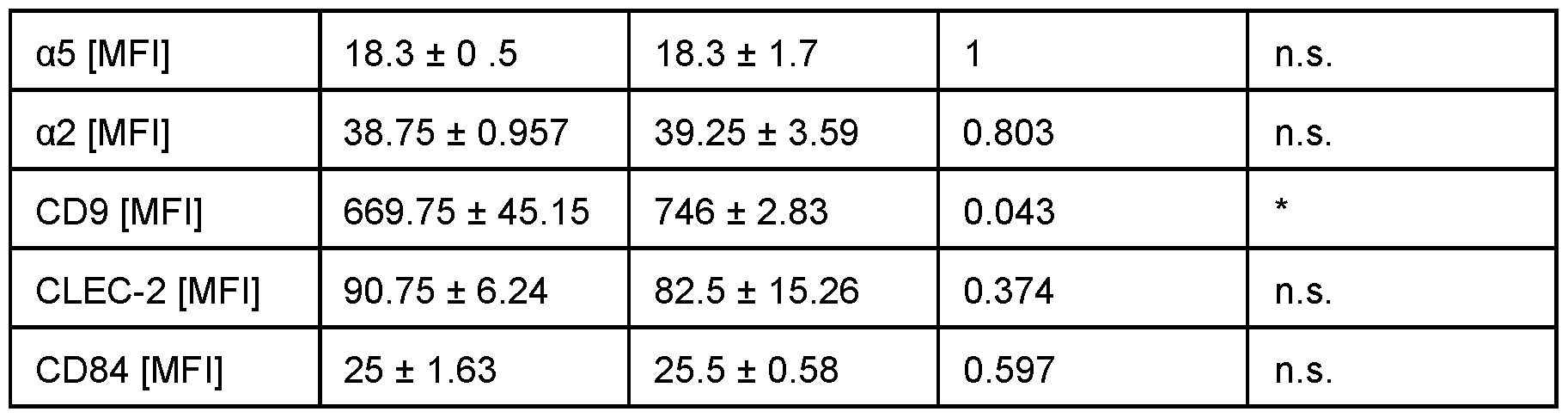

Claims
Priority Applications (3)
| Application Number | Priority Date | Filing Date | Title |
|---|---|---|---|
| AU2023408654A AU2023408654A1 (en) | 2022-12-22 | 2023-12-22 | Antibodies for use as coagulants |
| CN202380087836.7A CN120530134A (en) | 2022-12-22 | 2023-12-22 | Antibodies used as coagulants |
| EP23840946.0A EP4637920A1 (en) | 2022-12-22 | 2023-12-22 | Antibodies for use as coagulants |
Applications Claiming Priority (2)
| Application Number | Priority Date | Filing Date | Title |
|---|---|---|---|
| EP22216237 | 2022-12-22 | ||
| EP22216237.2 | 2022-12-22 |
Publications (1)
| Publication Number | Publication Date |
|---|---|
| WO2024133858A1 true WO2024133858A1 (en) | 2024-06-27 |
Family
ID=84569831
Family Applications (1)
| Application Number | Title | Priority Date | Filing Date |
|---|---|---|---|
| PCT/EP2023/087547 Ceased WO2024133858A1 (en) | 2022-12-22 | 2023-12-22 | Antibodies for use as coagulants |
Country Status (4)
| Country | Link |
|---|---|
| EP (1) | EP4637920A1 (en) |
| CN (1) | CN120530134A (en) |
| AU (1) | AU2023408654A1 (en) |
| WO (1) | WO2024133858A1 (en) |
Citations (41)
| Publication number | Priority date | Publication date | Assignee | Title |
|---|---|---|---|---|
| US4444887A (en) | 1979-12-10 | 1984-04-24 | Sloan-Kettering Institute | Process for making human antibody producing B-lymphocytes |
| WO1986001533A1 (en) | 1984-09-03 | 1986-03-13 | Celltech Limited | Production of chimeric antibodies |
| EP0239400A2 (en) | 1986-03-27 | 1987-09-30 | Medical Research Council | Recombinant antibodies and methods for their production |
| US4716111A (en) | 1982-08-11 | 1987-12-29 | Trustees Of Boston University | Process for producing human antibodies |
| US4816397A (en) | 1983-03-25 | 1989-03-28 | Celltech, Limited | Multichain polypeptides or proteins and processes for their production |
| US4816567A (en) | 1983-04-08 | 1989-03-28 | Genentech, Inc. | Recombinant immunoglobin preparations |
| EP0392745A2 (en) | 1989-04-05 | 1990-10-17 | Celltech Limited | Immunoconjugates and prodrugs and their use in association for drug delivery |
| WO1991009967A1 (en) | 1989-12-21 | 1991-07-11 | Celltech Limited | Humanised antibodies |
| WO1991010741A1 (en) | 1990-01-12 | 1991-07-25 | Cell Genesys, Inc. | Generation of xenogeneic antibodies |
| WO1992001047A1 (en) | 1990-07-10 | 1992-01-23 | Cambridge Antibody Technology Limited | Methods for producing members of specific binding pairs |
| EP0519596A1 (en) | 1991-05-17 | 1992-12-23 | Merck & Co. Inc. | A method for reducing the immunogenicity of antibody variable domains |
| US5219996A (en) | 1987-09-04 | 1993-06-15 | Celltech Limited | Recombinant antibodies and methods for their production in which surface residues are altered to cysteine residues for attachment of effector or receptor molecules |
| US5225539A (en) | 1986-03-27 | 1993-07-06 | Medical Research Council | Recombinant altered antibodies and methods of making altered antibodies |
| EP0592106A1 (en) | 1992-09-09 | 1994-04-13 | Immunogen Inc | Resurfacing of rodent antibodies |
| US5413923A (en) | 1989-07-25 | 1995-05-09 | Cell Genesys, Inc. | Homologous recombination for universal donor cells and chimeric mammalian hosts |
| US5530101A (en) | 1988-12-28 | 1996-06-25 | Protein Design Labs, Inc. | Humanized immunoglobulins |
| US5545806A (en) | 1990-08-29 | 1996-08-13 | Genpharm International, Inc. | Ransgenic non-human animals for producing heterologous antibodies |
| US5565332A (en) | 1991-09-23 | 1996-10-15 | Medical Research Council | Production of chimeric antibodies - a combinatorial approach |
| US5569825A (en) | 1990-08-29 | 1996-10-29 | Genpharm International | Transgenic non-human animals capable of producing heterologous antibodies of various isotypes |
| WO1996034096A1 (en) | 1995-04-28 | 1996-10-31 | Abgenix, Inc. | Human antibodies derived from immunized xenomice |
| WO1996033735A1 (en) | 1995-04-27 | 1996-10-31 | Abgenix, Inc. | Human antibodies derived from immunized xenomice |
| US5625126A (en) | 1990-08-29 | 1997-04-29 | Genpharm International, Inc. | Transgenic non-human animals for producing heterologous antibodies |
| US5633425A (en) | 1990-08-29 | 1997-05-27 | Genpharm International, Inc. | Transgenic non-human animals capable of producing heterologous antibodies |
| US5658570A (en) | 1991-07-25 | 1997-08-19 | Idec Pharmaceuticals Corporation | Recombinant antibodies for human therapy |
| US5661016A (en) | 1990-08-29 | 1997-08-26 | Genpharm International Inc. | Transgenic non-human animals capable of producing heterologous antibodies of various isotypes |
| WO1998016654A1 (en) | 1996-10-11 | 1998-04-23 | Japan Tobacco, Inc. | Production of a multimeric protein by cell fusion method |
| WO1998024893A2 (en) | 1996-12-03 | 1998-06-11 | Abgenix, Inc. | TRANSGENIC MAMMALS HAVING HUMAN IG LOCI INCLUDING PLURAL VH AND Vλ REGIONS AND ANTIBODIES PRODUCED THEREFROM |
| US5807715A (en) | 1984-08-27 | 1998-09-15 | The Board Of Trustees Of The Leland Stanford Junior University | Methods and transformed mammalian lymphocyte cells for producing functional antigen-binding protein including chimeric immunoglobulin |
| US5814318A (en) | 1990-08-29 | 1998-09-29 | Genpharm International Inc. | Transgenic non-human animals for producing heterologous antibodies |
| WO1998046645A2 (en) | 1997-04-14 | 1998-10-22 | Micromet Gesellschaft Für Biomedizinische Forschung Mbh | Method for the production of antihuman antigen receptors and uses thereof |
| WO1998050433A2 (en) | 1997-05-05 | 1998-11-12 | Abgenix, Inc. | Human monoclonal antibodies to epidermal growth factor receptor |
| US5885793A (en) | 1991-12-02 | 1999-03-23 | Medical Research Council | Production of anti-self antibodies from antibody segment repertoires and displayed on phage |
| EP0948544A1 (en) | 1996-12-10 | 1999-10-13 | Celltech Therapeutics Limited | Monovalent antibody fragments |
| WO2005117984A2 (en) | 2004-06-01 | 2005-12-15 | Celltech R & D Limited | Albumin-binding conjugates comprising a fatty acid and peg |
| WO2005123780A2 (en) | 2004-04-09 | 2005-12-29 | Protein Design Labs, Inc. | Alteration of fcrn binding affinities or serum half-lives of antibodies by mutagenesis |
| US7217797B2 (en) | 2002-10-15 | 2007-05-15 | Pdl Biopharma, Inc. | Alteration of FcRn binding affinities or serum half-lives of antibodies by mutagenesis |
| US7217798B2 (en) | 2003-10-15 | 2007-05-15 | Pdl Biopharma, Inc. | Alteration of Fc-fusion protein serum half-lives by mutagenesis |
| US20070280931A1 (en) | 1998-11-18 | 2007-12-06 | Chen Yvonne M | Antibody variants |
| US7361740B2 (en) | 2002-10-15 | 2008-04-22 | Pdl Biopharma, Inc. | Alteration of FcRn binding affinities or serum half-lives of antibodies by mutagenesis |
| US7365168B2 (en) | 2002-10-15 | 2008-04-29 | Pdl Biopharma, Inc. | Alteration of FcRn binding affinities or serum half-lives of antibodies by mutagenesis |
| WO2017109180A1 (en) | 2015-12-23 | 2017-06-29 | Julius-Maximilians-Universität Würzburg | Glycoprotein v inhibitors for use as coagulants |
-
2023
- 2023-12-22 EP EP23840946.0A patent/EP4637920A1/en active Pending
- 2023-12-22 CN CN202380087836.7A patent/CN120530134A/en active Pending
- 2023-12-22 WO PCT/EP2023/087547 patent/WO2024133858A1/en not_active Ceased
- 2023-12-22 AU AU2023408654A patent/AU2023408654A1/en active Pending
Patent Citations (49)
| Publication number | Priority date | Publication date | Assignee | Title |
|---|---|---|---|---|
| US4444887A (en) | 1979-12-10 | 1984-04-24 | Sloan-Kettering Institute | Process for making human antibody producing B-lymphocytes |
| US4716111A (en) | 1982-08-11 | 1987-12-29 | Trustees Of Boston University | Process for producing human antibodies |
| US4816397A (en) | 1983-03-25 | 1989-03-28 | Celltech, Limited | Multichain polypeptides or proteins and processes for their production |
| US4816567A (en) | 1983-04-08 | 1989-03-28 | Genentech, Inc. | Recombinant immunoglobin preparations |
| US5807715A (en) | 1984-08-27 | 1998-09-15 | The Board Of Trustees Of The Leland Stanford Junior University | Methods and transformed mammalian lymphocyte cells for producing functional antigen-binding protein including chimeric immunoglobulin |
| WO1986001533A1 (en) | 1984-09-03 | 1986-03-13 | Celltech Limited | Production of chimeric antibodies |
| EP0239400A2 (en) | 1986-03-27 | 1987-09-30 | Medical Research Council | Recombinant antibodies and methods for their production |
| US5225539A (en) | 1986-03-27 | 1993-07-06 | Medical Research Council | Recombinant altered antibodies and methods of making altered antibodies |
| US5219996A (en) | 1987-09-04 | 1993-06-15 | Celltech Limited | Recombinant antibodies and methods for their production in which surface residues are altered to cysteine residues for attachment of effector or receptor molecules |
| US5693761A (en) | 1988-12-28 | 1997-12-02 | Protein Design Labs, Inc. | Polynucleotides encoding improved humanized immunoglobulins |
| US5585089A (en) | 1988-12-28 | 1996-12-17 | Protein Design Labs, Inc. | Humanized immunoglobulins |
| US5693762A (en) | 1988-12-28 | 1997-12-02 | Protein Design Labs, Inc. | Humanized immunoglobulins |
| US5530101A (en) | 1988-12-28 | 1996-06-25 | Protein Design Labs, Inc. | Humanized immunoglobulins |
| US6180370B1 (en) | 1988-12-28 | 2001-01-30 | Protein Design Labs, Inc. | Humanized immunoglobulins and methods of making the same |
| EP0392745A2 (en) | 1989-04-05 | 1990-10-17 | Celltech Limited | Immunoconjugates and prodrugs and their use in association for drug delivery |
| US5413923A (en) | 1989-07-25 | 1995-05-09 | Cell Genesys, Inc. | Homologous recombination for universal donor cells and chimeric mammalian hosts |
| WO1991009967A1 (en) | 1989-12-21 | 1991-07-11 | Celltech Limited | Humanised antibodies |
| US5939598A (en) | 1990-01-12 | 1999-08-17 | Abgenix, Inc. | Method of making transgenic mice lacking endogenous heavy chains |
| WO1991010741A1 (en) | 1990-01-12 | 1991-07-25 | Cell Genesys, Inc. | Generation of xenogeneic antibodies |
| WO1992001047A1 (en) | 1990-07-10 | 1992-01-23 | Cambridge Antibody Technology Limited | Methods for producing members of specific binding pairs |
| US5545806A (en) | 1990-08-29 | 1996-08-13 | Genpharm International, Inc. | Ransgenic non-human animals for producing heterologous antibodies |
| US5569825A (en) | 1990-08-29 | 1996-10-29 | Genpharm International | Transgenic non-human animals capable of producing heterologous antibodies of various isotypes |
| US5814318A (en) | 1990-08-29 | 1998-09-29 | Genpharm International Inc. | Transgenic non-human animals for producing heterologous antibodies |
| US5625126A (en) | 1990-08-29 | 1997-04-29 | Genpharm International, Inc. | Transgenic non-human animals for producing heterologous antibodies |
| US5633425A (en) | 1990-08-29 | 1997-05-27 | Genpharm International, Inc. | Transgenic non-human animals capable of producing heterologous antibodies |
| US5661016A (en) | 1990-08-29 | 1997-08-26 | Genpharm International Inc. | Transgenic non-human animals capable of producing heterologous antibodies of various isotypes |
| EP0519596A1 (en) | 1991-05-17 | 1992-12-23 | Merck & Co. Inc. | A method for reducing the immunogenicity of antibody variable domains |
| US5658570A (en) | 1991-07-25 | 1997-08-19 | Idec Pharmaceuticals Corporation | Recombinant antibodies for human therapy |
| US5693780A (en) | 1991-07-25 | 1997-12-02 | Idec Pharmaceuticals Corporation | Recombinant antibodies for human therapy |
| US5681722A (en) | 1991-07-25 | 1997-10-28 | Idec Pharmaceuticals Corporation | Recombinant antibodies for human therapy |
| US5565332A (en) | 1991-09-23 | 1996-10-15 | Medical Research Council | Production of chimeric antibodies - a combinatorial approach |
| US5885793A (en) | 1991-12-02 | 1999-03-23 | Medical Research Council | Production of anti-self antibodies from antibody segment repertoires and displayed on phage |
| EP0592106A1 (en) | 1992-09-09 | 1994-04-13 | Immunogen Inc | Resurfacing of rodent antibodies |
| WO1996033735A1 (en) | 1995-04-27 | 1996-10-31 | Abgenix, Inc. | Human antibodies derived from immunized xenomice |
| WO1996034096A1 (en) | 1995-04-28 | 1996-10-31 | Abgenix, Inc. | Human antibodies derived from immunized xenomice |
| US5916771A (en) | 1996-10-11 | 1999-06-29 | Abgenix, Inc. | Production of a multimeric protein by cell fusion method |
| WO1998016654A1 (en) | 1996-10-11 | 1998-04-23 | Japan Tobacco, Inc. | Production of a multimeric protein by cell fusion method |
| WO1998024893A2 (en) | 1996-12-03 | 1998-06-11 | Abgenix, Inc. | TRANSGENIC MAMMALS HAVING HUMAN IG LOCI INCLUDING PLURAL VH AND Vλ REGIONS AND ANTIBODIES PRODUCED THEREFROM |
| EP0948544A1 (en) | 1996-12-10 | 1999-10-13 | Celltech Therapeutics Limited | Monovalent antibody fragments |
| WO1998046645A2 (en) | 1997-04-14 | 1998-10-22 | Micromet Gesellschaft Für Biomedizinische Forschung Mbh | Method for the production of antihuman antigen receptors and uses thereof |
| WO1998050433A2 (en) | 1997-05-05 | 1998-11-12 | Abgenix, Inc. | Human monoclonal antibodies to epidermal growth factor receptor |
| US20070280931A1 (en) | 1998-11-18 | 2007-12-06 | Chen Yvonne M | Antibody variants |
| US7365168B2 (en) | 2002-10-15 | 2008-04-29 | Pdl Biopharma, Inc. | Alteration of FcRn binding affinities or serum half-lives of antibodies by mutagenesis |
| US7361740B2 (en) | 2002-10-15 | 2008-04-22 | Pdl Biopharma, Inc. | Alteration of FcRn binding affinities or serum half-lives of antibodies by mutagenesis |
| US7217797B2 (en) | 2002-10-15 | 2007-05-15 | Pdl Biopharma, Inc. | Alteration of FcRn binding affinities or serum half-lives of antibodies by mutagenesis |
| US7217798B2 (en) | 2003-10-15 | 2007-05-15 | Pdl Biopharma, Inc. | Alteration of Fc-fusion protein serum half-lives by mutagenesis |
| WO2005123780A2 (en) | 2004-04-09 | 2005-12-29 | Protein Design Labs, Inc. | Alteration of fcrn binding affinities or serum half-lives of antibodies by mutagenesis |
| WO2005117984A2 (en) | 2004-06-01 | 2005-12-15 | Celltech R & D Limited | Albumin-binding conjugates comprising a fatty acid and peg |
| WO2017109180A1 (en) | 2015-12-23 | 2017-06-29 | Julius-Maximilians-Universität Würzburg | Glycoprotein v inhibitors for use as coagulants |
Non-Patent Citations (89)
| Title |
|---|
| "Bioconjugation Protein Coupling Techniques for the Biomedical Sciences", 1998, GROVE PUBLISHERS |
| "Poly(ethyleneglycol) Chemistry and Biological Applications", 1997, AMERICAN CHEMICAL SOCIETY |
| "Poly(ethyleneglycol) Chemistry, Biotechnical and Biomedical Applications", 1992, PLENUM PRESS |
| "Remington's Pharmaceutical Sciences", 1980 |
| AMICH, J. ET AL.: "Three-dimensional light sheet fluorescence microscopy of lungs to dissect local host immune-aspergillus fumigatus interactions.", MBIO, vol. 11, no. 1, 2020 |
| ASTER, R. H.BOUGIE, D. W.: "Drug-induced immune thrombocytopenia", N. ENGL. J. MED., vol. 357, no. 6, 2007, pages 580 - 587 |
| AZORSA DO ET AL.: "Measurement of GPV Released by Activated Platelets Using a Sensitive Immunocapture ELISA - Its Use to Follow Platelet Storage in Transfusion", THROMBOSIS AND HAEMOSTASIS., vol. 81, no. 1, 1999, pages 131 - 8, XP002754916 |
| BAKCHOUL, T.MARINI, I.: "Drug-associated thrombocytopenia", HEMATOLOGY AM. SOC. HEMATOL. EDUC. PROGRAM, 2018, pages 576 - 583 |
| BENDER, M. ET AL.: "Combined in vivo depletion of glycoprotein VI and C-type lectin-like receptor 2 severely compromises hemostasis and abrogates arterial thrombosis in mice.", ARTERIOSCLER. THROMB. VASC. BIOL., vol. 33, no. 5, 2013, pages 926 - 934 |
| BERGMEIER, W. ET AL.: "Flow cytometric detection of activated mouse integrin alphallbbeta3 with a novel monoclonal antibody", CYTOMETRY, vol. 48, no. 2, 2002, pages 80 - 86, XP009003714, DOI: 10.1002/cyto.10114 |
| BOCK, L. C.GRIFFIN, L. C.LATHAM, J. A.VERMAAS, E. H.TOOLE, J. J.: "Selection of single-stranded DNA molecules that bind and inhibit human thrombin.", NATURE, vol. 355, no. 6360, 1992, pages 564 - 566, XP000453533, DOI: 10.1038/355564a0 |
| BROUNS, S. L. N. ET AL.: "Platelet-primed interactions of coagulation and anticoagulation pathways in flow-dependent thrombus formation", SCI. REP., vol. 10, 2020, pages 11910 |
| CANFIELDMORRISON: "The Binding Affinity of Human IgG for its High Affinity Fc Receptor Is Determined by Multiple Amino Acids in the CH2 Domain and Is Modulated by the Hinge Region", J EXP MED., vol. 173, no. 6, 1991, pages 1483 - 1491 |
| CARSTAIRS, K. C.: "The identification of platelets and platelet antigens in histological sections", J. PATHOL. BACTERIAL., vol. 90, no. 1, 1965, pages 225 - 231 |
| CELIKEL, R. ET AL.: "Modulation of a-thrombin function by distinct interactions with platelet glycoprotein Iba", SCIENCE, vol. 301, no. 5630, 2003, pages 218 - 221, XP055134653, DOI: 10.1126/science.1084183 |
| CHAPMAN: "PEGylated antibodies and antibody fragments for improved therapy: a review", ADVANCED DRUG DELIVERY REVIEWS, vol. 54, no. 4, 2002, pages 531 - 545, XP001199533, DOI: 10.1016/S0169-409X(02)00026-1 |
| CLEMETSON, K. J.: "A short history of platelet glycoprotein Ib complex.", THROMB. HAEMOST., vol. 98, no. 1, 2007, pages 63 - 68 |
| DE WITT, S. M. ET AL.: "Identification of platelet function defects by multi-parameter assessment of thrombus formation", NAT COMMUN, vol. 5, 2014, pages 4257 |
| DEPPERMANN, C. ET AL.: "Gray platelet syndrome and defective thrombo-inflammation in Nbeal2-deficient mice", J. CLIN. INVEST., vol. 123, no. 8, 2013, pages 3331 - 3342, XP055322000, DOI: 10.1172/JCI69210 |
| DUBOWCHIK ET AL.: "Receptor-mediated and enzyme-dependent targeting of cytotoxic anticancer drugs", PHARMACOLOGY AND THERAPEUTICS, vol. 83, no. 2, 1999, pages 67 - 123, XP002391774, DOI: 10.1016/S0163-7258(99)00018-2 |
| DUMAS, J. J.KUMAR, R.SEEHRA, J.SOMERS, W. S.MOSYAK, L.: "Crystal structure of the Gplbalpha-thrombin complex essential for platelet aggregation", SCIENCE, vol. 301, no. 5630, 2003, pages 222 - 226 |
| ESTEVEZ, B. ET AL.: "Signaling-mediated cooperativity between glycoprotein Ib-IX and protease-activated receptors in thrombin-induced platelet activation", BLOOD, vol. 127, no. 5, 2016, pages 626 - 636, XP086694107, DOI: 10.1182/blood-2015-04-638387 |
| FREDENBURGH, J. C.STAFFORD, A. R.LESLIE, B. A.WEITZ, J. I.: "Bivalent binding to gammaA/gamma'-fibrin engages both exosites of thrombin and protects it from inhibition by the antithrombin-heparin complex", J. BIOL. CHEM., vol. 283, no. 5, 2008, pages 2470 - 2477 |
| FURIE, B.FURIE, B. C.: "The molecular basis of blood coagulation.", CELL, vol. 53, no. 4, 1988, pages 505 - 518, XP023908863, DOI: 10.1016/0092-8674(88)90567-3 |
| GILLIES ET AL.: "High-level expression of chimeric antibodies using adapted cDNA variable region cassettes", J. IMMUNOL. METHODS, vol. 125, no. 1-2, 1989, pages 191 - 202, XP023973835, DOI: 10.1016/0022-1759(89)90093-8 |
| GRUNER, S. ET AL.: "Anti-glycoprotein VI treatment severely compromises hemostasis in mice with reduced a201 levels or concomitant aspirin therapy", CIRCULATION, vol. 110, no. 18, 2004, pages 2946 - 2951, XP002486447, DOI: 10.1161/01.CIR.0000146341.63677.3C |
| HELLSTROM ET AL.: "Antibodies for Drug Delivery. Controlled Drug Delivery", 1987, pages: 623 - 53 |
| HEMKER, H. C. ET AL.: "Calibrated automated thrombin generation measurement in clotting plasma", PATHOPHYSIOL. HAEMOST. THROMB., vol. 33, no. 1, 2003, pages 4 - 15, XP008053802, DOI: 10.1159/000071636 |
| HEMKER, H. C.BEGUIN, S.: "Thrombin generation in plasma: its assessment via the endogenous thrombin potential", THROMB. HAEMOST., vol. 74, no. 1, 1995, pages 134 - 138, XP000195941 |
| HOLTKOTTER, O. ET AL.: "Integrin alpha 2-deficient mice develop normally, are fertile, but display partially defective platelet interaction with collagen.", J. BIOL. CHEM., vol. 277, no. 13, 2002, pages 10789 - 10794 |
| JACKSON, B. ET AL.: "RhoA is dispensable for skin development, but crucial for contraction and directed migration of keratinocytes", MOL. BIOL. CELL, vol. 22, no. 5, 2011, pages 593 - 605 |
| JESPERS ET AL.: "Guiding the Selection of Human Antibodies from Phage Display Repertories to a Single Epitope of an Antigen", NATURE BIOTECHNOLOGY, vol. 12, no. 9, 1994, pages 899 - 903 |
| KAHN, M. L. ET AL.: "A dual thrombin receptor system for platelet activation", NATURE, vol. 394, no. 6694, 1998, pages 690 - 694, XP002112287, DOI: 10.1038/29325 |
| KAHN, M. L. ET AL.: "Glycoprotein V-deficient platelets have undiminished thrombin responsiveness and do not exhibit a Bernard-Soulier phenotype", BLOOD, vol. 94, no. 12, 1999, pages 4112 - 4121, XP002212818 |
| KATSUTANI, S. ET AL.: "Cloning and characterization of the gene encoding the murine glycoprotein V: the conserved thrombin-cleavable protein on platelet surface", THROMB. RES., vol. 92, no. 1, 1998, pages 43 - 51 |
| KLEINSCHNITZ, C. ET AL.: "Targeting platelets in acute experimental stroke: impact of glycoprotein Ib, VI, and IIb/IIIa blockade on infarct size, functional outcome, and intracranial bleeding", CIRCULATION, vol. 115, no. 17, 2007, pages 2323 - 2330, XP002619576, DOI: 10.1161/CIRCULATIONAHA.107.691279 |
| KNIGHT, C. G. ET AL.: "Collagen-platelet interaction: Gly-Pro-Hyp is uniquely specific for platelet Gp VI and mediates platelet activation by collagen", CARDIOVASC. RES., vol. 41, no. 2, 1999, pages 450 - 457, XP002425787, DOI: 10.1016/S0008-6363(98)00306-X |
| LI, R.EMSLEY, J.: "The organizing principle of the platelet glycoprotein Ib-IX-V complex.", J. THROMB. HAEMOST., vol. 11, no. 4, 2013, pages 605 - 614 |
| LUND ET AL.: "Human Fc gamma RI and FC gamma RII interact with distinct but overlapping sites on human IgG", J IMMUNOL., vol. 147, no. 8, 1991, pages 2657 - 2662, XP009546324, DOI: 10.4049/jimmunol.147.8.2657 |
| MANGIN, P. ET AL.: "Thrombin overcomes the thrombosis defect associated with platelet GPVI/FcRy deficiency", BLOOD, vol. 107, no. 11, 2006, pages 4346 - 4353, XP055838356, DOI: 10.1182/blood-2005-10-4244 |
| MANGIN, P. H. ET AL.: "Immobilized fibrinogen activates human platelets through glycoprotein VI", HAEMATOLOGICA, vol. 103, no. 5, 2018, pages 898 - 907 |
| MASSBERG, S. ET AL.: "A crucial role of glycoprotein VI for platelet recruitment to the injured arterial wall in vivo.", J. EXP. MED., vol. 197, no. 1, 2003, pages 41 - 49, XP002329121, DOI: 10.1084/jem.20020945 |
| MAST, A. E. & RUF, W.: "Regulation of coagulation by tissue factor pathway inhibitor: Implications for haemophilia therapy.", J. THROMB. HAEMOST., vol. 20, no. 6, 2022, pages 1290 - 1300 |
| MCGOWAN, E. B.DING, A.DETWILER, T. C.: "Correlation of thrombin-induced glycoprotein V hydrolysis and platelet activation", J. BIOL. CHEM., vol. 258, no. 18, 1983, pages 11243 - 11248 |
| MO, X.LIU, L.LOPEZ, J. A.LI, R.: "Transmembrane domains are critical to the interaction between platelet glycoprotein V and glycoprotein Ib-IX complex", J. THROMB. HAEMOST., vol. 10, no. 9, 2012, pages 1875 - 1886 |
| MOOG, S. ET AL.: "Platelet glycoprotein V binds to collagen and participates in platelet adhesion and aggregation", BLOOD, vol. 98, no. 4, 2001, pages 1038 - 1046, XP055048230, DOI: 10.1182/blood.V98.4.1038 |
| MOROWSKI, M. ET AL.: "Only severe thrombocytopenia results in bleeding and defective thrombus formation in mice", BLOOD, vol. 121, no. 24, 2013, pages 4938 - 4947 |
| MORRISON, TRANSFECTOMAS PROVIDE NOVEL CHIMERIC ANTIBODIES SCIENCE, vol. 229, no. 4719, 1985, pages 1202 - 7 |
| MOSNIER, L. O.ZLOKOVIC, B. V.GRIFFIN, J. H.: "The cytoprotective protein C pathway.", BLOOD, vol. 109, no. 8, 2007, pages 3161 - 3172 |
| NAGY, M. ET AL.: "Comparative analysis of microfluidics thrombus formation in multiple genetically modified mice: link to thrombosis and hemostasis.", FRONT CARDIOVASC MED, vol. 6, 2019, pages 99 |
| NI, H.: "Increased thrombogenesis and embolus formation in mice lacking glycoprotein V", BLOOD, vol. 98, no. 2, 2001, pages 368 - 373, XP086704749, DOI: 10.1182/blood.V98.2.368 |
| NIESWANDT BERNHARD ET AL: "Identification of critical antigen-specific mechanisms in the development of immune thrombocytopenic purpura in mice", vol. 96, no. 7, 1 October 2000 (2000-10-01), US, pages 2520 - 2527, XP093046297, ISSN: 0006-4971, Retrieved from the Internet <URL:http://ashpublications.org/blood/article-pdf/96/7/2520/1668048/h8190002520.pdf> DOI: 10.1182/blood.V96.7.2520 * |
| NIESWANDT, B. ET AL.: "Acute systemic reaction and lung alterations induced by an antiplatelet integrin GPIIb/IIIa antibody in mice", BLOOD, vol. 94, no. 2, 1999, pages 684 - 693 |
| NIESWANDT, B. ET AL.: "Long-term antithrombotic protection by in vivo depletion of platelet glycoprotein VI in mice.", J. EXP. MED., vol. 193, no. 4, 2001, pages 459 - 469, XP002174652, DOI: 10.1084/jem.193.4.459 |
| NIESWANDT, B.BERGMEIER, W.RACKEBRANDT, K.GESSNER, J. E.ZIRNGIBL, H.: "Identification of critical antigen-specific mechanisms in the development of immune thrombocytopenic purpura in mice", BLOOD, vol. 96, no. 7, 2000, pages 2520 - 2527, XP093046297, DOI: 10.1182/blood.V96.7.2520 |
| NIESWANDT, B.WATSON, S. P.: "Platelet-collagen interaction: is GPVI the central receptor?", BLOOD, vol. 102, no. 2, 2003, pages 449 - 461, XP007911704 |
| NONNE, C., HECHLER, B., CAZENAVE, J. P., GACHET, C. & LANZA, F.: "Reassessment of in vivo thrombus formation in glycoprotein V deficient mice backcrossed on a C57B1/6 strain.", J. THROMB. HAEMOST., vol. 6, no. 1, 2008, pages 210 - 212 |
| OI ET AL., CHIMERIC ANTIBODIES BIOTECHNIQUES., vol. 4, no. 3, 1986, pages 214 - 221 |
| PADLAN: "A possible procedure for reducing the immunogenicity of antibody variable domains while preserving their ligand-binding properties", MOL IMMUNOL., vol. 28, no. 4-5, 1991, pages 489 - 498, XP023795271, DOI: 10.1016/0161-5890(91)90163-E |
| PETRERA, N. S. ET AL.: "Long range communication between exosites 1 and 2 modulates thrombin function", J. BIOL. CHEM., vol. 284, no. 38, 2009, pages 25620 - 25629 |
| PLEINES, I. ET AL.: "Megakaryocyte-specific RhoA deficiency causes macrothrombocytopenia and defective platelet activation in hemostasis and thrombosis.", BLOOD, vol. 119, no. 4, 2012, pages 1054 - 1063 |
| RABIE, T.STREHL, A.LUDWIG, A.NIESWANDT, B.: "Evidence for a role of ADAM17 (TACE) in the regulation of platelet glycoprotein V", J. BIOL. CHEM., vol. 280, no. 15, 2005, pages 14462 - 14468, XP055139629, DOI: 10.1074/jbc.M500041200 |
| RAMAKRISHNAN, V. ET AL.: "Increased thrombin responsiveness in platelets from mice lacking glycoprotein V.", PROC. NATL. ACAD. SCI. U. S. A., vol. 96, no. 23, 1999, pages 13336 - 13341, XP000869655, DOI: 10.1073/pnas.96.23.13336 |
| RAVANAT, C. ET AL.: "Gene cloning of rat and mouse platelet glycoprotein V: identification of megakaryocyte-specific promoters and demonstration of functional thrombin cleavage", BLOOD, vol. 89, no. 9, 1997, pages 3253 - 3262 |
| RIECHMANN ET AL.: "Reshaping human antibodies for therapy", NATURE, vol. 332, no. 6162, 1988, pages 323 - 7, XP002007067, DOI: 10.1038/332323a0 |
| ROGUSKA ET AL.: "Humanization of murine monoclonal antibodies through variable domain resurfacing", PNAS, vol. 91, no. 3, 1994, pages 969 - 973, XP002271704, DOI: 10.1073/pnas.91.3.969 |
| RUGGERI, Z. M. ET AL.: "Unravelling the mechanism and significance of thrombin binding to platelet glycoprotein Ib", THROMB. HAEMOST., vol. 104, no. 5, 2010, pages 894 - 902 |
| RYU, J. K. ET AL.: "Fibrin-targeting immunotherapy protects against neuroinflammation and neurodegeneration", NAT. IMMUNOL., vol. 19, 2018, pages 1212 - 1223, XP036617636, DOI: 10.1038/s41590-018-0232-x |
| S. MOOG: "Platelet glycoprotein V binds to collagen and participates in platelet adhesion and aggregation", BLOOD, vol. 98, no. 4, 15 August 2001 (2001-08-15), pages 1038 - 1046, XP055048230, ISSN: 0006-4971, DOI: 10.1182/blood.V98.4.1038 * |
| SANG, Y.ROEST, M.DE LAAT, B.DE GROOT, P. G.HUSKENS, D.: "Interplay between platelets and coagulation", BLOOD REV., vol. 46, 2021, pages 100733 |
| SCHINDELIN, J. ET AL.: "Fiji: an open-source platform for biological-image analysis", NAT. METHODS, vol. 9, no. 7, 2012, pages 676 - 682, XP055343835, DOI: 10.1038/nmeth.2019 |
| SCHULTE, V. ET AL.: "Targeting of the collagen-binding site on glycoprotein VI is not essential for in vivo depletion of the receptor", BLOOD, vol. 101, no. 10, 2003, pages 3948 - 3952 |
| SEKHON, U. D. S. ET AL.: "Platelet-mimicking procoagulant nanoparticles augment hemostasis in animal models of bleeding", SCI. TRANSL. MED., vol. 14, 2022, pages eabb8975 |
| SHIDA, Y. ET AL.: "Analysis of the role of von Willebrand factor, platelet glycoprotein VI- , and α2β1-mediated collagen binding in thrombus formation", BLOOD, vol. 124, no. 11, 2014, pages 1799 - 1807, XP086510409, DOI: 10.1182/blood-2013-09-521484 |
| SILVA, L. M. ET AL.: "Fibrin is a critical regulator of neutrophil effector function at the oral mucosal barrier", SCIENCE, vol. 374, no. 6575, 2021, pages eabl5450 |
| STALKER, T. J. ET AL.: "Hierarchical organization in the hemostatic response and its relationship to the platelet-signaling network.", BLOOD, vol. 121, no. 10, 2013, pages 1875 - 1885 |
| STEGNER, D. ET AL.: "Foudroyant cerebral venous (sinus) thrombosis triggered through CLEC-2 and GPIIb/IIIa dependent platelet activation", NATURE CARDIOVASCULAR RESEARCH, vol. 1, no. 2, 2022, pages 132 - 141 |
| STEGNER, D. ET AL.: "Thrombopoiesis is spatially regulated by the bone marrow vasculature", NAT COMMUN, vol. 8, 2017, pages 127 |
| STOLL, G.NIESWANDT, B.: "Thrombo-inflammation in acute ischaemic stroke - implications for treatment", NAT. REV. NEUROL., vol. 15, no. 8, 2019, pages 473 - 481, XP036847197, DOI: 10.1038/s41582-019-0221-1 |
| STUDNICKA ET AL.: "Human-engineered monoclonal antibodies retain full specific binding activity by preserving non-CDR complementaritymodulating residues", PROT ENG., vol. 7, no. 6, 1994, pages 805 - 814, XP009166823, DOI: 10.1093/protein/7.6.805 |
| SWIERINGA, F.SPRONK, H. M. H.HEEMSKERK, J. W. M.VAN DER MEIJDEN, P. E. J.: "Integrating platelet and coagulation activation in fibrin clot formation", RES PRACT THROMB HAEMOST, vol. 2, no. 3, 2018, pages 450 - 460 |
| TASSET, D. M.KUBIK, M. F.STEINER, W.: "Oligonucleotide inhibitors of human thrombin that bind distinct epitopes", J. MOL. BIOL., vol. 272, no. 5, 1997, pages 688 - 698, XP004453692, DOI: 10.1006/jmbi.1997.1275 |
| TATUSOVA ET AL.: "BLAST 2 SEQUENCES, a new tool for comparing protein and nucleotide sequences", FEMS MICROBIOL. LETT., vol. 174, 1999, pages 247 - 250 |
| THORPE ET AL.: "The Preparation and Cytotoxic Properties of Antibody-Toxin Conjugates", IMMUNOL REV., vol. 62, no. 1, 1982, pages 119 - 58, XP001179872, DOI: 10.1111/j.1600-065X.1982.tb00392.x |
| TOMAIUOLO, M.BRASS, L. F.STALKER, T. J.: "Regulation of platelet activation and coagulation and its role in vascular injury and arterial thrombosis", INTERV CARDIOL CLIN, vol. 6, no. 1, 2017, pages 1 - 12 |
| VAN DER WALT, S. ET AL.: "scikit-image: image processing in Python", PEERJ, vol. 2, 2014, pages e453 |
| VERSTEEG, H. H.HEEMSKERK, J. W.LEVI, M.REITSMA, P. H.: "New fundamentals in hemostasis.", PHYSIOL. REV., vol. 93, no. 1, 2013, pages 327 - 358 |
| VOLLENBERG, R. ET AL.: "Glycoprotein V is a relevant immune target in patients with immune thrombocytopenia", HAEMATOLOGICA, vol. 104, no. 6, 2019, pages 1237 - 1243 |
| WAHL ET AL.: "Improved Radioimaging andTumor Localization with Monoclonal F(ab", J. NUCL. MED., vol. 24, no. 4, 1983, pages 316 - 325 |
Also Published As
| Publication number | Publication date |
|---|---|
| EP4637920A1 (en) | 2025-10-29 |
| CN120530134A (en) | 2025-08-22 |
| AU2023408654A1 (en) | 2025-06-26 |
Similar Documents
| Publication | Publication Date | Title |
|---|---|---|
| US9409988B2 (en) | Anti-Axl antibodies and uses thereof | |
| RU2588668C2 (en) | PEPTIDE OR PEPTIDE COMPLEX BINDING TO alpha2- INTEGRIN, METHODS FOR PRODUCTION AND USE THEREOF | |
| Beck et al. | Platelet glycoprotein V spatio-temporally controls fibrin formation | |
| US20220089778A1 (en) | Coagulation factor binding proteins and uses thereof | |
| CN119119209A (en) | Matrix metalloproteinase-cleavable and serine protease-cleavable substrates and methods of use thereof | |
| WO2013126810A1 (en) | Anti sez6 antibodies and methods of use | |
| CN116063479A (en) | Compositions and methods for conjugating activatable antibodies | |
| AU2006315037A1 (en) | Anti-alpha2 integrin antibodies and their uses | |
| CA2994629A1 (en) | Novel anti-human gpvi antibodies and uses thereof | |
| CN104053673A (en) | Thrombin-binding Antibody Molecules And Uses Thereof | |
| JP2023116543A (en) | Binding protein for human thrombin receptor PAR4 | |
| EP2848633B1 (en) | Anti-cxadr antibody | |
| US7645592B2 (en) | Glycoprotein VI antibodies and methods of use thereof | |
| AU2023408654A1 (en) | Antibodies for use as coagulants | |
| US20190169307A1 (en) | Glycoprotein v inhibitors for use as coagulants | |
| WO2019200357A1 (en) | Biomarker for cd47 targeting therapeutics and uses therefor | |
| WO2016167227A1 (en) | Antibody against insoluble fibrin | |
| EP4431526A1 (en) | Anti-gpvi antibodies and functional fragments thereof | |
| JP2026509897A (en) | Anti-GPVI antibody and its functional fragment | |
| JP2008249552A (en) | Measuring system for soluble platelet membrane glycoprotein vi | |
| CA2567394C (en) | Glycoprotein vi antibodies and methods of use thereof |
Legal Events
| Date | Code | Title | Description |
|---|---|---|---|
| 121 | Ep: the epo has been informed by wipo that ep was designated in this application |
Ref document number: 23840946 Country of ref document: EP Kind code of ref document: A1 |
|
| WWE | Wipo information: entry into national phase |
Ref document number: AU2023408654 Country of ref document: AU |
|
| WWE | Wipo information: entry into national phase |
Ref document number: 202380087836.7 Country of ref document: CN |
|
| ENP | Entry into the national phase |
Ref document number: 2023408654 Country of ref document: AU Date of ref document: 20231222 Kind code of ref document: A |
|
| WWE | Wipo information: entry into national phase |
Ref document number: 2023840946 Country of ref document: EP |
|
| NENP | Non-entry into the national phase |
Ref country code: DE |
|
| ENP | Entry into the national phase |
Ref document number: 2023840946 Country of ref document: EP Effective date: 20250722 |
|
| ENP | Entry into the national phase |
Ref document number: 2023840946 Country of ref document: EP Effective date: 20250722 |
|
| ENP | Entry into the national phase |
Ref document number: 2023840946 Country of ref document: EP Effective date: 20250722 |
|
| WWP | Wipo information: published in national office |
Ref document number: 202380087836.7 Country of ref document: CN |
|
| WWP | Wipo information: published in national office |
Ref document number: 2023840946 Country of ref document: EP |