WO2021075451A1 - Modèle in vitro de barrière sang/tissu, et procédé d'évaluation de transfert de médicament à travers une barrière sang/tissu - Google Patents
Modèle in vitro de barrière sang/tissu, et procédé d'évaluation de transfert de médicament à travers une barrière sang/tissu Download PDFInfo
- Publication number
- WO2021075451A1 WO2021075451A1 PCT/JP2020/038721 JP2020038721W WO2021075451A1 WO 2021075451 A1 WO2021075451 A1 WO 2021075451A1 JP 2020038721 W JP2020038721 W JP 2020038721W WO 2021075451 A1 WO2021075451 A1 WO 2021075451A1
- Authority
- WO
- WIPO (PCT)
- Prior art keywords
- drug
- blood
- model
- vitro model
- tissue barrier
- Prior art date
- Legal status (The legal status is an assumption and is not a legal conclusion. Google has not performed a legal analysis and makes no representation as to the accuracy of the status listed.)
- Ceased
Links
Images


Classifications
-
- C—CHEMISTRY; METALLURGY
- C12—BIOCHEMISTRY; BEER; SPIRITS; WINE; VINEGAR; MICROBIOLOGY; ENZYMOLOGY; MUTATION OR GENETIC ENGINEERING
- C12Q—MEASURING OR TESTING PROCESSES INVOLVING ENZYMES, NUCLEIC ACIDS OR MICROORGANISMS; COMPOSITIONS OR TEST PAPERS THEREFOR; PROCESSES OF PREPARING SUCH COMPOSITIONS; CONDITION-RESPONSIVE CONTROL IN MICROBIOLOGICAL OR ENZYMOLOGICAL PROCESSES
- C12Q1/00—Measuring or testing processes involving enzymes, nucleic acids or microorganisms; Compositions therefor; Processes of preparing such compositions
- C12Q1/02—Measuring or testing processes involving enzymes, nucleic acids or microorganisms; Compositions therefor; Processes of preparing such compositions involving viable microorganisms
Definitions
- the present invention relates to an in vitro model of blood tissue barrier and a method for evaluating blood tissue barrier transferability of a drug.
- the present application claims priority based on Japanese Patent Application No. 2019-190832 filed in Japan on October 18, 2019, the contents of which are incorporated herein by reference.
- vascular endothelium that can extrapolate the permeability of chemical substances in the capillaries of normal human tissues such as the blood-brain barrier, skin, and liver sinusoids, and the capillaries of focal tissues such as cancer and inflammation.
- the development of a culture model has been renowned.
- various culture models have been developed by culturing vascular endothelial cells on a porous membrane (see, for example, Patent Documents 1 to 4 and Non-Patent Document 1).
- Patent Document 1 proposes an in vitro model of the blood-brain barrier constructed by proliferating endothelial cells of cerebral microvessels on a porous individual support until it becomes a confluent monolayer.
- Patent Document 2 proposes a blood-brain barrier reconstruction model by co-culture of an immortalized brain capillary endothelial cell line derived from a transgenic rat and an immortalized astrocyte cell line derived from the same rat.
- Patent Document 3 a blood-brain barrier in vitro model constructed by a co-culture system of brain capillary endothelial cells, astrocytes, and pericytes, and a pathological blood-brain barrier constructed by using a culture solution corresponding to a predetermined pathological condition are used.
- An in vitro model has been proposed.
- Patent Document 4 three types of cell layers, vascular endothelial cells, pericytes, and astrocytes, form a layer structure in a state where they can directly interact with each other, thereby reproducing the anatomical structure more accurately.
- An in vitro model of the blood-brain barrier has been proposed.
- Non-Patent Document 1 the world's first in vitro reconstruction model of the blood-brain barrier has been developed.
- Pericytes from brain microvessels strength the barrier integrity in primary cultures of rat brain Hayashi K, Nakaoke R, Kataoka Y, Niwa M.
- the present invention has been made in view of the above circumstances, and is an in vitro model of the blood tissue barrier of various human-type tissues including brain tissue, which can be easily constructed, and the blood tissue having simple and excellent reproducibility.
- the present invention includes the following aspects.
- [1] The collagen vitrigel membrane, human vascular endothelial cells arranged on the upper surface of the collagen vitrigel membrane, organ-derived cells arranged on the lower surface of the collagen vitrigel membrane, and a culture solution are included.
- a blood tissue barrier in vitro model in which human vascular endothelial cells are cells that can differentiate into vascular endothelial cells of the organ depending on the organ-derived cells.
- the human vascular endothelial cells include a collagen vitrigel membrane, human vascular endothelial cells arranged on the upper surface of the collagen vitrigel membrane, and an acclimatized culture solution of organ-derived cells, and the human vascular endothelial cells become the organ-derived cells.
- An in vitro model of the blood tissue barrier which is a cell that can differentiate into vascular endothelial cells of the organ in a dependent manner.
- the drug is added to the upper part, and after a certain period of time, the amount of the drug leaked to the lower part of the collagen in vitro gel membrane is measured and compared to evaluate the pathological tissue selective migration of the drug.
- an in vitro model of a blood tissue barrier of various human tissues including brain tissue which can be easily constructed, and a simple and excellent reproducible blood tissue barrier in vitro model of a drug
- a method for evaluating blood tissue barrier transferability can be provided.
- FIG. 100 It is a schematic diagram of the blood tissue barrier in vitro model 100 of the embodiment. It is a schematic diagram of the blood tissue barrier in vitro model 101 of the embodiment. It is a figure which shows the manufacturing process of the vascular endothelial culture model in Example 1.
- FIG. It is a photograph of the cell in the vascular endothelial culture model in Example 1. It is a measurement result of the transendothelium electric resistance value of the skin vascular endothelial culture model in Example 2. It is the result of examination of the effect of the conditioned culture solution derived from the "organ-like plate type" model of skin blood vessels in Example 3. It is a measurement result of the transendothelium electric resistance value of the inflammatory cutaneous vascular endothelial model in Example 4.
- Example 5 It is the permeability evaluation result of the fluorescent substance of the inflammatory skin vascular endothelial culture model in Example 5. It is a measurement result of the transendothelium electric resistance value of the tissue-specific vascular endothelial culture model in Example 6.
- A It is a photograph of the parent strain (Parent) and the clone cell line (clone A) of hCMEC / D3 in Example 7.
- B It is a measurement result of the transendothelium electrical resistance value of hCMEC / D3 (Partent).
- C It is a measurement result of the transendothelial electrical resistance value of hCMEC / D3 (clone A).
- the present invention comprises a collagen vitrigel membrane, human vascular endothelial cells arranged on the upper surface of the collagen vitrigel membrane, organ-derived cells arranged on the lower surface of the collagen vitrigel membrane, and a culture solution.
- human vascular endothelial cells are cells that can differentiate into vascular endothelial cells of the organ depending on the organ-derived cells.
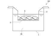
- FIG. 1 is a schematic view of the blood tissue barrier in vitro model 100 of the first embodiment.
- the incubator 1 contains the culture solution 2, and the collagen vitrigel membrane 3 is immersed in the culture solution 2 by a hanger 4 for suspending the collagen vitrigel membrane 3.
- Human vascular endothelial cells 5 are arranged on the upper surface of the collagen vitrigel membrane 3
- organ-derived cells 6 are arranged on the lower surface of the collagen vitrigel membrane 3, due to the porosity of the collagen vitrigel membrane 3. Cross talk between both cells is possible.
- the human vascular endothelial cell 5 is a cell that is in a proliferative state and has not yet undergone terminal differentiation, and is a cell that can differentiate into the vascular endothelial cell of the organ depending on the organ-derived cell 6.
- human vascular endothelial cells 5 are cells that differentiate into cerebral vascular endothelial cells constituting the blood-brain barrier.
- human vascular endothelial cells include human neonatal capsule-derived microvascular endothelial cells, various human stem cells (iPS cells, ES cells, etc.), and hCMEC / D3 cells (SV-40 rage T antigen-introduced human brain microvascular endothelium). Cell line) and the like.
- the conventional blood-brain barrier model having a barrier function is not an easily operable culture model because it is constructed by a co-culture system of vascular endothelial cells and heterologous cells such as pericytes and astrocytes.
- a method for producing a human blood-brain barrier model a vector containing a temperature-sensitive SV-40 large T antigen is isolated and cultured from the human blood-brain barrier, and brain microvascular endothelial cells, astrocytes, and astrosites are used.
- a method of introducing it into pericytes and producing it has been reported, it has not been easy to control the temperature of the culture.
- the blood tissue barrier in vitro model can be easily constructed by using the above-mentioned human vascular endothelial cells.
- the blood tissue barrier in vitro model 100 of this embodiment can be applied to the blood tissue barrier in vitro model of various human tissues including brain tissue.
- Applicable tissues include the blood-brain barrier, skin, liver sinusoids and the like.
- organ-derived cells 6 placed on the collagen vitrigel membrane 3 it is possible to support these blood tissue barrier in vitro models.
- Corresponding organ-derived cells 6 include kidney-derived cells, digestive organ-derived cells such as small intestine, urinary organ-derived cells such as bladder, skeletal muscle-derived cells, myocardial-derived cells, smooth muscle-derived cells, adipose-derived cells, lung-derived cells, and pancreas-derived cells.
- Examples thereof include cells, adrenal-derived cells, thyroid-derived cells, skin-derived cells, brain-derived cells, liver-derived cells and the like. More specifically, human skin-derived fibroblasts, C6 cells (cell line derived from rat nerve glial cell tumor), HepG2-NIAS cells (human hepatoma cell line) and the like can be mentioned.
- the "bitrigel” is a conventional hydrogel after the free water in the hydrogel is completely removed and then the bound water is partially removed to promote the trademarkization. It refers to a gel in a stable state obtained by rehydration, and has been named "vitrigel (registered trademark)" by the present inventor. In the present specification, when the term “Vitrigel” is used, the term “(registered trademark)” may be omitted. Further, as a more preferable raw material among collagen, native collagen or atelocollagen can be exemplified, and native collagen is further preferable.
- the culture solution 2 contained in the incubator 1 includes DMEM, Minimum Essential Medium (MEM), RPMI-1640, Glasgow Medium Eagle (BME), Dulvecco's Modified Eagle's Medium / NutritionM. -12), Glasgow Minimium Essential Medium (Glasgow MEM) and the like.
- MEM Minimum Essential Medium
- BME Glasgow Medium Eagle
- BME Dulvecco's Modified Eagle's Medium / NutritionM. -12
- Glasgow Minimium Essential Medium Glasgow MEM
- serum-free culture is performed from the viewpoint of avoiding non-specific adsorption of the compound to be evaluated to the protein. Liquid is preferred.
- FIG. 2 is a schematic view of the blood tissue barrier in vitro model 101 of the second embodiment. Unlike the blood tissue barrier in vitro model 100 of the first embodiment, the organ-derived cells 6 are not arranged on the lower surface of the collagen vitrigel membrane 3, and the conditioned culture solution 20 is used instead of the culture solution 2.
- Examples of the conditioned culture solution 20 include the culture solution used for culturing the organ-derived cells 6 for a certain period of time.
- the culture solution used in the blood tissue barrier in vitro model 100 of the first embodiment may be used in this embodiment.
- the culture solution used in the hanger in the blood tissue barrier in vitro model 100 is directly transferred into the hanger in the blood tissue barrier in vitro model 101, and the blood tissue barrier is transferred.
- the culture solution outside the hanger in the in vitro model 100 may be directly transferred to the outside of the hanger in the blood tissue barrier in vitro model 101, or the culture solution in the hanger in the blood tissue barrier in vitro model 100 may be transferred to the outside of the hanger in the blood tissue barrier in vitro model 101.
- the culture solution outside the hanger in the blood tissue barrier in vitro model 100 may be transferred into the hanger in the blood tissue barrier in vitro model 101, or the culture solution inside and outside the hanger in the blood tissue barrier in vitro model 100 may be mixed and used to mix the culture solution inside and outside the hanger in the blood tissue barrier in vitro model 101. You may move to.
- the conditioned culture solution 20 By using the conditioned culture solution 20, it is possible to save the trouble of arranging the organ-derived cells 6 on the lower surface of the collagen vitrigel membrane 3. Further, as will be described later, when the blood tissue barrier in vitro model 101 of the present embodiment is used in the method for evaluating the transferability of a drug to the blood tissue barrier, non-specific adsorption of the compound to be evaluated to the organ-derived cell 6 is performed. It can be avoided. Further, from the viewpoint of avoiding non-specific adsorption of the compound to be evaluated to the protein, it is preferable to use a serum-free culture solution as the acclimation culture solution 20.
- the present invention uses the blood tissue barrier in vitro model to add a drug to the upper part of the collagen vitrigel membrane, and after a certain period of time, the drug leaked to the lower part of the collagen vitrigel membrane.
- a method for evaluating the transferability of a drug to the blood tissue barrier to measure the amount of the drug is provided.
- the present invention uses the blood tissue barrier in vitro model to prepare an in vitro model of a normal state and an in vitro model of a pathological condition, and in both in vitro models, a drug is applied to the upper part of the collagen vitrigel membrane. After a certain period of time after addition, the amount of the drug leaked to the lower part of the collagen in vitro gel membrane was measured and compared to evaluate the pathological tissue selective migration of the drug. Provide a sex evaluation method.
- an in vitro model of a pathological condition can be prepared by using a culture solution corresponding to the pathological condition, or by using cells derived from a patient suffering from a disease. Then, by examining the tissue transferability of the drug in each of the in vitro model of the normal state and the in vitro model of the pathological condition, it is possible to screen the drug that selectively migrates to the pathological tissue.
- Example 1 Preparation of vascular endothelial culture model using human neonatal foramen-derived microvascular endothelial cells (HMVEC) and fibroblasts derived from the same (human dermal fibroblast; HDF)
- HMVEC human neonatal foramen-derived microvascular endothelial cells
- HDF human dermal fibroblast
- Pre-cultured HDF purchased from Kurabo Industries, KF-4009 was collected and mixed with the culture medium such that 1.3 ⁇ 10 5 cells / mL, to prepare a suspension of HDF.
- As the culture medium 10% fetal bovine serum (FBS), 20 mM HEPES, 100 units / mL penicillin, 100 ⁇ g / mL streptomycin-containing DMEM (purchased from Thermo Fisher Scientific, 11885-084) was used.
- FBS fetal bovine serum
- HEPES 100 units / mL penicillin
- streptomycin-containing DMEM purchased from Therm
- the culture medium was 5% FBS, 5 ng / mL recombinant human FGF-b, 50 ⁇ g / mL ascorbic acid, 1 ⁇ g / mL hydrocortisone hemicouccinate, 10 mM L-glutamine, 15 ng / mL recombinant human IGF-1, 5 ng / mL recombinant human EGF.
- a model in which HMVEC and HDF are cultured on both sides of the insert is an "organ-like plate type” model of skin blood vessels, and a model in which only one type of cells of HMVEC or HDF is cultured is a "tissue sheet type”. Defined as a model.
- the “organ-like plate type” model see (A) to (C) in FIG. 4
- the “tissue sheet type” model of HMVEC are used for day0, day2, and day6 (FIG. 4). See (D) to (F) of 4.).
- a "tissue sheet type” model of HDF was observed.
- Example 2 Measurement of changes over time in transendothelial electrical resistance (TEER) values of a cutaneous vascular endothelial culture model (1)
- the TER values of the three models prepared in Example 1 are set to day0, day1, and so on. Measurements were made on day2, day3, and day6. Before the measurement, the culture broth inside and outside the chamber of each model was removed, and 0.5 mL of the HMVEC culture broth that had been returned to room temperature in advance was added into the chamber and 1.5 mL outside the chamber.
- TEER transendothelial electrical resistance
- the "organ-like plate type” model showed higher TER values than day1, and the TER values of day1, day2, day3, and day6 were 22.62 ⁇ 3.06 ⁇ ⁇ cm 2 , 31.50 ⁇ 2, respectively. It was .46 ⁇ ⁇ cm 2 , 42.63 ⁇ 3.94 ⁇ ⁇ cm 2 , and 57.24 ⁇ 7.29 ⁇ ⁇ cm 2 . From the above, it was confirmed that the "organ-like plate type” model has a high endothelial barrier function.
- Example 3 Examination of the effect of the conditioned medium (CM) derived from the "organ-like plate type” model of skin blood vessels (1)
- the HMVEC "tissue sheet type” model of day 6 used in Example 2 The culture medium inside and outside the chamber was removed, and CM inside and outside the chamber of the "organ-like plate type” model of day 6 also used in Example 2 was added and cultured for 24 hours (see FIG. 6 (A)). ..
- the TER value of the HMVEC "tissue sheet type” model (HMVEC + culture solution) to which the culture solution was added was 14.5 ⁇ 1.51 ⁇ ⁇ cm 2.
- Example 4 Measurement of TER value of inflamed cutaneous vascular endothelial model (1) Cultivation inside and outside the chamber of the HMVEC "tissue sheet type” model and "organ-like plate type” model of day 6 used in Example 2. The liquid was removed, and 0.5 mL of VascuLife® Basal Medium containing no additives, which had been returned to room temperature in advance, was added into the chamber and 1.5 mL outside the chamber. (2) Next, an electrode was set in the chamber and the TER value was measured. The measured value at this time was taken as the value at the start of measurement (0 seconds).
- VascuLife (registered trademark) Basal Medium in the chamber was removed, and VascuLife (registered trademark) Basal Medium containing histamine dihydrochloride (purchased from Tokyo Chemical Industry Co., Ltd., H0146) was added.
- the TEER measurement was started, and the TEER value was continuously measured every 10 seconds for 180 seconds.
- the value at the start of measurement was about 50 ⁇ ⁇ cm 2 , but it decreased to about 32 ⁇ ⁇ cm 2 with the addition of 1 nM histamine, and this change was controlled without adding histamine. It was a statistically significant change in comparison ( *** P ⁇ 0.001, tested by Tukey-Kramer method after two-way ANOVA).
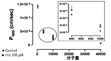
- Example 5 Evaluation of fluorescence substance permeability of inflamed skin vascular endothelial culture model (1) The culture medium inside and outside the chamber of the HMVEC “tissue sheet type” model of day 6 used in Example 2 was removed. Hank's Balanced Salt Solution (HBSS, purchased from Thermo Fisher Scientific, 14025-092), which had been returned to room temperature in advance, was washed twice by adding 0.5 mL into the chamber and 1.5 mL outside the chamber. (2) Next, 1.8 mL of HBSS was added to the outside of the chamber and 0.5 mL of HBSS containing 100 ⁇ M histamine was added to the inside of the chamber, and the mixture was incubated at room temperature for 10 minutes.
- HBSS Hank's Balanced Salt Solution
- P- app dQ / dt x 1 / A x 1 / C 0 dQ / dt; Transition amount per unit time ( ⁇ g / sec), A; Surface area (cm 2 ), C 0 ; Sodium fluorescein (molecular weight 376), FD-4 (molecular weight 376), by addition of 100 ⁇ M histamine initial concentration of each fluorescent substance. molecular weight of 4,000), and FD-40 (also P app of any molecules of molecular weight 40,000) was increased (FIG. 8 (a) - (C) see).
- an in vitro model of a normal state and an in vitro model of a pathological condition are prepared, and a drug having permeability selectively in the pathological tissue is screened by comparing and evaluating the transferability of the drug in both in vitro models. can do.
- Example 6 Preparation of tissue-specific vascular endothelial culture model and measurement of TEER value (1) HMVEC and rat in the same manner as the "organ-like plate type" model using HMVEC and HDF prepared in Example 1.
- Grioma cell line C6 (provided by RIKEN BioResource Center, RCB2854) or HMVEC and liver cancer cell line HepG2-NIAS (cell line in which HepG2 provided by RIKEN Bioresource Center was conditioned and re-deposited, RCB4679)
- the "organ-like plate type" model used was prepared.
- DMEM containing 10% FBS, 20 mM HEPES, 100 units / mL penicillin, and 100 ⁇ g / mL streptomycin was used.
- the change in the TEER value over time was measured by the same method as in Example 2. The results are shown in FIG. Not only skin-derived HDF (see FIG. 9 (A)) but also brain-derived C6 (see FIG. 9 (B)) and liver-derived HepG2-NIAS (see FIG. 9 (C)) can be used. It was confirmed that a vascular endothelial culture model could be prepared.
- hCMEC / D3 clone cell line and measurement of TEER value hCMEC / D3 is a human vascular endothelial cell line that has high proliferative ability and is widely used, but has a low endothelial barrier function, so that it is a blood tissue barrier model. Not suitable for building. Therefore, a cloned cell line having an endothelial barrier function was selected from the cells, and its inflammatory responsiveness was confirmed.
- Clone A was cultured in a collagen vitrigel membrane chamber for 10 days, 100 ⁇ M of histamine (His) was added into the chamber, and the TER value was measured every 1 minute. As a result, 15 minutes after the addition of histamine, the value at the start of the measurement was measured. Decreased by about 70% (see FIG. 11). This change was statistically significant compared to Control ( * *** P ⁇ 0.001 two-way ANOVA followed by Tukey-Kramer test). On the other hand, pretreatment with Y-27632, an inhibitor of ROCK, which is a downstream factor of histamine receptor, significantly suppressed the decrease in TER value due to histamine ( ### P ⁇ 0.001, binary). Tested by Tukey-Kramer method after ANOVA), it was suggested that clone A is a cell line with excellent histamine responsiveness (see FIG. 11).
- Clone A is cultured in a collagen vitrigel membrane chamber for 16 days, and 100 ⁇ M of histamine (His) and four kinds of fluorescent substances (Sodium fluorescein, FD-4, FD-10, and FD-40) are added to the chamber. After incubating for 1 hour, the amount of fluorescent substance transferred to the outside of the chamber was measured. As a result, histamine enhanced only the permeability of FD-4 (molecular weight 4,000) and FD-10 (molecular weight 10,000) (see FIG. 12). In particular, the increase in permeability of FD-10 was a statistically significant change ( * P ⁇ 0.05, tested by t-test).
- histamine-induced increase in vascular permeability in clone A has molecular size selectivity.
- this cloned cell line is suitable for constructing a model for evaluating the tissue transfer of a drug. ..
- Example 8 Examination of the effect of CM derived from the "tissue sheet type" model of HDF (1)
- the HDF cultured in advance was collected and mixed with the culture solution so as to have 8.0 ⁇ 10 4 cells / mL.
- a suspension of HDF was prepared.
- the culture medium is 5% FBS, 5 ng / mL recombinant human FGF-b, 50 ⁇ g / mL ascorbic acid, 1 ⁇ g / mL hydrocortisone heparinate, 10 mM L-glutamine, 15 ng / mL recombinant human IGF-1, 5 ng / mL recombinant.
- Human EGF 5 ng / mL recombinant human VEGF, 0.75 units / mL heparin sulfate, 30 mg / mL gentamicin, 15 ⁇ g / mL Amhotericin B-containing VascuLife® Basal Medium was used.
- the recovered CM (HDF-CM in and HDF-CM out) or culture solution was added from day 0 to day 7 inside and outside the chamber of the HMVEC “tissue sheet type” model prepared in Example 1.
- Day 4 and day 7 measured the TER value.
- the TEER value of the HMVEC "tissue sheet type" model (HMVEC + culture solution) to which the culture solution was added was 18.0 ⁇ 1 in day 4 (see FIG. 13 (C)) and day 7 (see FIG. 13 (D)), respectively. It was .41 ⁇ ⁇ cm 2 and 17.6 ⁇ 3.17 ⁇ ⁇ cm 2 .
- the TER values of the same model (HMVEC + (HDF-CM in)) to which HDF-CM in was added were 33.5 ⁇ 1.01 ⁇ ⁇ cm 2 and 33.0 ⁇ 2. There was 67 ⁇ ⁇ cm 2 .
- the TEER value of the same model (HMVEC + (HDF-CM out)) with HDF-CM out added is 33.9 ⁇ 3.68 ⁇ ⁇ cm 2 and 32.7 ⁇ 2.19 ⁇ ⁇ cm 2 on day 4 and day 7, respectively. Met.
- the TER value when HDF-CM in and HDF-CM out were added was statistically significantly higher in both day 4 and day 7 than when the culture medium was added ( ** P).
- Example 3 Comparing the above results with Example 3, it was shown that the CM of the HDF "tissue sheet type" model has a stronger effect of strengthening the endothelial barrier function than the CM of the "organ-like plate type” model.
- a tissue specificity using an easily constructable blood tissue barrier in vitro model of various human tissues including brain tissue and the blood tissue barrier in vitro model having simple and excellent reproducibility can be provided.
Landscapes
- Chemical & Material Sciences (AREA)
- Organic Chemistry (AREA)
- Life Sciences & Earth Sciences (AREA)
- Zoology (AREA)
- Wood Science & Technology (AREA)
- Proteomics, Peptides & Aminoacids (AREA)
- Health & Medical Sciences (AREA)
- Engineering & Computer Science (AREA)
- Microbiology (AREA)
- Biochemistry (AREA)
- Physics & Mathematics (AREA)
- Molecular Biology (AREA)
- Biotechnology (AREA)
- Biophysics (AREA)
- Analytical Chemistry (AREA)
- Immunology (AREA)
- Bioinformatics & Cheminformatics (AREA)
- General Engineering & Computer Science (AREA)
- General Health & Medical Sciences (AREA)
- Genetics & Genomics (AREA)
- Micro-Organisms Or Cultivation Processes Thereof (AREA)
- Measuring Or Testing Involving Enzymes Or Micro-Organisms (AREA)
Abstract
L'invention concerne un modèle in vitro de barrière sang/tissu qui contient un film de vitrigel de collagène, des cellules endothéliales vasculaires humaines disposées sur une face supérieure dudit film de vitrigel de collagène, des cellules dérivées d'un organe disposées sur une face inférieure dudit film de vitrigel de collagène, et une solution de culture. Lesdites cellules endothéliales vasculaires humaines sont dépendantes desdites cellules dérivées d'un organe, et peuvent être différenciées en cellules endothéliales vasculaires dudit organe.
Priority Applications (1)
| Application Number | Priority Date | Filing Date | Title |
|---|---|---|---|
| JP2021552405A JP7336154B2 (ja) | 2019-10-18 | 2020-10-14 | 血液組織関門インビトロモデル、及び薬物の血液組織関門移行性評価方法 |
Applications Claiming Priority (2)
| Application Number | Priority Date | Filing Date | Title |
|---|---|---|---|
| JP2019190832 | 2019-10-18 | ||
| JP2019-190832 | 2019-10-18 |
Publications (1)
| Publication Number | Publication Date |
|---|---|
| WO2021075451A1 true WO2021075451A1 (fr) | 2021-04-22 |
Family
ID=75538480
Family Applications (1)
| Application Number | Title | Priority Date | Filing Date |
|---|---|---|---|
| PCT/JP2020/038721 Ceased WO2021075451A1 (fr) | 2019-10-18 | 2020-10-14 | Modèle in vitro de barrière sang/tissu, et procédé d'évaluation de transfert de médicament à travers une barrière sang/tissu |
Country Status (2)
| Country | Link |
|---|---|
| JP (1) | JP7336154B2 (fr) |
| WO (1) | WO2021075451A1 (fr) |
Citations (5)
| Publication number | Priority date | Publication date | Assignee | Title |
|---|---|---|---|---|
| JPH05503920A (ja) * | 1989-09-27 | 1993-06-24 | エラン ファーマシューテイカルズ インコーポレイテッド | 血液脳関門のモデル |
| JP2001238681A (ja) * | 2000-03-03 | 2001-09-04 | Japan Science & Technology Corp | 共培養による血液脳関門再構築モデル |
| JP2007166915A (ja) * | 2005-12-19 | 2007-07-05 | Masami Niwa | 血液脳関門インヴィトロ・モデル、病態血液脳関門インヴィトロ・モデル、及びこれを用いた薬物スクリーニング方法、病態血液脳関門機能解析方法、病因解析方法 |
| JP2012115262A (ja) * | 2010-11-12 | 2012-06-21 | National Institute Of Agrobiological Sciences | 細胞培養チャンバーとその製造方法、および、この細胞培養チャンバーを利用した組織モデルとその作製方法 |
| WO2017179375A1 (fr) * | 2016-04-15 | 2017-10-19 | 国立大学法人山口大学 | Modèle in vitro de barrière hématoencéphalique ainsi que procédé d'élaboration de celui-ci |
Family Cites Families (2)
| Publication number | Priority date | Publication date | Assignee | Title |
|---|---|---|---|---|
| TWI523945B (zh) | 2013-03-29 | 2016-03-01 | Nat Univ Chung Cheng | Establishment of blood - brain barrier model in |
| CN110709503B (zh) | 2017-06-09 | 2023-09-08 | 富士胶片株式会社 | 细胞层叠体的制造方法 |
-
2020
- 2020-10-14 WO PCT/JP2020/038721 patent/WO2021075451A1/fr not_active Ceased
- 2020-10-14 JP JP2021552405A patent/JP7336154B2/ja active Active
Patent Citations (5)
| Publication number | Priority date | Publication date | Assignee | Title |
|---|---|---|---|---|
| JPH05503920A (ja) * | 1989-09-27 | 1993-06-24 | エラン ファーマシューテイカルズ インコーポレイテッド | 血液脳関門のモデル |
| JP2001238681A (ja) * | 2000-03-03 | 2001-09-04 | Japan Science & Technology Corp | 共培養による血液脳関門再構築モデル |
| JP2007166915A (ja) * | 2005-12-19 | 2007-07-05 | Masami Niwa | 血液脳関門インヴィトロ・モデル、病態血液脳関門インヴィトロ・モデル、及びこれを用いた薬物スクリーニング方法、病態血液脳関門機能解析方法、病因解析方法 |
| JP2012115262A (ja) * | 2010-11-12 | 2012-06-21 | National Institute Of Agrobiological Sciences | 細胞培養チャンバーとその製造方法、および、この細胞培養チャンバーを利用した組織モデルとその作製方法 |
| WO2017179375A1 (fr) * | 2016-04-15 | 2017-10-19 | 国立大学法人山口大学 | Modèle in vitro de barrière hématoencéphalique ainsi que procédé d'élaboration de celui-ci |
Non-Patent Citations (2)
| Title |
|---|
| HAMANAKA, YOSHIHIRO ET AL.: "Construction of vascular endothelial cell sheet using a collagen vitrigel membrane chamber and application concept for evaluating vascular permeability", ABSTRACTS OF THE 133RD ANNUAL MEETING OF THE PHARMACEUTICAL SOCIETY OF JAPAN, vol. 133, 2013, pages 99 * |
| OKAMOTO, CHIKA ET AL.: "The barrier function of the vascular endothelial model reconstructed by culturing human vascular endothelial cells in a collagen vitrigel membrane chamber", JOURNAL OF EXPERIMENTAL & APPLIED CELL CULTURE RESEARCH, vol. 31, no. 1, pages 27 * |
Also Published As
| Publication number | Publication date |
|---|---|
| JPWO2021075451A1 (fr) | 2021-04-22 |
| JP7336154B2 (ja) | 2023-08-31 |
Similar Documents
| Publication | Publication Date | Title |
|---|---|---|
| Ngo et al. | Mitochondrial morphology controls fatty acid utilization by changing CPT1 sensitivity to malonyl‐CoA | |
| Khodabukus | Tissue-engineered skeletal muscle models to study muscle function, plasticity, and disease | |
| Amano et al. | Development of vascularized iPSC derived 3D-cardiomyocyte tissues by filtration Layer-by-Layer technique and their application for pharmaceutical assays | |
| Blondot et al. | Intracellular transport and egress of hepatitis B virus | |
| Xu et al. | An cell-assembly derived physiological 3D model of the metabolic syndrome, based on adipose-derived stromal cells and a gelatin/alginate/fibrinogen matrix | |
| Richards et al. | Inspiration from heart development: Biomimetic development of functional human cardiac organoids | |
| Wang et al. | Coaxial extrusion bioprinted shell-core hydrogel microfibers mimic glioma microenvironment and enhance the drug resistance of cancer cells | |
| Mosadegh et al. | Three‐dimensional paper‐based model for cardiac ischemia | |
| Matsusaki et al. | Development of full‐thickness human skin equivalents with blood and lymph‐like capillary networks by cell coating technology | |
| Jang et al. | From single-to multi-organ-on-a-chip system for studying metabolic diseases | |
| CN108823145B (zh) | 一种人脑微血管生成模拟血脑屏障的体外构建方法 | |
| Olsen et al. | Manipulation of cellular spheroid composition and the effects on vascular tissue fusion | |
| Zhu et al. | Three-in-one customized bioink for islet organoid: GelMA/ECM/PRP orchestrate pro-angiogenic and immunoregulatory function | |
| US20140377862A1 (en) | Cell cultivation method and cell culture | |
| Wang et al. | The promoting effects of activated olfactory ensheathing cells on angiogenesis after spinal cord injury through the PI3K/Akt pathway | |
| Eyre et al. | A human retinal microvascular endothelial-pericyte co-culture model to study diabetic retinopathy in vitro | |
| Arisaka et al. | A heparin-modified thermoresponsive surface with heparin-binding epidermal growth factor-like growth factor for maintaining hepatic functions in vitro and harvesting hepatocyte sheets | |
| Liu et al. | Engineering neurovascular unit and Blood–Brain barrier for ischemic stroke modeling | |
| Koroleva et al. | Hydrogel-based microfluidics for vascular tissue engineering | |
| KR20190127041A (ko) | 간 오가노이드 및 그의 제조 방법 | |
| Auth et al. | Maintained function of primary human hepatocytes by cellular interactions in coculture: implications for liver support systems | |
| Takezawa et al. | A protein-permeable scaffold of a collagen vitrigel membrane useful for reconstructing crosstalk models between two different cell types | |
| Buonvino et al. | Modelling the disease: H2S-sensitivity and drug-resistance of triple negative breast cancer cells can be modulated by embedding in isotropic micro-environment | |
| Qu et al. | Construction of engineered cardiac tissue on a heart-on-a-chip device enables modeling of arrhythmogenic right ventricular cardiomyopathy | |
| Orbach et al. | Multi-cellular transitional organotypic models to investigate liver fibrosis |
Legal Events
| Date | Code | Title | Description |
|---|---|---|---|
| 121 | Ep: the epo has been informed by wipo that ep was designated in this application |
Ref document number: 20876906 Country of ref document: EP Kind code of ref document: A1 |
|
| ENP | Entry into the national phase |
Ref document number: 2021552405 Country of ref document: JP Kind code of ref document: A |
|
| NENP | Non-entry into the national phase |
Ref country code: DE |
|
| 122 | Ep: pct application non-entry in european phase |
Ref document number: 20876906 Country of ref document: EP Kind code of ref document: A1 |