KR101476769B1 - Biomarker composition for predicting progress from colorectal adenoma to carcinoma of human patients and biokit for dyagnosis thereof comprising the same - Google Patents
Biomarker composition for predicting progress from colorectal adenoma to carcinoma of human patients and biokit for dyagnosis thereof comprising the same Download PDFInfo
- Publication number
- KR101476769B1 KR101476769B1 KR1020130000392A KR20130000392A KR101476769B1 KR 101476769 B1 KR101476769 B1 KR 101476769B1 KR 1020130000392 A KR1020130000392 A KR 1020130000392A KR 20130000392 A KR20130000392 A KR 20130000392A KR 101476769 B1 KR101476769 B1 KR 101476769B1
- Authority
- KR
- South Korea
- Prior art keywords
- leu
- ser
- glu
- ala
- gly
- Prior art date
- Legal status (The legal status is an assumption and is not a legal conclusion. Google has not performed a legal analysis and makes no representation as to the accuracy of the status listed.)
- Active
Links
Images


Classifications
-
- G—PHYSICS
- G01—MEASURING; TESTING
- G01N—INVESTIGATING OR ANALYSING MATERIALS BY DETERMINING THEIR CHEMICAL OR PHYSICAL PROPERTIES
- G01N33/00—Investigating or analysing materials by specific methods not covered by groups G01N1/00 - G01N31/00
- G01N33/48—Biological material, e.g. blood, urine; Haemocytometers
- G01N33/50—Chemical analysis of biological material, e.g. blood, urine; Testing involving biospecific ligand binding methods; Immunological testing
- G01N33/68—Chemical analysis of biological material, e.g. blood, urine; Testing involving biospecific ligand binding methods; Immunological testing involving proteins, peptides or amino acids
- G01N33/6893—Chemical analysis of biological material, e.g. blood, urine; Testing involving biospecific ligand binding methods; Immunological testing involving proteins, peptides or amino acids related to diseases not provided for elsewhere
-
- G—PHYSICS
- G01—MEASURING; TESTING
- G01N—INVESTIGATING OR ANALYSING MATERIALS BY DETERMINING THEIR CHEMICAL OR PHYSICAL PROPERTIES
- G01N33/00—Investigating or analysing materials by specific methods not covered by groups G01N1/00 - G01N31/00
- G01N33/48—Biological material, e.g. blood, urine; Haemocytometers
- G01N33/50—Chemical analysis of biological material, e.g. blood, urine; Testing involving biospecific ligand binding methods; Immunological testing
- G01N33/68—Chemical analysis of biological material, e.g. blood, urine; Testing involving biospecific ligand binding methods; Immunological testing involving proteins, peptides or amino acids
- G01N33/6863—Cytokines, i.e. immune system proteins modifying a biological response such as cell growth proliferation or differentiation, e.g. TNF, CNF, GM-CSF, lymphotoxin, MIF or their receptors
-
- G—PHYSICS
- G01—MEASURING; TESTING
- G01N—INVESTIGATING OR ANALYSING MATERIALS BY DETERMINING THEIR CHEMICAL OR PHYSICAL PROPERTIES
- G01N2800/00—Detection or diagnosis of diseases
- G01N2800/06—Gastro-intestinal diseases
-
- G—PHYSICS
- G01—MEASURING; TESTING
- G01N—INVESTIGATING OR ANALYSING MATERIALS BY DETERMINING THEIR CHEMICAL OR PHYSICAL PROPERTIES
- G01N2800/00—Detection or diagnosis of diseases
- G01N2800/50—Determining the risk of developing a disease
-
- Y—GENERAL TAGGING OF NEW TECHNOLOGICAL DEVELOPMENTS; GENERAL TAGGING OF CROSS-SECTIONAL TECHNOLOGIES SPANNING OVER SEVERAL SECTIONS OF THE IPC; TECHNICAL SUBJECTS COVERED BY FORMER USPC CROSS-REFERENCE ART COLLECTIONS [XRACs] AND DIGESTS
- Y10—TECHNICAL SUBJECTS COVERED BY FORMER USPC
- Y10S—TECHNICAL SUBJECTS COVERED BY FORMER USPC CROSS-REFERENCE ART COLLECTIONS [XRACs] AND DIGESTS
- Y10S493/00—Manufacturing container or tube from paper; or other manufacturing from a sheet or web
- Y10S493/961—Tag, marker, or label
Landscapes
- Life Sciences & Earth Sciences (AREA)
- Health & Medical Sciences (AREA)
- Engineering & Computer Science (AREA)
- Immunology (AREA)
- Molecular Biology (AREA)
- Chemical & Material Sciences (AREA)
- Urology & Nephrology (AREA)
- Biomedical Technology (AREA)
- Hematology (AREA)
- Cell Biology (AREA)
- Medicinal Chemistry (AREA)
- Biochemistry (AREA)
- Biotechnology (AREA)
- Food Science & Technology (AREA)
- Proteomics, Peptides & Aminoacids (AREA)
- Physics & Mathematics (AREA)
- Analytical Chemistry (AREA)
- Microbiology (AREA)
- General Health & Medical Sciences (AREA)
- General Physics & Mathematics (AREA)
- Pathology (AREA)
- Investigating Or Analysing Biological Materials (AREA)
- Peptides Or Proteins (AREA)
- Measuring Or Testing Involving Enzymes Or Micro-Organisms (AREA)
Abstract
본 발명은 대장용종에서 대장암으로의 진행을 예측할 수 있는 단백질체 바이오마커 및 사이토카인 혈장 바이오마커를 포함하는 대장암 진단용 바이오키트에 관한 것으로, 구체적으로는 leucine-richα-2-glycoprotein(LRG), hemoglobin subunitβ(HBB), Igα-2 chain C region(IgA2C), complement factor B(CFB), afamin, zinc-α-2-glycoprotein(ZAG), vitronectin(VTN), α1-antichymotrypsin(AACT)중에서 적어도 하나 이상의 단백질을 포함하는 대장용종-대장암 진행예측 및 진단용 키트에 관한 것으로 상기 LRG, HBB, IgA2C, CFB 단백질은 대장암 환자에게서 증가하고, afamin, ZAG, VTN, AACT 단백질은 대장암 환자에게서 감소하는 상이한 특징이 있으며 또한 사이토카인 또는 케모카인인 IL-8, IP-10, TNF-α이 대장암 환자에게서 증가하는 특징이 있으며 상기 8종의 상기 단백질들은 대장용종에서 대장암으로의 진행을 예측할 수 있는 마커로 사용할 수 있으므로 대장암의 진행상태를 조기에 정확하게 구별하여 진단할 수 있는 뛰어난 효과가 있다. The present invention relates to a biocide for diagnosing colon cancer, which comprises a protein body biomarker capable of predicting progression from colon polyp to colorectal cancer and a cytokine plasma biomarker, and more specifically, a leukocyte-rich α-2-glycoprotein (LRG) at least one of hemoglobin subunit beta (HBB), Ig alpha-2 chain C region (IgA2C), complement factor B (CFB), afamin, zinc-alpha-2-glycoprotein (ZAG), vitronectin (VTN), alpha 1 -antichymotrypsin Wherein said LRG, HBB, IgA2C, and CFB proteins are increased in colorectal cancer patients, and afamin, ZAG, VTN, and AACT proteins are decreased in colorectal cancer patients. In addition, IL-8, IP-10, and TNF-alpha, which are cytokines or chemokines, are characterized by an increased number of patients with colorectal cancer, and these eight proteins are predictive of progression from colorectal to colorectal cancer It is possible to diagnose the progress of colorectal cancer in an early and accurate manner.
Description
본 발명은 대장용종에서 대장암으로의 진행을 예측진단할 수 있는 바이오마커에 관한 것으로, 더욱 상세하게는 대장용종 환자보다 대장암환자에게서 발현양이 감소 또는 증가되는 단백질체 및 사이토카인 혈장 바이오마커와 이를 이용한 대장용종-대장암 진행예측 진단용 바이오키트에 관한 것이다.The present invention relates to a biomarker capable of predicting the progression from colon to polyp, and more particularly, to a biomarker capable of predicting the progression from colon to polyp, The present invention relates to a biocide for the diagnosis of colorectal cancer-colon cancer progression using the same.
대장암(Colorectal cancer, CRC)이란 대장에 생긴 암세포로 이루어진 악성종양으로, 전세계적으로 가장 흔한 암들 중 하나이면서 악성종양에 의한 주요 사망원인 4위에 해당하는 암이다. 대장암은 높은 열량의 섭취, 동물성 지방 섭취, 섬유소 섭취 부족, 비만 등이 원인이 되어 발생하는 것으로 알려져 있다. 초기 대장암의 경우에는 아무런 증상도 나타나지 않으나, 눈에 띄지 않는 장 출혈로 혈액이 손실되어 빈혈이 생길 수 있으며 간혹 식욕부진과 체중감소가 나타난다. 암이 진행된 경우에는 배가 아프거나 설사 또는 변비가 생기는 등 배변습관의 변화가 나타나기도 하고 항문에서 피가 나오는 직장출혈의 증세가 나타나기도 한다.
Colorectal cancer (CRC) is a malignant tumor consisting of cancerous cells in the large intestine. It is one of the most common cancers in the world and the fourth leading cause of death by malignant tumors. It is known that colon cancer is caused by high calorie intake, animal fat intake, lack of fiber intake, and obesity. Early colorectal cancer does not show any symptoms, but it can cause anemia due to inconspicuous intestinal bleeding, resulting in anorexia and weight loss. When the cancer progresses, there is a change in bowel habits such as a stomach ache, diarrhea or constipation, and rectal bleeding from the anus.
대장암은 대장점막이 비정상적으로 자라 사마귀모양의 혹으로 보이는 조직인 대장 안의 용종(Polyp)으로부터 생긴다. 용종을 제거하지 않으면 결국 악성으로 변할 수 있고, 결장과 직장의 벽을 뚫고 다른 곳으로 전이될 수 있다. 대부분은 비종양성용종이지만, 선종성용종(종양성용종)은 대장 점막세포의 유전자에 돌연변이를 유발하는 것과 연관되어 있고 이러한 돌연변이가 대장암과 직결된다. 용종의 크기가 클수록 악성 세포를 포함할 가능성이 커진다.
Colorectal cancer arises from polyps in the large intestine, a tissue that looks abnormally enlarged in the large intestine and appears as a wart-shaped lump. If the polyp is not removed, it can eventually turn into malignancy, which can penetrate the walls of the colon and rectum and be transferred to another location. Most of them are non-polyps, but adenomatous polyps (tumorous polyps) are associated with mutations in the genes of the colon mucosal cells, and these mutations are directly related to colon cancer. The larger the polyp, the more likely it is to include malignant cells.
대장내시경(colonoscopy)이 대장암의 초기발견을 위한 가장 민감한 접근법이긴 하나, 침습적 시술이고 많은 비용이 들며 위험하고 불편한 방법이기도 하다. 따라서 대장암의 집단선별 프로그램을 더욱 증진할 수 있는 새로운 바이오마커가 필요하다.Although colonoscopy is the most sensitive approach to the early detection of colorectal cancer, it is invasive, costly, and dangerous and inconvenient. Therefore, new biomarkers are needed to further promote the colonoscopy screening program.
지금까지 대장암이 혈액 단백질체의 변화와 관련이 있음을 증명하는 수많은 보고서가 있었음에도 불구하고, 잠재적으로 치료가능한 초기단계의 대장암에 대하여 고감도 또는 높은 특이성을 가진 혈액 바이오마커가 치료용으로서 유효성이 승인된 적은 없었다. 현재 직접적인 육안검사를 제외하고 대장암을 발견하기 위한 2가지 간접적 진단도구들이 있다: 하나는 DNA 테스팅처럼 대변을 기반으로 한 접근법인 반면에, 다른 하나는 혈액 내 태아성 암 항원(CEA)의 레벨을 검사하는 것을 포함한다. 그러나 이 방법들은 초기단계 대장암 환자들에 대한 민감도가 낮다는 단점이 있다. 다른 주요 단점은 전구물 병변(선종)을 무시한 채 오직 대장암환자들과 건강한 대조구들에게만 제한된 방법인 것이다.
Although there have been so many reports to date that colon cancer is associated with changes in the blood protein body, blood biomarkers with high sensitivity or high specificity for potentially treatable early stage colorectal cancer I have never been approved. Currently, there are two indirect diagnostic tools to detect colon cancer, except direct visual inspection: one is a fecal-based approach like DNA testing, and the other is the level of fetal carcinoma antigen (CEA) in the blood Lt; / RTI > However, these methods have the disadvantage that they are less sensitive to early stage colorectal cancer patients. Another major disadvantage is that it is limited only to colorectal cancer patients and healthy controls, neglecting bulb water lesions (adenoma).
대장암의 발병 및 성장의 가장 중요한 패러다임은 대장용종-대장암 변화이다. 따라서 침습이 발생하기 전에 주요 전암성 병변(용종)이 감지되거나 제거된다면 대장암은 효과적으로 통제될 수 있다. 따라서 대장용종 형성의 기저를 이루는 분자적 메커니즘을 설명하는 것이 매우 중요하다. 대장암에 관련된 특정 단백질, 특히 용종-종양에 관련된 단백질의 동정은 충분히 대장암의 진단 및 치료를 발전시킬 수 있다. 현재 의학적 프로토콜이 대장내시경 및 변잠혈시험(fecal occult blood) 같은 다양한 검사를 사용하여 초기의 대장암을 감지할 수 있으나, 대장용종으로부터 대장암으로의 진행을 예측하는 바이오마커가 개발된다면 더욱 편리하고 유익할 것이다.The most important paradigm for the onset and growth of colorectal cancer is colorectal - colon cancer. Thus, colon cancer can be effectively controlled if major pre-cancerous lesions (polyps) are detected or eliminated before invasion occurs. Therefore, it is very important to explain the molecular mechanism underlying colonic polyp formation. Identification of certain proteins involved in colorectal cancer, particularly those associated with polypectomy-tumors, can sufficiently develop the diagnosis and treatment of colorectal cancer. Although current medical protocols can detect early colorectal cancer using a variety of tests such as colonoscopy and fecal occult blood, it is more convenient if biomarkers are developed that predict progress from colon polyps to colorectal cancer It will be beneficial.
지금까지 대장암의 새로운 바이오마커를 찾아내기 위해 단백질체적 접근법을 시도하여 왔다. 2-D 전기영동은 암 바이오마커를 추출할 뿐 아니라 기술적인 제한에 상관없이 대장암 형성의 기저를 이루는 복잡한 생물학적 사건을 연구하는 가장 흔한 접근법들 중 하나이다. 대장암의 단백질 바이오마커를 찾기 위해 바이오유체(biofluid) 또는 대장조직을 이용하여 많은 연구가 선행되어 왔다. 또, 환자들의 용종과 암 뿐만 아니라 건강한 대장점막에서의 차별되는 단백질분석을 증명하는 몇몇 선행연구들도 있다. 그러나 지금까지 대장용종에서 대장암으로 변하는 동안 특이적으로 조절되는 단백질들은 대장암의 진단 바이오마커로서 동정된 바 없었다.
So far, attempts have been made to approach protein volume to find new biomarkers for colorectal cancer. 2-D electrophoresis is one of the most common approaches to study complex biological events that underlie colon cancer formation, regardless of technical limitations, as well as extracting cancer biomarkers. Much research has been done using biofluids or colon tissue to find protein biomarkers for colon cancer. In addition, there are several previous studies that demonstrate differential protein analysis in healthy colon and mucosa as well as in patients' polyps and cancer. Until now, however, proteins specifically regulated during conversion from colon to colon have not been identified as diagnostic biomarkers for colorectal cancer.
혈액 내 단백질 암 바이오마커의 발견이 복잡한 것은 검체 전반에 걸친 높은 다양성과 혈청 및 혈장 내에 존재하는 광역동적영역(broaed dynamic range)의 단백질들때문이다. 그럼에도 불구하고 혈액을 이용하는 대장암 바이오마커의 동정은 임상적인 관점에서 점막검체보다 용이하다. 혈장은 기본적인 검체일 뿐만 아니라 어떤 샘플에도 존재하는 가장 크고 깊은 인간 단백질체를 나타내기 때문에, 암의 혈장 바이오마커를 찾는 매우 다양하고 체계적인 조사가 진행중이다. 혈장 단백질체는 병의 진단 및 치료목적의 모니터링을 근본적으로 변혁시킬 수 있기 때문에 많은 관심을 받고있다.
The complexity of the discovery of protein cancer biomarkers in the blood is due to the high diversity across the specimens and the proteins in the serum and plasma that are in the broad dynamic range. Nevertheless, identification of colon cancer biomarkers using blood is easier than mucosal specimens from a clinical point of view. Plasma is not only a basic sample, but it also represents the largest and deepest human protein body present in any sample, so a very diverse and systematic investigation is underway to find plasma biomarkers for cancer. Plasma proteomics has attracted much attention because it can fundamentally change the diagnosis and treatment of diseases.
수많은 예비보고서들이 혈청/혈장의 신호들을 원인으로 시사했음에도 불구하고 지금까지 대장용종에서 대장암으로의 순차적인 진행을 증명했던 강력한 마커는 없었다. 게다가 대부분의 생화학 및 단백질체 연구들은 대장암 그 자체마커의 동정에 제한하고 있었다. 그러나 소수의 단백질체 연구들은 선종성용종증(adenomatous polyposis)에서 대장암으로의 진행을 알려주는 유용한 바이오마커들을 증명하였다. 와일드 등(Clin . Cancer Res.2010)은 대장암 검출의 대안으로서 22종의 혈청 마커 및 복합마커를 평가하였다. 혈청 마커조합의 높은 성과는 최종진단을 위한 대장내시경의 후속장치를 촉발시키면서 신규한 대장암 선별도구를 제공한 것이다. 게다가 실험한 22종의 마커들 중에서 15종은 대장용종에서 대장암으로 진행 중에 순차적으로 증가됨을 보였다. 레너한 등(J. Clin . Endocrinol . Metab.2010)은 순환되는 양이 많은 인슐린유사성장인자Ⅱ(IGF-Ⅱ)가 대장용종의 종양마커가 될 수 있고 94%의 고 민감도로서 증가하는 대장암의 위험과 관련이 있을 수 있음을 보여주었다. 로블릭 등(Cell . Mol . Life Sci.2004)은 인간 대장조직의 생성 및 성장 동안에 순차적인 단백질체의 변형을 증명하였고 46종의 증가(up-regulated)단백질 및 26종의 감소(down-regulated)단백질을 동정하였다. 또한 대장암 환자에게 HSP10의 과발현은 대장용종에서 대장암으로의 진행을 알려주는 지표로서 동정되었다. 최근에는 프로가스트린의 과발현과 혈액 내 고순환농도가 증식성 대장용종의 예측변수로서 인식되었다.
Despite numerous preliminary reports suggesting serum / plasma signals, there has been no strong marker that has so far proven sequential progression from colon polyps to colorectal cancer. In addition, most biochemical and proteomic studies have limited the identification of colon cancer markers. However, a small number of protein body studies have demonstrated useful biomarkers to signal the progression from adenomatous polyposis to colorectal cancer. Wild, etc. ( Clin . Cancer Res. 2010) evaluated 22 serum markers and complex markers as an alternative to colon cancer detection. The high performance of the serum marker combination provides a novel colon cancer screening tool that triggers a subsequent device for colonoscopy for final diagnosis. In addition, of the 22 markers tested, 15 showed progressive progression from colon polyps to colon cancer. Renner et al . ( J. Clin . Endocrinol . Metab . 2010) demonstrated that circulating amounts of insulin-like growth factor II (IGF-Ⅱ) can be tumor markers for colon polyps and increased sensitivity of 94% And that the risk of death could be related to the risk of death. Loblik et al . ( Cell . Mol . Life Sci. 2004) demonstrated sequential protein body modifications during the production and growth of human colon tissues and identified 46 up-regulated proteins and 26 down-regulated proteins. Also, overexpression of HSP10 in colon cancer patients was identified as an indicator of progression from colon polyps to colorectal cancer. Recently, overgrowth of progesterone and high circulating concentrations in blood have been recognized as predictive variables of proliferative colonic polyps.
한편, 특정 단백질 또는 유전자 바이오마커에 관련된 국내특허로서 등록특허 제10-0892587호가 공지되어 있는 바, 이에는 대장암 특이적인 유전자의 메틸화된 프로모터를 함유하는 진단키트가 개시된 바 있다. 또, 국내등록특허 제10-1212024호에는 뉴클레오티드 서열로 코딩되는 단백질에 결합하는 항체를 포함하는 키트가 개시된 바 있다. 그리고 국내등록특허 제10-113605호에는 장암 특이적 마커유전자의 메틸화 검출키트가 개시된 바 있다. 이 밖에도 국내공개특허 제10-2011-0126570호에는 2개 이상의 유전자 조합으로 구성된 복합마커가 개시된 바 있다. 그러나 상기문헌 어디에도 대장용종에서 대장암으로 진행하는 동안 특이적으로 조절되는 본 발명의 단백질들은 개시되거나 암시된 바 없다.
On the other hand, as a domestic patent related to a specific protein or gene biomarker is known as Registration No. 10-0892587, a diagnostic kit containing a methylated promoter of a gene specific for colon cancer has been disclosed. In addition, Korean Patent No. 10-1212024 discloses a kit comprising an antibody that binds to a protein encoded by a nucleotide sequence. In addition, Korean Patent No. 10-113605 discloses a methylation detection kit of a cancer-specific marker gene. In addition, Korean Patent Laid-Open No. 10-2011-0126570 discloses a complex marker composed of two or more gene combinations. However, none of the above references discloses or suggests the proteins of the present invention that are specifically regulated during progression from colon polyps to colon cancer.
따라서 본 발명의 목적은 종양생성 및 종양진행의 기저에 있는 분자적 매커니즘에 대한 이해를 통하여 대장용종에서 대장암으로의 진단용 단백질체 및 사이토카인 혈장 바이오마커를 제공하는 데 있다.It is therefore an object of the present invention to provide diagnostic protein bodies and cytokine plasma biomarkers from colorectal to colorectal cancer through an understanding of molecular mechanisms underlying tumorigenesis and tumor progression.
본 발명의 상기 목적은 대장용종 환자들과 대장암 환자들 사이에 차등발현되는 사이토카인 및 케모카인을 생물학적 정량기술을 이용하여 프로파일하고, 2D 전기영동과 질량분석법을 이용한 혈장단백질들의 비교를 통하여 단백질체 분석을 수행함으로써 달성하였다.The above object of the present invention is achieved by a method of analyzing cytokines and chemokines differentially expressed between colonic polyp and colonic cancer patients by using biological quantitation technology and analyzing the protein by analyzing plasma proteins using 2D electrophoresis and mass spectrometry Lt; / RTI >
본 발명은 대장용종 환자의 혈장보다 대장암 환자의 혈장에서 발현이 상대적으로 감소하는 하기 군에서 선택된 단백질체를 유효성분으로 포함하는 대장용종으로부터 대장암으로의 진행예측 진단용 조성물을 제공한다:The present invention provides a composition for predicting the progression from a colorectal polyp into a colon cancer comprising as an active ingredient a protein body selected from the following group whose expression is relatively reduced in plasma of a patient suffering from colorectal cancer:
서열번호 1의 38 kDa의 분자량 및 6.45의 pI를 갖는 고 류신알파-2당단백질( Leucine-richα-2 glycoprotein 1(Homo sapiens), LRG), (Leucine-rich alpha-2 glycoprotein 1 (Homo sapiens), LRG) having a molecular weight of 38 kDa of SEQ ID NO: 1 and a pI of 6.45,
서열번호 2의 16 kDa의 분자량 및 6.75의 pI를 갖는 헤모글로빈 베타소단위( Hemoglobin subunit β(Homo sapiens), HBB), Hemoglobin subunit beta (Homo sapiens), HBB, having a molecular weight of 16 kDa of SEQ ID NO: 2 and a pI of 6.75,
서열번호 3의 24 kDa의 분자량 및 5.56의 pI를 갖는 면역글로불린-2사슬C(Igα-2 chain C (allotype A2m(2)), IgA2C), 2 chain C (allotype A2m (2)), IgA2C having a molecular weight of 24 kDa of SEQ ID NO: 3 and a pI of 5.56,
서열번호 4의 85 kDa의 분자량 및 6.81의 pI를 갖는 보체인자 B(Chain A, Human Complement Factor B, CFB), A complement factor B (Chain A, Human Complement Factor B, CFB) having a molecular weight of 85 kDa of SEQ ID NO: 4 and a pI of 6.81,
서열번호 5의 70 kDa의 분자량 및 5.64의 pI를 갖는 아파민(afamin precusor, Homo sapiens), An afamin precursor (Homo sapiens) having a molecular weight of 70 kDa of SEQ ID NO: 5 and a pI of 5.64,
서열번호 6의 31 kDa의 분자량 및 5.7의 pI를 갖는 아연알파2-당단백질(Chain B, Human Zinc-α-2-Glycoprotein, ZAG), A zinc alpha-2-glycoprotein (ZAG) having a molecular weight of 31 kDa of SEQ ID NO: 6 and a pI of 5.7,
서열번호 7의 55 kDa의 분자량 및 5.55의 pI를 갖는 비트로넥틴(Vitronectin(Homo sapiens), VTN), Vitronectin (Homo sapiens, VTN) having a molecular weight of 55 kDa of SEQ ID NO: 7 and a pI of 5.55,
서열번호 8의 49.9 kDa의 분자량 및 5.5의 pI를 갖는 알파1-안티키모트립신(α1-antichymotripsin(Homo sapiens), AACT).
Alpha 1-antichymotripsin (Homo sapiens), AACT, having a molecular weight of 49.9 kDa of SEQ ID NO: 8 and a pI of 5.5.
본 발명에 따른 LRG, HBB, IgA2G 및 CFB 단백질은 대장용종 환자에 비해 대장암 환자의 혈장에서 증가하고, afamin, ZAG, VTN 및 AACT 단백질은 대장용종 환자에 비해 대장암 환자의 혈장에서 감소하는 상이한 특징이 있으므로 대장용종에서 대장암으로의 진행을 예측할 수 있는 마커로 사용할 수 있는 효과가 있으므로 대장암을 조기에 정확하게 구별하여 진단할 수 있는 뛰어난 효과가 있다.The LRG, HBB, IgA2G and CFB proteins according to the present invention are increased in plasma of patients with colorectal cancer compared to patients with colorectal polyps, and afamin, ZAG, VTN and AACT proteins are different from those in colon cancer patients The colon cancer can be used as a marker for predicting progression from colon polyp to colon cancer, so that it is possible to accurately distinguish and diagnose colon cancer early.
도 1은 대장용종(왼쪽)과 대장암(오른쪽)의 두 그룹으로 나누어 실버-스테인한 인간 혈장단백질의 2D 전기영동 겔 영상이다.
도 2는 대장용종 환자(A) 및 대장암 환자(C) 사이의 전반적인 단백질 조절의 프로파일을 비교하는 산포도이다.
도 3은 대장용종 환자 및 대장암 환자 사이에 1.5-fold 이상(이하)의 차등 발현을 보여주는 단백질들의 도이다.
도 4는 대장용종 환자 및 대장암 환자 사이의 차등조절되는 혈장단백질들의 면역블롯 분석을 나타낸 도이다.
도 5는 대장용종 환자 및 대장암 환자 사이에 차등적으로 변한 사이토카인 및 케모카인을 나타낸 도이다.Fig. 1 is a 2D electrophoresis gel image of silver-stained human plasma protein divided into two groups, colonic polyp (left) and colorectal cancer (right).
Figure 2 is a scatter plot comparing the profile of overall protein regulation between patients with colonic polyp (A) and patients with colorectal cancer (C).
FIG. 3 is a map of proteins showing differential expression over 1.5-fold (or less) between patients with colonic polyps and patients with colon cancer.
Figure 4 shows immunoblot analysis of plasma proteins that are differentially regulated between colonic polyp and colon cancer patients.
Fig. 5 is a diagram showing cytokines and chemokines that have been differentially changed between colonic polyp and colon cancer patients.
최근의 단백질체 연구는 혈청 내 낮은 α-2-HS-glycoprotein(AHSG)의 농도가 증가하는 교모세포종(glioblastoma) 증식 및 간암과 연관성이 있음을 증명하였다. 더욱 중요한 것은 상기 단백질이 성장인자β를 변형시키는 데 반하여, 길항작용에 의해서 장 내 종양진행을 억제한다는 것이다. 그러나 정반대로, 선행 단백질체 연구에서는 여러 신경학적 진단을 받은 환자들로 구성된 대조군그룹과 비교하여 저등급 신경교종 환자들(low-grage gliomas)로부터 고농도 AHSG의 2가지 isoform을 발견하였다. 나아가 본 발명자들은 대장용종 환자에 비해 대장암 환자에게서 혈장 내 AHSG의 농도가 현저히 증가했음을 발견하였다.
Recent proteomic studies have demonstrated that glioblastoma proliferation and liver cancer are associated with increased serum α-2-HS-glycoprotein (AHSG) levels. More importantly, while the protein modifies growth factor beta, it inhibits intestinal tumor progression by antagonism. Conversely, a study of the protonated protein found two isoforms of high-grade AHSG from low-grade gliomas compared to the control group of patients with multiple neurological diagnoses. Furthermore, the present inventors found that the concentration of AHSG in plasma was significantly increased in patients with colorectal cancer compared with patients with colorectal polyps.
한편, 여러 라인의 증거들이 혈액 내 헤모글로빈 농도가 수많은 질병에서 중요한 임상적 역할을 한다고 증명해왔다. 예를 들어 헤모글로빈(Hb) 농도는 여러 암의 치료에서 방사선요법 또는 화학방사선요법의 결과에 대한 중요한 예측지수이다. 증가한 혈청단백질 헤모글로빈α소단위(HBA) 및 헤모글로빈β소단위(HBB)의 수준이 난소암 환자들에서 관찰되어왔다. 본 발명에서 서열번호 2의 헤모글로빈β소단위 증가(up-regulation)는 헵토글로빈(haptoglobin)이 혈장에서 에리스로사이트에서 해리된 자유Hb에 높은 친화력으로 결합하는 것을 고려할 때 혈장단백질 헵토글로빈α2 사슬(Hpα2)의 증가한 양에 밀접하게 관련되어 있으며 그 결과 산화활동을 저해한다고 할 수 있다. 3번의 개별적인 단백질체 연구를 통해 난소암 및 교모세포종의 순환하는 바이오마커로서 Hpα2를 동정하였다.
On the other hand, several lines of evidence have demonstrated that hemoglobin concentration in the blood plays an important clinical role in many diseases. For example, hemoglobin (Hb) concentrations are important predictors of the outcome of radiation or chemoradiotherapy in the treatment of multiple cancers. Increased levels of serum protein hemoglobin alpha subunit (HBA) and hemoglobin beta subunit (HBB) have been observed in patients with ovarian cancer. In the present invention, the up-regulation of hemoglobin in SEQ ID NO: 2 indicates that the plasma
혈청의 LRG 증가는 난소암(ovarian cancer) 및 담도암(biliary tract cancer)환자들에서 이미 관찰되었으나, 본 발명자들은 대장암 환자들에서 처음으로 서열번호 1의 혈장단백질 leucine-rich α-2-glycoprotein(LRG)의 증가를 관찰했다. LRG는 세포생존 및 세포사멸의 요인으로 제시되었다. 이는 LRG가 대장암 세포의 생존인자이며, 세포가 화학방사선요법에 더욱 저항하게 만든다는 것을 암시하였다.
The increase in serum LRG was observed in patients with ovarian cancer and biliary tract cancer. However, the inventors of the present invention found that the plasma protein leucine-rich α-2-glycoprotein (SEQ ID NO: 1) (LRG). LRG was suggested as a factor of cell survival and apoptosis. This suggests that LRG is a survival factor for colon cancer cells and makes the cells more resistant to chemoradiotherapy.
암환자의 높은 혈액 내 면역글로불린 항체 및 순환면역 복합체 지수는 계속 보고되었고, 수많은 암에서 조절패턴을 다양하게 할 수 있는 종양마커로서 제시되었었다. 그러나 Ig 사슬을 바이오마커로서 제시하는 증거는 없었다. 본 발명에서 대장암의 강력한 바이오마커 및 대장용종으로부터의 진행을 밝히면서 서열번호 3인 Igα-2사슬C부분(IgA2C)의 혈장 내 수준이 증가하였다.
The high intravenous immunoglobulin antibody and circulating immune complex indices of cancer patients have been reported and presented as tumor markers that can vary the regulatory pattern in many cancers. However, there was no evidence to suggest Ig chains as biomarkers. In the present invention, plasma levels of the Ig alpha-2 chain C portion (IgA2C) of SEQ ID NO: 3 were increased while progressing from the strong biomarker and colon polyp of the colon cancer.
대장암 또는 용종증 환자의 높은 혈장단백질 키니노젠1(KNG1)의 수준이 증명되었었다. 이 결과는 대장암 환자의 KNG1이 대장용종 환자의 그것보다 더 높다는 점에서 대장용종으로부터 대장암으로의 진행에 가능한 역할을 제시할 수 있었다. 그러나 대조군 및 유방암 환자에 비교했을 때 유의하게 감소한 KNG1의 수준이 난소암 환자의 소변에서 발견되었다.
The level of high plasma protein Kininogen 1 (KNG1) in patients with colorectal cancer or polyposis has been demonstrated. These results suggest that KNG1 in patients with colorectal cancer is higher than that in patients with colon polyps, suggesting a possible role for progression from colon polyps to colorectal cancer. However, the levels of KNG1 significantly decreased in the urine of patients with ovarian cancer when compared to control and breast cancer patients.
최근에 C3 및 C9처럼 보체 캐스케이드(complement cascade)에 관련된 단백질들이 면역반응을 반영하면서 대장암 환자들의 혈장에서 증가하는 것으로 보고되었다. 본 발명자들은 대장암 환자들이 보체 캐스케이드의 대체경로에서 중요한 구성요소인 서열번호 4인 혈장 내 보체인자 B(complement factor B)의 수준이 증가하는 것을 발견하였다. C9 및 factor B의 종양형성 효과는 세포외기질 분해를 포함하고 그 결과 종양세포의 침습 및 이동이 발생하였다. 따라서 대부분의 보체 단백질이 발암을 촉진함을 고려해볼 때 대장암 환자의 혈장 내 C9 및 factor B의 수준의 증가는 대장암의 강력한 바이오마커임을 나타내는 것으로 사료되었다. 이제까지 암에서 factor B 감소에 대한 보고는 없었다.
Recently, complement cascade-related proteins such as C3 and C9 have been reported to increase in plasma of patients with colorectal cancer reflecting the immune response. The inventors have found that levels of complement factor B in plasma, which is an important component in the alternative pathway of the complement cascade, are increased in patients with colorectal cancer. The tumorigenic effects of C9 and factor B involved extracellular matrix degradation resulting in invasion and migration of tumor cells. Therefore, considering that most of the complement proteins promote carcinogenesis, it is considered that the increase of plasma levels of C9 and factor B in colorectal cancer patients is a strong biomarker of colorectal cancer. There has been no report of factor B reduction in cancer so far.
본 발명의 단백질체 연구는 대장용종 및 대장암 환자 사이에 차등조절되는 패턴을 보이는 4종의 아포리포 단백질(Apo A-I, Apo C-Ⅲ, Apo E 및 Apo M)을 동정하였다. 이들 중에 Apo E만이 대장용종 환자보다 대장암 환자의 혈장에서 더 높은 수준을 보였던 반면에, 다른 3종(Apo A-I, Apo C-Ⅲ, Apo-M)의 수준은 감소되었다. 최근에 Apo E는 종양세포의 증식 및 생존에서의 역할 때문에 난소암 및 유방암(breast cancer)의 잠재적인 마커로서 동정되어 왔었다. 또한 혈장 내 Apo A-I, A-Ⅳ의 수준의 감소도 난소암의 단백질 마커이다. 선행 연구에서는 환자의 매치된 인접 정상세포 및 용종조직보다 대장암 종양조직에서 Apo A-I, Apo C-Ⅰ, Apo-M의 단백질 수준이 낮음을 보고했었다. 본 발명은 처음으로 Apo C-Ⅲ가 대장용종 환자보다 대장암 환자에서 낮은 수준을 보임을 발견하였다. 이전의 연구는 췌장암 환자의 혈정 내 Apo C-Ⅱ 수준이 더 높았던 반면에 Apo C-Ⅲ은 더 낮음을 암시하였다. 종합해보면 각 아포리포 단백질의 혈액 내 수준의 변화는 적어도 대장암에서는 특정한 것으로 사료되었다.
The proteomic studies of the present invention identified four types of apolipoproteins (Apo AI, Apo C-III, Apo E and Apo M) showing a pattern of differential regulation among colon polyp and colon cancer patients. Among these, only Apo E showed higher levels in the plasma of patients with colorectal cancer than in patients with colorectal polyps, while the levels of the other three (Apo AI, Apo C-III and Apo-M) were reduced. Recently, Apo E has been identified as a potential marker for ovarian cancer and breast cancer due to its role in tumor cell proliferation and survival. A decrease in levels of Apo AI, A-IV in plasma is also a protein marker of ovarian cancer. Previous studies have reported lower levels of Apo AI, Apo C-I, and Apo-M in colon cancer tumor tissues than matched adjacent normal cells and polyp tissues. The present invention has for the first time found that Apo C-Ⅲ is found to be lower in colorectal cancer patients than in colonic polyposis patients. Previous studies have shown that Apo C-Ⅱ levels in pancreas cancer patients are higher than those in patients with pancreatic cancer, while Apo C-Ⅲ is lower in pancreatic cancer patients. Taken together, changes in blood levels of apolipoproteins are considered to be specific for at least colorectal cancer.
최근에 혈액 내 레티놀 결합단백질(BRP4)수준의 변화는 여러 암과 관련이 있는 것으로 나타났다. 유의하게 감소된 RBP4 수준이 난소암, 유방암 환자들 및 새로 종양형성을 진단받은 아이들에게서 발견되었다. 반면에 췌장암(pancreatic cancer) 환자의 혈청 내 RBP4의 수준이 유의하게 증가함을 발견하였다. 감소한 혈장 내 BRP4의 수준은 그 결과 포유류에서 발암작용에 반하는 보호효과가 있는 레티노이드(retinoid)의 감소로 이어질 수 있다.
Recently, changes in blood levels of retinol binding protein (BRP4) have been associated with multiple cancers. Significantly reduced levels of RBP4 were found in ovarian cancer, breast cancer patients and children diagnosed with new tumors. Whereas the level of RBP4 in serum of pancreatic cancer patients was significantly increased. Decreased plasma levels of BRP4 may result in a reduction in retinoids that have a protective effect against carcinogenic effects in mammals.
최근에는 대장암 세포이동에서 겔솔린(GSN)의 자극하는 역할로 인하여 먼 장기전이 대장암 환자의 혈장 내 겔솔린의 수준이 증가되었음이 증명되었다. 그러나 본 발명에서는 용종증 환자에 비교했을 때 대장암 환자의 GSN 감소를 발견하였다. 이러한 관찰은 다른 유형의 종양에서 관찰된 이전의 발견점들, 예를 들면 GSN의 감소는 종양억제자로서 가능한 역할을 암시하면서 유방암 및 골육종의 발달을 초래함이 증명된 것과 일치한 것으로 사료되었다. 결론적으로 GSN의 감소는 대장발암현상의 초기에서부터 거의 만성적으로 일어나고 대장용종에서 대장암으로의 변화와 관련이 있음을 알 수 있었다.
Recently, it has been proven that the long - term prolongation of the level of gel solin in the plasma of colorectal cancer patients is due to the stimulating role of gelsolin (GSN) in colon cancer cell migration. However, the present invention has found a decrease in GSN in colon cancer patients as compared to polyposis patients. These observations seem to be consistent with previous findings found in other types of tumors, such as a reduction in GSN, suggesting a possible role as a tumor suppressor, leading to the development of breast cancer and osteosarcoma. In conclusion, the decrease of GSN was found to be almost chronic from the early stage of colonic carcinogenesis, and it was related to the change from colon polyp to colon cancer.
본 발명자들은 대장용종 및 대장암 환자들 사이에 차등 조절을 하면서 대장암 발병과는 관계가 없는 6종의 혈장 단백질을 조사하였다. The present inventors investigated six plasma proteins that were not related to the incidence of colorectal cancer while differentiating between colon and colon cancer patients.
A2M(α-2-macroglobulin)은 여러 병리학적 상태에 있는 환자들의 혈청에서 감소되는 것이 발견된 반면에, 혈청 내 A2M 수준의 증가는 일부 암의 경우에서 관찰되었다. 그러나 혈정 내 A2M의 수준감소가 유방암에서 일어난 것이 보고된 바 있다. 또 비타민E 결합단백질인 afamin의 혈액 내 감소가 난소암에서 관찰되었다.A2M (a-2-macroglobulin) was found to be reduced in the serum of patients in various pathological conditions, whereas elevated levels of A2M in serum were observed in some cancers. However, a decrease in the level of A2M in the blood has been reported to occur in breast cancer. In addition, a decrease in blood serum of afamin, a vitamin E binding protein, was observed in ovarian cancer.
대장암 환자의 세포성장 및 종양성장 및 전이같은 분화과정에 영향을 미칠 뿐 아니라 확산 및 이동를 유도하는 것으로 알려진 서열번호 7의 비트로넥틴(VTN)의 혈장 내 감소를 관찰하였다. 또한 유방암 환자의 혈액에서 감소한 VTN 수준도 검출되었다. 본 발명은 대장암 환자의 혈장 내 VTN 수준의 감소를 확인하는 신규한 발명이다. Platelet reduction of Vitonetin (VTN) of SEQ ID NO: 7, which is known to affect proliferation and migration as well as affecting cell growth and tumor growth and metastasis in colorectal cancer patients, was observed. Also, decreased levels of VTN were detected in the blood of patients with breast cancer. The present invention is a novel invention for confirming the reduction of the level of VTN in plasma of patients with colorectal cancer.
서열번호 8의 알파-1-안티트립신(AACT)은 단백질분해효소 활성을 억제하는 알파글로불린 당단백질이다. 이 단백질은 간에서 생성되고 염증반응 중에 유도되는 급성기 단백질(acute phage protein)로 알려져 있고, 나아가 이 단백질의 결핍은 간질환과 관련이 있어왔다. Alpha-1-antitrypsin (AACT) of SEQ ID NO: 8 is an alpha globulin glycoprotein that inhibits proteolytic enzyme activity. This protein is known to be an acute phage protein that is produced in the liver and induced during the inflammatory reaction. Furthermore, deficiency of this protein has been associated with liver disease.
이전의 증거에서는 서열번호 6의 ZAG(zinc-α-2-glycoprotein)가 전립선암 및 암 카켁시아(cancer cachexia)의 혈청과 소변 바이오마커로서 잠재성이 있음을 암시해왔다. 본 발명은 대장암 환자의 ZAG 감소를 최초로 관찰한 신규한 발명이다. Previous evidence has suggested that ZAG (zinc-alpha-2-glycoprotein) of SEQ ID NO: 6 has potential as a serum and urine biomarker for prostate cancer and cancer cachexia. The present invention is a novel invention for the first time observing the reduction of ZAG in patients with colorectal cancer.
혈청 내 TTR(transthyretin) 수준의 감소는 초기단계의 난소암과 대장암 전이의 지표로 알려져왔으나, 대장용종 환자보다 대장암 환자들의 TTR 수준의 감소가 대장암 진행의 증가한 위험을 잠재적으로 나타낼 수 있다.
Reduced levels of serum TTR (transthyretin) have been shown to be an indicator of early stage ovarian cancer and colorectal cancer metastasis, but a decrease in TTR levels in colorectal cancer patients may potentially represent an increased risk of colorectal cancer progression .
본 발명자들은 대장용종 또는 대장암 환자들의 혈장에서 사이토카인/케모카인에 대해 프로파일링한 후, 대장용종 환자와 비교했을 때 대장암 환자에게서 현저한 증가(p<0.05)를 보이는 3종의 사이토카인(IL-8,IP-10 및 TNF-α)을 성공적으로 동정하였다(표 3, 도 5). After profiling for cytokines / chemokines in the plasma of patients with colorectal or colorectal cancer, the present inventors have identified three cytokines (ILs) that show a significant increase (p < 0.05) in colorectal cancer patients as compared to colonic polyps -8, IP-10 and TNF-a) were successfully identified (Table 3, Fig. 5).
많은 증거들에서 종양성장이 미생물 주위의 염증성 신호에 의해 촉진됨을 암시한다. 따라서 만성염증 환자들은 대장암 발달의 위험이 증가한다. 대장암 환자들에서 전염증성 사이토카인 IL-8 수준이 높은 것은 다른 조사들에 의해 이미 보고되었다. 그러나 가장 중요한 것은 혈장 내 IL(인터루킨)-8 수준이 대장용종에서 대장암으로 변화하는 동안에 이를 조절하는 기능을 암시하면서 순차적으로 증가함을 발견된 것이다.Much evidence suggests that tumor growth is facilitated by inflammatory signals around the microbe. Patients with chronic inflammation are therefore at increased risk for colon cancer development. High levels of proinflammatory cytokine IL-8 in colorectal cancer patients have already been reported by other studies. But most importantly, it has been found that IL-8 levels in plasma increase sequentially, suggesting the ability to regulate IL-8 levels during colonoscopic to colorectal cancer.
유사하게도 종양촉진 중에 염증성 과정을 조정하는 주요 사이토카인은 TNF-α(tumor necrosis factor-α)이며, 수많은 암의 개시 및 진행에 관여하는 중요한 매개자이다. 혈액 내 TNF-α의 수준이 높은 것은 대장암 환자에게서 자주 발견되어 왔으나, 본 발명은 최초로 대장용종에서 대장암으로 변하는 중에 TNF-α이 증가하는 혈액 내의 수준을 관찰하는 것이다.
Similarly, the major cytokine that modulates inflammatory processes during tumor promotion is tumor necrosis factor-alpha (TNF-alpha), an important mediator involved in the initiation and progression of many cancers. High levels of TNF-a in the blood have been found frequently in patients with colorectal cancer, but the present invention is the first to observe levels of TNF-alpha in the blood during the transition from colorectal to colorectal cancer.
또한 IP(인터페론감마-유도단백질)-10, CXC 케모카인 리간드10(CXCL10)도 자가면역 질환에서 염증에 대한 마커로서 제시된 바 있다. IP-10은 사람의 대장암 세포의 진행 및 전이에 중요한 역할을 하는 것으로 알려져 있고, IP-10의 과발현은 대장암 환자들의 대장조직에서 발견된 바 있다. 본 발명은 최초로 대장암 환자의 혈장 내 IP-10의 증가한 수준을 보고하며, 동시에 대장용종에서 대장암으로의 진행에 대한 가능성 있는 진단 바이오마커임을 나타내었다.
In addition, IP (interferon gamma-inducible protein) -10 and CXC chemokine ligand 10 (CXCL10) have also been proposed as markers for inflammation in autoimmune diseases. IP-10 is known to play an important role in the progression and metastasis of human colon cancer cells, and overexpression of IP-10 has been found in the colon tissues of patients with colorectal cancer. The present invention first reports an increased level of IP-10 in plasma of patients with colorectal cancer, and at the same time a potential diagnostic biomarker for progression from colorectal to colorectal cancer.
이하, 실시예를 통하여 본 발명을 더욱 상세히 설명하기로 한다. 이들 실시예는 단지 본 발명을 예시하기 위한 것이므로, 본 발명의 범위가 이들 실시예에 의해 제한되는 것으로 해석되지는 않는다.
Hereinafter, the present invention will be described in more detail with reference to Examples. These embodiments are only for illustrating the present invention, and thus the scope of the present invention is not construed as being limited by these embodiments.
실시예Example 1: 공시재료준비 1: preparation of disclosure material
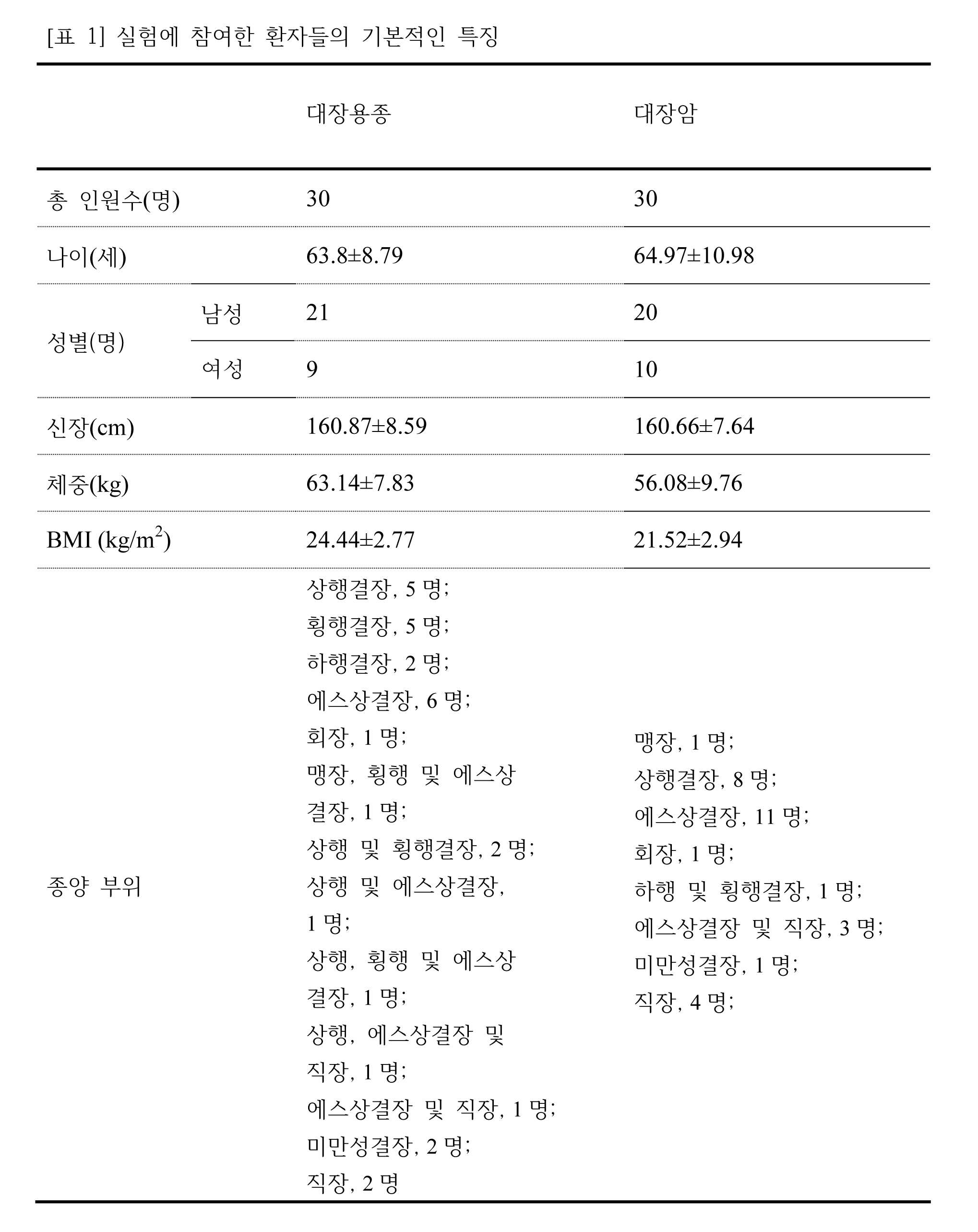
본 발명에서는 계명대학교 동산메디컬센터에서 공급된 30명의 대장용종 환자, 30명의 대장암 환자의 혈액을 사용하였다. 실험에 참여한 환자들의 기본적인 특징을 [표 1]에 나타내었다. 대장용종 환자들은 남성 21명(70%)과 여성 9명(30%)이었고, 대장암 환자들은 남성 20명(66.7%)과 여성 10명(33.3%)으로 구성되었다. BMI를 제외하고 나이,성별에 관해서 두 그룹사이에 유의한 차이는 없었다. 실험 프로토콜은 계명대학교 의과대학 기관생명윤리위원회의 윤리평가위원회에서 승인을 받았다. 혈액샘플은 EDTA-tube(BD,프랭클린 레이크,뉴저지,미국)에 담아서 원심분리(3,000×g, 10분)하여 혈장을 분리한 후 추가실험이 있을 때까지 -80℃에 보관하였다. 혈장의 단백질농도는 단백질분석 염색시약농축액(바이오라드,헤라클레스,캘리포니아,미국)을 이용하여 브래드포드 방법에 의해 결정되었다.In the present invention, 30 patients with colorectal polyps and 30 patients with colorectal cancer were supplied from Dongsan Medical Center of Keimyung University. The basic characteristics of the patients participating in the experiment are shown in [Table 1]. Colon cancer patients were composed of 21 males (70%) and 9 females (30%) and 20 males (66.7%) and 10 females (33.3%). There was no significant difference between the two groups with respect to age and gender except BMI. The experimental protocol was approved by the Ethics Evaluation Committee of the Institutional Bioethics Committee of Keimyung University Medical School. Blood samples were stored in EDTA-tube (BD, Franklin Lake, New Jersey, USA) and centrifuged (3,000 × g, 10 min) to separate plasma and stored at -80 ° C. until further experiments. Plasma protein concentrations were determined by the Bradford method using a protein assay stain reagent concentrate (BioRad, Hercules, CA, USA).
실시예Example 2: 이차원 전기영동 분석(2- 2: Two-dimensional electrophoresis analysis (2- DEDE analysis분석 ))
상기에서 수득한 총 60명 환자들(30명의 대장용종 환자들 및 30명의 대장암 환자들)의 혈장샘플을 이용하여 이차원 전기영동을 수행하였다. 간단히, IPG 스트립(pH 4-7,18cm; GE헬스케어,버킹엄셔,영국)을 효과적인 단백질체 분석을 위해 알부민을 제거하지 않은 완전한 혈장 단백질 43㎍ 및 7M 우레아(바이오베이직,온타리오,캐나다), 2M 티오우레아(시그마,세인트루이스,미주리,미국), 4% CHAPS(바이오베이직), 1mM PMSF(시그마), 20mM DTT(GE헬스케어), 2% IPG 버퍼(바이오라드)를 포함하는 재수화용액 350μL 을 담은 스트립홀더에서 26시간 동안 방치하여 재수화시켰다. IEF는 PROTEIN IEF Cell(바이오라드)을 따라 수행되었다: 250V에서 15분, 250-10,000V에서 3시간, 10,000V에서 6시간, 그 다음 2차원을 진행하기 전까지 500V에서 유지하였다. 초점을 맞춘 스트립은 6M 우레아, 2% SDS(제너리바이오텍,상하이,중국), 1% DTT, 30% 글리세롤(바이오베이직) 및 50mM Tris-HCl(pH 6.8)을 함유하는 용액에서 20분 동안 평형을 이루었고, 뒤이어 DTT를 2% 요오드아세트아미드 (바이오라드)로 대체한 것을 제외하고는 상기와 동일한 용액에 추가로 20분동안 더 배양하였다. 그 다음 2차원에서 분해를 위해 20×20cm 크기의 12% 폴리아크릴아미드겔 위에 겔스트립을 두었고, 14시간 동안 겔마다 15mA의 전압을 계속 흘려주면서 Laemmli SDS-불연속시스템을 이용하여 분획법을 수행하였다. 영상분석 및 펩티드 질량 핑거프린팅(PMF)을 위해 단백질이 분리된 총 60개의 겔을 실버스테이닝하여 시각화하였다.
Two-dimensional electrophoresis was performed using plasma samples from a total of 60 patients (30 colonic polyps and 30 colorectal cancer patients) obtained above. Briefly, IPG strips (pH 4-7, 18 cm; GE Healthcare, Buckinghamshire, UK) were coated with 43 μg of complete plasma protein and 7 M urea (BioBasic, Ontario, Canada) 350 μL of rehydration solution containing thiourea (Sigma, St. Louis, Missouri, USA), 4% CHAPS (Bio Basic), 1 mM PMSF (Sigma), 20 mM DTT (GE Healthcare), 2% IPG buffer The flesh was left in the strip holder for 26 hours to rehydrate. The IEF was performed according to PROTEIN IEF Cell (Biorad): 15 minutes at 250V, 3 hours at 250-10,000V, 6 hours at 10,000V, and 500V before proceeding to the second dimension. The focused strips were equilibrated for 20 min in a solution containing 6 M urea, 2% SDS (Generi Biotech, Shanghai, China), 1% DTT, 30% glycerol (BioBasic) and 50 mM Tris- , Followed by further incubation for 20 minutes in the same solution as above except that DTT was replaced with 2% iodoacetamide (biorad). The gel strips were then placed on 12% polyacrylamide gels of 20x20 cm size for dissolution in two dimensions and fractionation was carried out using a Laemmli SDS-discontinuous system with a constant flow of 15 mA per gel for 14 hours . A total of 60 gels separated from the protein for visual analysis and peptide mass fingerprinting (PMF) were visualized by silver staining.
1차원에서 pH 4-7 IEF 스트립을 이용하고, 2차원에서 12% w/v SDS-PAGE겔을 이용하여 인간 혈장단백질을 분리한 결과, 327개의 반점들이 감지되었다(도 1). 또, 두 그룹사이에 차등조절되는 24종의 단백질 변화가 두 겔에서 영상 및 PMF 분석을 이용하여 확인되었고(도 1), 확인된 세부사항은 [표 2]에 열거하였다. A human plasma protein was isolated using a pH 4-7 IEF strip in one dimension and a 12% w / v SDS-PAGE gel in two dimensions, resulting in 327 spots (FIG. 1). In addition, 24 protein modifications that were differentially modulated between the two groups were identified using image and PMF analysis on two gels (Figure 1), and the identified details are listed in Table 2.

대장용종 및 대장암 환자들 사이의 혈장단백질 조절 프로파일에 대한 전반적인 비교를 위해서 각 그룹의 30개 겔에 나타나는 반점들을 이용하여 산포도를 만들었다(도 2). 도 2의 산포도는 두 환자그룹 사이의 반점 밀도를 한눈에 비교하기 위해 작성되었다. X- 및 Y-축은 각 반점의 volume(%)를 나타낸다. 각 빨간색 반점 및 파란색 반점은 각각 대장용종 환자보다 대장암 환자의 활성화(C>A) 및 활성억제(C<A)된 단백질을 각각 나타낸다(p<0.05).
For an overall comparison of plasma protein regulation profiles between colonic polyp and colorectal cancer patients, scatterplots were made using spots appearing in 30 gels of each group (Fig. 2). The scatter plot of FIG. 2 was created to compare the density of spots between two patient groups at a glance. The X- and Y-axes represent the volume (%) of each spot. Each red spot and blue spot showed protein activation (C> A) and inhibition (C <A) protein in colorectal cancer patients, respectively (p <0.05).
실시예Example 3: 영상수집 및 자료분석 3: Image collection and data analysis
겔은 UMAX PowerLook 1120(맥시엄 테스놀로지,애크론,오하이오,미국)로 이미지화되었고, modified ImageMaster 2D 소프트웨어V4.95(GE 헬스케어)을 이용하여 수행된 그룹들 간의 이미지들을 비교하였다. 모든 겔에서 검출된 반점들은 대장용종 환자그룹의 겔에서 선택된 참조 겔의 반점과 매치되었다. 상대흡광도(relative optical density) 및 상대부피(relative volume)는 겔 염색 중간에 차이를 수정할 목적으로 산출되었고, 각 반점 강도부피(intensity volume)는 배경제거(background subtraction)와 전체 반점부피의 표준화(total spot volume normalization)에 의해 처리되었다. 그 결과 초래된 반점 부피비율(spot volume percentage)은 그룹간의 비교를 위해 사용되었다.
The gels were imaged with a UMAX PowerLook 1120 (Maxi Tester, Akron, Ohio, USA) and images were compared between groups performed using the modified ImageMaster 2D software V4.95 (GE Healthcare). The spots detected in all the gels matched the spots of the reference gels selected in the gel of the colonic polyp group. The relative optical density and relative volume were calculated for the purpose of correcting the difference in the middle of the gel staining and the intensity intensity of each spot was calculated by subtracting the background subtraction and the standardization of the total spot volume spot volume normalization. The resulting spot volume percentage was used for comparison between groups.
실시예Example 4: 펩티드 질량 핑거프린팅( 4: Peptide mass fingerprinting ( PMFPMF ))
페르난데즈 등(1998)이 서술한대로 단백질 반점을 절제하여 트립신으로 분해한 다음 α-cyano-4-hydroxycinnamic acid(CHCA;시그마)를 포함한 50% 아세토니트릴/0.1% 트리플루오로아세트산과 혼합하여 MALDI-TOF분석(마이크로플렉스 LRF20, 브룩커 달토닉스)에 이용하였다. 600-3,000 m/z범위 이상인 스펙트럼마다 300shots을 수집하였고 트립신 자동분해 피크(m/z 842.5099, 2211.1046)를 이용하여 두 점 내부눈금에 의해 눈금이 매겨졌다. 피크 리스트는 Flex Analysis 3.0으로 만들었다. 피크를 뽑는데 이용한 역치는 단동위질량의 최소분리능은 500, S/N은 5였다. 매트릭스사이언스(http://www.matrixscience.com)에서 만든 검색 프로그램 MASCOT(마스콧서버2.3)이 사용되었고, 확률기반의 MOWSE(분자량검색)점수가 PMF를 위해 산출되었다. 자료검색을 위해 절단효소로서 트립신, 최대 하나의 손실된 절단, 완전한 변형으로서 요오드아세트아미드(Cys), 부분적 변형으로서 산화(Met), 단동위질량 및 질량 오차범위±0.1Da을 포함하는 파라미터들을 이용하였다. PMF의 허용기준은 확률점수이다:-10*Log(P), P는 관찰된 매치가 우연한 일이고 66이상인 점수는 유의한 확률이다(p<0.05).
Protein spots were excised and digested with trypsin as described by Fernandez et al. (1998) and then mixed with 50% acetonitrile / 0.1% trifluoroacetic acid containing α-cyano-4-hydroxycinnamic acid (CHCA; Sigma) Analysis (Microflex LRF20, Brockerdaltonics). 300 shots were collected for spectra above the range of 600-3,000 m / z and were scaled by two point internal scales using the trypsin autolysis peak (m / z 842.5099, 2211.1046). The peak list was created with Flex Analysis 3.0. The minimum threshold for single peak mass was 500 and S / N was 5. A search program MASCOT (Mascot Server 2.3) created by Matrix Science (http://www.matrixscience.com) was used, and probability based MOWSE (Molecular Weight Search) scores were calculated for the PMF. For data retrieval, we used parameters including trypsin as a cleavage enzyme, maximal one lost cleavage, iodoacetamide (Cys) as a complete strain, oxidation (Met) as a partial strain, single-acting mass and mass error range ± 0.1 Da Respectively. The PMF acceptance criterion is the probability score: -10 * Log (P), P is the probability that the observed match is a coincidence and a score of 66 or more is significant (p <0.05).
두 환자그룹 사이에 차별적으로 조절되는 24종의 단백질을 동정하였고, 그의 증가 및 감소되는 패턴을 비교하였다. 이 중에서 대장용종 환자보다 대장암 환자에게서 11종의 단백질들이 유의하게 증가한 반면에, 13종의 단백질들은 유의하게 감소하였다. 증가한 단백질들은 서열번호 1의 LRG, 서열번호 2의 HBB, AHSG, 서열번호 3의 IgA2C, 보체인자 9, Apo E, Hpα2, A2M, 서열번호 4의 CFB, A1AT 및 KNG1을 포함한 반면에, 감소한 단백질들은 TTR, 서열번호 6의 ZAG, RBP4, Apo A-I, GSN, IgH, 서열번호 6의 afamin, 서열번호 8의 AACT, afamin, 서열번호 7의 VTN, ITIH2, Apo M, Apo C-Ⅲ 및 Agt이었다(표 2). 상기 24종의 단백질 중에서, 두 그룹사이에 1.5-fold(p<0.05)change 이상을 보이는 9종의 단백질 혈장의 레벨은 GraphPad Presm 6을 이용하여 상자 및 수염그래프로 나타냈다(도 3). Twenty - four differentially regulated proteins were identified among the two patient groups, and their increased and decreased patterns were compared. Of these, 11 proteins were significantly increased in colorectal cancer patients compared to colon polyps, while 13 proteins were significantly decreased. The increased proteins include the LRG of SEQ ID NO: 1, the HBB of SEQ ID NO: 2, the IgG2C of SEQ ID NO: 3, the
데이터는 각 그룹마다 30개의 겔에서 변화된 volume density(%)의 평균±표준편차로 나타냈고 3D 도형 및 줌인 영상은 modified ImageMaster 2D 소프트웨어 V4.95를 이용하여 제작되었다.
Data were expressed as mean ± standard deviation of volume density (%) in 30 gels per group, and 3D shapes and zoom in images were generated using modified ImageMaster 2D software V4.95.
실시예Example 5: 5: 면역블롯팅Immunoblotting 분석( analysis( ImmunoblotImmunoblot analysis분석 ))
단백질체 분석을 통해 동정된 단백질 6종의 레벨을 면역블롯팅 분석하여 유효성을 검사하였다. 혈장의 부분표본(70㎍)을 5X 샘플버퍼(pH 6.8인 50mM Tris, 2% SDS, 10% 글리세롤, 5% β-머캅토에탄올 및 0.1% 브로모페놀블루)에 희석하였고 95℃에서 3분 동안 열처리하였다. SDS-폴리아크릴아미드 겔 전기영동한 후에 단백질을 폴리비닐리덴 디플루오라이드(PVEF,산타크루즈 바이오테크놀로지,산타크루즈,캘리포니아,미국) 막에 옮긴다음 5% 탈지분유를 함유한 TBS-T버퍼로 1시간 동안 블록킹하였다. 그 다음 TBS-T버퍼를 3회 바꾸어가며 막을 15분 동안 세척하였고, 뒤이어 5% 탈지분유를 함유한 TBS-T버퍼에 1:1,000으로 희석한 주요 다중클론항체 (anti-LRG, anti-HBB, anti-AHSG, anti-ZAG, anti-RBP4, anti-Apo A-I 또는 anti-β-actin; 산타크루즈 바이오테크놀로지)와 함께 90분 동안 배양하였다. 3회 세척한 후, 막을 horseradish peroxidase-conjugated anti-goat IgG, anti-mouse IgG 또는 anti-rabbit IgG 2차항체(산타크루즈)와 함께 1% 탈지분유를 함유한 TBS-T버퍼에 1:1,000으로 희석하여 1시간 동안 배양하였고 UMAX PowerLook 1120으로 스캔함으로써 밴드들을 디지털화한 후 영상분석 소프트웨어(KODAK 1D,애머샴 코닥,로체스터,뉴욕,미국)를 이용하여 분석하였다.
The levels of the six proteins identified through protein body analysis were analyzed by immunoblotting and validated. A partial sample (70 μg) of plasma was diluted in 5 × sample buffer (50 mM Tris, 2% SDS, 10% glycerol, 5% β-mercaptoethanol and 0.1% bromophenol blue at pH 6.8) Lt; / RTI > After SDS-polyacrylamide gel electrophoresis, proteins were transferred to polyvinylidene difluoride (PVEF, Santa Cruz Biotechnology, Santa Cruz, Calif., USA) membranes and incubated in TBS-T buffer containing 5% Lt; / RTI > The membranes were then washed for 15 minutes with 3 changes of TBS-T buffer followed by the addition of a primary polyclonal antibody (anti-LRG, anti-HBB, anti-HBs) diluted 1: 1,000 in TBS- anti-AHSG, anti-ZAG, anti-RBP4, anti-Apo AI or anti-beta-actin; Santa Cruz Biotechnology) for 90 minutes. After washing three times, the membranes were incubated with 1: 1,000 TBS-T buffer containing 1% skimmed milk powder with horseradish peroxidase-conjugated anti-goat IgG, anti-mouse IgG or anti-rabbit IgG secondary antibody (Santa Cruz) Bands were digitized by scanning with UMAX PowerLook 1120 and analyzed using image analysis software (KODAK 1D, Amersham Kodak, Rochester, New York, USA).
본 단백질체 분석에 있어서 기술적 오차 및 인위적인 영향의 가능성을 배제하기 위해서, 대장암 환자그룹에서 11종의 증가한 단백질 중의 3종(LRG, HBB 및 AHSG)과 13종의 감소한 단백질 중의 3종(ZAG, RBP4 및 Apo A-I)의 레벨을 각 그룹에서 환자 30명에서 수득한 풀링 플라즈마를 이용하여 3회 반복하여 면역블롯팅 분석을 수행하였다. 상기 6종의 단백질 혈장의 레벨은 단백질체 분석 결과대로 일렬로 위치하였다(도 4). 데이터는 3번의 독립적인 실험을 통해 얻었고 Relative intensity(%)는 β-actin 수준으로 정상화된 이후의 단백질값이다.
(LRG, HBB, and AHSG) and three of the 13 reduced proteins (ZAG, RBP4, and RBP4) in eleven proteins in colorectal cancer patients to exclude the possibility of technical errors and artifacts ≪ / RTI > and Apo < RTI ID = 0.0 > AI) < / RTI > were repeated three times using pooling plasmids obtained from 30 patients in each group for immunoblot analysis. The levels of the six protein plasma samples were aligned in line with the results of the protein assay (Fig. 4). Data were obtained from three independent experiments and the relative intensity (%) was the protein value after normalization to the β-actin level.
실시예Example 6: 사이토카인/ 6: cytokine / 케모카인Chemokine 배열 Arrangement
대장용종(20명) 및 대장암(20명) 환자들 각각에서 26종의 사이토카인 및 케모카인을 Milliplex MAP Human Cytokine/Chemokine Kit 96 Well Plate Assay(Pre-mixed 26 Plex kit;Millipore Corp.,St.Charles,일리노이,미국)으로 중복 측정하였다. Pre-mixed 26 Plex Kit는 Eotaxin, G-CSF, GM-CSF, IFN-α2, IFN-γ, IL-1α,IL-1β, IL-2, IL-3, IL-4 , IL-5, IL-6, IL-7, IL-8, IL-10, IL-12(p40), IL-12(p70), IL-13, IL-15, IL-17 ,IP-10, MCP-1, MIP-1α, MIP-1β, TNF-α 및 TNF-β를 포함한다. 형광은 Luminex 200 reader(Luminex,오스틴,텍사스,미국)로 측정하였고, 데이터는 MasterPlex QT 2010(히타치솔루션 아메리카의 미래바이오그룹, 샌프란시스코, 캘리포니아, 미국)으로 분석하였다. 측정 및 분석은 제조업자의 지시에 따라 수행되었다.
Twenty-six cytokines and chemokines were measured in each of the colonic polyps (20 patients) and colorectal cancer patients (20 patients) using the Milliplex MAP Human Cytokine / Chemokine Kit 96 Well Plate Assay (Pre-mixed 26 Plex kit; Charles, Illinois, USA). Pre-mixed 26 Plex Kits were used for the treatment of Eotaxin, G-CSF, GM-CSF, IFN-α2, IFN-γ, IL-1α, IL-1β, IL-2, IL-3, IL- 10, MCP-1, MIP-6, IL-7, IL-8, IL-10, IL-12 (p40), IL- -1 alpha, MIP-1 [beta], TNF- [alpha] and TNF- [beta]. Fluorescence was measured with a
대장암과 관련있거나 대장용종에서 대장암으로의 진행에 관련된 사이토카인 및 케모카인을 동정하기 위해서 멀티플렉스 비드 분석을 이용하여 혈장 내 26종의 사이토카인 및 케모카인 농도를 측정한 결과, 그 중에서 IL-8(1.89-fold;p=0.003), IP-10(1.75-fold;p=0.024) 및 TNF-α(1.62-fold;p=0.042)의 수준이 대장용종 환자보다 대장암 환자의 혈장에서 유의적으로 높았고(도 5), IL-10(1.73-fold 증가), G-CSF(1.51-fold 감소), GM-CSF(1.9-fold 감소) 및 IL-6(2.06-fold 감소)을 포함하여 다른 분자들은 통계적 유의성없이 두 그룹사이에 검출할 수 있는 fold change를 보였다(표 3). 도 5에서 각 반점은 중복실험의 평균값을 나타낸다. In order to identify cytokines and chemokines involved in colorectal cancer-related or colorectal to colorectal cancer progression, 26 cytokines and chemokines in plasma were measured using multiplex bead analysis. Among them, IL-8 (1.98-fold; p = 0.003), IP-10 (1.75-fold; p = 0.024) and TNF-α (1.62-fold; p = 0.042) were significantly higher in patients with colorectal cancer (Fig. 5), and other (including increased IL-10 (1.73-fold), G-CSF (1.51-fold reduction), GM-CSF (1.9- fold reduction) and IL- The molecules showed detectable fold changes between the two groups without statistical significance (Table 3). In FIG. 5, each spot represents the average value of the duplicate experiments.

실시예Example 7: 통계적 분석 7: Statistical analysis
모든 데이터는 Statistical Package of Social Science(SPSS, 버전14.0K)프로그램을 이용하여 일원분산분석(ANOVA)에 의해 비교되었다:결과는 평균±표준편차로 나타내었다. 그룹평균은 ANOVA가 전체적이고 유의미한 치료효과(p<0.05)를 나타낼 때 보호되는 최소유의차(LSD)기술에 의해 결정되었듯이 p<0.05에서 현저히 달랐다.
All data were compared by one-way analysis of variance (ANOVA) using the Statistical Package of Social Science (SPSS, version 14.0K) program: Results are expressed as mean ± SD. Group mean was significantly different at p <0.05 as determined by the least significant difference (LSD) technique protected when ANOVA had a global and significant therapeutic effect (p <0.05).
<110> Daugu University IACF For Advanced Technology <120> Proteomic and cytokine plasma biomarkers for predicting progression from colorectal adenoma to carcinoma in human patients <130> p5000 <160> 8 <170> KopatentIn 2.0 <210> 1 <211> 347 <212> PRT <213> Homo sapiens <400> 1 Met Ser Ser Trp Ser Arg Gln Arg Pro Lys Ser Pro Gly Gly Ile Gln 1 5 10 15 Pro His Val Ser Arg Thr Leu Phe Leu Leu Leu Leu Leu Ala Ala Ser 20 25 30 Ala Trp Gly Val Thr Leu Ser Pro Lys Asp Cys Gln Val Phe Arg Ser 35 40 45 Asp His Gly Ser Ser Ile Ser Cys Gln Pro Pro Ala Glu Ile Pro Gly 50 55 60 Tyr Leu Pro Ala Asp Thr Val His Leu Ala Val Glu Phe Phe Asn Leu 65 70 75 80 Thr His Leu Pro Ala Asn Leu Leu Gln Gly Ala Ser Lys Leu Gln Glu 85 90 95 Leu His Leu Ser Ser Asn Gly Leu Glu Ser Leu Ser Pro Glu Phe Leu 100 105 110 Arg Pro Val Pro Gln Leu Arg Val Leu Asp Leu Thr Arg Asn Ala Leu 115 120 125 Thr Gly Leu Pro Ser Gly Leu Phe Gln Ala Ser Ala Thr Leu Asp Thr 130 135 140 Leu Val Leu Lys Glu Asn Gln Leu Glu Val Leu Glu Val Ser Trp Leu 145 150 155 160 His Gly Leu Lys Ala Leu Gly His Leu Asp Leu Ser Gly Asn Arg Leu 165 170 175 Arg Lys Leu Pro Pro Gly Leu Leu Ala Asn Phe Thr Leu Leu Arg Thr 180 185 190 Leu Asp Leu Gly Glu Asn Gln Leu Glu Thr Leu Pro Pro Asp Leu Leu 195 200 205 Arg Gly Pro Leu Gln Leu Glu Arg Leu His Leu Glu Gly Asn Lys Leu 210 215 220 Gln Val Leu Gly Lys Asp Leu Leu Leu Pro Gln Pro Asp Leu Arg Tyr 225 230 235 240 Leu Phe Leu Asn Gly Asn Lys Leu Ala Arg Val Ala Ala Gly Ala Phe 245 250 255 Gln Gly Leu Arg Gln Leu Asp Met Leu Asp Leu Ser Asn Asn Ser Leu 260 265 270 Ala Ser Val Pro Glu Gly Leu Trp Ala Ser Leu Gly Gln Pro Asn Trp 275 280 285 Asp Met Arg Asp Gly Phe Asp Ile Ser Gly Asn Pro Trp Ile Cys Asp 290 295 300 Gln Asn Leu Ser Asp Leu Tyr Arg Trp Leu Gln Ala Gln Lys Asp Lys 305 310 315 320 Met Phe Ser Gln Asn Asp Thr Arg Cys Ala Gly Pro Glu Ala Val Lys 325 330 335 Gly Gln Thr Leu Leu Ala Val Ala Lys Ser Gln 340 345 <210> 2 <211> 147 <212> PRT <213> Homo sapiens <400> 2 Met Val His Leu Thr Pro Glu Glu Lys Ser Ala Val Thr Ala Leu Trp 1 5 10 15 Gly Lys Val Asn Val Asp Glu Val Gly Gly Glu Ala Leu Gly Arg Leu 20 25 30 Leu Val Val Tyr Pro Trp Thr Gln Arg Phe Phe Glu Ser Phe Gly Asp 35 40 45 Leu Ser Thr Pro Asp Ala Val Met Gly Asn Pro Lys Val Lys Ala His 50 55 60 Gly Lys Lys Val Leu Gly Ala Phe Ser Asp Gly Leu Ala His Leu Asp 65 70 75 80 Asn Leu Lys Gly Thr Phe Ala Thr Leu Ser Glu Leu His Cys Asp Lys 85 90 95 Leu His Val Asp Pro Glu Asn Phe Arg Leu Leu Gly Asn Val Leu Val 100 105 110 Cys Val Leu Ala His His Phe Gly Lys Glu Phe Thr Pro Pro Val Gln 115 120 125 Ala Ala Tyr Gln Lys Val Val Ala Gly Val Ala Asn Ala Leu Ala His 130 135 140 Lys Tyr His 145 <210> 3 <211> 220 <212> PRT <213> Homo sapiens <400> 3 Leu Glu Asp Leu Leu Leu Gly Ser Glu Ala Asn Leu Thr Cys Thr Leu 1 5 10 15 Thr Gly Leu Arg Asp Ala Ser Gly Ala Thr Phe Thr Trp Thr Pro Ser 20 25 30 Ser Gly Lys Ser Ala Val Gln Gly Pro Pro Glu Arg Asp Leu Cys Gly 35 40 45 Cys Tyr Ser Val Ser Ser Val Leu Pro Gly Cys Ala Gln Pro Trp Asn 50 55 60 His Gly Glu Thr Phe Thr Cys Thr Ala Ala His Pro Glu Leu Lys Thr 65 70 75 80 Pro Leu Thr Ala Asn Ile Thr Lys Ser Gly Asn Thr Phe Arg Pro Glu 85 90 95 Val His Leu Leu Pro Pro Pro Ser Glu Glu Leu Ala Leu Asn Glu Leu 100 105 110 Val Thr Leu Thr Cys Leu Ala Arg Gly Phe Ser Pro Lys Asp Val Leu 115 120 125 Val Arg Trp Leu Gln Gly Ser Gln Glu Leu Pro Arg Glu Lys Tyr Leu 130 135 140 Thr Trp Ala Ser Arg Gln Glu Pro Ser Gln Gly Thr Thr Thr Tyr Ala 145 150 155 160 Val Thr Ser Ile Leu Arg Val Ala Ala Glu Asp Trp Lys Lys Gly Glu 165 170 175 Thr Phe Ser Cys Met Val Gly His Glu Ala Leu Pro Leu Ala Phe Thr 180 185 190 Gln Lys Thr Ile Asp Arg Met Ala Gly Lys Pro Thr His Ile Asn Val 195 200 205 Ser Val Val Met Ala Glu Ala Asp Gly Thr Cys Tyr 210 215 220 <210> 4 <211> 752 <212> PRT <213> Homo sapiens <400> 4 Gly Ser His His His His His His Gly Ser Thr Pro Trp Ser Leu Ala 1 5 10 15 Arg Pro Gln Gly Ser Cys Ser Leu Glu Gly Val Glu Ile Lys Gly Gly 20 25 30 Ser Phe Arg Leu Leu Gln Glu Gly Gln Ala Leu Glu Tyr Val Cys Pro 35 40 45 Ser Gly Phe Tyr Pro Tyr Pro Val Gln Thr Arg Thr Cys Arg Ser Thr 50 55 60 Gly Ser Trp Ser Thr Leu Lys Thr Gln Asp Gln Lys Thr Val Arg Lys 65 70 75 80 Ala Glu Cys Arg Ala Ile His Cys Pro Arg Pro His Asp Phe Glu Asn 85 90 95 Gly Glu Tyr Trp Pro Arg Ser Pro Tyr Tyr Asn Val Ser Asp Glu Ile 100 105 110 Ser Phe His Cys Tyr Asp Gly Tyr Thr Leu Arg Gly Ser Ala Asn Arg 115 120 125 Thr Cys Gln Val Asn Gly Arg Trp Ser Gly Gln Thr Ala Ile Cys Asp 130 135 140 Asn Gly Ala Gly Tyr Cys Ser Asn Pro Gly Ile Pro Ile Gly Thr Arg 145 150 155 160 Lys Val Gly Ser Gln Tyr Arg Leu Glu Asp Ser Val Thr Tyr His Cys 165 170 175 Ser Arg Gly Leu Thr Leu Arg Gly Ser Gln Arg Arg Thr Cys Gln Glu 180 185 190 Gly Gly Ser Trp Ser Gly Thr Glu Pro Ser Cys Gln Asp Ser Phe Met 195 200 205 Tyr Asp Thr Pro Gln Glu Val Ala Glu Ala Phe Leu Ser Ser Leu Thr 210 215 220 Glu Thr Ile Glu Gly Val Asp Ala Glu Asp Gly His Gly Pro Gly Glu 225 230 235 240 Gln Gln Lys Arg Lys Ile Val Leu Asp Pro Ser Gly Ser Met Asn Ile 245 250 255 Tyr Leu Val Leu Asp Gly Ser Asp Ser Ile Gly Ala Ser Asn Phe Thr 260 265 270 Gly Ala Lys Lys Cys Leu Val Asn Leu Ile Glu Lys Val Ala Ser Tyr 275 280 285 Gly Val Lys Pro Arg Tyr Gly Leu Val Thr Tyr Ala Thr Tyr Pro Lys 290 295 300 Ile Trp Val Lys Val Ser Glu Ala Asp Ser Ser Asn Ala Asp Trp Val 305 310 315 320 Thr Lys Gln Leu Asn Glu Ile Asn Tyr Glu Asp His Lys Leu Lys Ser 325 330 335 Gly Thr Asn Thr Lys Lys Ala Leu Gln Ala Val Tyr Ser Met Met Ser 340 345 350 Trp Pro Asp Asp Val Pro Pro Glu Gly Trp Asn Arg Thr Arg His Val 355 360 365 Ile Ile Leu Met Thr Asp Gly Leu His Asn Met Gly Gly Asp Pro Ile 370 375 380 Thr Val Ile Asp Glu Ile Arg Asp Leu Leu Tyr Ile Gly Lys Asp Arg 385 390 395 400 Lys Asn Pro Arg Glu Asp Tyr Leu Asp Val Tyr Val Phe Gly Val Gly 405 410 415 Pro Leu Val Asn Gln Val Asn Ile Asn Ala Leu Ala Ser Lys Lys Asp 420 425 430 Asn Glu Gln His Val Phe Lys Val Lys Asp Met Glu Asn Leu Glu Asp 435 440 445 Val Phe Tyr Gln Met Ile Asp Glu Ser Gln Ser Leu Ser Leu Cys Gly 450 455 460 Met Val Trp Glu His Arg Lys Gly Thr Asp Tyr His Lys Gln Pro Trp 465 470 475 480 Gln Ala Lys Ile Ser Val Ile Arg Pro Ser Lys Gly His Glu Ser Cys 485 490 495 Met Gly Ala Val Val Ser Glu Tyr Phe Val Leu Thr Ala Ala His Cys 500 505 510 Phe Thr Val Asp Asp Lys Glu His Ser Ile Lys Val Ser Val Gly Gly 515 520 525 Glu Lys Arg Asp Leu Glu Ile Glu Val Val Leu Phe His Pro Asn Tyr 530 535 540 Asn Ile Asn Gly Lys Lys Glu Ala Gly Ile Pro Glu Phe Tyr Asp Tyr 545 550 555 560 Asp Val Ala Leu Ile Lys Leu Lys Asn Lys Leu Lys Tyr Gly Gln Thr 565 570 575 Ile Arg Pro Ile Cys Leu Pro Cys Thr Glu Gly Thr Thr Arg Ala Leu 580 585 590 Arg Leu Pro Pro Thr Thr Thr Cys Gln Gln Gln Lys Glu Glu Leu Leu 595 600 605 Pro Ala Gln Asp Ile Lys Ala Leu Phe Val Ser Glu Glu Glu Lys Lys 610 615 620 Leu Thr Arg Lys Glu Val Tyr Ile Lys Asn Gly Asp Lys Lys Gly Ser 625 630 635 640 Cys Glu Arg Asp Ala Gln Tyr Ala Pro Gly Tyr Asp Lys Val Lys Asp 645 650 655 Ile Ser Glu Val Val Thr Pro Arg Phe Leu Cys Thr Gly Gly Val Ser 660 665 670 Pro Tyr Ala Asp Pro Asn Thr Cys Arg Gly Asp Ser Gly Gly Pro Leu 675 680 685 Ile Val His Lys Arg Ser Arg Phe Ile Gln Val Gly Val Ile Ser Trp 690 695 700 Gly Val Val Asp Val Cys Lys Asn Gln Lys Arg Gln Lys Gln Val Pro 705 710 715 720 Ala His Ala Arg Asp Phe His Ile Asn Leu Phe Gln Val Leu Pro Trp 725 730 735 Leu Lys Glu Lys Leu Gln Asp Glu Asp Leu Gly Phe Leu Ala Ala Ala 740 745 750 <210> 5 <211> 599 <212> PRT <213> Homo sapiens <400> 5 Met Lys Leu Leu Lys Leu Thr Gly Phe Ile Phe Phe Leu Phe Phe Leu 1 5 10 15 Thr Glu Ser Leu Thr Leu Pro Thr Gln Pro Arg Asp Ile Glu Asn Phe 20 25 30 Asn Ser Thr Gln Lys Phe Ile Glu Asp Asn Ile Glu Tyr Ile Thr Ile 35 40 45 Ile Ala Phe Ala Gln Tyr Val Gln Glu Ala Thr Phe Glu Glu Met Glu 50 55 60 Lys Leu Val Lys Asp Met Val Glu Tyr Lys Asp Arg Cys Met Ala Asp 65 70 75 80 Lys Thr Leu Pro Glu Cys Ser Lys Leu Pro Asn Asn Val Leu Gln Glu 85 90 95 Lys Ile Cys Ala Met Glu Gly Leu Pro Gln Lys His Asn Phe Ser His 100 105 110 Cys Cys Ser Lys Val Asp Ala Gln Arg Arg Leu Cys Phe Phe Tyr Asn 115 120 125 Lys Lys Ser Asp Val Gly Phe Leu Pro Pro Phe Pro Thr Leu Asp Pro 130 135 140 Glu Glu Lys Cys Gln Ala Tyr Glu Ser Asn Arg Glu Ser Leu Leu Asn 145 150 155 160 His Phe Leu Tyr Glu Val Ala Arg Arg Asn Pro Phe Val Phe Ala Pro 165 170 175 Thr Leu Leu Thr Val Ala Val His Phe Glu Glu Val Ala Lys Ser Cys 180 185 190 Cys Glu Glu Gln Asn Lys Val Asn Cys Leu Gln Thr Arg Ala Ile Pro 195 200 205 Val Thr Gln Tyr Leu Lys Ala Phe Ser Ser Tyr Gln Lys His Val Cys 210 215 220 Gly Ala Leu Leu Lys Phe Gly Thr Lys Val Val His Phe Ile Tyr Ile 225 230 235 240 Ala Ile Leu Ser Gln Lys Phe Pro Lys Ile Glu Phe Lys Glu Leu Ile 245 250 255 Ser Leu Val Glu Asp Val Ser Ser Asn Tyr Asp Gly Cys Cys Glu Gly 260 265 270 Asp Val Val Gln Cys Ile Arg Asp Thr Ser Lys Val Met Asn His Ile 275 280 285 Cys Ser Lys Gln Asp Ser Ile Ser Ser Lys Ile Lys Glu Cys Cys Glu 290 295 300 Lys Lys Ile Pro Glu Arg Gly Gln Cys Ile Ile Asn Ser Asn Lys Asp 305 310 315 320 Asp Arg Pro Lys Asp Leu Ser Leu Arg Glu Gly Lys Phe Thr Asp Ser 325 330 335 Glu Asn Val Cys Gln Glu Arg Asp Ala Asp Pro Asp Thr Phe Phe Ala 340 345 350 Lys Phe Thr Phe Glu Tyr Ser Arg Arg His Pro Asp Leu Ser Ile Pro 355 360 365 Glu Leu Leu Arg Ile Val Gln Ile Tyr Lys Asp Leu Leu Arg Asn Cys 370 375 380 Cys Asn Thr Glu Asn Pro Pro Gly Cys Tyr Arg Tyr Ala Glu Asp Lys 385 390 395 400 Phe Asn Glu Thr Thr Glu Lys Ser Leu Lys Met Val Gln Gln Glu Cys 405 410 415 Lys His Phe Gln Asn Leu Gly Lys Asp Gly Leu Lys Tyr His Tyr Leu 420 425 430 Ile Arg Leu Thr Lys Ile Ala Pro Gln Leu Ser Thr Glu Glu Leu Val 435 440 445 Ser Leu Gly Glu Lys Met Val Thr Ala Phe Thr Thr Cys Cys Thr Leu 450 455 460 Ser Glu Glu Phe Ala Cys Val Asp Asn Leu Ala Asp Leu Val Phe Gly 465 470 475 480 Glu Leu Cys Gly Val Asn Glu Asn Arg Thr Ile Asn Pro Ala Val Asp 485 490 495 His Cys Cys Lys Thr Asn Phe Ala Phe Arg Arg Pro Cys Phe Glu Ser 500 505 510 Leu Lys Ala Asp Lys Thr Tyr Val Pro Pro Pro Phe Ser Gln Asp Leu 515 520 525 Phe Thr Phe His Ala Asp Met Cys Gln Ser Gln Asn Glu Glu Leu Gln 530 535 540 Arg Lys Thr Asp Arg Phe Leu Val Asn Leu Val Lys Leu Lys His Glu 545 550 555 560 Leu Thr Asp Glu Glu Leu Gln Ser Leu Phe Thr Asn Phe Ala Asn Val 565 570 575 Val Asp Lys Cys Cys Lys Ala Glu Ser Pro Glu Val Cys Phe Asn Glu 580 585 590 Glu Ser Pro Lys Ile Gly Asn 595 <210> 6 <211> 274 <212> PRT <213> Homo sapiens <400> 6 Asp Gly Arg Tyr Ser Leu Thr Tyr Ile Tyr Thr Gly Leu Ser Lys His 1 5 10 15 Val Glu Asp Val Pro Ala Phe Gln Ala Leu Gly Ser Leu Asn Asp Leu 20 25 30 Gln Phe Phe Arg Tyr Asn Ser Lys Asp Arg Lys Ser Gln Pro Met Gly 35 40 45 Leu Trp Arg Gln Val Glu Gly Met Glu Asp Trp Lys Gln Asp Ser Gln 50 55 60 Leu Gln Lys Ala Arg Glu Asp Ile Phe Met Glu Thr Leu Lys Asp Ile 65 70 75 80 Val Glu Tyr Tyr Asn Asp Ser Asn Gly Ser His Val Leu Gln Gly Arg 85 90 95 Phe Gly Cys Glu Ile Glu Asn Asn Arg Ser Ser Gly Ala Phe Trp Lys 100 105 110 Tyr Tyr Tyr Asp Gly Lys Asp Tyr Ile Glu Phe Asn Lys Glu Ile Pro 115 120 125 Ala Trp Val Pro Phe Asp Pro Ala Ala Gln Ile Thr Lys Gln Lys Trp 130 135 140 Glu Ala Glu Pro Val Tyr Val Gln Arg Ala Lys Ala Tyr Leu Glu Glu 145 150 155 160 Glu Cys Pro Ala Thr Leu Arg Lys Tyr Leu Lys Tyr Ser Lys Asn Ile 165 170 175 Leu Asp Arg Gln Asp Pro Pro Ser Val Val Val Thr Ser His Gln Ala 180 185 190 Pro Gly Glu Lys Lys Lys Leu Lys Cys Leu Ala Tyr Asp Phe Tyr Pro 195 200 205 Gly Lys Ile Asp Val His Trp Thr Arg Ala Gly Glu Val Gln Glu Pro 210 215 220 Glu Leu Arg Gly Asp Val Leu His Asn Gly Asn Gly Thr Tyr Gln Ser 225 230 235 240 Trp Val Val Val Ala Val Pro Pro Gln Asp Thr Ala Pro Tyr Ser Cys 245 250 255 His Val Gln His Ser Ser Leu Ala Gln Pro Leu Val Val Pro Trp Glu 260 265 270 Ala Ser <210> 7 <211> 478 <212> PRT <213> Homo sapiens <400> 7 Met Ala Pro Leu Arg Pro Leu Leu Ile Leu Ala Leu Leu Ala Trp Val 1 5 10 15 Ala Leu Ala Asp Gln Glu Ser Cys Lys Gly Arg Cys Thr Glu Gly Phe 20 25 30 Asn Val Asp Lys Lys Cys Gln Cys Asp Glu Leu Cys Ser Tyr Tyr Gln 35 40 45 Ser Cys Cys Thr Asp Tyr Thr Ala Glu Cys Lys Pro Gln Val Thr Arg 50 55 60 Gly Asp Val Phe Thr Met Pro Glu Asp Glu Tyr Thr Val Tyr Asp Asp 65 70 75 80 Gly Glu Glu Lys Asn Asn Ala Thr Val His Glu Gln Val Gly Gly Pro 85 90 95 Ser Leu Thr Ser Asp Leu Gln Ala Gln Ser Lys Gly Asn Pro Glu Gln 100 105 110 Thr Pro Val Leu Lys Pro Glu Glu Glu Ala Pro Ala Pro Glu Val Gly 115 120 125 Ala Ser Lys Pro Glu Gly Ile Asp Ser Arg Pro Glu Thr Leu His Pro 130 135 140 Gly Arg Pro Gln Pro Pro Ala Glu Glu Glu Leu Cys Ser Gly Lys Pro 145 150 155 160 Phe Asp Ala Phe Thr Asp Leu Lys Asn Gly Ser Leu Phe Ala Phe Arg 165 170 175 Gly Gln Tyr Cys Tyr Glu Leu Asp Glu Lys Ala Val Arg Pro Gly Tyr 180 185 190 Pro Lys Leu Ile Arg Asp Val Trp Gly Ile Glu Gly Pro Ile Asp Ala 195 200 205 Ala Phe Thr Arg Ile Asn Cys Gln Gly Lys Thr Tyr Leu Phe Lys Gly 210 215 220 Ser Gln Tyr Trp Arg Phe Glu Asp Gly Val Leu Asp Pro Asp Tyr Pro 225 230 235 240 Arg Asn Ile Ser Asp Gly Phe Asp Gly Ile Pro Asp Asn Val Asp Ala 245 250 255 Ala Leu Ala Leu Pro Ala His Ser Tyr Ser Gly Arg Glu Arg Val Tyr 260 265 270 Phe Phe Lys Gly Lys Gln Tyr Trp Glu Tyr Gln Phe Gln His Gln Pro 275 280 285 Ser Gln Glu Glu Cys Glu Gly Ser Ser Leu Ser Ala Val Phe Glu His 290 295 300 Phe Ala Met Met Gln Arg Asp Ser Trp Glu Asp Ile Phe Glu Leu Leu 305 310 315 320 Phe Trp Gly Arg Thr Ser Ala Gly Thr Arg Gln Pro Gln Phe Ile Ser 325 330 335 Arg Asp Trp His Gly Val Pro Gly Gln Val Asp Ala Ala Met Ala Gly 340 345 350 Arg Ile Tyr Ile Ser Gly Met Ala Pro Arg Pro Ser Leu Ala Lys Lys 355 360 365 Gln Arg Phe Arg His Arg Asn Arg Lys Gly Tyr Arg Ser Gln Arg Gly 370 375 380 His Ser Arg Gly Arg Asn Gln Asn Ser Arg Arg Pro Ser Arg Ala Met 385 390 395 400 Trp Leu Ser Leu Phe Ser Ser Glu Glu Ser Asn Leu Gly Ala Asn Asn 405 410 415 Tyr Asp Asp Tyr Arg Met Asp Trp Leu Val Pro Ala Thr Cys Glu Pro 420 425 430 Ile Gln Ser Val Phe Phe Phe Ser Gly Asp Lys Tyr Tyr Arg Val Asn 435 440 445 Leu Arg Thr Arg Arg Val Asp Thr Val Asp Pro Pro Tyr Pro Arg Ser 450 455 460 Ile Ala Gln Tyr Trp Leu Gly Cys Pro Ala Pro Gly His Leu 465 470 475 <210> 8 <211> 442 <212> PRT <213> Homo sapiens <400> 8 Thr Thr Pro Asp Arg Arg Leu Trp Asn Pro Pro Ala Thr Ser Ser Ser 1 5 10 15 Leu Arg Gln Met Glu Arg Met Leu Pro Leu Leu Thr Leu Gly Leu Leu 20 25 30 Ala Ala Gly Phe Cys Pro Ala Val Leu Cys His Pro Asn Ser Pro Leu 35 40 45 Asp Glu Glu Asn Leu Thr Gln Glu Asn Gln Asp Arg Gly Thr His Val 50 55 60 Asp Leu Gly Leu Ala Ser Ala Asn Val Asp Phe Ala Phe Ser Leu Tyr 65 70 75 80 Lys Gln Leu Val Leu Lys Ala Pro Asp Lys Asn Val Ile Phe Ser Pro 85 90 95 Leu Ser Ile Ser Thr Ala Leu Ala Phe Leu Ser Leu Gly Ala His Asn 100 105 110 Thr Thr Leu Thr Glu Ile Leu Lys Gly Leu Lys Phe Asn Leu Thr Glu 115 120 125 Thr Ser Glu Ala Glu Ile His Gln Ser Phe Gln His Leu Leu Arg Thr 130 135 140 Leu Asn Gln Ser Ser Asp Glu Leu Gln Leu Ser Met Gly Asn Ala Met 145 150 155 160 Phe Val Lys Glu Gln Leu Ser Leu Leu Asp Arg Phe Thr Glu Asp Ala 165 170 175 Lys Arg Leu Tyr Gly Ser Glu Ala Phe Ala Thr Asp Phe Gln Asp Ser 180 185 190 Ala Ala Ala Lys Lys Leu Ile Asn Asp Tyr Val Lys Asn Gly Thr Arg 195 200 205 Gly Lys Ile Thr Asp Leu Ile Lys Asp Leu Asp Ser Gln Thr Met Met 210 215 220 Val Leu Val Asn Tyr Ile Phe Phe Lys Ala Lys Trp Glu Met Pro Phe 225 230 235 240 Asp Pro Gln Asp Thr His Gln Ser Arg Phe Tyr Leu Ser Lys Lys Lys 245 250 255 Trp Val Met Val Pro Met Met Ser Leu His His Leu Thr Ile Pro Tyr 260 265 270 Phe Arg Asp Glu Glu Leu Ser Cys Thr Val Val Glu Leu Lys Tyr Thr 275 280 285 Gly Asn Ala Ser Ala Leu Phe Ile Leu Pro Asp Gln Asp Lys Met Glu 290 295 300 Glu Val Glu Ala Met Leu Leu Pro Glu Thr Leu Lys Arg Trp Arg Asp 305 310 315 320 Ser Leu Glu Phe Arg Glu Ile Gly Glu Leu Tyr Leu Pro Lys Phe Ser 325 330 335 Ile Ser Arg Asp Tyr Asn Leu Asn Asp Ile Leu Leu Gln Leu Gly Ile 340 345 350 Glu Glu Ala Phe Thr Ser Lys Ala Asp Leu Ser Gly Ile Thr Gly Ala 355 360 365 Arg Asn Leu Ala Val Ser Gln Val Val His Lys Ala Val Leu Asp Val 370 375 380 Phe Glu Glu Gly Thr Glu Ala Ser Ala Ala Thr Ala Val Lys Ile Thr 385 390 395 400 Leu Leu Ser Ala Leu Val Glu Thr Arg Thr Ile Val Arg Phe Asn Arg 405 410 415 Pro Phe Leu Met Ile Ile Val Pro Thr Asp Thr Gln Asn Ile Phe Phe 420 425 430 Met Ser Lys Val Thr Asn Pro Lys Gln Ala 435 440 <110> Daugu University IACF For Advanced Technology <120> Proteomic and cytokine plasma biomarkers for predicting progression from colorectal adenoma to carcinoma in human patients <130> p5000 <160> 8 <170> Kopatentin 2.0 <210> 1 <211> 347 <212> PRT <213> Homo sapiens <400> 1 Met Ser Ser Trp Ser Arg Gln Arg Pro Lys Ser Pro Gly Gly Ile Gln 1 5 10 15 Pro His Val Ser Arg Thr Leu Phe Leu Leu Leu Leu Leu Ala Ala Ser 20 25 30 Ala Trp Gly Val Thr Leu Ser Pro Lys Asp Cys Gln Val Phe Arg Ser 35 40 45 Asp His Gly Ser Ser Ile Ser Cys Gln Pro Pro Ala Glu Ile Pro Gly 50 55 60 Tyr Leu Pro Ala Asp Thr Val His Leu Ala Val Glu Phe Phe Asn Leu 65 70 75 80 Thr His Leu Pro Ala Asn Leu Leu Gln Gly Ala Ser Lys Leu Gln Glu 85 90 95 Leu His Leu Ser Ser Asn Gly Leu Glu Ser Leu Ser Pro Glu Phe Leu 100 105 110 Arg Pro Val Pro Gln Leu Arg Val Leu Asp Leu Thr Arg Asn Ala Leu 115 120 125 Thr Gly Leu Pro Ser Gly Leu Phe Gln Ala Ser Ala Thr Leu Asp Thr 130 135 140 Leu Val Leu Lys Glu Asn Gln Leu Glu Val Leu Glu Val Ser Trp Leu 145 150 155 160 His Gly Leu Lys Ala Leu Gly His Leu Asp Leu Ser Gly Asn Arg Leu 165 170 175 Arg Lys Leu Pro Pro Gly Leu Leu Ala Asn Phe Thr Leu Leu Arg Thr 180 185 190 Leu Asp Leu Gly Glu Asn Gln Leu Glu Thr Leu Pro Pro Asp Leu Leu 195 200 205 Arg Gly Pro Leu Gln Leu Glu Arg Leu His Leu Glu Gly Asn Lys Leu 210 215 220 Gln Val Leu Gly Lys Asp Leu Leu Leu Pro Gln Pro Asp Leu Arg Tyr 225 230 235 240 Leu Phe Leu Asn Gly Asn Lys Leu Ala Arg Val Ala Ala Gly Ala Phe 245 250 255 Gln Gly Leu Arg Gln Leu Asp Met Leu Asp Leu Ser Asn Asn Ser Leu 260 265 270 Ala Ser Val Pro Glu Gly Leu Trp Ala Ser Leu Gly Gln Pro Asn Trp 275 280 285 Asp Met Arg Asp Gly Phe Asp Ile Ser Gly Asn Pro Trp Ile Cys Asp 290 295 300 Gln Asn Leu Ser Asp Leu Tyr Arg Trp Leu Gln Ala Gln Lys Asp Lys 305 310 315 320 Met Phe Ser Gln Asn Asp Thr Arg Cys Ala Gly Pro Glu Ala Val Lys 325 330 335 Gly Gln Thr Leu Leu Ala Val Ala Lys Ser Gln 340 345 <210> 2 <211> 147 <212> PRT <213> Homo sapiens <400> 2 Met Val His Leu Thr Pro Glu Glu Lys Ser Ala Val Thr Ala Leu Trp 1 5 10 15 Gly Lys Val Asn Val Asp Glu Val Gly Gly Glu Ala Leu Gly Arg Leu 20 25 30 Leu Val Val Tyr Pro Trp Thr Gln Arg Phe Phe Glu Ser Phe Gly Asp 35 40 45 Leu Ser Thr Pro Asp Ala Val Met Gly Asn Pro Lys Val Lys Ala His 50 55 60 Gly Lys Lys Val Leu Gly Ala Phe Ser Asp Gly Leu Ala His Leu Asp 65 70 75 80 Asn Leu Lys Gly Thr Phe Ala Thr Leu Ser Glu Leu His Cys Asp Lys 85 90 95 Leu His Val Asp Pro Glu Asn Phe Arg Leu Leu Gly Asn Val Leu Val 100 105 110 Cys Val Leu Ala His His Phe Gly Lys Glu Phe Thr Pro Pro Val Gln 115 120 125 Ala Ala Tyr Gln Lys Val Ala Gly Val Ala Asn Ala Leu Ala His 130 135 140 Lys Tyr His 145 <210> 3 <211> 220 <212> PRT <213> Homo sapiens <400> 3 Leu Glu Asp Leu Leu Leu Gly Ser Glu Ala Asn Leu Thr Cys Thr Leu 1 5 10 15 Thr Gly Leu Arg Asp Ala Ser Gly Ala Thr Phe Thr Trp Thr Pro Ser 20 25 30 Ser Gly Lys Ser Ala Val Gln Gly Pro Pro Glu Arg Asp Leu Cys Gly 35 40 45 Cys Tyr Ser Val Ser Ser Val Leu Pro Gly Cys Ala Gln Pro Trp Asn 50 55 60 His Gly Glu Thr Phe Thr Cys Thr Ala Ala His Pro Glu Leu Lys Thr 65 70 75 80 Pro Leu Thr Ala Asn Ile Thr Lys Ser Gly Asn Thr Phe Arg Pro Glu 85 90 95 Val His Leu Leu Pro Pro Ser Glu Glu Leu Ala Leu Asn Glu Leu 100 105 110 Val Thr Leu Thr Cys Leu Ala Arg Gly Phe Ser Pro Lys Asp Val Leu 115 120 125 Val Arg Trp Leu Gln Gly Ser Glu Glu Leu Pro Arg Glu Lys Tyr Leu 130 135 140 Thr Trp Ala Ser Arg Gln Glu Pro Ser Gln Gly Thr Thr Thr Tyr Ala 145 150 155 160 Val Thr Ser Ile Leu Arg Val Ala Glu Asp Trp Lys Lys Gly Glu 165 170 175 Thr Phe Ser Cys Met Val Gly His Glu Ala Leu Pro Leu Ala Phe Thr 180 185 190 Gln Lys Thr Ile Asp Arg Met Ala Gly Lys Pro Thr His Ile Asn Val 195 200 205 Ser Val Val Met Ala Glu Ala Asp Gly Thr Cys Tyr 210 215 220 <210> 4 <211> 752 <212> PRT <213> Homo sapiens <400> 4 Gly Ser His His His His His Gly Ser Thr Pro Trp Ser Leu Ala 1 5 10 15 Arg Pro Gln Gly Ser Cys Ser Leu Glu Gly Val Glu Ile Lys Gly Gly 20 25 30 Ser Phe Arg Leu Leu Gln Glu Gly Gln Ala Leu Glu Tyr Val Cys Pro 35 40 45 Ser Gly Phe Tyr Pro Tyr Pro Val Gln Thr Arg Thr Cys Arg Ser Thr 50 55 60 Gly Ser Trp Ser Thr Leu Lys Thr Gln Asp Gln Lys Thr Val Arg Lys 65 70 75 80 Ala Glu Cys Arg Ala Ile His Cys Pro Arg Pro His Asp Phe Glu Asn 85 90 95 Gly Glu Tyr Trp Pro Arg Ser Pro Tyr Tyr Asn Val Ser Asp Glu Ile 100 105 110 Ser Phe His Cys Tyr Asp Gly Tyr Thr Leu Arg Gly Ser Ala Asn Arg 115 120 125 Thr Cys Gln Val Asn Gly Arg Trp Ser Gly Gln Thr Ala Ile Cys Asp 130 135 140 Asn Gly Ala Gly Tyr Cys Ser Asn Pro Gly Ile Pro Ile Gly Thr Arg 145 150 155 160 Lys Val Gly Ser Gln Tyr Arg Leu Glu Asp Ser Val Thr Tyr His Cys 165 170 175 Ser Arg Gly Leu Thr Leu Arg Gly Ser Gln Arg Arg Thr Cys Gln Glu 180 185 190 Gly Gly Ser Trp Ser Gly Thr Glu Pro Ser Cys Gln Asp Ser Phe Met 195 200 205 Tyr Asp Thr Pro Gln Glu Val Ala Glu Ala Phe Leu Ser Ser Leu Thr 210 215 220 Glu Thr Ile Glu Gly Val Asp Ala Glu Asp Gly His Gly Pro Gly Glu 225 230 235 240 Gln Gln Lys Arg Lys Ile Val Leu Asp Pro Ser Gly Ser Met Asn Ile 245 250 255 Tyr Leu Val Leu Asp Gly Ser Asp Ser Ile Gly Ala Ser Asn Phe Thr 260 265 270 Gly Ala Lys Lys Cys Leu Val Asn Leu Ile Glu Lys Val Ala Ser Tyr 275 280 285 Gly Val Lys Pro Arg Tyr Gly Leu Val Thr Tyr Ala Thr Tyr Pro Lys 290 295 300 Ile Trp Val Lys Val Ser Glu Ala Asp Ser Ser Asn Ala Asp Trp Val 305 310 315 320 Thr Lys Gln Leu Asn Glu Ile Asn Tyr Glu Asp His Lys Leu Lys Ser 325 330 335 Gly Thr Asn Thr Lys Lys Ala Leu Gln Ala Val Tyr Ser Met Met Ser 340 345 350 Trp Pro Asp Asp Val Pro Pro Glu Gly Trp Asn Arg Thr Arg His Val 355 360 365 Ile Ile Leu Met Thr Asp Gly Leu His Asn Met Gly Gly Asp Pro Ile 370 375 380 Thr Val Ile Asp Glu Ile Arg Asp Leu Leu Tyr Ile Gly Lys Asp Arg 385 390 395 400 Lys Asn Pro Arg Glu Asp Tyr Leu Asp Val Tyr Val Phe Gly Val Gly 405 410 415 Pro Leu Val Asn Gln Val Asn Ile Asn Ala Leu Ala Ser Lys Lys Asp 420 425 430 Asn Glu Gln His Val Phe Lys Val Lys Asp Met Glu Asn Leu Glu Asp 435 440 445 Val Phe Tyr Gln Met Ile Asp Glu Ser Gln Ser Leu Ser Leu Cys Gly 450 455 460 Met Val Trp Glu His Arg Lys Gly Thr Asp Tyr His Lys Gln Pro Trp 465 470 475 480 Gln Ala Lys Ile Ser Val Ile Arg Pro Ser Lys Gly His Glu Ser Cys 485 490 495 Met Gly Ala Val Val Ser Glu Tyr Phe Val Leu Thr Ala Ala His Cys 500 505 510 Phe Thr Val Asp Asp Lys Glu His Ser Ile Lys Val Ser Val Gly Gly 515 520 525 Glu Lys Arg Asp Leu Glu Ile Glu Val Val Leu Phe His Pro Asn Tyr 530 535 540 Asn Ile Asn Gly Lys Lys Glu Ala Gly Ile Pro Glu Phe Tyr Asp Tyr 545 550 555 560 Asp Val Ala Leu Ile Lys Leu Lys Asn Lys Leu Lys Tyr Gly Gln Thr 565 570 575 Ile Arg Pro Ile Cys Leu Pro Cys Thr Glu Gly Thr Thr Arg Ala Leu 580 585 590 Arg Leu Pro Pro Thr Thr Thr Cys Gln Gln Gln Lys Glu Glu Leu Leu 595 600 605 Pro Ala Gln Asp Ile Lys Ala Leu Phe Val Ser Glu Glu Glu Lys Lys 610 615 620 Leu Thr Arg Lys Glu Val Tyr Ile Lys Asn Gly Asp Lys Lys Gly Ser 625 630 635 640 Cys Glu Arg Asp Ala Gln Tyr Ala Pro Gly Tyr Asp Lys Val Lys Asp 645 650 655 Ile Ser Glu Val Val Thr Pro Arg Phe Leu Cys Thr Gly Gly Val Ser 660 665 670 Pro Tyr Ala Asp Pro Asn Thr Cys Arg Gly Asp Ser Gly Gly Pro Leu 675 680 685 Ile Val His Lys Arg Ser Ser Phe Ile Gln Val Gly Val Ser Serp 690 695 700 Gly Val Val Asp Val Cys Lys Asn Gln Lys Arg Gln Lys Gln Val Pro 705 710 715 720 Ala His Ala Arg Asp Phe His Ile Asn Leu Phe Gln Val Leu Pro Trp 725 730 735 Leu Lys Glu Lys Leu Glu Asp Glu Asp Leu Gly Phe Leu Ala Ala Ala 740 745 750 <210> 5 <211> 599 <212> PRT <213> Homo sapiens <400> 5 Met Lys Leu Leu Lys Leu Thr Gly Phe Ile Phe Phe Leu Phe Phe Leu 1 5 10 15 Thr Glu Ser Leu Thr Leu Pro Thr Gln Pro Arg Asp Ile Glu Asn Phe 20 25 30 Asn Ser Thr Gln Lys Phe Ile Glu Asp Asn Ile Glu Tyr Ile Thr Ile 35 40 45 Ile Ala Phe Ala Gln Tyr Val Gln Glu Ala Thr Phe Glu Glu Met Glu 50 55 60 Lys Leu Val Lys Asp Met Val Glu Tyr Lys Asp Arg Cys Met Ala Asp 65 70 75 80 Lys Thr Leu Pro Glu Cys Ser Lys Leu Pro Asn Asn Val Leu Gln Glu 85 90 95 Lys Ile Cys Ala Met Glu Gly Leu Pro Gln Lys His Asn Phe Ser His 100 105 110 Cys Cys Ser Lys Val Asp Ala Gln Arg Arg Leu Cys Phe Phe Tyr Asn 115 120 125 Lys Lys Ser Asp Val Gly Phe Leu Pro Pro Phe Pro Thr Leu Asp Pro 130 135 140 Glu Glu Lys Cys Gln Ala Tyr Glu Ser Asn Arg Glu Ser Leu Leu Asn 145 150 155 160 His Phe Leu Tyr Glu Val Ala Arg Arg Asn Pro Phe Val Phe Ala Pro 165 170 175 Thr Leu Leu Thr Val Ala Val His Phe Glu Glu Val Ala Lys Ser Cys 180 185 190 Cys Glu Glu Gln Asn Lys Val Asn Cys Leu Gln Thr Arg Ala Ile Pro 195 200 205 Val Thr Gln Tyr Leu Lys Ala Phe Ser Ser Tyr Gln Lys His Val Cys 210 215 220 Gly Ala Leu Leu Lys Phe Gly Thr Lys Val Val His Phe Ile Tyr Ile 225 230 235 240 Ala Ile Leu Ser Gln Lys Phe Pro Lys Ile Glu Phe Lys Glu Leu Ile 245 250 255 Ser Leu Val Glu Asp Val Ser Ser Asn Tyr Asp Gly Cys Cys Glu Gly 260 265 270 Asp Val Val Gln Cys Ile Arg Asp Thr Ser Lys Val Met Asn His Ile 275 280 285 Cys Ser Lys Gln Asp Ser Ser Ser Ser Lys Ile Lys Glu Cys Cys Glu 290 295 300 Lys Lys Ile Pro Glu Arg Gly Gln Cys Ile Ile Asn Ser Asn Lys Asp 305 310 315 320 Asp Arg Pro Lys Asp Leu Ser Leu Arg Glu Gly Lys Phe Thr Asp Ser 325 330 335 Glu Asn Val Cys Gln Glu Arg Asp Ala Asp Pro Asp Thr Phe Phe Ala 340 345 350 Lys Phe Thr Phe Glu Tyr Ser Arg Arg His Pro Asp Leu Ser Ile Pro 355 360 365 Glu Leu Leu Arg Ile Val Gln Ile Tyr Lys Asp Leu Leu Arg Asn Cys 370 375 380 Cys Asn Thr Glu Asn Pro Pro Gly Cys Tyr Arg Tyr Ala Glu Asp Lys 385 390 395 400 Phe Asn Glu Thr Thr Glu Lys Ser Leu Lys Met Val Gln Gln Glu Cys 405 410 415 Lys His Phe Gln Asn Leu Gly Lys Asp Gly Leu Lys Tyr His Tyr Leu 420 425 430 Ile Arg Leu Thr Lys Ile Ala Pro Gln Leu Ser Thr Glu Glu Leu Val 435 440 445 Ser Leu Gly Glu Lys Met Val Thr Ala Phe Thr Thr Cys Cys Thr Leu 450 455 460 Ser Glu Glu Phe Ala Cys Val Asp Asn Leu Ala Asp Leu Val Phe Gly 465 470 475 480 Glu Leu Cys Gly Val Asn Glu Asn Arg Thr Ile Asn Pro Ala Val Asp 485 490 495 His Cys Cys Lys Thr Asn Phe Ala Phe Arg Arg Pro Cys Phe Glu Ser 500 505 510 Leu Lys Ala Asp Lys Thr Tyr Val Pro Pro Phe Ser Gln Asp Leu 515 520 525 Phe Thr Phe His Ala Asp Met Cys Gln Ser Gln Asn Glu Glu Leu Gln 530 535 540 Arg Lys Thr Asp Arg Phe Leu Val Asn Leu Val Lys Leu Lys His Glu 545 550 555 560 Leu Thr Asp Glu Glu Leu Gln Ser Leu Phe Thr Asn Phe Ala Asn Val 565 570 575 Val Asp Lys Cys Cys Lys Ala Glu Ser Pro Glu Val Cys Phe Asn Glu 580 585 590 Glu Ser Pro Lys Ile Gly Asn 595 <210> 6 <211> 274 <212> PRT <213> Homo sapiens <400> 6 Asp Gly Arg Tyr Ser Leu Thr Tyr Ile Tyr Thr Gly Leu Ser Lys His 1 5 10 15 Val Glu Asp Val Ala Phe Gln Ala Leu Gly Ser Leu Asn Asp Leu 20 25 30 Gln Phe Phe Arg Tyr Asn Ser Lys Asp Arg Lys Ser Gln Pro Met Gly 35 40 45 Leu Trp Arg Gln Val Glu Gly Met Glu Asp Trp Lys Gln Asp Ser Gln 50 55 60 Leu Gln Lys Ala Arg Glu Asp Ile Phe Met Glu Thr Leu Lys Asp Ile 65 70 75 80 Val Glu Tyr Tyr Asn Asp Ser Asn Gly Ser His Val Val Leu Gln Gly Arg 85 90 95 Phe Gly Cys Glu Ile Glu Asn Asn Arg Ser Ser Gly Ala Phe Trp Lys 100 105 110 Tyr Tyr Tyr Asp Gly Lys Asp Tyr Ile Glu Phe Asn Lys Glu Ile Pro 115 120 125 Ala Trp Val Pro Phe Asp Pro Ala Ala Gln Ile Thr Lys Gln Lys Trp 130 135 140 Glu Ala Glu Pro Val Tyr Val Gln Arg Ala Lys Ala Tyr Leu Glu Glu 145 150 155 160 Glu Cys Pro Ala Thr Leu Arg Lys Tyr Leu Lys Tyr Ser Lys Asn Ile 165 170 175 Leu Asp Arg Gln Asp Pro Pro Ser Val Val Val Thr Ser His Gln Ala 180 185 190 Pro Gly Glu Lys Lys Lys Leu Lys Cys Leu Ala Tyr Asp Phe Tyr Pro 195 200 205 Gly Lys Ile Asp Val His Trp Thr Arg Ala Gly Glu Val Gln Glu Pro 210 215 220 Glu Leu Arg Gly Asp Val Leu His Asn Gly Asn Gly Thr Tyr Gln Ser 225 230 235 240 Trp Val Val Ala Val Pro Pro Gln Asp Thr Ala Pro Tyr Ser Cys 245 250 255 His Val Gln His Ser Ser Leu Ala Gln Pro Leu Val Val Pro Trp Glu 260 265 270 Ala Ser <210> 7 <211> 478 <212> PRT <213> Homo sapiens <400> 7 Met Ala Pro Leu Arg Pro Leu Leu Ile Leu Ala Leu Leu Ala Trp Val 1 5 10 15 Ala Leu Ala Asp Gln Glu Ser Cys Lys Gly Arg Cys Thr Glu Gly Phe 20 25 30 Asn Val Asp Lys Lys Cys Gln Cys Asp Glu Leu Cys Ser Tyr Tyr Gln 35 40 45 Ser Cys Cys Thr Asp Tyr Thr Ala Glu Cys Lys Pro Gln Val Thr Arg 50 55 60 Gly Asp Val Phe Thr Met Pro Glu Asp Glu Tyr Thr Val Tyr Asp Asp 65 70 75 80 Gly Glu Glu Lys Asn Asn Ala Thr Val Glu Gln Val Gly Gly Pro 85 90 95 Ser Leu Thr Ser Asp Leu Gln Ala Gln Ser Lys Gly Asn Pro Glu Gln 100 105 110 Thr Pro Val Leu Lys Pro Glu Glu Glu Ala Pro Ala Pro Glu Val Gly 115 120 125 Ala Ser Lys Pro Glu Gly Ile Asp Ser Arg Pro Glu Thr Leu His Pro 130 135 140 Gly Arg Pro Gln Pro Pro Ala Glu Glu Glu Leu Cys Ser Gly Lys Pro 145 150 155 160 Phe Asp Ala Phe Thr Asp Leu Lys Asn Gly Ser Leu Phe Ala Phe Arg 165 170 175 Gly Gln Tyr Cys Tyr Glu Leu Asp Glu Lys Ala Val Arg Pro Gly Tyr 180 185 190 Pro Lys Leu Ile Arg Asp Val Trp Gly Ile Glu Gly Pro Ile Asp Ala 195 200 205 Ala Phe Thr Arg Ile Asn Cys Gln Gly Lys Thr Tyr Leu Phe Lys Gly 210 215 220 Ser Gln Tyr Trp Arg Phe Glu Asp Gly Val Leu Asp Pro Asp Tyr Pro 225 230 235 240 Arg Asn Ile Ser Asp Gly Phe Asp Gly Ile Pro Asp Asn Val Asp Ala 245 250 255 Ala Leu Ala Leu Pro Ala His Ser Tyr Ser Gly Arg Glu Arg Val Tyr 260 265 270 Phe Phe Lys Gly Lys Gln Tyr Trp Glu Tyr Gln Phe Gln His Gln Pro 275 280 285 Ser Gln Glu Glu Cys Glu Gly Ser Ser Leu Ser Ala Val Phe Glu His 290 295 300 Phe Ala Met Met Gln Arg Asp Ser Trp Glu Asp Ile Phe Glu Leu Leu 305 310 315 320 Phe Trp Gly Arg Thr Ser Ala Gly Thr Arg Gln Pro Gln Phe Ile Ser 325 330 335 Arg Asp Trp His Gly Val Pro Gly Gln Val Asp Ala Ala Met Ala Gly 340 345 350 Arg Ile Tyr Ile Ser Gly Met Ala Pro Arg Ser Ser Leu Ala Lys Lys 355 360 365 Gln Arg Phe Arg His Arg Asn Arg Lys Gly Tyr Arg Ser Gln Arg Gly 370 375 380 His Ser Arg Gly Arg Asn Gln Asn Ser Arg Arg Pro Ser Arg Ala Met 385 390 395 400 Trp Leu Ser Leu Phe Ser Ser Glu Glu Ser Asn Leu Gly Ala Asn Asn 405 410 415 Tyr Asp Asp Tyr Arg Met Asp Trp Leu Val Pro Ala Thr Cys Glu Pro 420 425 430 Ile Gln Ser Val Phe Phe Phe Ser Gly Asp Lys Tyr Tyr Arg Val Asn 435 440 445 Leu Arg Thr Arg Arg Val Asp Thr Val Asp Pro Pro Tyr Pro Arg Ser 450 455 460 Ile Ala Gln Tyr Trp Leu Gly Cys Pro Ala Pro Gly His Leu 465 470 475 <210> 8 <211> 442 <212> PRT <213> Homo sapiens <400> 8 Thr Thr Pro Asp Arg Arg Leu Trp Asn Pro Pro Ala Thr Ser Ser Ser 1 5 10 15 Leu Arg Gln Met Glu Arg Met Leu Pro Leu Leu Thr Leu Gly Leu Leu 20 25 30 Ala Ala Gly Phe Cys Pro Ala Val Leu Cys His Pro Asn Ser Pro Leu 35 40 45 Asp Glu Glu Asn Leu Thr Gln Glu Asn Gln Asp Arg Gly Thr His Val 50 55 60 Asp Leu Gly Leu Ala Ser Ala Asn Val Asp Phe Ala Phe Ser Leu Tyr 65 70 75 80 Lys Gln Leu Val Leu Lys Ala Pro Asp Lys Asn Val Ile Phe Ser Pro 85 90 95 Leu Ser Ile Ser Thr Ala Leu Ala Phe Leu Ser Leu Gly Ala His Asn 100 105 110 Thr Thr Leu Thr Glu Ile Leu Lys Gly Leu Lys Phe Asn Leu Thr Glu 115 120 125 Thr Ser Glu Ala Glu Ile His Gln Ser Phe Gln His Leu Leu Arg Thr 130 135 140 Leu Asn Gln Ser Ser Asp Glu Leu Gln Leu Ser Met Gly Asn Ala Met 145 150 155 160 Phe Val Lys Glu Gln Leu Ser Leu Leu Asp Arg Phe Thr Glu Asp Ala 165 170 175 Lys Arg Leu Tyr Gly Ser Glu Ala Phe Ala Thr Asp Phe Gln Asp Ser 180 185 190 Ala Ala Ala Lys Lys Leu Ile Asn Asp Tyr Val Lys Asn Gly Thr Arg 195 200 205 Gly Lys Ile Thr Asp Leu Ile Lys Asp Leu Asp Ser Gln Thr Met Met 210 215 220 Val Leu Val Asn Tyr Ile Phe Phe Lys Ala Lys Trp Glu Met Pro Phe 225 230 235 240 Asp Pro Gln Asp Thr His Gln Ser Arg Phe Tyr Leu Ser Lys Lys Lys 245 250 255 Trp Val Met Met Pro Met Met Ser Leu His His Leu Thr Ile Pro Tyr 260 265 270 Phe Arg Asp Glu Glu Leu Ser Cys Thr Val Val Glu Leu Lys Tyr Thr 275 280 285 Gly Asn Ala Ser Ala Leu Phe Ile Leu Pro Asp Gln Asp Lys Met Glu 290 295 300 Glu Val Glu Ala Met Leu Leu Pro Glu Thr Leu Lys Arg Trp Arg Asp 305 310 315 320 Ser Leu Glu Phe Arg Glu Ile Gly Glu Leu Tyr Leu Pro Lys Phe Ser 325 330 335 Ile Ser Arg Asp Tyr Asn Leu Asn Asp Ile Leu Leu Gln Leu Gly Ile 340 345 350 Glu Glu Ala Phe Thr Ser Lys Ala Asp Leu Ser Gly Ile Thr Gly Ala 355 360 365 Arg Asn Leu Ala Val Ser Gln Val Val His Lys Ala Val Leu Asp Val 370 375 380 Phe Glu Glu Gly Thr Glu Ala Ser Ala Ala Thr Ala Val Lys Ile Thr 385 390 395 400 Leu Leu Ser Ala Leu Val Glu Thr Arg Thr Ile Val Arg Phe Asn Arg 405 410 415 Pro Phe Leu Met Ile Ile Val Pro Thr Asp Thr Gln Asn Ile Phe Phe 420 425 430 Met Ser Lys Val Thr Asn Pro Lys Gln Ala 435 440
Claims (4)
1. A biomarker composition for diagnosing colon cancer, comprising leucine-rich?-2 glyprotein represented by SEQ ID NO: 1.
A biocide for colon cancer diagnosis comprising the biomarker composition of claim 1.
Priority Applications (1)
| Application Number | Priority Date | Filing Date | Title |
|---|---|---|---|
| KR1020130000392A KR101476769B1 (en) | 2013-01-02 | 2013-01-02 | Biomarker composition for predicting progress from colorectal adenoma to carcinoma of human patients and biokit for dyagnosis thereof comprising the same |
Applications Claiming Priority (1)
| Application Number | Priority Date | Filing Date | Title |
|---|---|---|---|
| KR1020130000392A KR101476769B1 (en) | 2013-01-02 | 2013-01-02 | Biomarker composition for predicting progress from colorectal adenoma to carcinoma of human patients and biokit for dyagnosis thereof comprising the same |
Related Child Applications (3)
| Application Number | Title | Priority Date | Filing Date |
|---|---|---|---|
| KR1020140082124A Division KR101438519B1 (en) | 2014-07-01 | 2014-07-01 | Biomarker composition for predicting progress from colorectal adenoma to carcinoma of human patients and biokit for dyagnosis thereof comprising the same |
| KR1020140082114A Division KR20140092796A (en) | 2014-07-01 | 2014-07-01 | Biomarker composition for predicting progress from colorectal adenoma to carcinoma of human patients and biokit for dyagnosis thereof comprising the same |
| KR1020140082120A Division KR101512678B1 (en) | 2014-07-01 | 2014-07-01 | Biomarker composition for predicting progress from colorectal adenoma to carcinoma of human patients and biokit for dyagnosis thereof comprising the same |
Publications (2)
| Publication Number | Publication Date |
|---|---|
| KR20140088468A KR20140088468A (en) | 2014-07-10 |
| KR101476769B1 true KR101476769B1 (en) | 2014-12-26 |
Family
ID=51737015
Family Applications (1)
| Application Number | Title | Priority Date | Filing Date |
|---|---|---|---|
| KR1020130000392A Active KR101476769B1 (en) | 2013-01-02 | 2013-01-02 | Biomarker composition for predicting progress from colorectal adenoma to carcinoma of human patients and biokit for dyagnosis thereof comprising the same |
Country Status (1)
| Country | Link |
|---|---|
| KR (1) | KR101476769B1 (en) |
Families Citing this family (1)
| Publication number | Priority date | Publication date | Assignee | Title |
|---|---|---|---|---|
| KR101641825B1 (en) * | 2015-03-10 | 2016-07-21 | 대구한의대학교산학협력단 | The peptide probes high specific and high selective for target biomarker, and the biochip for clinical prediction of colon polyp and carcinoma |
Citations (4)
| Publication number | Priority date | Publication date | Assignee | Title |
|---|---|---|---|---|
| JP2008014937A (en) * | 2006-06-05 | 2008-01-24 | Shimadzu Corp | Tumor marker and method for identifying cancer disease |
| KR20080011287A (en) * | 2005-04-15 | 2008-02-01 | 에피제노믹스 아게 | Methods and Nucleic Acids for Analyzing Cell Proliferative Diseases |
| KR101077275B1 (en) * | 2009-05-07 | 2011-10-27 | 한국기초과학지원연구원 | A method for the diagnosis of cancers by using glycosylation of glycoprotein |
| WO2011148669A1 (en) * | 2010-05-25 | 2011-12-01 | 株式会社 島津製作所 | Colorectal cancer marker vitronectin and method for analyzing vitronectin concentration in blood sample |
-
2013
- 2013-01-02 KR KR1020130000392A patent/KR101476769B1/en active Active
Patent Citations (4)
| Publication number | Priority date | Publication date | Assignee | Title |
|---|---|---|---|---|
| KR20080011287A (en) * | 2005-04-15 | 2008-02-01 | 에피제노믹스 아게 | Methods and Nucleic Acids for Analyzing Cell Proliferative Diseases |
| JP2008014937A (en) * | 2006-06-05 | 2008-01-24 | Shimadzu Corp | Tumor marker and method for identifying cancer disease |
| KR101077275B1 (en) * | 2009-05-07 | 2011-10-27 | 한국기초과학지원연구원 | A method for the diagnosis of cancers by using glycosylation of glycoprotein |
| WO2011148669A1 (en) * | 2010-05-25 | 2011-12-01 | 株式会社 島津製作所 | Colorectal cancer marker vitronectin and method for analyzing vitronectin concentration in blood sample |
Also Published As
| Publication number | Publication date |
|---|---|
| KR20140088468A (en) | 2014-07-10 |
Similar Documents
| Publication | Publication Date | Title |
|---|---|---|
| Cheng et al. | Overexpression of CXCL1 and its receptor CXCR2 promote tumor invasion in gastric cancer | |
| ES2653646T3 (en) | Diagnosis for colorectal cancer | |
| EP2948769B1 (en) | Platelet biomarkers in cancer diagnosis | |
| AU2006251380B2 (en) | Annexin for cancer risk assessment | |
| Heawchaiyaphum et al. | Peroxiredoxin-2 and zinc-alpha-2-glycoprotein as potentially combined novel salivary biomarkers for early detection of oral squamous cell carcinoma using proteomic approaches | |
| EP2525227B1 (en) | A method for detecting pancreatic cancer using the serological marker ULBP2 | |
| US20250146075A1 (en) | Methods and uses of inflammatory bowel disease biomarkers | |
| US7732148B2 (en) | Annexin A3 for prostate cancer diagnosis | |
| CN109342727B (en) | Esophageal squamous cell carcinoma autoantibody molecular marker model and application thereof | |
| Sen et al. | Role of polypeptide inflammatory biomarkers in the diagnosis and monitoring of COVID-19 | |
| KR101438519B1 (en) | Biomarker composition for predicting progress from colorectal adenoma to carcinoma of human patients and biokit for dyagnosis thereof comprising the same | |
| Medkova et al. | Multifarious diagnostic possibilities of the S100 protein family: predominantly in pediatrics and neonatology | |
| TWI399541B (en) | Method for detecting the prognosis of a gastric cancer | |
| KR101476769B1 (en) | Biomarker composition for predicting progress from colorectal adenoma to carcinoma of human patients and biokit for dyagnosis thereof comprising the same | |
| JP6361943B2 (en) | Pancreatic cancer diagnostic kit comprising an antibody that specifically binds to complement factor B protein and an antibody that specifically binds to sugar chain antigen 19-9 protein | |
| EP2793028B1 (en) | Thioredoxin-1 as diagnostic marker for ovarian cancer | |
| KR101512678B1 (en) | Biomarker composition for predicting progress from colorectal adenoma to carcinoma of human patients and biokit for dyagnosis thereof comprising the same | |
| KR20140092796A (en) | Biomarker composition for predicting progress from colorectal adenoma to carcinoma of human patients and biokit for dyagnosis thereof comprising the same | |
| US20240110916A1 (en) | Method for treating hepatocellular carcinoma | |
| JP6339814B2 (en) | Cancer metastasis marker and analysis method thereof | |
| KR20180117918A (en) | Composition or kit comprising MFAP5 measuring agent for diagnosis of colorectal cancer and method for diagnosing colorectal cancer the same | |
| CN119023972A (en) | A diagnostic marker for hepatocellular carcinoma and its application | |
| Shaaban et al. | Salivary levels of Cathelicidin LL-37 in patients with oral potentially malignant lesions, A case control study | |
| CN108061803B (en) | Liver cancer serum marker CCT3 | |
| WO2023111329A1 (en) | Prostate cancer clinical status markers |
Legal Events
| Date | Code | Title | Description |
|---|---|---|---|
| A201 | Request for examination | ||
| PA0109 | Patent application |
Patent event code: PA01091R01D Comment text: Patent Application Patent event date: 20130102 |
|
| PA0201 | Request for examination | ||
| E902 | Notification of reason for refusal | ||
| PE0902 | Notice of grounds for rejection |
Comment text: Notification of reason for refusal Patent event date: 20140401 Patent event code: PE09021S01D |
|
| A107 | Divisional application of patent | ||
| PA0107 | Divisional application |
Comment text: Divisional Application of Patent Patent event date: 20140701 Patent event code: PA01071R01D |
|
| PG1501 | Laying open of application | ||
| E701 | Decision to grant or registration of patent right | ||
| PE0701 | Decision of registration |
Patent event code: PE07011S01D Comment text: Decision to Grant Registration Patent event date: 20141126 |
|
| GRNT | Written decision to grant | ||
| PR0701 | Registration of establishment |
Comment text: Registration of Establishment Patent event date: 20141219 Patent event code: PR07011E01D |
|
| PR1002 | Payment of registration fee |
Payment date: 20141222 End annual number: 3 Start annual number: 1 |
|
| PG1601 | Publication of registration | ||
| FPAY | Annual fee payment |
Payment date: 20171102 Year of fee payment: 4 |
|
| PR1001 | Payment of annual fee |
Payment date: 20171102 Start annual number: 4 End annual number: 4 |
|
| FPAY | Annual fee payment |
Payment date: 20190104 Year of fee payment: 5 |
|
| PR1001 | Payment of annual fee |
Payment date: 20190104 Start annual number: 5 End annual number: 5 |
|
| FPAY | Annual fee payment |
Payment date: 20191219 Year of fee payment: 6 |
|
| PR1001 | Payment of annual fee |
Payment date: 20191219 Start annual number: 6 End annual number: 6 |
|
| PR1001 | Payment of annual fee |
Payment date: 20201221 Start annual number: 7 End annual number: 7 |
|
| PR1001 | Payment of annual fee |
Payment date: 20220405 Start annual number: 8 End annual number: 8 |
|
| PR1001 | Payment of annual fee |
Payment date: 20230208 Start annual number: 9 End annual number: 9 |