JP7492336B2 - Multimeric IL-15-Based Molecules - Google Patents
Multimeric IL-15-Based Molecules Download PDFInfo
- Publication number
- JP7492336B2 JP7492336B2 JP2019521081A JP2019521081A JP7492336B2 JP 7492336 B2 JP7492336 B2 JP 7492336B2 JP 2019521081 A JP2019521081 A JP 2019521081A JP 2019521081 A JP2019521081 A JP 2019521081A JP 7492336 B2 JP7492336 B2 JP 7492336B2
- Authority
- JP
- Japan
- Prior art keywords
- human
- cells
- domain
- txm
- fusion protein
- Prior art date
- Legal status (The legal status is an assumption and is not a legal conclusion. Google has not performed a legal analysis and makes no representation as to the accuracy of the status listed.)
- Active
Links
Images


Classifications
-
- A—HUMAN NECESSITIES
- A61—MEDICAL OR VETERINARY SCIENCE; HYGIENE
- A61K—PREPARATIONS FOR MEDICAL, DENTAL OR TOILETRY PURPOSES
- A61K39/00—Medicinal preparations containing antigens or antibodies
- A61K39/395—Antibodies; Immunoglobulins; Immune serum, e.g. antilymphocytic serum
- A61K39/39533—Antibodies; Immunoglobulins; Immune serum, e.g. antilymphocytic serum against materials from animals
- A61K39/3955—Antibodies; Immunoglobulins; Immune serum, e.g. antilymphocytic serum against materials from animals against proteinaceous materials, e.g. enzymes, hormones, lymphokines
-
- C—CHEMISTRY; METALLURGY
- C07—ORGANIC CHEMISTRY
- C07K—PEPTIDES
- C07K14/00—Peptides having more than 20 amino acids; Gastrins; Somatostatins; Melanotropins; Derivatives thereof
- C07K14/435—Peptides having more than 20 amino acids; Gastrins; Somatostatins; Melanotropins; Derivatives thereof from animals; from humans
- C07K14/52—Cytokines; Lymphokines; Interferons
- C07K14/54—Interleukins [IL]
- C07K14/5443—IL-15
-
- A—HUMAN NECESSITIES
- A61—MEDICAL OR VETERINARY SCIENCE; HYGIENE
- A61K—PREPARATIONS FOR MEDICAL, DENTAL OR TOILETRY PURPOSES
- A61K38/00—Medicinal preparations containing peptides
- A61K38/005—Enzyme inhibitors
-
- A—HUMAN NECESSITIES
- A61—MEDICAL OR VETERINARY SCIENCE; HYGIENE
- A61K—PREPARATIONS FOR MEDICAL, DENTAL OR TOILETRY PURPOSES
- A61K38/00—Medicinal preparations containing peptides
- A61K38/16—Peptides having more than 20 amino acids; Gastrins; Somatostatins; Melanotropins; Derivatives thereof
- A61K38/17—Peptides having more than 20 amino acids; Gastrins; Somatostatins; Melanotropins; Derivatives thereof from animals; from humans
- A61K38/19—Cytokines; Lymphokines; Interferons
- A61K38/20—Interleukins [IL]
- A61K38/2086—IL-13 to IL-16
-
- A—HUMAN NECESSITIES
- A61—MEDICAL OR VETERINARY SCIENCE; HYGIENE
- A61P—SPECIFIC THERAPEUTIC ACTIVITY OF CHEMICAL COMPOUNDS OR MEDICINAL PREPARATIONS
- A61P31/00—Antiinfectives, i.e. antibiotics, antiseptics, chemotherapeutics
-
- A—HUMAN NECESSITIES
- A61—MEDICAL OR VETERINARY SCIENCE; HYGIENE
- A61P—SPECIFIC THERAPEUTIC ACTIVITY OF CHEMICAL COMPOUNDS OR MEDICINAL PREPARATIONS
- A61P31/00—Antiinfectives, i.e. antibiotics, antiseptics, chemotherapeutics
- A61P31/12—Antivirals
- A61P31/14—Antivirals for RNA viruses
- A61P31/18—Antivirals for RNA viruses for HIV
-
- A—HUMAN NECESSITIES
- A61—MEDICAL OR VETERINARY SCIENCE; HYGIENE
- A61P—SPECIFIC THERAPEUTIC ACTIVITY OF CHEMICAL COMPOUNDS OR MEDICINAL PREPARATIONS
- A61P35/00—Antineoplastic agents
-
- A—HUMAN NECESSITIES
- A61—MEDICAL OR VETERINARY SCIENCE; HYGIENE
- A61P—SPECIFIC THERAPEUTIC ACTIVITY OF CHEMICAL COMPOUNDS OR MEDICINAL PREPARATIONS
- A61P35/00—Antineoplastic agents
- A61P35/02—Antineoplastic agents specific for leukemia
-
- A—HUMAN NECESSITIES
- A61—MEDICAL OR VETERINARY SCIENCE; HYGIENE
- A61P—SPECIFIC THERAPEUTIC ACTIVITY OF CHEMICAL COMPOUNDS OR MEDICINAL PREPARATIONS
- A61P37/00—Drugs for immunological or allergic disorders
- A61P37/02—Immunomodulators
-
- C—CHEMISTRY; METALLURGY
- C07—ORGANIC CHEMISTRY
- C07K—PEPTIDES
- C07K14/00—Peptides having more than 20 amino acids; Gastrins; Somatostatins; Melanotropins; Derivatives thereof
- C07K14/435—Peptides having more than 20 amino acids; Gastrins; Somatostatins; Melanotropins; Derivatives thereof from animals; from humans
- C07K14/705—Receptors; Cell surface antigens; Cell surface determinants
- C07K14/715—Receptors; Cell surface antigens; Cell surface determinants for cytokines; for lymphokines; for interferons
- C07K14/7155—Receptors; Cell surface antigens; Cell surface determinants for cytokines; for lymphokines; for interferons for interleukins [IL]
-
- C—CHEMISTRY; METALLURGY
- C07—ORGANIC CHEMISTRY
- C07K—PEPTIDES
- C07K16/00—Immunoglobulins [IG], e.g. monoclonal or polyclonal antibodies
- C07K16/08—Immunoglobulins [IG], e.g. monoclonal or polyclonal antibodies against material from viruses
- C07K16/10—RNA viruses
- C07K16/112—Retroviridae (F), e.g. leukemia viruses
- C07K16/114—Lentivirus (G), e.g. human immunodeficiency virus [HIV], feline immunodeficiency virus [FIV] or simian immunodeficiency virus [SIV]
-
- C—CHEMISTRY; METALLURGY
- C07—ORGANIC CHEMISTRY
- C07K—PEPTIDES
- C07K16/00—Immunoglobulins [IG], e.g. monoclonal or polyclonal antibodies
- C07K16/18—Immunoglobulins [IG], e.g. monoclonal or polyclonal antibodies against material from animals or humans
- C07K16/28—Immunoglobulins [IG], e.g. monoclonal or polyclonal antibodies against material from animals or humans against receptors, cell surface antigens or cell surface determinants
- C07K16/2803—Immunoglobulins [IG], e.g. monoclonal or polyclonal antibodies against material from animals or humans against receptors, cell surface antigens or cell surface determinants against the immunoglobulin superfamily
-
- C—CHEMISTRY; METALLURGY
- C07—ORGANIC CHEMISTRY
- C07K—PEPTIDES
- C07K16/00—Immunoglobulins [IG], e.g. monoclonal or polyclonal antibodies
- C07K16/18—Immunoglobulins [IG], e.g. monoclonal or polyclonal antibodies against material from animals or humans
- C07K16/28—Immunoglobulins [IG], e.g. monoclonal or polyclonal antibodies against material from animals or humans against receptors, cell surface antigens or cell surface determinants
- C07K16/2803—Immunoglobulins [IG], e.g. monoclonal or polyclonal antibodies against material from animals or humans against receptors, cell surface antigens or cell surface determinants against the immunoglobulin superfamily
- C07K16/2818—Immunoglobulins [IG], e.g. monoclonal or polyclonal antibodies against material from animals or humans against receptors, cell surface antigens or cell surface determinants against the immunoglobulin superfamily against CD28 or CD152
-
- C—CHEMISTRY; METALLURGY
- C07—ORGANIC CHEMISTRY
- C07K—PEPTIDES
- C07K16/00—Immunoglobulins [IG], e.g. monoclonal or polyclonal antibodies
- C07K16/18—Immunoglobulins [IG], e.g. monoclonal or polyclonal antibodies against material from animals or humans
- C07K16/28—Immunoglobulins [IG], e.g. monoclonal or polyclonal antibodies against material from animals or humans against receptors, cell surface antigens or cell surface determinants
- C07K16/2803—Immunoglobulins [IG], e.g. monoclonal or polyclonal antibodies against material from animals or humans against receptors, cell surface antigens or cell surface determinants against the immunoglobulin superfamily
- C07K16/2827—Immunoglobulins [IG], e.g. monoclonal or polyclonal antibodies against material from animals or humans against receptors, cell surface antigens or cell surface determinants against the immunoglobulin superfamily against B7 molecules, e.g. CD80, CD86
-
- C—CHEMISTRY; METALLURGY
- C07—ORGANIC CHEMISTRY
- C07K—PEPTIDES
- C07K16/00—Immunoglobulins [IG], e.g. monoclonal or polyclonal antibodies
- C07K16/18—Immunoglobulins [IG], e.g. monoclonal or polyclonal antibodies against material from animals or humans
- C07K16/36—Immunoglobulins [IG], e.g. monoclonal or polyclonal antibodies against material from animals or humans against blood coagulation factors
-
- A—HUMAN NECESSITIES
- A61—MEDICAL OR VETERINARY SCIENCE; HYGIENE
- A61K—PREPARATIONS FOR MEDICAL, DENTAL OR TOILETRY PURPOSES
- A61K39/00—Medicinal preparations containing antigens or antibodies
- A61K2039/505—Medicinal preparations containing antigens or antibodies comprising antibodies
-
- A—HUMAN NECESSITIES
- A61—MEDICAL OR VETERINARY SCIENCE; HYGIENE
- A61K—PREPARATIONS FOR MEDICAL, DENTAL OR TOILETRY PURPOSES
- A61K38/00—Medicinal preparations containing peptides
-
- A—HUMAN NECESSITIES
- A61—MEDICAL OR VETERINARY SCIENCE; HYGIENE
- A61K—PREPARATIONS FOR MEDICAL, DENTAL OR TOILETRY PURPOSES
- A61K38/00—Medicinal preparations containing peptides
- A61K38/16—Peptides having more than 20 amino acids; Gastrins; Somatostatins; Melanotropins; Derivatives thereof
- A61K38/17—Peptides having more than 20 amino acids; Gastrins; Somatostatins; Melanotropins; Derivatives thereof from animals; from humans
-
- C—CHEMISTRY; METALLURGY
- C07—ORGANIC CHEMISTRY
- C07K—PEPTIDES
- C07K2317/00—Immunoglobulins specific features
- C07K2317/60—Immunoglobulins specific features characterized by non-natural combinations of immunoglobulin fragments
-
- C—CHEMISTRY; METALLURGY
- C07—ORGANIC CHEMISTRY
- C07K—PEPTIDES
- C07K2317/00—Immunoglobulins specific features
- C07K2317/60—Immunoglobulins specific features characterized by non-natural combinations of immunoglobulin fragments
- C07K2317/62—Immunoglobulins specific features characterized by non-natural combinations of immunoglobulin fragments comprising only variable region components
- C07K2317/622—Single chain antibody (scFv)
-
- C—CHEMISTRY; METALLURGY
- C07—ORGANIC CHEMISTRY
- C07K—PEPTIDES
- C07K2319/00—Fusion polypeptide
- C07K2319/01—Fusion polypeptide containing a localisation/targetting motif
- C07K2319/035—Fusion polypeptide containing a localisation/targetting motif containing a signal for targeting to the external surface of a cell, e.g. to the outer membrane of Gram negative bacteria, GPI- anchored eukaryote proteins
-
- C—CHEMISTRY; METALLURGY
- C07—ORGANIC CHEMISTRY
- C07K—PEPTIDES
- C07K2319/00—Fusion polypeptide
- C07K2319/30—Non-immunoglobulin-derived peptide or protein having an immunoglobulin constant or Fc region, or a fragment thereof, attached thereto
-
- C—CHEMISTRY; METALLURGY
- C07—ORGANIC CHEMISTRY
- C07K—PEPTIDES
- C07K2319/00—Fusion polypeptide
- C07K2319/70—Fusion polypeptide containing domain for protein-protein interaction
Landscapes
- Health & Medical Sciences (AREA)
- Chemical & Material Sciences (AREA)
- Life Sciences & Earth Sciences (AREA)
- Organic Chemistry (AREA)
- Immunology (AREA)
- Medicinal Chemistry (AREA)
- General Health & Medical Sciences (AREA)
- Proteomics, Peptides & Aminoacids (AREA)
- Molecular Biology (AREA)
- Genetics & Genomics (AREA)
- Biophysics (AREA)
- Biochemistry (AREA)
- Pharmacology & Pharmacy (AREA)
- Veterinary Medicine (AREA)
- Public Health (AREA)
- Animal Behavior & Ethology (AREA)
- Virology (AREA)
- Zoology (AREA)
- Gastroenterology & Hepatology (AREA)
- General Chemical & Material Sciences (AREA)
- Nuclear Medicine, Radiotherapy & Molecular Imaging (AREA)
- Chemical Kinetics & Catalysis (AREA)
- Toxicology (AREA)
- Bioinformatics & Cheminformatics (AREA)
- Engineering & Computer Science (AREA)
- Epidemiology (AREA)
- Oncology (AREA)
- Communicable Diseases (AREA)
- Hematology (AREA)
- Cell Biology (AREA)
- AIDS & HIV (AREA)
- Tropical Medicine & Parasitology (AREA)
- Endocrinology (AREA)
- Microbiology (AREA)
- Mycology (AREA)
- Peptides Or Proteins (AREA)
- Medicines That Contain Protein Lipid Enzymes And Other Medicines (AREA)
- Medicines Containing Antibodies Or Antigens For Use As Internal Diagnostic Agents (AREA)
- Medicinal Preparation (AREA)
- Micro-Organisms Or Cultivation Processes Thereof (AREA)
Description
関連出願
本出願は、2017年6月1日に出願された米国特許仮出願第62/513,964号及び2016年10月21日に出願された米国特許仮出願第62/411,216号の優先権を主張する。これらの出願の全内容は参照によりその全体が本明細書に組み込まれる。
RELATED APPLICATIONS This application claims priority to U.S. Provisional Patent Application No. 62/513,964, filed June 1, 2017, and U.S. Provisional Patent Application No. 62/411,216, filed October 21, 2016, the entire contents of which are incorporated herein by reference in their entireties.
本発明は概して多量体融合分子の分野に関する。 The present invention relates generally to the field of multimeric fusion molecules.
本明細書に記載の発明に先立ち、疾患の部位に対して多様なエフェクター分子を標的化し、非特異的免疫活性と関連のある副作用を伴わずに治療上の利益を提供するための新たな戦略を発展させることが急務であった。 Prior to the inventions described herein, there was an urgent need to develop new strategies to target diverse effector molecules to sites of disease and provide therapeutic benefit without the side effects associated with non-specific immune activity.
本発明は、少なくとも部分的には、複数特異的なIL-15に基づくタンパク質複合体が免疫細胞の刺激を高め、疾患細胞に対してそれらの活性を促進させ、それにより疾患の低減又は予防をもたらすという驚くべき発見に基づく。これらのIL-15に基づくタンパク質複合体は疾患及び標的抗原との増加した結合をまた示す。本明細書では、IL-15又は機能性多様体を含む1つのドメイン、及び疾患抗原、免疫チェックポイント又はシグナル分子に特異的な結合ドメインを有する複数特異的なタンパク質複合体を提供する。具体的には、本明細書に記載のタンパク質複合体は、プログラム細胞死リガンド1(PD-L1)、プログラム細胞死1(PD-1)、細胞傷害性Tリンパ球に関連したタンパク質4(CTLA-4)、分化抗原群47(CD47)、T細胞イムノグロブリン及びムチンドメイン含有-3(TIM-3、TIM3)、若しくはグルココルチコイドに誘発された腫瘍壊死因子受容体(TNFR)群に関する遺伝子(GITR)を認識する結合ドメインと融合したIL-15N72D:IL-15RαSu-Ig Fc骨格(スキャフォールド)を含む。これらの複合体はIL-15活性を介してNK細胞及びT細胞反応を誘発し、抗PD-L1、PD-1、CTLA-4、CD47、TIM3又はGITR結合ドメインを介して免疫チェックポイント阻害を通じ免疫反応を増加させる(図1)。一部の場合では、これらの複合体は疾患細胞上に発現した抗原、例えばPD-L1、一本鎖デオキシリボ核酸(ssDNA)、CD20、ヒト上皮細胞成長因子受容体2(HER2)、上皮細胞成長因子受容体(EGFR)、CD19、CD38、CD52、ジシアロガングリオシド(GD2)、CD33、Notch 1、細胞間接着分子1(ICAM-1)、組織因子又はHIVエンベロープをまた認識し、Fc結合ドメインを介して疾患細胞に対し抗体依存性細胞傷害(ADCC)及び補体依存性細胞傷害(CDC)を刺激する。 The present invention is based, at least in part, on the surprising discovery that multispecific IL-15-based protein complexes enhance stimulation of immune cells and promote their activity against diseased cells, thereby resulting in the reduction or prevention of disease. These IL-15-based protein complexes also exhibit increased binding to disease and target antigens. Provided herein are multispecific protein complexes having one domain comprising IL-15 or a functional variant, and a binding domain specific for a disease antigen, immune checkpoint or signaling molecule. Specifically, the protein complexes described herein comprise an IL-15N72D:IL-15RαSu-Ig Fc scaffold fused to binding domains that recognize programmed cell death ligand 1 (PD-L1), programmed cell death 1 (PD-1), cytotoxic T lymphocyte-associated protein 4 (CTLA-4), cluster of differentiation 47 (CD47), T-cell immunoglobulin and mucin domain-containing-3 (TIM-3, TIM3), or glucocorticoid-induced gene for tumor necrosis factor receptor (TNFR) (GITR). These complexes induce NK and T cell responses via IL-15 activity and increase immune responses through immune checkpoint inhibition via anti-PD-L1, PD-1, CTLA-4, CD47, TIM3, or GITR binding domains (FIG. 1). In some cases, these complexes also recognize antigens expressed on diseased cells, such as PD-L1, single-stranded deoxyribonucleic acid (ssDNA), CD20, human epidermal growth factor receptor 2 (HER2), epidermal growth factor receptor (EGFR), CD19, CD38, CD52, disialoganglioside (GD2), CD33, Notch 1, intercellular adhesion molecule 1 (ICAM-1), tissue factor, or HIV envelope, and stimulate antibody-dependent cellular cytotoxicity (ADCC) and complement-dependent cytotoxicity (CDC) against diseased cells via the Fc binding domain.
本明細書に提供される単離された可溶性融合タンパク質複合体は、少なくとも2つの可溶性タンパク質を含む。例えば、第一のタンパク質はインターロイキン-15(IL-15)ポリペプチド、例えば、N72D変異を含む多様体IL-15ポリペプチド(IL-15N72D)を含む。第二のタンパク質はイムノグロブリンFcドメイン(IL-15RαSu/Fc)と融合した可溶性IL-15受容体αスシ結合ドメイン(IL-15RαSu)を含む。単離された可溶性融合タンパク質複合体の第三の成分は、疾患抗原、免疫チェックポイント分子又はシグナル分子、例えばPD-L1、PD-1、CTLA-4、CD47、TIM3又はGITRを認識する結合ドメインを含み、そこで該結合ドメインはIL-15N72D又はIL-15RαSu/Fcタンパク質のどちらかと融合している。一部の場合では、これらの結合ドメインはIL-15N72D及びIL-15RαSu/Fcタンパク質の両方と結合している。他の場合では、これらの結合ドメインのうちの1つが、IL-15N72D又はIL-15RαSu/Fcタンパク質と融合しており、第二の結合ドメイン、すなわち免疫チェックポイント又はシグナル分子又は疾患抗原に特異的な結合ドメインは他のタンパク質と融合している。一態様において、疾患抗原は新生組織形成、感染症又は自己免疫性疾患と関連している。一部の場合では、第一又は第二の可溶性タンパク質は、疾患細胞上に発現した疾患抗原、例えばPD-L1、ssDNA、CD20、HER2、EGFR、CD19、CD38、CD52、GD2、CD33、Notch 1、細胞間接着分子1(ICAM-1)、組織因子又はHIVエンベロープ又は他の既知の抗原を認識する結合ドメインをさらに含む。あるいは、IL-15N72D又はIL-15RαSu/Fcタンパク質のどちらかが疾患抗原、免疫チェックポイント又はシグナル分子に特異的な結合ドメインを含み、他方のタンパク質(それぞれIL-15RαSu/Fc又はIL-15N72Dタンパク質)が追加の融合した結合ドメインを含まない。第一のタンパク質のIL-15N72Dドメインは第二のタンパク質の可溶性IL-15RαSuドメインと結合し、可溶性融合タンパク質複合体を形成する。例示的な融合タンパク質複合体は、IL-15N72D及び/又はIL-15RαSu/Fc融合タンパク質と共有結合した抗PD-L1抗体を含む(図1及び図2)。あるいは、第一のタンパク質がイムノグロブリンFcドメインと融合した可溶性IL-15受容体αスシ結合ドメイン(IL-15RαSu)と共有結合した抗PD-L1抗体を含む一方、第二のタンパク質はN72D変異を含む多様体インターロイキン-15(IL-15)ポリペプチド(IL-15N72D)と共有結合した疾患抗原を認識する結合ドメインを含む。 The isolated soluble fusion protein complexes provided herein include at least two soluble proteins. For example, a first protein includes an interleukin-15 (IL-15) polypeptide, e.g., a variant IL-15 polypeptide including an N72D mutation (IL-15N72D). A second protein includes a soluble IL-15 receptor alpha sushi binding domain (IL-15RαSu) fused to an immunoglobulin Fc domain (IL-15RαSu/Fc). A third component of the isolated soluble fusion protein complex includes a binding domain that recognizes a disease antigen, immune checkpoint molecule, or signaling molecule, e.g., PD-L1, PD-1, CTLA-4, CD47, TIM3, or GITR, where the binding domain is fused to either the IL-15N72D or IL-15RαSu/Fc protein. In some cases, these binding domains bind to both the IL-15N72D and IL-15RαSu/Fc proteins. In other cases, one of these binding domains is fused to the IL-15N72D or IL-15RαSu/Fc protein, and the second binding domain, i.e., a binding domain specific for an immune checkpoint or signaling molecule or disease antigen, is fused to another protein. In one embodiment, the disease antigen is associated with neoplasia, infectious disease, or autoimmune disease. In some cases, the first or second soluble protein further comprises a binding domain that recognizes a disease antigen expressed on disease cells, such as PD-L1, ssDNA, CD20, HER2, EGFR, CD19, CD38, CD52, GD2, CD33, Notch 1, intercellular adhesion molecule 1 (ICAM-1), tissue factor, or HIV envelope, or other known antigens. Alternatively, either the IL-15N72D or IL-15RαSu/Fc protein contains a binding domain specific for a disease antigen, immune checkpoint or signaling molecule, and the other protein (IL-15RαSu/Fc or IL-15N72D protein, respectively) does not contain an additional fused binding domain. The IL-15N72D domain of the first protein binds to a soluble IL-15RαSu domain of the second protein to form a soluble fusion protein complex. An exemplary fusion protein complex comprises an anti-PD-L1 antibody covalently linked to an IL-15N72D and/or IL-15RαSu/Fc fusion protein (Figures 1 and 2). Alternatively, the first protein comprises an anti-PD-L1 antibody covalently linked to a soluble IL-15 receptor alpha sushi binding domain (IL-15RαSu) fused to an immunoglobulin Fc domain, while the second protein comprises a binding domain recognizing a disease antigen covalently linked to a variant interleukin-15 (IL-15) polypeptide containing the N72D mutation (IL-15N72D).
一部の場合では、該結合ドメインが一本鎖抗体(scAb又はscFv)を含み、そこでイムノグロブリン軽鎖可変ドメインがポリペプチドリンカー配列によってイムノグロブリン重鎖可変ドメインと共有結合している。あるいは、該結合ドメインは免疫チェックポイント阻害剤又は免疫アゴニストとして作用可能な可溶性又は細胞外リガンド又は受容体ドメインを含む。 In some cases, the binding domain comprises a single chain antibody (scAb or scFv) in which an immunoglobulin light chain variable domain is covalently linked to an immunoglobulin heavy chain variable domain by a polypeptide linker sequence. Alternatively, the binding domain comprises a soluble or extracellular ligand or receptor domain capable of acting as an immune checkpoint inhibitor or immune agonist.
例示的な第一のタンパク質は配列番号:2、6、10、18、20、24、28、32又は38に示されたアミノ酸配列を含む。例示的な第二のタンパク質は配列番号:4、8、12、14、16、22、26、30、34、36、40、42、44、46、51、52、53又は54に示されたアミノ酸配列を含む。第一のタンパク質をコードする例示的な核酸配列は、配列番号:1、5、9、17、19、23、27、31又は37に示された配列を含む。第二のタンパク質をコードする例示的な核酸配列は配列番号:3、7、11、13、15、21、25、29、33、35、39、41、43、45、47、48、49又は50に示された配列を含む。一態様において、該核酸配列は該融合タンパク質をコードする配列と作動可能に結合されたプロモーター、翻訳開始シグナル及びリーダー配列をさらに含む。本明細書に記載の核酸配列を含むDNAベクターもまた提供される。例えば、該核酸配列は複製、発現又はその両方のためにベクター内に存在する。 Exemplary first proteins include the amino acid sequence set forth in SEQ ID NO: 2, 6, 10, 18, 20, 24, 28, 32, or 38. Exemplary second proteins include the amino acid sequence set forth in SEQ ID NO: 4, 8, 12, 14, 16, 22, 26, 30, 34, 36, 40, 42, 44, 46, 51, 52, 53, or 54. Exemplary nucleic acid sequences encoding the first proteins include the sequences set forth in SEQ ID NO: 1, 5, 9, 17, 19, 23, 27, 31, or 37. Exemplary nucleic acid sequences encoding the second proteins include the sequences set forth in SEQ ID NO: 3, 7, 11, 13, 15, 21, 25, 29, 33, 35, 39, 41, 43, 45, 47, 48, 49, or 50. In one embodiment, the nucleic acid sequence further includes a promoter, a translation initiation signal, and a leader sequence operably linked to the sequence encoding the fusion protein. DNA vectors comprising the nucleic acid sequences described herein are also provided. For example, the nucleic acid sequence is present in a vector for replication, expression, or both.
第二の可溶性融合タンパク質複合体と共有結合した第一の可溶性融合タンパク質複合体を含む可溶性融合タンパク質複合体もまた提供される。例えば、本発明の可溶性融合タンパク質複合体は多量体化され、例えば、二量体化、三量体化又はその他に多量体化(例えば、4つの複合体、5つの複合体等)される。例えば、多量体はホモ多量体又はヘテロ多量体である。該可溶性融合タンパク質複合体は共有結合、例えばジスルフィド結合、化学架橋剤によって結合される。一部の場合では、ある可溶性融合タンパク質は、第一の可溶性タンパク質のFcドメインと第二の可溶性タンパク質のFcドメインを結合するジスルフィド結合によって、別の可溶性融合タンパク質と共有結合している。 Also provided are soluble fusion protein complexes comprising a first soluble fusion protein complex covalently linked to a second soluble fusion protein complex. For example, the soluble fusion protein complexes of the invention are multimerized, e.g., dimerized, trimerized, or otherwise multimerized (e.g., 4-complex, 5-complex, etc.). For example, the multimers are homomultimers or heteromultimers. The soluble fusion protein complexes are linked by covalent bonds, e.g., disulfide bonds, chemical crosslinkers. In some cases, a soluble fusion protein is covalently linked to another soluble fusion protein by a disulfide bond linking the Fc domain of the first soluble protein to the Fc domain of the second soluble protein.
該Fcドメイン又はその機能性フラグメントは、IgG Fcドメイン、ヒトIgG1 Fcドメイン、ヒトIgG2 Fcドメイン、ヒトIgG3 Fcドメイン、ヒトIgG4 Fcドメイン、IgA Fcドメイン、IgD Fcドメイン、IgE Fcドメイン及びIgM Fcドメイン;マウスIgG2Aドメインからなる群から選択されるFcドメイン、又はそれらの任意の組み合わせを含む。任意選択的に、該Fcドメインは、変化した相補体若しくはFc受容体結合特性、又は変化した二量体化又はグリコシル化プロフィールを有するFcドメインをもたらすアミノ酸変化を含む。変化した相補体若しくはFc受容体結合特性、又は変化した二量体化又はグリコシル化プロフィールを有するFcドメインを産生するアミノ酸変化が当該分野において知られている。例えば、IgG1 CH2の234位と235位のロイシン残基(抗体コンセンサス配列に基づくナンバリング)(すなわち、……PELLGG……)とアラニン残基(すなわち、……PEAAGG……)の置換は、Fcγ受容体結合の損失をもたらす一方、IgG1 CH2の322位のリジン残基(抗体コンセンサス配列に基づくナンバリング)(すなわち、……KCKSL……)とアラニン残基(すなわち、……KCASL……)の置換は、補体活性化の損失をもたらす。一部の場合では、かかる変異は組み合わさっている。 The Fc domain or functional fragment thereof comprises an Fc domain selected from the group consisting of an IgG Fc domain, a human IgG1 Fc domain, a human IgG2 Fc domain, a human IgG3 Fc domain, a human IgG4 Fc domain, an IgA Fc domain, an IgD Fc domain, an IgE Fc domain, and an IgM Fc domain; a mouse IgG2A domain, or any combination thereof. Optionally, the Fc domain comprises an amino acid change that results in an Fc domain with altered complement or Fc receptor binding characteristics, or an altered dimerization or glycosylation profile. Amino acid changes that produce Fc domains with altered complement or Fc receptor binding characteristics, or an altered dimerization or glycosylation profile are known in the art. For example, substitution of the leucine residues at positions 234 and 235 (numbering based on the antibody consensus sequence) (i.e., ...PELLGG...) with an alanine residue (i.e., ...PEAAGG...) in IgG1 CH2 results in loss of Fcγ receptor binding, while substitution of the lysine residue at position 322 (numbering based on the antibody consensus sequence) (i.e., ...KCKSL...) with an alanine residue (i.e., ...KCASL...) in IgG1 CH2 results in loss of complement activation. In some cases, such mutations are combined.
一部の態様において、該結合ドメインはポリペプチドリンカー配列によってIL-15ポリペプチド(又はその機能性フラグメント)と共有結合している。同様に、該結合ドメインはポリペプチドリンカー配列によってIL-15Rαポリペプチド(又はその機能性フラグメント)と共有結合している。任意選択的に、該IL-15Rαポリペプチド(又はその機能性フラグメント)はポリペプチドリンカー配列によって該Fcドメイン(又はその機能性フラグメント)と共有結合している。各ポリペプチドリンカー配列は独立して選択され得る。任意選択的に、ポリペプチドリンカー配列は同一である。あるいは、それらは異なっている。 In some embodiments, the binding domain is covalently linked to the IL-15 polypeptide (or a functional fragment thereof) by a polypeptide linker sequence. Similarly, the binding domain is covalently linked to the IL-15Rα polypeptide (or a functional fragment thereof) by a polypeptide linker sequence. Optionally, the IL-15Rα polypeptide (or a functional fragment thereof) is covalently linked to the Fc domain (or a functional fragment thereof) by a polypeptide linker sequence. Each polypeptide linker sequence may be independently selected. Optionally, the polypeptide linker sequences are the same. Alternatively, they are different.
任意選択的に、少なくとも1つの可溶性融合タンパク質が検出可能な標識を含む本発明の可溶性融合タンパク質複合体が提供される。検出可能な標識は、限定されないが、ビオチン、ストレプトアビジン、酵素、又はその触媒活性フラグメント、放射性核種、ナノ粒子、常磁性金属イオン、又は蛍光、リン光性若しくは化学発光分子、又はそれらの任意の組み合わせを含む。 Optionally, soluble fusion protein complexes of the invention are provided in which at least one soluble fusion protein comprises a detectable label. Detectable labels include, but are not limited to, biotin, streptavidin, an enzyme or catalytically active fragment thereof, a radionuclide, a nanoparticle, a paramagnetic metal ion, or a fluorescent, phosphorescent or chemiluminescent molecule, or any combination thereof.
一部の実施形態では、第一の可溶性タンパク質をコードする核酸配列は配列番号:1、5、9、17、19、23、27、31又は37に示された配列を含む。一部の実施形態では、第二の可溶性タンパク質をコードする核酸配列は配列番号:3、7、11、13、15、21、25、29、33、35、39、41、43、45、47、48、49又は50に示された配列を含む。 In some embodiments, the nucleic acid sequence encoding the first soluble protein comprises the sequence set forth in SEQ ID NO: 1, 5, 9, 17, 19, 23, 27, 31, or 37. In some embodiments, the nucleic acid sequence encoding the second soluble protein comprises the sequence set forth in SEQ ID NO: 3, 7, 11, 13, 15, 21, 25, 29, 33, 35, 39, 41, 43, 45, 47, 48, 49, or 50.
一部の実施形態では、核酸配列は配列番号:1、3、5、7、9、11、13、15、17、19、21、23、25、27、29、31、33、35、37、39、41、43、45、47、48、49又は50を含む。 In some embodiments, the nucleic acid sequence comprises SEQ ID NO: 1, 3, 5, 7, 9, 11, 13, 15, 17, 19, 21, 23, 25, 27, 29, 31, 33, 35, 37, 39, 41, 43, 45, 47, 48, 49, or 50.
該核酸配列は、該可溶性タンパク質をコードする配列と作動可能に結合されたプロモーター、翻訳開始シグナル及びリーダー配列をさらに含む。 The nucleic acid sequence further comprises a promoter, a translation initiation signal, and a leader sequence operably linked to the sequence encoding the soluble protein.
他の実施形態では、ポリペプチドは配列番号:2、4、6、8、10、12、14、16、18、20、22、24、26、28、30、32、34、36、38、40、42、44、46、51、52、53又は54を含む。 In other embodiments, the polypeptide comprises SEQ ID NO: 2, 4, 6, 8, 10, 12, 14, 16, 18, 20, 22, 24, 26, 28, 30, 32, 34, 36, 38, 40, 42, 44, 46, 51, 52, 53, or 54.
本発明は、本発明の可溶性融合タンパク質複合体を製造する方法を提供する。該方法は以下の段階を含む:a)第一の宿主細胞内に第一のタンパク質をコードする適切な制御配列を有するDNAベクターを導入すること、b)第一のタンパク質が細胞内又は培地内で発現するのに十分な状況下で宿主細胞を培地で培養すること、c)宿主細胞又は培地から第一のタンパク質を精製すること、d)第二の宿主細胞内に第二のタンパク質をコードする適切な制御配列を有するDNAベクターを導入すること、e)第二のタンパク質が細胞内又は培地内で発現するのに十分な状況下で宿主細胞を培地で培養すること;及びf)宿主細胞又は培地から第二のタンパク質を精製すること、及びg)第一と第二のタンパク質を、第一のタンパク質のIL-15ドメインと第二のタンパク質の可溶性IL-15Rαドメイン間が結合して可溶性融合タンパク質複合体を形成するのに十分な状況下で精製すること。 The present invention provides a method for producing a soluble fusion protein complex of the present invention. The method includes the steps of: a) introducing into a first host cell a DNA vector having a suitable control sequence encoding a first protein; b) culturing the host cell in a medium under conditions sufficient for the first protein to be expressed in the cell or in the medium; c) purifying the first protein from the host cell or medium; d) introducing into a second host cell a DNA vector having a suitable control sequence encoding a second protein; e) culturing the host cell in a medium under conditions sufficient for the second protein to be expressed in the cell or in the medium; and f) purifying the second protein from the host cell or medium; and g) purifying the first and second proteins under conditions sufficient for binding between the IL-15 domain of the first protein and the soluble IL-15Rα domain of the second protein to form a soluble fusion protein complex.
一部の場合では、該方法は発現ベクターから発現したポリペプチド間のジスルフィド結合を形成するのに十分な状況下で第一と第二のタンパク質を混合することをさらに含む。 In some cases, the method further comprises mixing the first and second proteins under conditions sufficient to form disulfide bonds between the polypeptides expressed from the expression vector.
あるいは、本発明の可溶性融合タンパク質複合体を製造する方法は、a)宿主細胞内に第一のタンパク質をコードする適切な制御配列を有するDNAベクター、及び第二のタンパク質をコードする適切な制御配列を有するDNAベクターを導入すること、b)タンパク質が細胞内又は培地内で発現し、第一のタンパク質のIL-15ドメインと第二のタンパク質のIL-15Rαドメイン間を関連させて可溶性融合タンパク質複合体を形成させるのに十分な状況下で宿主細胞を培地で培養すること、及びc)宿主細胞又は培地から可溶性融合タンパク質複合体を精製することによって実行される。 Alternatively, the method of producing a soluble fusion protein complex of the present invention is carried out by: a) introducing into a host cell a DNA vector having an appropriate control sequence encoding a first protein and a DNA vector having an appropriate control sequence encoding a second protein; b) culturing the host cell in a medium under conditions sufficient to express the proteins in the cell or medium and to cause association between the IL-15 domain of the first protein and the IL-15Rα domain of the second protein to form a soluble fusion protein complex; and c) purifying the soluble fusion protein complex from the host cell or medium.
一態様において、該方法は発現ベクターから発現されたポリペプチド間のジスルフィド結合を形成するのに十分な状況下で第一及び第二のタンパク質を混合することをさらに含む。 In one embodiment, the method further comprises mixing the first and second proteins under conditions sufficient to form disulfide bonds between the polypeptides expressed from the expression vector.
以下を含む可溶性融合タンパク質複合体を製造する方法がまた提供される:a)宿主細胞に第一及び第二のタンパク質をコードする適切な制御配列を有するDNAベクターを導入すること、b)タンパク質が細胞内又は培地内で発現し、第一のタンパク質のIL-15ドメインと第二のタンパク質のIL-15Rαドメイン間を関連させて可溶性融合タンパク質複合体を形成させ、ポリペプチド間のジスルフィド結合を形成させるのに十分な状況下で宿主細胞を培地で培養すること;c)宿主細胞又は培地から可溶性融合タンパク質複合体を精製すること。 Also provided is a method for producing a soluble fusion protein complex, comprising: a) introducing into a host cell a DNA vector having appropriate control sequences encoding a first and second protein; b) culturing the host cell in a medium under conditions sufficient to express the proteins within the cell or medium and to allow association between the IL-15 domain of the first protein and the IL-15Rα domain of the second protein to form a soluble fusion protein complex and to allow disulfide bonds to form between the polypeptides; and c) purifying the soluble fusion protein complex from the host cell or medium.
任意選択的に、該方法は発現ベクターから発現されたポリペプチド間のジスルフィド結合を形成するのに十分な状況下で第一及び第二のタンパク質を混合することをさらに含む。 Optionally, the method further includes mixing the first and second proteins under conditions sufficient to form disulfide bonds between the polypeptides expressed from the expression vector.
治療を必要とする対象の新生組織形成、感染症又は自己免疫性疾患を治療する方法は、本明細書に記載の可溶性融合タンパク質複合体、例えば可溶性抗PD-L1 scAb/IL-15N72D:抗PD-L1 scAb/IL-15RαSu/Fc融合タンパク質複合体を含む有効量の医薬組成物を対象に投与し、それにより新生組織形成、感染症又は自己免疫性疾患を治療することにより実行される。例えば、治療を必要とする対象の固形又は血液悪性腫瘍を治療する方法は、可溶性抗ヒトPD-L1 scAb/huIL-15N72D:抗ヒトPD-L1 scAb/huIL-15RαSu/huIgG1 Fc融合タンパク質複合体を対象に投与し、それにより悪性腫瘍を治療することにより実行される。例示的な抗ヒトPD-L1 scAb/huIL-15N72Dタンパク質は、配列番号:2及び6に示されたアミノ酸配列を含む。例示的な抗ヒトPD-L1 scAb/huIL-15RαSu/huIgG1 Fcタンパク質は、配列番号:4及び8に示されたアミノ酸配列を含む。 A method of treating a neoplasia, infectious disease, or autoimmune disease in a subject in need of treatment is carried out by administering to the subject an effective amount of a pharmaceutical composition comprising a soluble fusion protein complex described herein, e.g., a soluble anti-PD-L1 scAb/IL-15N72D:anti-PD-L1 scAb/IL-15RαSu/Fc fusion protein complex, thereby treating the neoplasia, infectious disease, or autoimmune disease. For example, a method of treating a solid or hematological malignancy in a subject in need of treatment is carried out by administering to the subject a soluble anti-human PD-L1 scAb/huIL-15N72D:anti-human PD-L1 scAb/huIL-15RαSu/huIgG1 Fc fusion protein complex, thereby treating the malignancy. Exemplary anti-human PD-L1 scAb/huIL-15N72D proteins include the amino acid sequences set forth in SEQ ID NOs:2 and 6. An exemplary anti-human PD-L1 scAb/huIL-15RαSu/huIgG1 Fc protein comprises the amino acid sequences shown in SEQ ID NOs: 4 and 8.
本明細書に記載の方法による治療に適した新生組織形成は、膠芽腫、前立腺癌、急性骨髄性白血病、B細胞新生物、多発性骨髄腫、B細胞リンパ腫、B細胞非ホジキンリンパ腫、ホジキンリンパ腫、急性リンパ性白血病、急性骨髄性白血病、皮膚T細胞性リンパ腫、T細胞リンパ種、充実性腫瘍、尿路上皮/膀胱腫瘍、黒色腫、肺癌、腎細胞癌、乳癌、胃癌及び食道癌、頭部及び頸部癌、前立腺癌、膵癌、結腸直腸癌、卵巣癌、非小細胞肺癌、並びに頭部及び頸部の扁平上皮癌を含む。 Neoplasias suitable for treatment by the methods described herein include glioblastoma, prostate cancer, acute myeloid leukemia, B cell neoplasms, multiple myeloma, B cell lymphoma, B cell non-Hodgkin's lymphoma, Hodgkin's lymphoma, acute lymphocytic leukemia, acute myeloid leukemia, cutaneous T cell lymphoma, T cell lymphoma, solid tumors, urothelial/bladder tumors, melanoma, lung cancer, renal cell carcinoma, breast cancer, gastric and esophageal cancer, head and neck cancer, prostate cancer, pancreatic cancer, colorectal cancer, ovarian cancer, non-small cell lung cancer, and squamous cell carcinoma of the head and neck.
本明細書に記載の方法を用いる治療のための例示的な感染は、ヒト免疫不全ウイルス(HIV)を有する感染である。例示的な核酸配列は、配列番号:47、48、49又は50を含む。例示的なアミノ酸配列は、配列番号:51、52、53又は54を含む。本明細書に記載の方法は細菌感染(例えばグラム陽性菌又はグラム陰性菌)を治療することにまた有用である(Oleksiewicz et al. 2012. Arch Biochem Biophys. 526:124-31)。本明細書に記載の方法を用いる治療のための例示的な自己免疫性疾患は、B細胞によって介在される自己免疫性疾患である。かかる自己免疫性疾患は、関節リウマチ、多発性硬化症、突発性血小板増加症、IgM介在性多発ニューロパチー、第VIII因子欠乏症、全身性エリテマトーデス、シェーグレン症候群、炎症性筋炎、尋常性天疱瘡、視神経脊髄炎、ANCA関連血管炎、慢性炎症性脱髄性多発ニューロパチー、自己免疫性貧血、赤芽球癆、血栓性血小板減少性紫斑病(TTP)、特発性血小板減少性紫斑病(ITP)、エバンス症候群、血管炎(例えばかつてウェゲナー肉芽腫症と呼ばれた多発血管炎性肉芽腫症)、水疱性皮膚疾患(例えば天疱瘡、類天疱瘡)、1型糖尿病、抗NMDA受容体抗体脳炎及びデビック病、グレイブス眼病、自己免疫性膵炎、オプソクローヌス・ミオクローヌス症候群(OMS)並びにIgG4に関する疾患を含む。
An exemplary infection for treatment using the methods described herein is an infection with human immunodeficiency virus (HIV). Exemplary nucleic acid sequences include SEQ ID NOs: 47, 48, 49, or 50. Exemplary amino acid sequences include SEQ ID NOs: 51, 52, 53, or 54. The methods described herein are also useful for treating bacterial infections (e.g., gram-positive or gram-negative) (Oleksiewicz et al. 2012. Arch Biochem Biophys. 526:124-31). An exemplary autoimmune disease for treatment using the methods described herein is an autoimmune disease mediated by B cells. Such autoimmune diseases include rheumatoid arthritis, multiple sclerosis, idiopathic thrombocytosis, IgM-mediated polyneuropathy, factor VIII deficiency, systemic lupus erythematosus, Sjogren's syndrome, inflammatory myositis, pemphigus vulgaris, neuromyelitis optica, ANCA-associated vasculitis, chronic inflammatory demyelinating polyneuropathy, autoimmune anemia, pure red cell aplasia, thrombotic thrombocytopenic purpura (TTP), idiopathic thrombocytopenic purpura (ITP), Evans syndrome, vasculitis (e.g., granulomatosis with polyangiitis, formerly known as Wegener's granulomatosis), bullous skin diseases (e.g., pemphigus, pemphigoid),
融合タンパク質複合体を含む医薬組成物は有効量で投与される。例えば、該医薬組成物の有効量は約1μg/kgと約100μg/kgの間、例えば1、5、10、15、20、25、30、35、40、45、50、55、60、65、70、75、80、85、90、95又は100μg/kgである。あるいは、TxM複合体は固定用量又は体表面積(すなわち、m2あたり)に基づいて投与される。 The pharmaceutical composition comprising the fusion protein conjugate is administered in an effective amount. For example, the effective amount of the pharmaceutical composition is between about 1 μg/kg and about 100 μg/kg, e.g., 1, 5, 10, 15, 20, 25, 30, 35, 40, 45, 50, 55, 60, 65, 70, 75, 80, 85, 90, 95, or 100 μg/kg. Alternatively, the TxM conjugate is administered on a fixed dose or body surface area (i.e., per m2 ) basis.
該融合タンパク質複合体を含む医薬組成物は少なくとも1ヶ月に1回、例えば1ヶ月に2回、1週間に1回、1週間に2回、1日に1回、1日に2回、8時間ごとに、4時間ごとに、2時間ごとに又は1時間ごとに投与される。該医薬組成物のための適した投与の様式は全身投与、静脈内投与、局所投与、皮下投与、筋肉内投与、腫瘍内投与、吸入及び腹腔内投与である。 The pharmaceutical composition comprising the fusion protein complex is administered at least once a month, e.g., twice a month, once a week, twice a week, once a day, twice a day, every 8 hours, every 4 hours, every 2 hours, or every hour. Suitable modes of administration for the pharmaceutical composition are systemic administration, intravenous administration, topical administration, subcutaneous administration, intramuscular administration, intratumoral administration, inhalation, and intraperitoneal administration.
好ましくは、該融合タンパク質複合体はインターフェロンγ(IFN-γ)の血中濃度を増加させる、及び/又はCD4+細胞及びCD8+細胞及びNK細胞を刺激して対象の疾患細胞又は腫瘍細胞を殺傷する。 Preferably, the fusion protein complex increases blood levels of interferon gamma (IFN-γ) and/or stimulates CD4 + cells, CD8 + cells and NK cells to kill diseased or tumor cells in a subject.
本発明の可溶性タンパク質複合体の一部の態様において、該IL-15ポリペプチドは天然のIL-15ポリペプチドとは異なるアミノ酸配列を有するIL-15R多様体である。ヒトのIL-15ポリペプチドは、本明細書中ではhuIL-15、hIL-15、huIL15、hIL15、IL-15野生型(wt)を指し、それらの多様体は天然のアミノ酸を用いること、成熟配列中のその位置及び多様体アミノ酸を指す。例えば、huIL15N72Dは72位においてNのDへの置換を含むヒトIL-15を指す。一態様において、IL-15アゴニストとしてのIL-15多様体機能は、例えば天然のIL-15ポリペプチドと比較してIL-15/IL-2βγc受容体(IL-15R)の増加した結合活性によって明らかにされている。あるいは、IL-15アンタゴニストとしてのIL-15多様体機能は、例えば天然のIL-15ポリペプチドと比較してIL-15Rの減少した結合活性によって明らかにされている。 In some embodiments of the soluble protein complexes of the invention, the IL-15 polypeptide is an IL-15R variant having an amino acid sequence that differs from a native IL-15 polypeptide. Human IL-15 polypeptide is referred to herein as huIL-15, hIL-15, huIL15, hIL15, IL-15 wild type (wt), which variants refer to the use of native amino acids, their position in the mature sequence, and the variant amino acid. For example, huIL15N72D refers to human IL-15 containing an N to D substitution at position 72. In one embodiment, the IL-15 variant functions as an IL-15 agonist, e.g., as compared to a native IL-15 polypeptide. Alternatively, the IL-15 variant functions as an IL-15 antagonist, e.g., as compared to a native IL-15 polypeptide, by a decreased binding activity of the IL-15R.
標的細胞を殺傷する方法は、a)複数の細胞を本発明の可溶性融合タンパク質と接触させ、そこで複数の細胞がIL-15ドメインによって認識されるIL-15R鎖を保有する免疫細胞、又はチェックポイント阻害剤若しくは免疫アゴニスト結合ドメインによって調節されるチェックポイント又はシグナル分子を保有する免疫細胞、及び標的疾患細胞をさらに含むこと;b)IL-15R若しくはシグナル分子を介して、又はチェックポイント分子の阻害を介して免疫細胞を活性化させること;c)活性化免疫細胞によって標的疾患細胞を殺傷することによって実行される。例えば、該標的細胞は腫瘍細胞、自己免疫性細胞又はウイルス感染細胞である。一部の場合では、該結合ドメインは抗PD-L1抗体を含む。 The method of killing a target cell is carried out by: a) contacting a plurality of cells with a soluble fusion protein of the invention, where the plurality of cells further comprises an immune cell bearing an IL-15R chain recognized by the IL-15 domain, or an immune cell bearing a checkpoint or signaling molecule regulated by the checkpoint inhibitor or immune agonist binding domain, and a target disease cell; b) activating the immune cell via the IL-15R or signaling molecule, or via inhibition of a checkpoint molecule; and c) killing the target disease cell with the activated immune cell. For example, the target cell is a tumor cell, an autoimmune cell, or a virally infected cell. In some cases, the binding domain comprises an anti-PD-L1 antibody.
標的細胞を殺傷する方法は、a)複数の細胞を本発明の可溶性融合タンパク質複合体に接触させ、そこで複数の細胞がFcドメインによって認識されるFc受容体鎖を保有する免疫細胞、及び結合ドメイン、例えば抗原に特異的なscAbによって認識される抗原を保有する標的疾患細胞をさらに含むこと;b)免疫細胞を結合して活性化させるのに十分な標的細胞上の抗原と免疫細胞上のFc受容体鎖間の特異的な結合複合体(架橋)を形成させること;c)結合した活性化免疫細胞によって標的細胞を殺傷することをさらに含む。例えば、該標的細胞は腫瘍細胞、自己免疫性細胞又はウイルス感染細胞である。一部の場合では、該結合ドメインは抗PD-L1抗体を含む。 The method of killing a target cell further includes a) contacting a plurality of cells with a soluble fusion protein complex of the invention, where the plurality of cells further includes an immune cell bearing an Fc receptor chain recognized by the Fc domain, and a target disease cell bearing an antigen recognized by the binding domain, e.g., an scAb specific for the antigen; b) forming a specific binding complex (crosslinking) between the antigen on the target cell and the Fc receptor chain on the immune cell sufficient to bind and activate the immune cell; and c) killing the target cell with the bound activated immune cell. For example, the target cell is a tumor cell, an autoimmune cell, or a virus-infected cell. In some cases, the binding domain includes an anti-PD-L1 antibody.
患者の疾患を予防又は治療する方法がまた提供され、該方法は以下の段階を含む:a)本発明の可溶性融合タンパク質複合体を患者に投与すること;b)患者の免疫細胞を活性化すること;c)患者の疾患を予防又は治療するのに十分な活性化免疫細胞を介して疾患細胞を損傷又は殺傷すること。 Also provided is a method of preventing or treating a disease in a patient, the method comprising the steps of: a) administering to the patient a soluble fusion protein complex of the invention; b) activating immune cells in the patient; and c) damaging or killing the disease cells via the activated immune cells sufficient to prevent or treat the disease in the patient.
本発明により、疾患細胞を有する患者の疾患を予防又は治療する方法であって、以下の段階を含む方法がまた提供される:a)IL-15R鎖又はチェックポイント又はシグナル分子を保有する免疫細胞を本発明の可溶性融合タンパク質と混合すること;b)免疫細胞を活性化させること;c)活性化免疫細胞を患者に投与すること;及びd)患者の疾患を予防又は治療するのに十分な活性化免疫細胞を介して疾患細胞を損傷又は殺傷すること。 The present invention also provides a method for preventing or treating a disease in a patient having disease cells, the method comprising the steps of: a) mixing immune cells bearing an IL-15R chain or a checkpoint or signal molecule with a soluble fusion protein of the present invention; b) activating the immune cells; c) administering the activated immune cells to the patient; and d) damaging or killing the disease cells via the activated immune cells sufficient to prevent or treat the disease in the patient.
本発明の融合タンパク質複合体の投与は対象の免疫反応を誘発する。例えば、本発明の融合タンパク質複合体の投与は新生組織形成、感染症又は自己免疫性疾患と関連した細胞に対して免疫反応を誘発する。一態様において、本発明の融合タンパク質複合体は免疫細胞増殖を増加させる。 Administration of the fusion protein complex of the invention elicits an immune response in a subject. For example, administration of the fusion protein complex of the invention elicits an immune response against cells associated with neoplasia, infection, or autoimmune disease. In one embodiment, the fusion protein complex of the invention increases immune cell proliferation.
本発明により、本発明の可溶性融合タンパク質複合体の有効量を哺乳類に投与することによって哺乳類の免疫反応を刺激する方法が提供される。本発明により、本発明のいずれかの可溶性融合タンパク質複合体の有効量を哺乳類に投与することによって哺乳類の免疫反応を抑制する方法がまた提供される。 The present invention provides a method for stimulating an immune response in a mammal by administering to the mammal an effective amount of a soluble fusion protein complex of the present invention. The present invention also provides a method for suppressing an immune response in a mammal by administering to the mammal an effective amount of any soluble fusion protein complex of the present invention.
他に定義されていない限り、本明細書に使用されているすべての技術及び科学用語は、本発明が属する分野の当業者によって通常理解される意味を有する。以下の参考文献は本発明で使用されている多くの用語の一般的な定義を当業者に提供する:Singleton et al., Dictionary of Microbiology and Molecular Biology (2nd ed. 1994); The Cambridge Dictionary of Science and Technology (Walker ed., 1988); The Glossary of Genetics, 5th Ed., R. Rieger et al. (eds.), Springer Verlag (1991)及びHale & Marham, The Harper Collins Dictionary of Biology (1991)。本明細書に使用されているように、以下の用語は、他に指定されていない限り、以下のそれらが帰する意味を有する。 Unless otherwise defined, all technical and scientific terms used herein have the meanings commonly understood by one of ordinary skill in the art to which this invention belongs. The following references provide one of ordinary skill in the art with general definitions of many of the terms used in this invention: Singleton et al., Dictionary of Microbiology and Molecular Biology (2nd ed. 1994); The Cambridge Dictionary of Science and Technology (Walker ed., 1988); The Glossary of Genetics, 5th Ed., R. Rieger et al. (eds.), Springer Verlag (1991) and Hale & Marham, The Harper Collins Dictionary of Biology (1991). As used herein, the following terms have the meanings ascribed to them below, unless otherwise specified.
「剤」はペプチド、核酸分子又は小さな化合物を意味する。 "Agent" means a peptide, a nucleic acid molecule or a small chemical compound.
「TxM」は結合ドメインに結合されたIL-15N72D:IL-15RαSu/Fc骨格を含む複合体を意味する(図2)。例示的なTxMは、PD-L1を特異的に認識する結合ドメインとの融合を含むIL-15N72D:IL-15RαSu複合体である(PD-L1 TxM)。 "TxM" refers to a complex that includes an IL-15N72D:IL-15RαSu/Fc scaffold linked to a binding domain (Figure 2). An exemplary TxM is an IL-15N72D:IL-15RαSu complex that includes a fusion with a binding domain that specifically recognizes PD-L1 (PD-L1 TxM).
「改善する」は疾患の発展又は進行を減少させること、抑制させること、衰えさせること、減退させること、阻止させること又は安定させることを意味する。 "Ameliorate" means to lessen, inhibit, abate, diminish, arrest, or stabilize the development or progression of a disease.
「類似体」は、同一でないが類似の機能的又は構造的特徴を有する分子を意味する。例えば、ポリペプチド類似体は対応する自然発生的なポリペプチドの生物学的活性を保有する一方、自然発生的なポリペプチドと関連した類似体の機能を高める特定の生化学的修飾を有する。かかる生化学的修飾は類似体のプロテアーゼ抵抗性、膜透過性、又は半減期を、例えばリガンド結合を変化させることなしに増加させることができる。類似体は非天然アミノ酸を含んでいてもよい。 "Analog" refers to a molecule that is not identical but has similar functional or structural characteristics. For example, a polypeptide analog retains the biological activity of a corresponding naturally occurring polypeptide while having certain biochemical modifications that enhance the function of the analog relative to the naturally occurring polypeptide. Such biochemical modifications can increase the analog's protease resistance, membrane permeability, or half-life, for example, without altering ligand binding. Analogs may also contain unnatural amino acids.
用語「結合ドメイン」は抗体、一本鎖抗体、Fab、Fv、T細胞受容体結合ドメイン、リガンド結合ドメイン、受容体結合ドメイン又は当該分野に知られている他の抗原に特異的なポリペプチドを包含することを意図している。 The term "binding domain" is intended to include antibodies, single chain antibodies, Fabs, Fvs, T-cell receptor binding domains, ligand binding domains, receptor binding domains, or other antigen-specific polypeptides known in the art.
本発明は、抗体又はかかる抗体のフラグメントであって、それらが望ましい生物学的活性を示す限りのものを含む。本発明にはキメラ抗体、例えばヒト化抗体もまた含まれる。概して、ヒト化抗体は非ヒトである源から導入される1又はそれ以上のアミノ酸残基を有する。ヒト化は、例えば当該分野に記載の方法を用いて、齧歯類の相補性決定領域の少なくとも一部をヒト抗体の対応する領域に置き換えることによって実行される。 The present invention includes antibodies or fragments of such antibodies so long as they exhibit the desired biological activity. The present invention also includes chimeric antibodies, e.g., humanized antibodies. Generally, humanized antibodies have one or more amino acid residues introduced from a source that is non-human. Humanization is performed by replacing at least a portion of a rodent complementarity determining region with the corresponding region of a human antibody, e.g., using methods known in the art.
用語「抗体」又は「イムノグロブリン」はポリクローナル抗体及びモノクローナル抗体の両方を包含することを意図している。好ましい抗体は抗原に反応性のあるモノクローナル抗体である。用語「抗体」は1つ以上の抗原に反応性のある抗体の混合物(例えば、抗原に反応性のある異なった型のモノクローナル抗体のカクテル)を包含することをまた意図している。用語「抗体」は全抗体、その生物学的に機能性のあるフラグメント、一本鎖抗体及び遺伝子操作された抗体、例えば1種以上からの部分を含むキメラ抗体、バイファンクショナル抗体、抗体共役体、ヒト化抗体及びヒト抗体を包含することをさらに意図している。生物学的に機能性のある抗体フラグメントをまた使用することができ、それらは抗原との結合に十分な抗体に由来するペプチドフラグメントである。本明細書に使用される「抗体」は目的のエピトープ、抗原又は抗原性フラグメントを結合可能な完全な抗体及び任意の抗体フラグメント(例えば、F(ab’)2、Fab’、Fab、Fv)を含むことを意味する。 The term "antibody" or "immunoglobulin" is intended to include both polyclonal and monoclonal antibodies. Preferred antibodies are monoclonal antibodies reactive to an antigen. The term "antibody" is also intended to include a mixture of antibodies reactive to one or more antigens (e.g., a cocktail of different types of monoclonal antibodies reactive to an antigen). The term "antibody" is further intended to include whole antibodies, biologically functional fragments thereof, single chain antibodies, and genetically engineered antibodies, such as chimeric antibodies containing portions from one or more types, bifunctional antibodies, antibody conjugates, humanized antibodies, and human antibodies. Biologically functional antibody fragments can also be used, which are peptide fragments derived from an antibody sufficient to bind to the antigen. As used herein, "antibody" is meant to include intact antibodies and any antibody fragments (e.g., F(ab') 2 , Fab', Fab, Fv) capable of binding the epitope, antigen, or antigenic fragment of interest.
「結合している」はその分子に対して物理化学的親和性を有する分子を意味する。 "Bound" means a molecule that has a physicochemical affinity for the molecule.
「検出する」は検出されるべき検体の存在、非存在又は量を特定することを指す。 "Detect" refers to identifying the presence, absence, or amount of the analyte to be detected.
「疾患」は細胞、組織又は器官の通常の機能を損傷又は干渉する任意の状態又は障害を意味する。疾患の例には新生組織形成、自己免疫性疾患及びウイルス感染が含まれる。 "Disease" means any condition or disorder that damages or interferes with the normal function of a cell, tissue, or organ. Examples of diseases include neoplasia, autoimmune diseases, and viral infections.
製剤又は製剤の成分の用語「有効量」及び「治療上の有効量」は、単独又は組み合わせで望ましい効果を提供するのに十分な製剤又は成分の量を意味する。例えば、「有効量」は単独又は組み合わせで未治療の患者に関連する疾患の症状を改善するために必要な化合物の量を意味する。疾患の治療的処置に本発明を実践するために使用される活性化合物の有効量は、投与の様式、対象の年齢、体重及び全体的な健康状態によって異なる。最終的には、主治医又は主治獣医が適切な量と投薬量のレジメンを決定するであろう。かかる量は「有効」量として称される。 The terms "effective amount" and "therapeutically effective amount" of a formulation or a component of a formulation refer to an amount of the formulation or component sufficient, alone or in combination, to provide a desired effect. For example, "effective amount" refers to the amount of a compound, alone or in combination, necessary to ameliorate the symptoms of a disease associated with an untreated patient. The effective amount of an active compound used to practice the present invention for the therapeutic treatment of a disease will vary depending on the mode of administration, the age, weight, and general health of the subject. Ultimately, the attending physician or veterinarian will determine the appropriate amount and dosage regimen. Such an amount is referred to as an "effective" amount.
「フラグメント」はポリペプチド又は核酸分子の一部を意味する。この一部は、好ましくは参考核酸分子又はポリペプチドの全長の少なくとも10%、20%、30%、40%、50%、60%、70%、80%又は90%を含む。例えば、フラグメントは10、20、30、40、50、60、70、80、90又は100、200、300、400、500、600、700、800、900又は1000のヌクレオチド又はアミノ酸を含んでいてもよい。しかしながら、本発明はポリペプチド及び核酸フラグメントを、それらがそれぞれ全長のポリペプチド及び核酸の望ましい生物学的活性を示す範囲内でまた含む。ほとんどいかなる長さの核酸フラグメントも用いられる。例えば、実例として、長さにして約10,000、約5,000、約3,000、約2,000、約1,000、約500、約200、約100、約50の塩基対の全長(すべての中間の長さを含む)を有するポリヌクレオチドのセグメントが本発明の多くの実施に含まれる。同様に、ほとんどいかなる長さのポリペプチドのフラグメントも用いられる。例えば、実例として、長さにして約10,000、約5,000、約3,000、約2,000、約1,000、約5,000、約1,000、約500、約200、約100又は約50のアミノ酸の全長(すべての中間の長さを含む)を有するポリヌクレオチドのセグメントが本発明の多くの実施に含まれる。 "Fragment" refers to a portion of a polypeptide or nucleic acid molecule. The portion preferably comprises at least 10%, 20%, 30%, 40%, 50%, 60%, 70%, 80% or 90% of the full length of the reference nucleic acid molecule or polypeptide. For example, a fragment may comprise 10, 20, 30, 40, 50, 60, 70, 80, 90 or 100, 200, 300, 400, 500, 600, 700, 800, 900 or 1000 nucleotides or amino acids. However, the invention also includes polypeptide and nucleic acid fragments to the extent that they exhibit the desired biological activity of the full length polypeptide and nucleic acid, respectively. Nucleic acid fragments of almost any length may be used. For example, illustrative polynucleotide segments having overall lengths of about 10,000, about 5,000, about 3,000, about 2,000, about 1,000, about 500, about 200, about 100, about 50 base pairs in length (including all intermediate lengths) are included in many implementations of the invention. Similarly, polypeptide fragments of almost any length may be used. For example, illustrative polynucleotide segments having overall lengths of about 10,000, about 5,000, about 3,000, about 2,000, about 1,000, about 5,000, about 1,000, about 500, about 200, about 100, or about 50 amino acids in length (including all intermediate lengths) are included in many implementations of the invention.
用語「単離された」、「精製された」及び「生物学的に純粋な」は、その天然状態中に見いだせるような通常それに付随する成分とは異なる度合の含まれていない物質を指す。「単離する」は元の源又は環境からの分離の度合を意味する。「精製する」は単離より高度な分離の度合を意味する。 The terms "isolated," "purified," and "biologically pure" refer to material that is free to a different degree from components that normally accompany it as found in its native state. "Isolate" refers to a degree of separation from the original source or environment. "Purify" refers to a greater degree of separation than isolation.
「精製された」又は「生物学的に純粋な」タンパク質は、任意の不純物が実質的にそのタンパク質の生物学的特性に影響したり、他の反する結果を引き起こしたりするのに十分なほどの他の物質を含まない。すなわち、本発明の核酸又はポリペプチドは、組み換えDNA技術によって産生される際の細胞物質、ウイルス物質若しくは培養培地、又は化学合成された際の化学的前駆体若しくは他の化学物質を実質的に伴わない場合に精製される。純度及び均一性は、典型的には分析的化学技術、例えばポリアクリルアミドゲル電気泳動法又は高パフォーマンス液体クロマトグラフィーを用いて決定される。用語「精製された」は核酸又はタンパク質が電気泳動ゲルに実質的に1本のバンドを生じさせることを意味してもよい。修飾、例えばリン酸化反応又はグリコシル化を受けたタンパク質では、異なる修飾が異なる単離されたタンパク質を生じ得るし、それらは分離して精製され得る。 A "purified" or "biologically pure" protein is free from other substances to such an extent that any impurities are substantially free of the biological properties of the protein or cause other adverse results. That is, a nucleic acid or polypeptide of the invention is purified when it is substantially free of cellular material, viral material, or culture medium when produced by recombinant DNA techniques, or chemical precursors or other chemicals when chemically synthesized. Purity and homogeneity are typically determined using analytical chemistry techniques, such as polyacrylamide gel electrophoresis or high performance liquid chromatography. The term "purified" may mean that the nucleic acid or protein gives rise to substantially one band in an electrophoretic gel. For proteins that have undergone modifications, such as phosphorylation or glycosylation, different modifications may give rise to different isolated proteins, which may be separated and purified.
同様に、「実質的に純粋な」は自然に付随する成分から分離したヌクレオチド又はポリペプチドを意味する。典型的には、該ヌクレオチド及びポリペプチドは少なくとも重量にして60%、70%、80%、90%、95%又は99%でさえあり、それらが自然に関連するタンパク質及び自然発生的な有機分子を伴わない場合、実質的に純粋である。 Similarly, "substantially pure" refers to a nucleotide or polypeptide that has been separated from components that naturally accompany it. Typically, the nucleotides and polypeptides are at least 60%, 70%, 80%, 90%, 95% or even 99%, by weight, free from the proteins and naturally-occurring organic molecules with which they are naturally associated, and are substantially pure.
「単離された核酸」はその核酸が由来する生物の自然発生的なゲノム中で、その側面に位置する遺伝子を伴わない核酸を意味する。該用語は例えば以下をカバーする:(a)自然発生的なゲノムDNA分子の一部であるDNAだが、自然に発生する生物のゲノム中では分子のその部分の側面に位置している核酸配列の両方が側面に位置していない;(b)ある様式で原核生物又は真核生物のベクター又はゲノムDNAに組み込まれた核酸で、生じる分子がいずれの自然発生的なベクター又はゲノムDNAとも同一でない核酸;(c)分離された分子、例えばcDNA、ゲノムフラグメント、ポリメラーゼ連鎖反応(PCR)によって産生されたフラグメント、又は制限フラグメント;(d)ハイブリッド遺伝子、すなわち融合タンパク質をコードする遺伝子の一部である組み換えヌクレオチド配列。単離された核酸分子は、本発明によって合成により産生された分子及び化学的に変化された、及び/又は修飾された骨格を有する任意の核酸をさらに含む。例えば、単離された核酸は精製されたcDNA又はRNAポリヌクレオチドである。単離された核酸分子はメッセンジャーリボ核酸(mRNA)分子をまた含む。 "Isolated nucleic acid" means a nucleic acid that is free of the genes flanking it in the naturally occurring genome of the organism from which it is derived. The term covers, for example: (a) DNA that is part of a naturally occurring genomic DNA molecule, but is not flanked by both of the nucleic acid sequences that flank that portion of the molecule in the genome of the naturally occurring organism; (b) a nucleic acid that has been incorporated into a prokaryotic or eukaryotic vector or genomic DNA in some manner such that the resulting molecule is not identical to any naturally occurring vector or genomic DNA; (c) an isolated molecule, such as a cDNA, genomic fragment, a fragment produced by polymerase chain reaction (PCR), or a restriction fragment; (d) a recombinant nucleotide sequence that is part of a hybrid gene, i.e., a gene that encodes a fusion protein. Isolated nucleic acid molecules further include molecules produced synthetically according to the invention and any nucleic acid that has been chemically altered and/or has a modified backbone. For example, an isolated nucleic acid is a purified cDNA or RNA polynucleotide. Isolated nucleic acid molecules also include messenger ribonucleic acid (mRNA) molecules.
「単離されたポリペプチド」は自然に付随する成分から分離された本発明のポリペプチドを意味する。典型的には、該ポリペプチドは少なくとも重量にして60%であり、自然に付随するタンパク質及び自然発生的な有機分子を伴わない場合に単離されている。好ましくは、調製は重量にして本発明のポリペプチドの少なくとも75%、より好ましくは少なくとも90%、最も好ましくは少なくとも99%である。本発明の単離されたポリペプチドは、例えば自然源からの抽出により、かかるポリペプチドをコードする組み換え核酸の発現により、又はタンパク質の化学合成により入手され得る。任意の適切な方法によって、例えば、カラムクロマトグラフィー、ポリアクリルアミドゲル電気泳動法又はHPLC分析によって純度を測定することができる。 "Isolated polypeptide" refers to a polypeptide of the invention separated from components that naturally accompany it. Typically, the polypeptide is isolated when it is at least 60%, by weight, free from associated proteins and naturally occurring organic molecules with which it is naturally associated. Preferably, a preparation is at least 75%, more preferably at least 90%, and most preferably at least 99%, by weight, of the polypeptide of the invention. An isolated polypeptide of the invention can be obtained, for example, by extraction from a natural source, by expression of a recombinant nucleic acid encoding such a polypeptide, or by chemical synthesis of the protein. Purity can be measured by any suitable method, for example, by column chromatography, polyacrylamide gel electrophoresis, or HPLC analysis.
「マーカー」は、疾患又は障害に関連している発現レベル又は活性に変化を有する任意のタンパク質又はポリヌクレオチドを意味する。 "Marker" means any protein or polynucleotide that has an alteration in expression level or activity that is associated with a disease or disorder.
「新生組織形成」は、過剰な増殖又は減少したアポトーシスによって特徴づけられる疾患又は障害を意味する。本発明を使用できる例示的な新生組織形成は、限定されないが、白血病(例えば急性白血病、急性リンパ性白血病、急性骨髄性白血病、急性骨髄芽球性白血病、急性前骨髄球性白血病、急性骨髄単球性白血病、急性単球性白血病、急性赤白血病、慢性白血病、慢性骨髄性白血病、慢性リンパ性白血病)、真性赤血球増加症、リンパ腫(ホジキン病、非ホジキン病)、ワルデンシュトレームマクログロブリン血症、重鎖病、及び充実性腫瘍、例えば肉腫及び癌腫(例えば線維肉腫、粘液肉腫、脂肪肉腫、軟骨肉腫、骨肉腫、脊索腫、血管肉腫、内皮肉種、リンパ管肉腫、リンパ管内皮肉腫、滑液腫瘍、中皮腫、ユーイング腫瘍、平滑筋肉腫、横紋筋肉腫、結腸癌、膵癌、乳癌、卵巣癌、前立腺癌、扁平上皮癌、基底細胞癌、腺癌、汗腺癌、脂腺癌、乳頭癌、乳頭腺癌、嚢胞腺癌、髄様癌、気管支癌、腎細胞癌、肝癌、胆管癌(nile duct cancer)、絨毛腫、精上皮腫、胎生期癌、ウィルムス腫瘍、子宮頸癌、子宮癌、精巣癌、肺癌、小細胞肺癌、膀胱癌、上皮癌、神経膠腫、多形性膠芽腫、星状細胞腫、髄芽腫、頭蓋咽頭腫、上衣腫、松果体腫、血管芽細胞腫、聴神経腫、乏突起膠腫(oligodenroglioma)、神経鞘腫、髄膜腫、黒色腫、神経芽細胞腫及び網膜芽腫)を含む。特定の実施形態では、該新生組織形成は多発性骨髄腫、β細胞リンパ腫、尿路上皮/膀胱癌又は黒色腫である。本明細書で使用されているように、「剤を入手する」の「入手する」はその剤を合成すること、購入すること、又は他の方法で獲得することを含む。 "Neoplasia" refers to a disease or disorder characterized by excessive proliferation or decreased apoptosis. Exemplary neoplasias for which the present invention can be used include, but are not limited to, leukemia (e.g., acute leukemia, acute lymphocytic leukemia, acute myelogenous leukemia, acute myeloblastic leukemia, acute promyelocytic leukemia, acute myelomonocytic leukemia, acute monocytic leukemia, acute erythroleukemia, chronic leukemia, chronic myelogenous leukemia, chronic lymphocytic leukemia), polycythemia vera, lymphoma (Hodgkin's disease, non-Hodgkin's disease), Waldenstrom's macroglobulinemia, heavy chain leukemia, and leukemia (e.g., acute myelogenous leukemia, acute myeloblastic leukemia, acute promyelocytic leukemia, acute myelomonocytic leukemia, acute monocytic leukemia, acute erythroleukemia, chronic leukemia, chronic myelogenous leukemia, and chronic lymphocytic leukemia). and solid tumors, such as sarcomas and carcinomas (e.g., fibrosarcoma, myxosarcoma, liposarcoma, chondrosarcoma, osteosarcoma, chordoma, angiosarcoma, endothelial tumor, lymphangiosarcoma, lymphangioendothelial tumor, synovial tumor, mesothelioma, Ewing's tumor, leiomyosarcoma, rhabdomyosarcoma, colon cancer, pancreatic cancer, breast cancer, ovarian cancer, prostate cancer, squamous cell carcinoma, basal cell carcinoma, adenocarcinoma, sweat gland carcinoma, sebaceous gland carcinoma, papillary carcinoma, papillary adenocarcinoma, cystadenocarcinoma, medullary carcinoma, bronchial carcinoma, renal cell carcinoma, hepatic carcinoma, cholangiocarcinoma (nile duct cancer), choriomas, seminoma, embryonal carcinomas, Wilms' tumors, cervical cancer, uterine cancer, testicular cancer, lung cancer, small cell lung cancer, bladder cancer, epithelial carcinomas, gliomas, glioblastoma multiforme, astrocytoma, medulloblastomas, craniopharyngiomas, ependymoma, pinealomas, hemangioblastomas, acoustic neuromas, oligodendroglioma, schwannoma, meningiomas, melanomas, neuroblastomas, and retinoblastomas). In certain embodiments, the neoplasia is multiple myeloma, beta cell lymphoma, urothelial/bladder cancer, or melanoma. As used herein, "obtaining" in "obtaining an agent" includes synthesizing, purchasing, or otherwise acquiring the agent.
「減少する」は少なくとも5%、10%、25%、50%、75%又は100%の負の変化を意味する。 "Decrease" means a negative change of at least 5%, 10%, 25%, 50%, 75% or 100%.
「参考」は標準的な、又は対照の状態を意味する。 "Reference" means standard or control condition.
「参考配列」は配列比較のための基準として用いられる定義された配列である。参考配列は特定の配列の全体のサブセット;例えば、全長のcDNA又は遺伝子配列のセグメント、又は完全なcDNA又は遺伝子配列であってもよい。ポリペプチドでは、参考ポリペプチド配列の長さは概して少なくとも約16のアミノ酸、好ましくは少なくとも約20のアミノ酸、より好ましくは少なくとも約25のアミノ酸、さらにより好ましくは約35のアミノ酸、約50のアミノ酸又は約100のアミノ酸であろう。核酸では、参考核酸配列の長さは概して少なくとも約50のヌクレオチド、好ましくは少なくとも約60のヌクレオチド、より好ましくは少なくとも約75のヌクレオチド、さらに好ましくは少なくとも約100のヌクレオチド又は約300のヌクレオチド又はおよその整数又はそれらの間であろう。 A "reference sequence" is a defined sequence used as a basis for sequence comparison. A reference sequence may be an entire subset of a particular sequence; for example, a segment of a full-length cDNA or gene sequence, or a complete cDNA or gene sequence. For polypeptides, the length of a reference polypeptide sequence will generally be at least about 16 amino acids, preferably at least about 20 amino acids, more preferably at least about 25 amino acids, even more preferably about 35 amino acids, about 50 amino acids, or about 100 amino acids. For nucleic acids, the length of a reference nucleic acid sequence will generally be at least about 50 nucleotides, preferably at least about 60 nucleotides, more preferably at least about 75 nucleotides, even more preferably at least about 100 nucleotides, or about 300 nucleotides, or any approximate integer or interval therebetween.
「特異的に結合する」は、本発明のポリペプチドを認識して結合するが、実質的に試料、例えば本発明のポリペプチドを自然に含む生物学的試料内の他の分子を認識して結合するのではない、化合物又は抗体を意味する。 "Specifically binds" means that a compound or antibody recognizes and binds to a polypeptide of the invention, but does not substantially recognize and bind to other molecules within a sample, e.g., a biological sample that naturally contains the polypeptide of the invention.
本発明の方法において有用な核酸分子は、本発明のポリペプチド又はそのフラグメントをコードする任意の核酸分子を含む。かかる核酸分子は内因性核酸配列と100%同一である必要はないが、典型的には実質的な同一性を示すであろう。内因性配列に対して「実質的な同一性」を有するポリヌクレオチドは、典型的には二本鎖核酸分子の少なくとも一本鎖をハイブリダイゼーションすることができる。「ハイブリダイゼーションする」は、様々な緊縮の状況下で、相補的なポリヌクレオチド配列(例えば、本明細書に記載の遺伝子)又はその部分間で二本鎖の核酸分子を形成する組を意味する。(例えば、Wahl, G.M. and S. L. Berger (1987) Methods Enzymol. 152:399; Kimmel, A. R. (1987) Methods Enzymol. 152:507を参照されたい)。 Nucleic acid molecules useful in the methods of the present invention include any nucleic acid molecule that encodes a polypeptide of the present invention or a fragment thereof. Such nucleic acid molecules need not be 100% identical to an endogenous nucleic acid sequence, but will typically exhibit substantial identity. A polynucleotide having "substantial identity" to an endogenous sequence is typically capable of hybridizing at least one strand of a double-stranded nucleic acid molecule . " Hybridizing" refers to a pairing that forms a double-stranded nucleic acid molecule between complementary polynucleotide sequences (e.g., genes described herein) or portions thereof under various stringency conditions. (See, e.g., Wahl, GM and SL Berger (1987) Methods Enzymol. 152:399; Kimmel, AR (1987) Methods Enzymol. 152:507).
例えば、緊縮塩分濃度は通常750mMのNaCl及び75mMのクエン酸三ナトリウム未満、好ましくは500mMのNaCl及び50mMのクエン酸三ナトリウム未満、より好ましくは250mMのNaCl及び25mMのクエン酸三ナトリウム未満であろう。低緊縮のハイブリダイゼーションは有機溶媒、例えばホルムアミドの非存在下で入手され得る一方、高緊縮のハイブリダイゼーションは少なくとも約35%のホルムアミド、好ましくは少なくとも約50%のホルムアミドの存在下で入手され得る。緊縮温度状態は通常少なくとも約30℃、より好ましくは少なくとも約37℃、最も好ましくは少なくとも約42℃の温度を含む。異なる追加のパラメーター、例えばハイブリダイゼーションの時間、洗浄剤の濃度、例えばドデシル硫酸ナトリウム(SDS)、及びキャリアDNAの内包又は排除が当該分野の当業者によく知られている。好ましい実施形態では、ハイブリダイゼーションは30℃の750mMのNaCl、75mMのクエン酸三ナトリウム及び1%のSDS内で生じるであろう。より好ましい実施形態では、ハイブリダイゼーションは37℃の500mMのNaCl、50mMのクエン酸三ナトリウム、1%のSDS、35%のホルムアミド及び100μg/mlの変性したサケの精子DNA(ssDNA)内で生じるであろう。最も好ましい実施形態では、ハイブリダイゼーションは42℃の250mMのNaCl、25mMのクエン酸三ナトリウム、1%のSDS、50%のホルムアミド及び200μg/mlのssDNA内で生じるであろう。これらの状況における有用な多様性が当業者には容易に明白であろう。 For example, stringent salt concentrations will usually be less than 750 mM NaCl and 75 mM trisodium citrate, preferably less than 500 mM NaCl and 50 mM trisodium citrate, more preferably less than 250 mM NaCl and 25 mM trisodium citrate. Low stringency hybridization can be obtained in the absence of organic solvents, such as formamide, while high stringency hybridization can be obtained in the presence of at least about 35% formamide, preferably at least about 50% formamide. Stringent temperature conditions usually include a temperature of at least about 30° C., more preferably at least about 37° C., and most preferably at least about 42° C. Different additional parameters, such as hybridization time, detergent concentration, such as sodium dodecyl sulfate (SDS), and inclusion or exclusion of carrier DNA, are well known to those skilled in the art. In a preferred embodiment, hybridization will occur in 750 mM NaCl, 75 mM trisodium citrate, and 1% SDS at 30° C. In a more preferred embodiment, hybridization will occur in 500 mM NaCl, 50 mM trisodium citrate, 1% SDS, 35% formamide, and 100 μg /ml denatured salmon sperm DNA (ssDNA) at 37° C. In a most preferred embodiment, hybridization will occur in 250 mM NaCl, 25 mM trisodium citrate, 1% SDS, 50% formamide, and 200 μg/ml ssDNA at 42° C. Useful variations in these conditions will be readily apparent to one of skill in the art.
多くの適用では、ハイブリダイゼーションに続く洗浄段階は緊縮においてまた異なるであろう。洗浄緊縮状態は塩分濃度によって、及び温度によって定義され得る。上記のように、洗浄緊縮は塩分濃度を下げることによって、又は温度を上げることによって増加され得る。例えば、洗浄段階の緊縮塩分濃度は、好ましくは約30mMのNaCl及び3mMのクエン酸三ナトリウム未満、最も好ましくは約15mMのNaCl及び1.5mMのクエン酸三ナトリウム未満であろう。洗浄段階の緊縮温度状態は通常少なくとも約25℃、より好ましくは少なくとも約42℃、さらにより好ましくは少なくとも約68℃の温度を含むであろう。好ましい実施形態では、洗浄段階は25℃の30mMのNaCl、3mMのクエン酸三ナトリウム、0.1%のSDS内で生じるであろう。より好ましい実施状況では、洗浄段階は42℃の15mMのNaCl、1.5mMのクエン酸三ナトリウム、及び0.1%のSDS内で生じるであろう。より好ましい実施状況では、洗浄段階は68℃の15mMのNaCl、1.5mMのクエン酸三ナトリウム、及び0.1%のSDS内で生じるであろう。これらの状況における追加の多様性が当業者には容易に明白であろう。ハイブリダイゼーション技術は当該分野の当業者にはよく知られており、例えば、Benton and Davis (Science 196:180, 1977), Grunstein and Hogness (Proc. Natl. Acad. Sci., USA 72:3961, 1975); Ausubel et al. (Current Protocols in Molecular Biology, Wiley Interscience, New York, 2001); Berger and Kimmel (Guide to Molecular Cloning Techniques, 1987, Academic Press, New York)及びSambrook et al., Molecular Cloning: A Laboratory Manual, Cold Spring Harbor Laboratory Press, New Yorkに記載されている。 In many applications, the washing steps following hybridization will also differ in stringency. Wash stringency conditions can be defined by salt concentration and by temperature. As noted above, washing stringency can be increased by lowering the salt concentration or by raising the temperature. For example, the stringent salt concentration of the washing step will preferably be less than about 30 mM NaCl and 3 mM trisodium citrate, most preferably less than about 15 mM NaCl and 1.5 mM trisodium citrate. Stringent temperature conditions of the washing step will usually include a temperature of at least about 25°C, more preferably at least about 42°C, even more preferably at least about 68°C. In a preferred embodiment, the washing step will occur in 30 mM NaCl, 3 mM trisodium citrate, 0.1% SDS at 25°C. In a more preferred implementation, the washing step will occur in 15 mM NaCl, 1.5 mM trisodium citrate, and 0.1% SDS at 42 °C . In a more preferred implementation, the wash steps will occur in 15 mM NaCl, 1.5 mM trisodium citrate, and 0.1% SDS at 68° C. Additional variations in these conditions will be readily apparent to those of skill in the art. Hybridization techniques are well known to those of skill in the art and are described, for example, in Benton and Davis (Science 196:180, 1977), Grunstein and Hogness (Proc. Natl. Acad. Sci., USA 72:3961, 1975); Ausubel et al. (Current Protocols in Molecular Biology, Wiley Interscience, New York, 2001); Berger and Kimmel (Guide to Molecular Cloning Techniques, 1987, Academic Press, New York) and Sambrook et al., Molecular Cloning: A Laboratory Manual, Cold Spring Harbor Laboratory Press, New York.
「実質的に同一な」は参考アミノ酸配列(例えば、本明細書に記載のいずれか1つのアミノ酸配列)又は核酸配列(例えば、本明細書に記載のいずれか1つの核酸配列)に対して少なくとも50%の同一性を示すポリペプチド又は核酸分子を意味する。好ましくは、かかる配列は比較のために用いられる配列に対して少なくとも60%、より好ましくは80%又は85%、より好ましくは90%、95%又は99%さえもアミノ酸レベル又は核酸が同一である。 "Substantially identical" refers to a polypeptide or nucleic acid molecule that exhibits at least 50% identity to a reference amino acid sequence (e.g., any one of the amino acid sequences described herein) or nucleic acid sequence (e.g., any one of the nucleic acid sequences described herein). Preferably, such sequences are at least 60%, more preferably 80% or 85%, more preferably 90%, 95% or even 99% identical at the amino acid or nucleic acid level to the sequence used for comparison.
配列同一性は典型的に配列分析ソフトウェア(例えば、Sequencher, Gene Codes Corporation, 775 Technology Drive, Ann Arbor, MI; Vector NTI, Life Technologies, 3175 Staley Rd. Grand Island, NY)を用いて測定される。かかるソフトウェアは多様な置換、欠失及び/又は他の修飾に対する相同の度合を割り当てることによって同一又は類似の配列とマッチングさせる。同類置換は典型的に以下の群の範囲内の置換を含む:グリシン、アラニン;バリン、イソロイシン、ロイシン;アスパラギン酸、グルタミン酸、アスパラギン、グルタミン;セリン、トレオニン;リジン、アルギニン;及びフェニルアラニン、チロシン。同一性の度合を決定するための例示的なアプローチでは、密接に関連した配列を示すe-3とe-100間の可能性スコアを用いてBLASTプログラムを使用してもよい。 Sequence identity is typically measured using sequence analysis software (e.g., Sequencher, Gene Codes Corporation, 775 Technology Drive, Ann Arbor, MI; Vector NTI, Life Technologies, 3175 Staley Rd. Grand Island, NY). Such software matches identical or similar sequences by assigning degrees of homology to various substitutions, deletions, and/or other modifications. Conservative substitutions typically include substitutions within the following groups: glycine, alanine; valine, isoleucine, leucine; aspartic acid, glutamic acid, asparagine, glutamine; serine, threonine; lysine, arginine; and phenylalanine, tyrosine. An exemplary approach for determining the degree of identity may use the BLAST program, with a likelihood score between e -3 and e -100 indicating closely related sequences.
「対象」は限定されないが、ヒト又は非ヒト哺乳類、例えばウシ属、ウマ科、イヌ科、ヒツジ属又はネコ科を含む哺乳類を意味する。該対象は、好ましくはかかる治療を必要とする哺乳類、例えばB細胞リンパ腫又はその傾向があると診断された対象である。該哺乳類は任意の哺乳類であり、例えばヒト、霊長類、マウス、ラット、イヌ、ネコ、ウマ及び家畜又は食品消費のために育てられる動物、例えばウシ、ヒツジ、ブタ、ニワトリ及びヤギである。好ましい実施形態では、該哺乳類はヒトである。 "Subject" means a mammal, including but not limited to a human or non-human mammal, such as a bovine, equine, canine, ovine or feline. The subject is preferably a mammal in need of such treatment, such as a subject diagnosed with or prone to B-cell lymphoma. The mammal may be any mammal, such as humans, primates, mice, rats, dogs, cats, horses, and livestock or animals raised for food consumption, such as cows, sheep, pigs, chickens and goats. In a preferred embodiment, the mammal is a human.
本明細書に提供される範囲は、範囲内のすべての値の簡潔な表現であることが理解される。例えば、1~50までの範囲は1、2、3、4、5、6、7、8、9、10、11、12、13、14、15、16、17、18、19、20、21、22、23、24、25、26、27、28、29、30、31、32、33、34、35、36、37、38、39、40、41、42、43、44、45、46、47、48、49又は50からなる群からの任意の数字、数字の組み合わせ又はサブ範囲を含むと理解される。 It is understood that the ranges provided herein are shorthand representations of all values within the range. For example, a range of 1 to 50 is understood to include any number, combination of numbers, or subrange from the group consisting of 1, 2, 3, 4, 5, 6, 7, 8, 9, 10, 11, 12, 13, 14, 15, 16, 17, 18, 19, 20, 21, 22, 23, 24, 25, 26, 27, 28, 29, 30, 31, 32, 33, 34, 35, 36, 37, 38, 39, 40, 41, 42, 43, 44, 45, 46, 47, 48, 49, or 50.
本明細書で使用される用語「治療する」や「治療」は逆の状態、障害又は疾患に苦しめられる臨床的な症状を示す個体に対して、症状の重篤さ及び/又は頻度の減少に影響するための、症状及び/又は根本原因を取り除くための、及び/又は損傷の改良又は改善を容易にするための剤又は製剤の投与を指す。排除できないとしても、疾患又は状態を治療することが、それらと関連した障害、状態又は症状を完全に取り除くことが要求されるわけではないことが理解されるであろう。 As used herein, the terms "treat" and "treatment" refer to the administration of an agent or preparation to an individual exhibiting clinical symptoms afflicted with an adverse condition, disorder, or disease to affect a decrease in the severity and/or frequency of symptoms, to eliminate the symptoms and/or underlying causes, and/or to facilitate the amelioration or amelioration of damage. It will be understood that treating a disease or condition does not require the complete elimination of, if not elimination, the disorder, condition, or symptoms associated therewith.
用語「予防する」又は「予防」は特定の逆の状態、障害又は疾患に感染しやすい又は罹患しやすい臨床的な症状を示す個体への、剤又は組成物の投与を指し、それゆえに症状及び/又は根本原因の発生を予防することに関する。 The term "prevent" or "prevention" refers to the administration of an agent or composition to an individual who exhibits clinical symptoms that make him or her susceptible to or susceptible to a particular adverse condition, disorder, or disease, and thus relates to preventing the onset of the symptoms and/or underlying causes.
具体的に記載されている、又は内容から明白である場合を除き、本明細書に使用されるように、用語「又は」は包括的であると理解される。具体的に記載されている、又は内容から明白である場合を除き、本明細書に使用されるように、用語「1の」(“a,” “an”)及び「その」(“the”)は単数又は複数であると理解される。 As used herein, the term "or" is understood to be inclusive unless specifically stated or clear from the context. As used herein, the terms "a," "an," and "the" are understood to be singular or plural unless specifically stated or clear from the context.
具体的に記載されている、又は内容から明白である場合を除き、本明細書に使用されるように、用語「約」は当該分野において通常の許容範囲内、例えば平均値の2の標準偏差内として理解される。約は記載された値の10%、9%、8%、7%、6%、5%、4%、3%、2%、1%、0.5%、0.1%、0.05%又は0.01%と理解されてもよい。内容から明らかでない場合は、本明細書に記載のすべての数値は用語「約」で修飾される。 Unless specifically stated or clear from the context, as used herein, the term "about" is understood to mean within normal tolerances in the art, e.g., within 2 standard deviations of the mean. About may be understood as 10%, 9%, 8%, 7%, 6%, 5%, 4%, 3%, 2%, 1%, 0.5%, 0.1%, 0.05% or 0.01% of the stated value. Unless otherwise clear from the context, all numerical values stated herein are modified by the term "about."
本明細書の変数の任意の定義における化学基のリストの列挙は、その変数がリストアップされた基の任意の単独の基又は組み合わせであるという定義を含む。本明細書の変数又は態様のための一実施形態の列挙は、その実施形態が任意の単独の実施形態又は任意の他の実施形態又はその部分との組み合わせであるものを含む。 The recitation of a list of chemical groups in any definition of a variable herein includes a definition where that variable is any single group or combination of the listed groups. The recitation of an embodiment for a variable or aspect herein includes that embodiment being any single embodiment or combination with any other embodiment or portion thereof.
本明細書に提供される組成物又は方法は、本明細書に提供される1又はそれ以上の任意のその他の組成物及び方法と組み合わせることができる。 Any of the compositions or methods provided herein may be combined with one or more of any of the other compositions and methods provided herein.
暫定的な用語「含む」(comprising)は、「含む」(including)、「含む」(containing)又は「によって特徴づけられる」と類義語であり、包括的又は制限がなく、追加の、列挙されていない要素又は方法段階を除外しない。対照的に、暫定的な語句「からなる」(consisting of)は請求項で具体的に述べられていない任意の要素、段階又は材料を除外する。暫定的な語句「本質的に……からなる」(consisting essentially of)は本発明の請求項に具体的に述べられた物質又は段階「及び基本かつ新たな特徴に物質的に影響しないもの」に請求項の範囲を限定する。 The transitional term "comprising" is synonymous with "including," "containing," or "characterized by" and is not inclusive or limiting and does not exclude additional, unrecited elements or method steps. In contrast, the transitional phrase "consisting of" excludes any element, step, or material not specifically recited in the claim. The transitional phrase "consisting essentially of" limits the scope of the claim to those materials or steps specifically recited in the claim "and which do not materially affect the basic and novel characteristics" of the invention.
本発明の他の特徴及び利点は、以下の好ましい実施形態の記述から、及び請求項から明白であろう。他に定義されていない場合、本明細書に使用されるすべての技術用語及び科学用語は、本発明が属する分野の当業者に通常理解されるのと同じ意味を有する。本明細書に記載のものと類似又は等価の方法及び物質を本発明の実践又は試験において使用することができるが、適した方法と物質が下記に記載されている。本明細書に引用されているすべての公開された外国特許及び特許出願は参照により本明細書中に組み込まれる。 Other features and advantages of the present invention will be apparent from the following description of the preferred embodiments and from the claims. Unless otherwise defined, all technical and scientific terms used herein have the same meaning as commonly understood by one of ordinary skill in the art to which this invention belongs. Although methods and materials similar or equivalent to those described herein can be used in the practice or testing of the present invention, suitable methods and materials are described below. All published foreign patents and patent applications cited herein are incorporated herein by reference.
本明細書にアクセッション番号によって引用されるGenbank及びNCBI寄託が参照により本明細書中に組み込まれる。本明細書に引用される他のすべての公開された参考文献、文書、原稿及び科学文献は参照により本明細書中に組み込まれる。不一致があった場合、定義を含む本明細書は制御されるであろう。加えて、物質、方法及び例示は説明のためだけのものであり、限定することを意図しない。 Genbank and NCBI deposits cited herein by accession number are incorporated herein by reference. All other published references, documents, manuscripts, and scientific literature cited herein are incorporated herein by reference. In case of conflict, the present specification, including definitions, will control. In addition, the materials, methods, and examples are illustrative only and are not intended to be limiting.
本発明は、少なくとも一部は、複数特異的なIL-15に基づくタンパク質複合体が免疫細胞の活性を高め、疾患細胞に対するそれらの活性を促進し、それにより疾患の低減又は予防をもたらすという驚くべき発見に基づいている。これらのタンパク質複合体は、疾患及び標的抗原への増加した結合をまた示す。IL-15又は機能性多様体を含む1つのドメイン、及び疾患特異的結合ドメイン、免疫チェックポイント阻害剤又は免疫アゴニストを含む結合ドメインを有する複数特異的タンパク質複合体が本明細書に提供される。かかるタンパク質複合体は対象の新生組織形成、感染症又は自己免疫性疾患を治療するための方法に有用性がある。具体的には、以下に詳細に記載されているように、可溶性抗PD-L1 scAb/huIL-15N72D:抗PD-L1 scAb/huIL-15RαSu/huIgG1 Fc複合体(「PD-L1 TxM」)が免疫細胞を刺激して腫瘍標的細胞を殺傷した(図1)。このように、PD-L1 TxMを取り上げた組成物、及び新生組織形成(例えば、充実性及び血液腫瘍)に対して免疫反応を高めるためにかかる組成物を用いる方法が本明細書に提供される。 The present invention is based, at least in part, on the surprising discovery that multispecific IL-15-based protein complexes enhance the activity of immune cells and promote their activity against diseased cells, thereby resulting in the reduction or prevention of disease. These protein complexes also exhibit increased binding to disease and target antigens. Provided herein are multispecific protein complexes having one domain comprising IL-15 or a functional variant, and a binding domain comprising a disease-specific binding domain, an immune checkpoint inhibitor, or an immune agonist. Such protein complexes have utility in methods for treating neoplasia, infection, or autoimmune disease in a subject. Specifically, as described in detail below, a soluble anti-PD-L1 scAb/huIL-15N72D:anti-PD-L1 scAb/huIL-15RαSu/huIgG1 Fc complex ("PD-L1 TxM") stimulated immune cells to kill tumor target cells (Figure 1). Thus, provided herein are compositions that address PD-L1 TxM and methods of using such compositions to enhance immune responses to neoplastic tissue formations (e.g., solid and hematologic tumors).
本明細書に記載のように、宿主免疫認識及び反応のための疾患細胞を標的化する能力を有するタンパク質の使用は、がん、感染症及び自己免疫性疾患に効果的な戦略である。米国特許第8,507,222号(参照により本明細書に組み込まれる)に記載のように、IL-15及びIL-15受容体αドメインを含むタンパク質骨格は、疾患細胞上の抗原及び免疫細胞上の受容体を認識できる複数特異的タンパク質を生成するために使用されてきた。米国特許第8,507,222号の実施例15を参照されたい。免疫チェックポイント又はシグナル分子を認識する1又はそれ以上の結合ドメインと結合したIL-15及びIL-15受容体αを含む可溶性複数特異的タンパク質複合体の生成が本明細書に記載される。一部の場合では、これらの複合体は疾患細胞上に発現する抗原、例えばPD-L1、ssDNA、CD20、HER2、EGFR、CD19、CD38、CD52、GD2、CD33、Notch 1、細胞間接着分子1(ICAM-1)、組織因子、HIVエンベロープ又は他の腫瘍抗原を認識する結合ドメインをまた含む。
As described herein, the use of proteins capable of targeting diseased cells for host immune recognition and response is an effective strategy for cancer, infectious diseases, and autoimmune diseases. As described in U.S. Pat. No. 8,507,222 (herein incorporated by reference), protein scaffolds containing IL-15 and IL-15 receptor alpha domains have been used to generate multispecific proteins capable of recognizing antigens on diseased cells and receptors on immune cells. See Example 15 of U.S. Pat. No. 8,507,222. Described herein is the generation of soluble multispecific protein complexes containing IL-15 and IL-15 receptor alpha associated with one or more binding domains that recognize immune checkpoint or signaling molecules. In some cases, these complexes also contain binding domains that recognize antigens expressed on diseased cells, such as PD-L1, ssDNA, CD20, HER2, EGFR, CD19, CD38, CD52, GD2, CD33,
一部の場合では、該結合ドメインは一本鎖抗体を含み、そこでイムノグロブリン軽鎖可変ドメインがポリペプチドリンカー配列によってイムノグロブリン重鎖可変ドメインと共有結合している。該一本鎖抗体ドメインはVH-リンカー-VL又はVL-リンカー-VHフォーマットのどちらかで配置される。あるいは、該結合ドメインは免疫チェックポイント阻害剤又は免疫アゴニストとして作用することができる可溶性若しくは細胞外リガンド又は受容体ドメインを含む。免疫チェックポイント又はシグナル分子を認識する結合ドメインは、結合活性が維持される限り、追加のリンカー配列を伴う又は伴わないIL-15又はIL-15受容体αタンパク質のN末端又はC末端のどちらかと結合する。好ましくは、該結合ドメインはヒトIL-15N72Dスーパーアゴニストタンパク質(huIL-15N72D)のN末端と結合している。あるいは、該結合ドメインはヒトIL-15N72Dタンパク質のC末端と結合している。好ましくは、該結合ドメインはヒトIL-15受容体αスシドメイン(huIL-15RαSu)のN末端と結合している。あるいは、該結合ドメインはhuIL-15RαSuFcタンパク質のC末端と結合している。一部の場合では、本発明の複数特異的タンパク質複合体は、免疫細胞上のタンパク質二量化及びCD16受容体の認識のためのIgG Fcドメインをさらに含む。かかるドメインは、標的細胞に対して、抗体依存性細胞傷害性(ADCC)、抗体依存性細胞食作用(ADCP)及び補体依存性細胞傷害性(CDC)の刺激を介在する。一部の例では、高められた、又は減少したCD16結合活性を有するFcドメインを用いることが有用である。一態様では、該FcドメインはADCC活性を低減するが、ジスルフィド結合二量体を形成する能力を保有するアミノ酸置換L234A及びL235A(LALA)(Fcコンセンサス配列に基づく番号)を含む。 In some cases, the binding domain comprises a single chain antibody, in which an immunoglobulin light chain variable domain is covalently linked to an immunoglobulin heavy chain variable domain by a polypeptide linker sequence. The single chain antibody domain is arranged in either a VH-linker-VL or VL-linker-VH format. Alternatively, the binding domain comprises a soluble or extracellular ligand or receptor domain that can act as an immune checkpoint inhibitor or immune agonist. The binding domain that recognizes an immune checkpoint or signaling molecule is bound to either the N-terminus or C-terminus of an IL-15 or IL-15 receptor alpha protein, with or without an additional linker sequence, so long as binding activity is maintained. Preferably, the binding domain is bound to the N-terminus of human IL-15N72D superagonist protein (huIL-15N72D). Alternatively, the binding domain is bound to the C-terminus of human IL-15N72D protein. Preferably, the binding domain is linked to the N-terminus of the human IL-15 receptor alpha sushi domain (huIL-15RαSu). Alternatively, the binding domain is linked to the C-terminus of the huIL-15RαSuFc protein. In some cases, the multispecific protein complex of the present invention further comprises an IgG Fc domain for protein dimerization and recognition of the CD16 receptor on immune cells. Such domains mediate stimulation of antibody-dependent cellular cytotoxicity (ADCC), antibody-dependent cellular phagocytosis (ADCP) and complement-dependent cytotoxicity (CDC) against target cells. In some cases, it is useful to use an Fc domain with increased or decreased CD16 binding activity. In one aspect, the Fc domain contains amino acid substitutions L234A and L235A (LALA) (numbering based on the Fc consensus sequence) that reduce ADCC activity but retain the ability to form disulfide-bonded dimers.
インターロイキン-15
インターロイキン-15(IL-15)はエフェクターNK細胞及びCD8+記憶T細胞の発展、増殖及び活性化のための重大なサイトカインである。IL-15はIL-15受容体α(IL-15Rα)と結合し、エフェクター細胞上のIL-2/IL-15受容体βコモンγ鎖(IL-15Rβγc)複合体にトランスで提供される。IL-15及びIL-2はIL-15Rβγcとの結合を共有し、STAT3及びSTAT5経路を通してシグナルを送る。しかしながら、IL-2とは異なり、IL-15はCD4+CD25+FoxP3+調節T(Treg)細胞の維持を支持しないし、活性化CD+8T細胞の細胞死、多発性骨髄腫に対してIL-2の治療的活性を制限していたかもしれない効果を誘発しない。さらに、IL-15はエフェクターCD8+T細胞へ抗アポトーシスシグナルを提供することが知られている唯一のサイトカインである。単独又はIL-15Rαとの複合体としてのどちらかで投与されるIL-15は、実験動物モデル内に定着した充実性腫瘍に対する潜在的抗腫瘍活性を示し、このように、潜在的にがんを治癒することができる最も有望な免疫治療薬の1つとして特定された。
Interleukin-15
Interleukin-15 (IL-15) is a critical cytokine for the development, proliferation, and activation of effector NK cells and CD8 + memory T cells. IL-15 binds to IL-15 receptor α (IL-15Rα) and is delivered in trans to the IL-2/IL-15 receptor β common γ chain (IL-15Rβγ c ) complex on effector cells. IL-15 and IL-2 share binding to IL-15Rβγ c and signal through the STAT3 and STAT5 pathways. However, unlike IL-2, IL-15 does not support the maintenance of CD4 + CD25 + FoxP3 + regulatory T (Treg) cells and does not induce cell death of activated CD8 + T cells, effects that may have limited the therapeutic activity of IL-2 against multiple myeloma. Moreover, IL-15 is the only cytokine known to provide an antiapoptotic signal to effector CD8 + T cells. IL-15, administered either alone or in complex with IL-15Rα, has demonstrated potential antitumor activity against established solid tumors in experimental animal models and thus has been identified as one of the most promising immunotherapeutic agents that can potentially cure cancer.
IL-15に基づくがん治療の臨床的発展を容易にするために、IL-15と比較して増加した生物学的活性を有するIL-15変異体(IL-15N72D)が特定された(Zhu et al., J Immunol, 183: 3598-3607, 2009)。このIL-15スーパーアゴニスト(IL-15N72D)の薬物動態学及び生物学的活性は、IL-15N72D:IL-15Rα/Fc融合複合体(ALT-803)の創出によってさらに改良され、該スーパーアゴニスト複合体がインビボでの天然のサイトカインの少なくとも25倍の活性を有するほどである(Han et al., Cytokine, 56:804-810, 2011)。 To facilitate the clinical development of IL-15-based cancer therapy, an IL-15 variant (IL-15N72D) was identified that has increased biological activity compared to IL-15 (Zhu et al., J Immunol, 183: 3598-3607, 2009). The pharmacokinetics and biological activity of this IL-15 superagonist (IL-15N72D) were further improved by the creation of an IL-15N72D:IL-15Rα/Fc fusion complex (ALT-803), such that the superagonist complex is at least 25-fold more active than the native cytokine in vivo (Han et al., Cytokine, 56:804-810, 2011).
免疫チェックポイント阻害剤及び免疫アゴニストドメイン
他の実施形態では、該結合ドメインは免疫チェックポイント又はシグナル分子又はそのリガンドに特異的であり、免疫チェックポイント抑制活性の阻害剤として、又は免疫刺激活性のアゴニストとして作用する。かかる免疫チェックポイント及びシグナル分子及びリガンドは、PD-1、PD-L1、PD-L2、CTLA-4、CD28、CD80、CD86、B7-H3、B7-H4、B7-H5、ICOS-L、ICOS、BTLA、CD137L、CD137、HVEM、KIR、4-1BB、OX40L、CD70、CD27、CD47、CIS、OX40、GITR、IDO、TIM3、GAL9、VISTA、CD155、TIGIT、LIGHT、LAIR-1、シグレック(Siglecs)及びA2aRを含む(Pardoll DM. 2012. Nature Rev Cancer 12:252-264, Thaventhiran T, et al. 2012. J Clin Cell Immunol S12:004)。さらに、本発明の好ましい抗体ドメインは、イピリムマブ及び/又はトレメリムマブ(抗CTLA4)、ニボルマブ、ペンブロリズマブ、ピジリズマブ、TSR-042、ANB011、AMP-514及びAMP-224(リガンド-Fc融合)(抗PD1)、アテゾリズマブ(MPDL3280A)、アベルマブ(MSB0010718C)、デュルバルマブ(MEDI4736)、MEDI0680及びBMS-9365569(抗PDL1)、MEDI6469(抗OX40アゴニスト)、BMS-986016、IMP701、IMP731、IMP321(抗LAG3)及びGITRリガンドを含んでいてもよい。
Immune Checkpoint Inhibitor and Immune Agonist Domains In other embodiments, the binding domains are specific for immune checkpoint or signaling molecules or their ligands and act as inhibitors of immune checkpoint suppressive activity or as agonists of immune stimulatory activity. Such immune checkpoint and signaling molecules and ligands include PD-1, PD-L1, PD-L2, CTLA-4, CD28, CD80, CD86, B7-H3, B7-H4, B7-H5, ICOS-L, ICOS, BTLA, CD137L, CD137, HVEM, KIR, 4-1BB, OX40L, CD70, CD27, CD47, CIS, OX40, GITR, IDO, TIM3, GAL9, VISTA, CD155, TIGIT, LIGHT, LAIR-1, Siglecs, and A2aR (Pardoll DM. 2012. Nature Rev Cancer 12:252-264, Thaventhiran T, et al. 2012. J Clin Cell Immunol S12:004). Additionally, preferred antibody domains of the invention may include ipilimumab and/or tremelimumab (anti-CTLA4), nivolumab, pembrolizumab, pidilizumab, TSR-042, ANB011, AMP-514 and AMP-224 (ligand-Fc fusion) (anti-PD1), atezolizumab (MPDL3280A), avelumab (MSB0010718C), durvalumab (MEDI4736), MEDI0680 and BMS-9365569 (anti-PDL1), MEDI6469 (anti-OX40 agonist), BMS-986016, IMP701, IMP731, IMP321 (anti-LAG3) and GITR ligands.
抗体特異的結合ドメイン
抗体特異的結合ドメインは疾患細胞上の標的と特異的に結合するポリペプチドからなる。あるいは、これらのドメインは疾患状態を支持する他の細胞上の標的、例えば腫瘍の成長を支持する間質細胞上の標的又は疾患介在性免疫抑制を支持する免疫細胞上の標的と結合してもよい。抗原特異的結合ドメインは当該分野で知られている抗体、一本鎖抗体、Fabs、Fv、T細胞受容体結合ドメイン、リガンド結合ドメイン、受容体結合ドメイン、ドメイン抗体、単一ドメイン抗体、ミニボディ、ナノボディ、ペプチボディ又は多様な他の抗体模倣体(例えばアフィマー(affimers)、アフィチン(affitin)、アルファボディ(alphabodies)、アトリマー(atrimers)、CTLA-4に基づく分子、アドネクチン(adnectins)、アンチカリン(anticalins)、クニッツ(Kunitz)ドメインに基づくタンパク質、アビマー(avimers)、ノッチン(knottins)、フィノマー(fynomers)、ダルピン(darpins)、アフィボディ(affibodies)、アフィリン(affilins)、モノボディ及びアルマジロ反復タンパク質に基づくタンパク質(Weidle, UH, et al. 2013. Cancer Genomics & Proteomics 10:155-168))を含む。
Antibody specific binding domains Antibody specific binding domains consist of a polypeptide that specifically binds to a target on a diseased cell. Alternatively, these domains may bind to targets on other cells that support the disease state, such as targets on stromal cells that support tumor growth or targets on immune cells that support disease-mediated immune suppression. Antigen-specific binding domains can be any of the antibodies, single chain antibodies, Fabs, Fvs, T cell receptor binding domains, ligand binding domains, receptor binding domains, domain antibodies, single domain antibodies, minibodies, nanobodies, peptibodies or a variety of other antibody mimetics known in the art (e.g., affimers, affitins, alphabodies, atrimers, CTLA-4 based molecules, adnectins, anticalins, Kunitz domain based proteins, avimers, knottins, fynomers, darpins, affibodies, affilins, monobodies and armadillo repeat protein based proteins (Weidle, UH, et al. 2013. Cancer Genomics & Proteomics 10:155-168).
特定の実施形態では、抗原特異的結合ドメインのための抗原は、細胞表面受容体又はリガンドを含む。さらなる実施形態では、該抗原はCD抗原、サイトカイン又はケモカイン受容体又はリガンド、成長因子受容体又はリガンド、組織因子、細胞間接着分子、MHC/MHC様分子、Fc受容体、トル(Toll)様受容体、NK受容体、TCR、BCR、陽性/陰性共刺激受容体又はリガンド、死受容体又はリガンド、腫瘍に関連した抗原又はウイルスがコードした抗原を含む。 In certain embodiments, the antigen for the antigen-specific binding domain comprises a cell surface receptor or ligand. In further embodiments, the antigen comprises a CD antigen, a cytokine or chemokine receptor or ligand, a growth factor receptor or ligand, a tissue factor, an intercellular adhesion molecule, an MHC/MHC-like molecule, an Fc receptor, a Toll-like receptor, an NK receptor, a TCR, a BCR, a positive/negative co-stimulatory receptor or ligand, a death receptor or ligand, a tumor-associated antigen, or a virally encoded antigen.
好ましくは、抗原特異的結合ドメインは腫瘍細胞上の抗原と結合することができる。腫瘍特異的結合ドメインはがんを有する患者の治療のために承認された抗体に由来していてもよく、以下のものを含む:リツキシマブ、オファツムマブ及びオビヌツズマブ(抗CD20 Abs);トラスツズマブ及びペルツズマブ(抗HER2 Abs);セツキシマブ及びパニツムマブ(抗EGFR Abs);並びにアレムツズマブ(抗CD52 Ab)。同様に、CD20(90Y標識化イブリツモマブチウキセタン、131I標識化トシツモマブ)、HER2(アド-トラスツズマブエムタンシン)、CD30(ブレンツキシマブベドチン)及びCD33(ゲムツズマブオゾガマイシン)(Sliwkowski MX, Mellman I. 2013 Science 341:1192)に特異的な承認された抗体-エフェクター分子複合体からの結合ドメインを使用し得る。 Preferably, the antigen-specific binding domain is capable of binding to an antigen on a tumor cell. The tumor-specific binding domain may be derived from an antibody approved for the treatment of patients with cancer, including: rituximab, ofatumumab and obinutuzumab (anti-CD20 Abs); trastuzumab and pertuzumab (anti-HER2 Abs); cetuximab and panitumumab (anti-EGFR Abs); and alemtuzumab (anti-CD52 Ab). Similarly, binding domains from approved antibody-effector molecule complexes specific for CD20 ( 90 Y-labeled ibritumomab tiuxetan, 131 I-labeled tositumomab), HER2 (ado-trastuzumab emtansine), CD30 (brentuximab vedotin) and CD33 (gemtuzumab ozogamicin) (Sliwkowski MX, Mellman I. 2013 Science 341:1192) may be used.
さらに、本発明の好ましい結合ドメインは当該分野で知られている多様な他の腫瘍特異的抗体ドメインを含んでいてもよい。がんの治療のための抗体及びそれらの各標的は、限定されないが以下を含む:ニボルマブ(抗PD-1 Ab)、TA99(抗gp75)、3F8(抗GD2)、8H9(抗B7-H3)、アバゴボマブ(抗CA-125(模造))、アデカツムマブ(抗EpCAM)、アフツズマブ(抗CD20)、アラシズマブペゴル(抗VEGFR2)、ペンテト酸アルツモマブ(抗CEA)、アマツキシマブ(抗メソテリン)、AME-133(抗CD20)、アナツモマブマフェナトックス(抗TAG-72)、アポリズマブ(抗HLA-DR)、アルシツモマブ(抗CEA)、バビツキシマブ(抗ホスファチジルセリン)、ベクツモマブ(抗CD22)、ベリムマブ(抗BAFF)、ベシレソマブ(抗CEA関連抗原)、ベバシズマブ(抗VEGF-A)、ビバツズマブメルタンシン(抗CD44 v6)、ブリナツモマブ(抗CD19)、BMS-663513(抗CD137)、ブレンツキシマブベドチン(抗CD30(TNFRSF8))、カンツズマブメルタンシン(抗ムチンCanAg)、カンツズマブラブタンシン(抗MUC1)、カプロマブペンデチド(抗前立腺癌細胞)、カルルマブ(抗MCP-1)、カツマキソマブ(抗EpCAM、CD3)、cBR96-ドキソルビシン免疫抱合体(抗ルイスY抗原)、CC49(抗TAG-72)、セデリズマブ(抗CD4)、Ch.14.18(抗GD2)、ch-TNT(抗DNA関連抗原)、シタツズマブボガトックス(抗EpCAM)、シキスツムマブ(抗IGF-1受容体)、クリバツズマブテトラキセタン(抗MUC1)、コナツムマブ(抗TRAIL-R2)、CP-870893(抗CD40)、ダセツズマブ(抗CD40)、ダクリズマブ(抗CD25)、ダロツズマブ(抗インスリン様成長因子I受容体)、ダラツムマブ(抗CD38(環状ADPリボースヒドロラーゼ))、デムシズマブ(抗DLL4)、デツモマブ(抗Bリンパ腫細胞)、ドロジツマブ(抗DR5)、ズリゴツマブ(抗HER3)、ズシギツマブ(抗ILGF2)、エクロメキシマブ(抗GD3ガングリオシド)、エドレコロマブ(抗EpCAM)、エロツズマブ(抗SLAMF7)、エルシリモマブ(抗IL-6)、エナバツズマブ(抗TWEAK受容体)、エノチクマブ(抗DLL4)、エンシツキシマブ(抗5AC)、エピツモマブシツキセタン(抗エピシアリン)、エプラツズマブ(抗CD22)、エルツマキソマブ(抗HER2/neu、CD3)、エタラシズマブ(抗インテグリンανβ3)、ファラリモマブ(抗インターフェロン受容体)、ファルレツズマブ(抗葉酸受容体1)、FBTA05(抗CD20)、フィクラツズマブ(抗HGF)、フィギツムマブ(抗IGF-1受容体)フランボツマブ(抗TYRP1(糖タンパク質75))、フレソリムマブ(抗TGF β)、フツキシマブ(抗EGFR)、ガリキシマブ(抗CD80)、ガニツマブ(抗IGF-1)、ゲムツズマブオゾガマイシン(抗CD33)、ギレンツキシマブ(抗炭酸脱水酵素9(CA-IX))、グレムバツムマブベドチン(抗GPNMB)、グセルクマブ(抗IL13)、イバリズマブ(抗CD4)、イブリツモマブチウキセタン(抗CD20)、イクルクマブ(抗VEGFR-1)、イゴボマブ(抗CA-125)、IMAB362(抗CLDN18.2)、IMC-CS4(抗CSF1R)、IMC-TR1(TGFβRII)、イムガツズマブ(抗EGFR)、インクラクマブ(抗セレクチンP)、インダツキシマブラブタンシン(抗SDC1)、イノツズマブオゾガマイシン(抗CD22)、インテツムマブ(抗CD51)、イピリムマブ(抗CD152)、イラツムマブ(抗CD30(TNFRSF8))、KM3065(抗CD20)、KW-0761(抗CD194)、LY2875358(抗MET)、ラベツズマブ(抗CEA)、ラムブロリズマブ(抗PDCD1)、レキサツムマブ(抗TRAIL-R1)、リンツズマブ(抗CD33)、リリルマブ(抗KIR2D)、ロルボツズマブメルタンシン(抗CD56)、ルカツムマブ(抗CD40)、ルミリキシマブ(抗CD23(IgE受容体))、マパツムマブ(抗TRAIL-R1)、マルゲツキシマブ(抗ch4D5)、マツズマブ(抗EGFR)、マブリリムマブ(抗GMCSF受容体α鎖)、ミラツズマブ(抗CD74)、ミンレツモマブ(抗TAG-72)、ミツモマブ(抗GD3ガングリオシド)、モガムリズマブ(抗CCR4)、モキセツモマブパスドトックス(抗CD22)、ナコロマブタフェナトックス(抗C242抗原)、ナプツモマブエスタフェナトックス(抗5T4)、ナルナツマブ(抗RON)、ネシツムマブ(抗EGFR)、ネスバクマブ(抗アンジオポイエチン2)、ニモツズマブ(抗EGFR)、ニボルマブ(抗IgG4)、ノフェツモマブメルペンタン、オクレリズマブ(抗CD20)、オカラツズマブ(抗CD20)、オララツマブ(抗PDGF-R α)、オナルツズマブ(抗c-MET)、オンツキシズマブ(抗TEM1)、オポルツズマブモナトックス(抗EpCAM)、オレゴボマブ(抗CA-125)、オトレルツズマブ(抗CD37)、パンコマブ(MUC1の抗腫瘍特異的グリコシル化)、パルサツズマブ(抗EGFL7)、パスコリズマブ(抗IL-4)、パルチツマブ(抗HER3)、ペムツモマブ(抗MUC1)、ペルツズマブ(抗HER2/neu)、ピジリズマブ(抗PD-1)、ピナツズマブベドチン(抗CD22)、ピンツモマブ(抗腺癌抗原)、ポラツズマブベドチン(抗CD79B)、プリツムマブ(抗ビメンチン)、PRO131921(抗CD20)、キリズマブ(抗IGHE)、ラコツモマブ(抗Nグリコリルノイラミン酸)、ラドレツマブ(抗フィブロネクチンエキストラドメイン-B)、ラムシルマブ(抗VEGFR2)、リロツムマブ(抗HGF)、ロバツムマブ(抗IGF-1受容体)、ロレズマブ(抗RHD)、ロベリズマブ(抗CD11&CD18)、サマリズマブ(抗CD200)、サツモマブペンデチド(抗TAG-72)、セリバンツマブ(抗ERBB3)、SGN-CD19A(抗CD19)、SGN-CD33A(抗CD33)、シブロツズマブ(抗FAP)、シルツキシマブ(抗IL-6)、ソリトマブ(抗EpCAM)、ソンツズマブ(抗エピシアリン)、タバルマブ(抗BAFF)、タカツズマブテトラキセタン(抗αフェトプロテイン)、タプリツモマブパプトックス(抗CD19)、テリモマブアリトックス、テナツモマブ(抗テネイシンC)、テネリキシマブ(抗CD40)、テプロツムマブ(抗CD221)、TGN1412(抗CD28)、チシリムマブ(抗CTLA-4)、チガツズマブ(抗TRAIL-R2)、TNX-650(抗IL-13)、トシツモマブ(抗CS20)、トベツマブ(抗CD140a)、TRBS07(抗GD2)、トレガリズマブ(抗CD4)、トレメリムマブ(抗CTLA-4)、TRU-016(抗CD37)、ツコツズマブセルモロイキン(抗EpCAM)、ウブリツキシマブ(抗CD20)、ウレルマブ(抗4-1BB)、バンチクツマブ(抗フリズルド(Frizzled)受容体)、バパリキシマブ(抗AOC3(VAP-1))、バテリズマブ(抗ITGA2)、ベルツズマブ(抗CD20)、ベセンクマブ(抗NRP1)、ビシリズマブ(抗CD3)、ボロシキシマブ(抗インテグリンα5β1)、ボルセツズマブマフォドチン(抗CD70)、ボツムマブ(抗腫瘍抗原CTAA16.88)、ザルツムマブ(抗EGFR)、ザノリムマブ(抗CD4)、ザツキシマブ(抗HER1)、ジラリムマブ(抗CD147ベイシジン(basigin))、RG7636(抗ETBR)、RG7458(抗MUC16)、RG7599(抗NaPi2b)、MPDL3280A(抗PD-L1)、RG7450(抗STEAP1)及びGDC-0199(抗Bcl-2)。 Additionally, preferred binding domains of the present invention may include a variety of other tumor-specific antibody domains known in the art. Antibodies and their respective targets for the treatment of cancer include, but are not limited to, nivolumab (anti-PD-1 Ab), TA99 (anti-gp75), 3F8 (anti-GD2), 8H9 (anti-B7-H3), abagovomab (anti-CA-125 (mimetic)), adecatumumab (anti-EpCAM), afutuzumab (anti-CD20), alacizumab pegol (anti-VEGFR2), altumomab pentetate (anti-CEA), amatuximab (anti-mesothelin), AME-133 (anti-CD20 ), anatumomab mafenatox (anti-TAG-72), apolizumab (anti-HLA-DR), arcitumomab (anti-CEA), bavituximab (anti-phosphatidylserine), bectumomab (anti-CD22), belimumab (anti-BAFF), besilesomab (anti-CEA-associated antigen), bevacizumab (anti-VEGF-A), bivatuzumab mertansine (anti-CD44 v6), blinatumomab (anti-CD19), BMS-663513 (anti-CD137), brentuximab vedotin (anti-CD30 (TNFRSF8)), cantuzumab mertansine (anti-mucin CanAg), cantuzumab ravtansine (anti-MUC1), capromab pendetide (anti-prostate cancer cells), carlumab (anti-MCP-1), catumaxomab (anti-EpCAM, CD3), cBR96-doxorubicin immunoconjugate (anti-Lewis Y antigen), CC49 (anti-TAG-72), cedelizumab (anti-CD4), Ch. 14.18 (anti-GD2), ch-TNT (anti-DNA-related antigen), sitatuzumab bogatox (anti-EpCAM), sikisumumab (anti-IGF-1 receptor), clivatuzumab tetraxetan (anti-MUC1), conatumumab (anti-TRAIL-R2), CP-870893 (anti-CD40), dacetuzumab (anti-CD40), daclizumab (anti-CD25), darotuzumab (anti-insulin-like growth factor I receptor), daratumumab (anti-CD38 (cyclic ADP-ribose hydrolase)), demcizumab (anti-DLL4), detumomab (anti-B lymphoma cells), drozitumab (anti-DR5), zurigotumab (anti-HER3), zusigitumab (anti-ILGF2), ecromeximab (anti-GD3 ganglioside) , edrecolomab (anti-EpCAM), elotuzumab (anti-SLAMF7), ersilimomab (anti-IL-6), enavatuzumab (anti-TWEAK receptor), enoticumab (anti-DLL4), encituximab (anti-5AC), epitumomab-sitaxetan (anti-episialin), epratuzumab (anti-CD22), ertumaxomab (anti-HER2/neu, CD3), etaracizumab (anti-integrin ανβ3), faralimomab (anti-interferon receptor), farletuzumab (anti-folate receptor 1), FBTA05 (anti-CD20), ficlatuzumab (anti-HGF), figitumumab (anti-IGF-1 receptor), framvotumab (anti-TYRP1 (glycoprotein 75)), fresolimumab (anti-TGF β), futuximab (anti-EGFR), galiximab (anti-CD80), ganitumab (anti-IGF-1), gemtuzumab ozogamicin (anti-CD33), girentuximab (anti-carbonic anhydrase 9 (CA-IX)), grembatumumab vedotin (anti-GPNMB), guselkumab (anti-IL13), ibalizumab (anti-CD4), ibritumomab tiuxetan (anti-CD20), icrucumab (anti-VEGFR-1), igovomab (anti-CA-125), IMAB362 (anti-CLDN18.2), IMC-CS4 (anti-C SF1R), IMC-TR1 (TGFβRII), imgatuzumab (anti-EGFR), inlacumab (anti-selectin P), indatuximab ravtansine (anti-SDC1), inotuzumab ozogamicin (anti-CD22), intetumumab (anti-CD51), ipilimumab (anti-CD152), iratumumab (anti-CD30 (TNFRSF8)), KM3065 (anti-CD20), KW-0761 (anti-CD194), LY2875358 (anti-MET), labetuzumab (anti-CEA), lambrolizumab (anti-PD CD1), lexatumumab (anti-TRAIL-R1), lintuzumab (anti-CD33), lirilumab (anti-KIR2D), lorvotuzumab mertansine (anti-CD56), lucatumumab (anti-CD40), rumiliximab (anti-CD23 (IgE receptor)), mapatumumab (anti-TRAIL-R1), margetuximab (anti-ch4D5), matuzumab (anti-EGFR), mavrilimumab (anti-GMCSF receptor α chain), milatuzumab (anti-CD74), minretumomab (anti-TAG-72), mitumomab (anti-GD3 cancer gliosides), mogamulizumab (anti-CCR4), moxetumomab passudotox (anti-CD22), nacolomab butafenatox (anti-C242 antigen), naptumomab estafenatox (anti-5T4), nalunatumab (anti-RON), necitumumab (anti-EGFR), nesbacumab (anti-angiopoietin 2), nimotuzumab (anti-EGFR), nivolumab (anti-IgG4), nofetumomab merpentane, ocrelizumab (anti-CD20), okaratumab (anti-CD20), olaratumab (anti-PDGF-R α), onartuzumab (anti-c-MET), ontuxizumab (anti-TEM1), oportuzumab monatox (anti-EpCAM), oregovomab (anti-CA-125), otlertuzumab (anti-CD37), vancomab (anti-tumor specific glycosylation of MUC1), palsatuzumab (anti-EGFL7), pascolizumab (anti-IL-4), partizumab (anti-H ER3), pemtumomab (anti-MUC1), pertuzumab (anti-HER2/neu), pidilizumab (anti-PD-1), pinatuzumab vedotin (anti-CD22), pintumomab (anti-adenocarcinoma antigen), polatuzumab vedotin (anti-CD79B), pritumumab (anti-vimentin), PRO131921 (anti-CD20), kirisumab (anti-IGHE), racotumomab (anti N-glycolylneuraminic acid), radletumab (anti-fibronectin extra domain-B), ramucirumab (anti-VEGFR2), rilotumumab (anti-HGF), lobatumumab (anti-IGF-1 receptor), lorezumab (anti-RHD), rovelizumab (anti-CD11 & CD18), samaryzumab (anti-CD200), satumomab pendetide (anti-TAG-72), seribantumab (anti-ERBB3), SGN-CD19A (anti-CD19), SGN-CD33A (anti-CD33), sibrotuzumab (anti-FAP), siltuximab (anti-IL-6), solitomab (anti-EpCAM), sontuzumab (anti-episialin), tabalumab (anti-BAFF), tacatuzumab tetraxetan (anti-alpha-fetoprotein), tapritum Mabpaptox (anti-CD19), terimomab alitox, tenatumomab (anti-tenascin-C), teneliximab (anti-CD40), teprotumumab (anti-CD221), TGN1412 (anti-CD28), ticilimumab (anti-CTLA-4), tigatuzumab (anti-TRAIL-R2), TNX-650 (anti-IL-13), tositumomab (anti-CS20), Tobetumab (anti-CD140a), TRBS07 (anti-GD2), Tregalizumab (anti-CD4), Tremelimumab (anti-CTLA-4), TRU-016 (anti-CD37), Tucotuzumab-cell moleukin (anti-EpCAM), Ublituximab (anti-CD20), Urelumab (anti-4-1BB), Vanticutumab (anti-Frizzled receptor), Bapaliximab (anti-AOC3 (VAP-1)), Batelizumab (anti-ITGA2), Veltuzumab (anti-CD20), Besenkumab (anti-NRP1), Vicilizumab (anti-CD3), Volociximab (anti-integrin α5β1), Borsetuzumab-mafodotin (anti-CD70), Votumumab (anti-tumor antigen CTAA16.88), Zalutumumab (anti-EGFR) , zanolimumab (anti-CD4), zatuximab (anti-HER1), diralimumab (anti-CD147 basigin), RG7636 (anti-ETBR), RG7458 (anti-MUC16), RG7599 (anti-NaPi2b), MPDL3280A (anti-PD-L1), RG7450 (anti-STEAP1) and GDC-0199 (anti-Bcl-2).
本発明に有用な他の抗体ドメイン又は腫瘍標的結合タンパク質(例えばTCRドメイン)は、限定されないが、以下の抗原を結合するものを含む(表示されているがんの表示は限定しない例を表していることに注意されたい):アミノペプチダーゼN(CD13)、アネキシンA1、B7-H3(CD276、様々ながん)、CA125(卵巣癌)、CA15-3(がん)、CA19-9(がん)、L6(がん)、ルイスY(がん)、ルイスX(がん)、αフェトプロテイン(がん)、CA242(結腸直腸癌)、胎盤アルカリ性ホスファターゼ(がん)、前立腺特異的抗原(前立腺)、前立腺酸性ホスファターゼ(前立腺)、上皮細胞成長因子(がん)、CD2(ホジキン病、NHLリンパ腫、多発性骨髄腫)、CD3ε(T細胞リンパ腫、肺癌、乳癌、胃癌、卵巣癌、自己免疫性疾患、悪性腹水)、CD19(B細胞悪性腫瘍)、CD20(非ホジキンリンパ腫、B細胞新生組織形成(neoplasmas)、自己免疫性疾患)、CD21(B細胞リンパ腫)、CD22(白血病、リンパ腫、多発性骨髄腫、SLE)、CD30(ホジキンリンパ腫)、CD33(白血病、自己免疫性疾患)、CD38(多発性骨髄腫)、CD40(リンパ腫、多発性骨髄腫、白血病(CLL))、CD51(転移性黒色腫、肉腫)、CD52(白血病)、CD56(小細胞肺癌、卵巣癌、メルケル細胞がん、液体腫瘍、多発性骨髄腫)、CD66e(がん)、CD70(転移性腎細胞癌及び非ホジキンリンパ腫)、CD74(多発性骨髄腫)、CD80(リンパ腫)、CD98(がん)、CD123(白血病)、ムチン(がん)、CD221(充実性腫瘍)、CD227(乳癌、卵巣癌)、CD262(NSCLC及び他のがん)、CD309(卵巣癌)、CD326(充実性腫瘍)、CEACAM3(直腸結腸癌、胃癌)、CEACAM5(CEA、CD66e)(乳癌、直腸結腸癌及び肺癌)、DLL4(A様4)、EGFR(多様ながん)、CTLA4(黒色腫)、CXCR4(CD184、ヘム腫瘍学(heme-oncology)、充実性腫瘍)、エンドグリン(CD105、充実性腫瘍)、EPCAM(上皮細胞間接着分子、膀胱癌、頭部癌、頸部癌、結腸癌、NHL前立腺癌及び卵巣癌)、ERBB2(肺癌、乳癌、前立腺癌)、FCGR1(自己免疫性疾患)、FOLR(葉酸受容体、卵巣癌)、FGFR(がん)、GD2ガングリオシド(がん)、G-28(細胞表面抗原糖脂質、黒色腫)、GD3イディオタイプ(がん)、ヒートショックプロテイン(がん)、HER1(肺癌、胃癌)、HER2(乳癌、肺癌及び卵巣癌)、HLA-DR10(NHL)、HLA-DRB(NHL、B細胞白血病)、ヒト絨毛性ゴナドトロピン(がん)、IGF1R(充実性腫瘍、血液のがん)、IL-2受容体(T細胞白血病及びリンパ腫)、IL-6R(多発性骨髄腫、RA、キャッスルマン病、IL6依存性腫瘍)、インテグリン(ανβ3、α5β1、α6β4、α11β3、α5β5、ανβ5、多様ながん)、MAGE-1(がん)、MAGE-2(がん)、MAGE-3(がん)、MAGE4(がん)、抗トランスフェリン受容体(がん)、p97(黒色腫)、MS4A1(膜貫通4ドメイン亜科A構成員1、非ホジキンB細胞リンパ腫、白血病)、MUC1(乳癌、卵巣癌、頸癌、気管支癌及び消化管癌)、MUC16(CA125)(卵巣癌)、CEA(直腸結腸癌)、gp100(黒色腫)、MARTI(黒色腫)、MPG(黒色腫)、MS4A1(膜貫通4ドメイン亜科A、小細胞肺癌、NHL)、ヌクレオリン、Neuがん遺伝子産物(がん)、P21(がん)、ネクチン-4(がん)、抗(Nグリコリルノイラミン酸、乳癌、黒色腫癌)のパラトープ、PLAP様精巣アルカリ性ホスファターゼ(卵巣癌、精巣癌)、PSMA(前立腺腫瘍)、PSA(前立腺)、ROB04、TAG72(腫瘍関連糖タンパク質72、AML、胃癌、直腸結腸癌、卵巣癌)、T細胞膜貫通タンパク質(がん)、Tie(CD202b)、組織因子、TNFRSF10B(腫瘍壊死因子受容体上科構成員10B、がん)、TNFRSF13B(腫瘍壊死因子受容体上科構成員13B、多発性骨髄腫、NHL、他のがん、RA及びSLE)、TPBG(トロホブラスト糖タンパク質、腎細胞癌)、TRAIL-R1(リガンド受容体1、リンパ腫、NHL、直腸結腸癌、肺癌を含む腫瘍壊死アポトーシス)、VCAM-1(CD106、黒色腫)、VEGF、VEGF-A、VEGF-2(CD309)(多様ながん)。一部の他の腫瘍関連抗原標的が精査されている(Gerber, et al, mAbs 2009 1:247-253; Novellino et al, Cancer Immunol Immunother. 2005 54:187-207, Franke, et al, Cancer Biother Radiopharm. 2000, 15:459-76, Guo, et al., Adv Cancer Res. 2013; 119:421-475, Parmiani et al. J Immunol. 2007 178:1975-9)。これらの抗原の例は分化抗原群(CD4、CD5、CD6、CD7、CD8、CD9、CD10、CD11a、CD11b、CD11c、CD12w、CD14、CD15、CD16、CDw17、CD18、CD21、CD23、CD24、CD25、CD26、CD27、CD28、CD29、CD31、CD32、CD34、CD35、CD36、CD37、CD41、CD42、CD43、CD44、CD45、CD46、CD47、CD48、CD49b、CD49c、CD53、CD54、CD55、CD58、CD59、CD61、CD62E、CD62L、CD62P、CD63、CD68、CD69、CD71、CD72、CD79、CD81、CD82、CD83、CD86、CD87、CD88、CD89、CD90、CD91、CD95、CD96、CD100、CD103、CD105、CD106、CD109、CD117、CD120、CD127、CD133、CD134、CD135、CD138、CD141、CD142、CD143、CD144、CD147、CD151、CD152、CD154、CD156、CD158、CD163、CD166、CD168、CD184、CDw186、CD195、CD202(a、b)、CD209、CD235a、CD271、CD303、CD304)、アネキシンA1、ヌクレオリン、エンドグリン(CD105)、ROB04、アミノペプチダーゼN、様4(DLL4)、VEGFR-2(CD309)、CXCR4(CD184)、Tie2、B7-H3、WT1、MUC1、LMP2、HPV E6 E7、EGFRvIII、HER-2/neu、イディオタイプ、MAGE A3、p53非変異体(nonmutant)、NY-ESO-1、GD2、CEA、MelanA/MART1、Ras変異体、gp100、p53変異体、プロテイナーゼ3(PR1)、bcr-abl、チロシナーゼ、スルビビン、hTERT、肉腫転座切断点、EphA2、PAP、ML-IAP、AFP、EpCAM、ERG(TMPRSS2 ETS融合遺伝子)、NA17、PAX3、ALK、アンドロゲン受容体、サイクリンB1、ポリシアル酸、MYCN、RhoC、TRP-2、GD3、フコシルGM1、メソテリン、PSCA、MAGE A1、sLe(a)、CYPIB I、PLAC1、GM3、BORIS、Tn、GloboH、ETV6-AML、NY-BR-1、RGS5、SART3、STn、炭酸脱水酵素IX、PAX5、OY-TES1、精子タンパク質17、LCK、HMWMAA、AKAP-4、SSX2、XAGE1、B7H3、レグマイン、Tie2、Page4、VEGFR2、MAD-CT-1、FAP、PDGFR-β、MAD-CT-2、Notch 1、ICAM及びFos関連抗原1を含む。 Other antibody domains or tumor target binding proteins (e.g., TCR domains) useful in the present invention include, but are not limited to, those that bind the following antigens (note that the cancer designations shown represent non-limiting examples): aminopeptidase N (CD13), annexin A1, B7-H3 (CD276, various cancers), CA125 (ovarian cancer), CA15-3 (cancer), CA19-9 (cancer), L6 (cancer), Lewis Y (cancer), Lewis X (cancer), alpha-fetoprotein (cancer), CA242 (colorectal cancer), placental alkaline phosphatase (CAP) phosphatase (cancer), prostate specific antigen (prostate), prostatic acid phosphatase (prostate), epidermal growth factor (cancer), CD2 (Hodgkin's disease, NHL lymphoma, multiple myeloma), CD3ε (T cell lymphoma, lung cancer, breast cancer, gastric cancer, ovarian cancer, autoimmune diseases, malignant ascites), CD19 (B cell malignancies), CD20 (non-Hodgkin's lymphoma, B cell neoplasms, autoimmune diseases), CD21 (B cell lymphoma), CD22 (leukemia, lymphoma, multiple myeloma, SLE), CD30 (Hodgkin's lymphoma) , CD33 (leukemia, autoimmune diseases), CD38 (multiple myeloma), CD40 (lymphoma, multiple myeloma, leukemia (CLL)), CD51 (metastatic melanoma, sarcoma), CD52 (leukemia), CD56 (small cell lung cancer, ovarian cancer, Merkel cell carcinoma, liquid tumors, multiple myeloma), CD66e (cancer), CD70 (metastatic renal cell carcinoma and non-Hodgkin's lymphoma), CD74 (multiple myeloma), CD80 (lymphoma), CD98 (cancer), CD123 (leukemia), mucin (cancer), CD221 (solid tumors), CD227 (breast cancer, ovarian cancer), CD 262 (NSCLC and other cancers), CD309 (ovarian cancer), CD326 (solid tumors), CEACAM3 (colorectal cancer, gastric cancer), CEACAM5 (CEA, CD66e) (breast cancer, colorectal cancer and lung cancer), DLL4 (A-like 4), EGFR (multiple cancers), CTLA4 (melanoma), CXCR4 (CD184, heme-oncology, solid tumors), endoglin (CD105, solid tumors), EPCAM (epithelial cell-cell adhesion molecule, bladder cancer, head, neck, colon cancer, NHL prostate cancer and ovarian cancer), ERBB2 (lung cancer, breast cancer, prostate cancer), FCGR1 (autoimmune disease), FOLR (folate receptor, ovarian cancer), FGFR (cancer), GD2 ganglioside (cancer), G-28 (cell surface antigen glycolipid, melanoma), GD3 idiotype (cancer), heat shock protein (cancer), HER1 (lung cancer, gastric cancer), HER2 (breast cancer, lung cancer and ovarian cancer), HLA-DR10 (NHL), HLA-DRB (NHL, B-cell leukemia), human chorionic gonadotropin (cancer), IGF1R (solid tumor, blood cancer), IL-2 receptor (T-cell leukemia and lymphoma) , IL-6R (multiple myeloma, RA, Castleman's disease, IL6-dependent tumors), integrins (ανβ3, α5β1, α6β4, α11β3, α5β5, ανβ5, various cancers), MAGE-1 (cancer), MAGE-2 (cancer), MAGE-3 (cancer), MAGE4 (cancer), anti-transferrin receptor (cancer), p97 (melanoma), MS4A1 (transmembrane 4 domain subfamily A member 1, non-Hodgkin's B-cell lymphoma, leukemia), MUC1 (breast, ovarian, cervical, bronchial and gastrointestinal cancers), MUC16 (CA125) (ovarian cancer), CEA (colorectal cancer), gp100 (melanoma), MARTI (melanoma), MPG (melanoma), MS4A1 (transmembrane 4 domain subfamily A, small cell lung cancer, NHL), nucleolin, Neu oncogene product (cancer), P21 (cancer), nectin-4 (cancer), paratope of anti-(N-glycolylneuraminic acid, breast cancer, melanoma cancer), PLAP-like testicular alkaline phosphatase (ovarian cancer, testicular cancer), PSMA (prostate tumor), PSA (prostate), ROB04, TAG72 (tumor-associated glycoprotein 72, AML, gastric cancer, colorectal cancer, ovarian cancer), T-cell transmembrane Protein (cancer), Tie (CD202b), Tissue Factor, TNFRSF10B (Tumor Necrosis Factor Receptor Superfamily Member 10B, cancer), TNFRSF13B (Tumor Necrosis Factor Receptor Superfamily Member 13B, multiple myeloma, NHL, other cancers, RA and SLE), TPBG (Trophoblast Glycoprotein, renal cell carcinoma), TRAIL-R1 (Tumor Necrosis Apoptosis Ligand Receptor 1, including lymphoma, NHL, colorectal cancer, and lung cancer), VCAM-1 (CD106, melanoma), VEGF, VEGF-A, VEGF-2 (CD309) (diverse cancers). Several other tumor-associated antigen targets have been investigated (Gerber, et al, mAbs 2009 1:247-253; Novellino et al, Cancer Immunol Immunother. 2005 54:187-207, Franke, et al, Cancer Biother Radiopharm. 2000, 15:459-76, Guo, et al., Adv Cancer Res. 2013; 119:421-475, Parmiani et al. J Immunol. 2007 178:1975-9). Examples of these antigens include cluster of differentiation antigens (CD4, CD5, CD6, CD7, CD8, CD9, CD10, CD11a, CD11b, CD11c, CD12w, CD14, CD15, CD16, CDw17, CD18, CD21, CD23, CD24, CD25, CD26, CD27, CD28, CD29, CD31, CD32, CD34, CD35, CD36, CD37, CD41, , CD42, CD43, CD44, CD45, CD46, CD47, CD48, CD49b, CD49c, CD53, CD54, CD55, CD58, CD59, CD61, CD62E, CD62L, CD62P, CD63, CD68, CD69, CD71, CD72, CD79, CD81, CD82, CD83, CD86, CD87, CD88, CD89, CD90, CD 91, CD95, CD96, CD100, CD103, CD105, CD106, CD109, CD117, CD120, CD127, CD133, CD134, CD135, CD138, CD141, CD142, CD143, CD144, CD147, CD151, CD152, CD154, CD156, CD158, CD163, CD166, CD168, CD184 , CDw186, CD195, CD202(a,b), CD209, CD235a, CD271, CD303, CD304), annexin A1, nucleolin, endoglin (CD105), ROB04, aminopeptidase N,-like 4 (DLL4), VEGFR-2 (CD309), CXCR4 (CD184), Tie2, B7-H3, WT1, MUC1, LMP2, HPV E6 E7, EGFRvIII, HER-2/neu, idiotype, MAGE A3, p53 nonmutant, NY-ESO-1, GD2, CEA, MelanA/MART1, Ras mutant, gp100, p53 mutant, proteinase 3 (PR1), bcr-abl, tyrosinase, survivin, hTERT, sarcoma translocation breakpoint, EphA2, PAP, ML-IAP, AFP, EpCAM, ERG (TMPRSS2 ETS fusion gene), NA17, PAX3, ALK, androgen receptor, cyclin B1, polysialic acid, MYCN, RhoC, TRP-2, GD3, fucosyl GM1, mesothelin, PSCA, MAGE A1, sLe(a), CYPIB I, PLAC1, GM3, BORIS, Tn, GloboH, ETV6-AML, NY-BR-1, RGS5, SART3, STn, carbonic anhydrase IX, PAX5, OY-TES1, sperm protein 17, LCK, HMWMAA, AKAP-4, SSX2, XAGE1, B7H3, legumain, Tie2, Page4, VEGFR2, MAD-CT-1, FAP, PDGFR-β, MAD-CT-2, Notch 1, ICAM and Fos-related antigen 1.
さらに、本発明の好ましい結合ドメインは、当該分野で知られている感染した細胞に関連する抗原及びエピトープ標的に特異的なものを含む。かかる標的は、限定されないが、以下の目的の病原菌に由来するものを含む:HIVウイルス(特にHIVエンベロープスパイク及び/又はgp120及びgp41エピトープに由来する抗原)、ヒトパピローマウイルス(HPV)、ヒト結核菌、Streptococcus agalactiae、メチシリン耐性黄色ブドウ球菌、Legionella pneumophilia、化膿レンサ球菌、大腸菌、淋菌、髄膜炎菌、肺炎球菌、Cryptococcus neoformans、Histoplasma capsulatum、Histoplasma influenzae B、梅毒トレポネーマ、ライム病スピロヘータ、緑膿菌、ハンセン菌、Brucella abortus、狂犬病ウイルス、インフルエンザウイルス、サイトメガロウイルス、単純ヘルペスウイルスI、単純ヘルペスウイルスII、ヒト血清パルボ様ウイルス、呼吸器合胞体ウイルス、水痘帯状ヘルペスウイルス、B型肝炎ウイルス、C型肝炎ウイルス、麻疹ウイルス、アデノウイルス、ヒトT細胞白血病ウイルス、エプスタイン・バーウイルス、マウス白血病ウイルス、ムンプスウイルス、水疱性口内炎ウイルス、シンドビスウイルス、リンパ球性脈絡髄膜炎ウイルス、疣ウイルス、ブルータングウイルス、センダイウイルス、ネコ白血病ウイルス、レオウイルス、ポリオウイルス、シミアンウイルス40、マウス乳腺腫瘍ウイルス、デングウイルス、風疹ウイルス、ウエストナイルウイルス、熱帯熱マラリア原虫、三日熱マラリア原虫、トキソプラズマ原虫、Trypanosoma rangeli、Trypanosoma cruzi、Trypanosoma rhodesiensei、Trypanosoma brucei、マンソン住血吸虫、日本住血吸虫、Babesia bovis、Elmeria tenella、回旋糸状虫、熱帯リーシュマニア、旋毛虫、Theileria parva、Taenia hydatigena、Taenia ovis、無鉤条虫、単包条虫、Mesocestoides corti、Mycoplasma arthritidis、M.hyorhinis、M.orale、M.arginini、Acholeplasma laidlawii、M.salivarium及びM.pneumoniae。 Additionally, preferred binding domains of the present invention include those specific for antigen and epitope targets associated with infected cells known in the art. Such targets include, but are not limited to, those derived from the following pathogens of interest: HIV virus (particularly antigens derived from the HIV envelope spike and/or gp120 and gp41 epitopes), human papillomavirus (HPV), Mycobacterium tuberculosis, Streptococcus agalactiae, Methicillin-resistant Staphylococcus aureus, Legionella pneumophilia, Streptococcus pyogenes, Escherichia coli, Neisseria gonorrhoeae, Neisseria meningitidis, Streptococcus pneumoniae, Cryptococcus neoformans, Histoplasma capsulatum, Histoplasma influenzae B, Treponema pallidum, Lyme disease spirochete, Pseudomonas aeruginosa, Mycobacterium leprae, Brucella abortus, rabies virus, influenza virus, cytomegalovirus, herpes simplex virus I, herpes simplex virus II, human serum parvo-like virus, respiratory syncytial virus, varicella-zoster virus, hepatitis B virus, hepatitis C virus, measles virus, adenovirus, human T-cell leukemia virus, Epstein-Barr virus, murine leukemia virus, mumps virus, vesicular stomatitis virus, Sindbis virus, lymphocytic choriomeningitis virus, wart virus, bluetongue virus, Sendai virus, feline leukemia virus, reovirus, poliovirus, simian virus 40, mouse mammary tumor virus, dengue virus, rubella virus, West Nile virus, Plasmodium falciparum, Plasmodium vivax, Toxoplasma gondii, Trypanosoma rangeli, Trypanosoma cruzi, Trypanosoma rhodesiensei, Trypanosoma brucei, Schistosoma mansoni, Schistosoma japonicum, Babesia bovis, Elmeria tenella, Onchocerca volvulus, Leishmania tropica, Trichinella spiralis, Theileria parva, Taenia hydatigena, Taenia ovis, Taenia saginata, Echinococcus granulosus, Mesocestoides corti, Mycoplasma arthritidis, M. hyorhinis, M. orale, M. arginini, Acholeplasma laidlawii, M. salivarium, and M. pneumoniae.
T細胞受容体(TCRs)
T細胞は他の免疫細胞型(多核白血球、好酸球、好塩基球、マスト細胞、B細胞、NK細胞)とともに、免疫系の細胞成分を構成する細胞のサブグループである。生理学的状態下で、T細胞は免疫学的監視及び外来抗原の除外において機能する。しかしながら、病的状態下では、T細胞が疾患の因果関係及び伝播において主要な役割を果たしているとの有力な証拠がある。これらの障害において、中央又は末梢いずれかのT細胞の免疫寛容の故障が自己免疫性疾患の因果関係における基本的な経過である。
T cell receptors (TCRs)
T cells are a subgroup of cells that, together with other immune cell types (polymorphonuclear leukocytes, eosinophils, basophils, mast cells, B cells, NK cells), constitute the cellular component of the immune system. Under physiological conditions, T cells function in immunological surveillance and in the elimination of foreign antigens. However, under pathological conditions, there is strong evidence that T cells play a key role in the pathogenesis and propagation of disease. In these disorders, the breakdown of immune tolerance of either central or peripheral T cells is a fundamental process in the pathogenesis of autoimmune diseases.
該TCR複合体は少なくとも7の膜貫通タンパク質から構成される。ジスルフィド結合(αβ又はγδ)ヘテロ二量体は単一型抗原認識ユニットを形成する一方、ε、γ、δ、ζ及びη鎖を含むCD3の不変鎖は、T細胞活性化及び細胞性免疫反応の生成をもたらすシグナル経路と結合しているリガンドと結合することに責任を負う。TCR鎖の遺伝子多様性にかかわらず、2の構造特性はすべての知られているサブユニットに共通である。第一に、それらは単独の膜貫通ドメインを有する膜貫通タンパク質――推定するにαヘリックスである。第二に、すべてのTCR鎖は予測された膜貫通ドメイン内に荷電アミノ酸を保有するという普通でない特徴を有する。該不変鎖はマウスとヒトの間で保たれる単独の負電荷を有し、該可変鎖は1つ(TCR-β)又は2つ(TCR-α)の正電荷を有する。TCR-αの膜貫通配列は多くの種に高度に保存されており、それゆえに系統発生学的に重大な機能的役割を果たしていてもよい。親水性アミノ酸アルギニン及びリジンを含むオクタペプチド配列はそれらの種の間で同一である。 The TCR complex is composed of at least seven transmembrane proteins. Disulfide-linked (αβ or γδ) heterodimers form a single type of antigen recognition unit, while the invariant chain of CD3, including the ε, γ, δ, ζ, and η chains, is responsible for binding ligands that are linked to signaling pathways leading to T cell activation and the generation of a cellular immune response. Despite the genetic diversity of the TCR chains, two structural features are common to all known subunits. First, they are transmembrane proteins with a single transmembrane domain - presumably α-helical. Second, all TCR chains have the unusual feature of possessing a charged amino acid within the predicted transmembrane domain. The invariant chain has a single negative charge that is conserved between mouse and human, while the variable chain has one (TCR-β) or two (TCR-α) positive charges. The transmembrane sequence of TCR-α is highly conserved in many species and may therefore play a phylogenetically important functional role. The octapeptide sequence, which contains the hydrophilic amino acids arginine and lysine, is identical between the species.
T細胞反応はTCRと結合する抗原によって調節される。TCRの1つの型はイムノグロブリン可変(V)及び不変(C)領域に類似したα及びβ鎖からなる膜結合ヘテロ二量体である。該TCRα鎖は共有結合したV-α及びC-α鎖を含み、該β鎖はC-β鎖と共有結合したV-β鎖を含む。該V-α鎖及びV-β鎖は、主要組織適合遺伝子複合体(MHC)(ヒトにおいてはHLA複合体として知られる)との関連で、スーパー抗原又は抗原と結合できるポケット又は裂け目を形成する。Davis Ann. Rev. of Immunology 3: 537 (1985); Fundamental Immunology 3rd Ed., W. Paul Ed. Rsen Press LTD. New York (1993)を参照されたい。 T cell responses are regulated by antigen binding to the TCR. One type of TCR is a membrane-bound heterodimer consisting of α and β chains similar to immunoglobulin variable (V) and constant (C) regions. The TCR α chain contains covalently linked V-α and C-α chains, and the β chain contains a V-β chain covalently linked to a C-β chain. The V-α and V-β chains, in the context of the major histocompatibility complex (MHC) (known in humans as the HLA complex), form a pocket or cleft that can bind superantigens or antigens. See Davis Ann. Rev. of Immunology 3: 537 (1985); Fundamental Immunology 3rd Ed., W. Paul Ed. Rsen Press LTD. New York (1993).
TCR鎖(αβ又はγδ)の細胞外ドメインは、細胞表面上での発現のために異種膜貫通ドメインとの融合としてまた設計されてもよい。かかるTCRsはCD3、CD28、CD8、4-1BB及び/又はキメラ活性化受容体(CAR)膜貫通又は活性化ドメインとの融合を含んでいてもよい。TCRsはαβ又はγδ鎖の1又はそれ以上の抗原結合ドメインを含む可溶性タンパク質であってもまたよい。かかるTCRsはTCR不変ドメインを有する、又は有さないTCR可変ドメイン又はその機能フラグメントを含んでいてもよい。可溶性TCRsはヘテロ二量体又は一本鎖分子であってもよい。 The extracellular domains of the TCR chains (αβ or γδ) may also be engineered as fusions with heterologous transmembrane domains for expression on the cell surface. Such TCRs may include fusions with CD3, CD28, CD8, 4-1BB and/or chimeric activating receptor (CAR) transmembrane or activation domains. The TCRs may also be soluble proteins comprising one or more antigen-binding domains of the αβ or γδ chains. Such TCRs may comprise TCR variable domains or functional fragments thereof with or without TCR constant domains. Soluble TCRs may be heterodimers or single chain molecules.
Fcドメイン
本発明のタンパク質複合体はFcドメインを含んでいてもよい。例えば、PD-L1 TxMは抗PD-L1 scAb/huIL-15N72D:抗PD-L1 scAb/huIL-15RαSu/huIgG1 Fc融合複合体を含む。IgGのFc領域と別のタンパク質のドメインを結合させる融合タンパク質、例えば多様なサイトカイン及び可溶性受容体が報告されている(例えば、Capon et al., Nature, 337:525-531, 1989; Chamow et al., Trends Biotechnol., 14:52-60, 1996);米国特許第5,116,964号及び第5,541,087号を参照されたい)。該プロトタイプ融合タンパク質はIgG Fcのヒンジ領域でシステイン残基を介して結合したホモ二量体タンパク質であり、重鎖可変及びCH1ドメイン及び軽鎖を伴わない、IgG分子と類似した分子をもたらす。Fcドメインを含む融合タンパク質の二量体の性質は、他の分子との高次相互作用(二価結合又は二重特異性結合)を提供するのに好都合であり得る。構造的相同性のために、Fc融合タンパク質は、類似のアイソトープを有するヒトIgGのそれと比較可能な、薬物動態プロフィールをインビボで示す。IgGクラスのイムノグロブリンはヒトの血液中で最も豊富なタンパク質であり、循環半減期は21日にも届くことがある。IL-15又はIL-15融合タンパク質の循環半減期を引き伸ばすため、及び/又は、その生物学的活性を増加させるために、ヒト重鎖IgGタンパク質のFc部分と共有結合したIL-15αと非共有結合したIL-15ドメインを含む融合タンパク質複合体が本明細書に記載される。
Fc Domain The protein complexes of the invention may comprise an Fc domain. For example, PD-L1 TxM comprises an anti-PD-L1 scAb/huIL-15N72D:anti-PD-L1 scAb/huIL-15RαSu/huIgG1 Fc fusion complex. Fusion proteins combining the Fc region of IgG with a domain of another protein, such as various cytokines and soluble receptors, have been reported (e.g., Capon et al., Nature, 337:525-531, 1989; Chamow et al., Trends Biotechnol., 14:52-60, 1996; see U.S. Patent Nos. 5,116,964 and 5,541,087). The prototype fusion protein is a homodimeric protein linked through cysteine residues in the hinge region of IgG Fc, resulting in a molecule similar to an IgG molecule, without the heavy chain variable and
用語「Fc」はFc受容体と呼ばれる細胞表面受容体及び補体系の一部のタンパク質と相互作用する抗体の不変領域であるフラグメント結晶化可能領域を指す。かかる「Fc」は二量体形である。天然のFcの元のイムノグロブリン源は好ましくはヒト由来であり、IgG1及びIgG2が好ましいが、任意のイムノグロブリンであってもよい。天然のFc’sは共有会合(すなわちジスルフィド結合)及び非共有会合によって二量体形又は多量体形と結合していてもよい単量体ポリペプチドから構成される。天然のFc分子の単量体サブユニット間の分子間ジスルフィド結合の数は、クラス(例えばIgG、IgA、IgE)又はサブクラス(例えばIgG1、IgG2、IgG3、IgA1、IgGA2)によって1から4の幅がある。天然のFcの一例はIgGのパパイン分解から生じるジスルフィド結合二量体である(Ellison et al. (1982), Nucleic Acids Res. 10: 4071-9を参照されたい)。プロテインA、プロテインG、多様なFc受容体及び補体タンパク質の結合部位を含むFcドメイン。一部の実施形態では、該複合体のFcドメインはFc受容体と相互作用して抗体依存性細胞介在性細胞傷害性(ADCC)及び/又は抗体依存性細胞食作用(ADCP)を介在することができる。他の適用では、該複合体は効果的にADCC又はADCPを介在することのできないFcドメイン(例えば、IgG4 Fc)を含む。 The term "Fc" refers to the fragment crystallizable region, which is the constant region of an antibody that interacts with cell surface receptors called Fc receptors and with proteins that are part of the complement system. Such "Fc" is in dimeric form. The original immunoglobulin source of native Fc is preferably human, and may be any immunoglobulin, although IgG1 and IgG2 are preferred. Native Fc's are composed of monomeric polypeptides that may be linked into dimeric or multimeric forms by covalent associations (i.e., disulfide bonds) and non-covalent associations. The number of intermolecular disulfide bonds between monomeric subunits of native Fc molecules ranges from 1 to 4, depending on the class (e.g., IgG, IgA, IgE) or subclass (e.g., IgG1, IgG2, IgG3, IgA1, IgGA2). One example of a native Fc is a disulfide-bonded dimer resulting from papain digestion of IgG (see Ellison et al. (1982), Nucleic Acids Res. 10: 4071-9). The Fc domain includes binding sites for protein A, protein G, various Fc receptors, and complement proteins. In some embodiments, the Fc domain of the complex can interact with an Fc receptor to mediate antibody-dependent cell-mediated cytotoxicity (ADCC) and/or antibody-dependent cellular phagocytosis (ADCP). In other applications, the complex includes an Fc domain that cannot effectively mediate ADCC or ADCP (e.g., IgG4 Fc).
一部の実施形態では、用語「Fc変異体」は天然のFcから修飾された分子又は配列を指すが、サルベージ受容体FcRnのための結合部位をまだ含んでいる。参照によって本明細書に組み込まれる公開PCT出願第WO97/34631号及び第WO96/32478号により、例示的なFc多様体及びサルベージ受容体との相互作用が記載されている。このように、用語「Fc多様体」は非ヒト天然Fcからヒト化された分子又は配列を含む。さらに、天然のFcは本発明の融合分子には必要とされない構造特性又は生物学的活性を提供するために、除去されてもよい部位を含む。このように、特定の実施形態では、用語「Fc多様体」は、(1)ジスルフィド結合構造、(2)選択された宿主細胞との不和合性、(3)選択された宿主細胞における発現のN末端異質性、(4)グリコシル化、(5)補体との相互作用、(6)サルベージ受容体以外のFc受容体との結合、(7)抗体依存性細胞傷害性(ADCC)、若しくは(8)抗体依存性細胞食作用(ADCP)に影響する、又は関与する1又はそれ以上の天然のFc部位若しくは残基を変更する分子又は配列を含む。かかる変化は、これらのFc特性の任意の1つ又はそれ以上を増加又は減少させることがある。Fc多様体は以下にさらに詳細に記載される。 In some embodiments, the term "Fc variant" refers to a molecule or sequence that has been modified from a native Fc, but still contains a binding site for the salvage receptor FcRn. Exemplary Fc variants and their interactions with the salvage receptor are described in published PCT applications WO 97/34631 and WO 96/32478, which are incorporated herein by reference. Thus, the term "Fc variant" includes a molecule or sequence that has been humanized from a non-human native Fc. Additionally, the native Fc contains sites that may be removed to provide structural features or biological activity not required for the fusion molecules of the invention. Thus, in certain embodiments, the term "Fc variant" includes molecules or sequences that alter one or more native Fc sites or residues that affect or are involved in (1) disulfide bond structure, (2) incompatibility with a selected host cell, (3) N-terminal heterogeneity of expression in a selected host cell, (4) glycosylation, (5) interaction with complement, (6) binding to Fc receptors other than salvage receptors, (7) antibody-dependent cellular cytotoxicity (ADCC), or (8) antibody-dependent cellular phagocytosis (ADCP). Such alterations may increase or decrease any one or more of these Fc properties. Fc variants are described in more detail below.
用語「Fcドメイン」は、上記に定義された天然のFc及びFc多様体分子及び配列を包含する。Fc多様体及び天然のFc’sと同様に、用語「Fcドメイン」は、全抗体から分解されるか、又は組み換え遺伝子発現により産生されるか、又は他の方法のいずれかによって、単量体形又は多量体形の分子を含む。 The term "Fc domain" encompasses native Fc and Fc variant molecules and sequences as defined above. As with Fc variants and native Fc's, the term "Fc domain" includes molecules in monomeric or multimeric form, whether resolved from a whole antibody or produced by recombinant gene expression or by other methods.
融合タンパク質複合体
本発明は融合タンパク質複合体を提供する(図1及び図2)。一部の場合では、第一のタンパク質がインターロイキン-15(IL-15)又はその機能性フラグメントと共有結合した第一の生物学的に活性なポリペプチドを含み;第二のタンパク質が可溶性インターロイキン-15受容体α(IL-15Rα)ポリペプチド又はその機能性フラグメントと共有結合した第二の生物学的に活性なポリペプチドを含み、そこで第一のタンパク質のIL-15ドメインが第二のタンパク質の可溶性IL-15Rαドメインと結合して可溶性融合タンパク質複合体を形成する。本発明の融合タンパク質複合体は、第一及び第二のタンパク質の1つ又は両方と結合したイムノグロブリンFcドメイン又はその機能性フラグメントをまた含む。好ましくは、該融合タンパク質と結合したFcドメインは相互作用して融合タンパク質複合体を形成する。かかる複合体はイムノグロブリンFcドメイン間のジスルフィド結合構造によって安定していてもよい。一態様において、本発明の可溶性融合タンパク質複合体はIL-15ポリペプチド、IL-15多様体又はその機能性フラグメント及び可溶性IL-15Rαポリペプチド又はその機能性フラグメントを含み、そこでIL-15及びIL-15Rαポリペプチドの1つ又は両方がイムノグロブリンFcドメイン又はその機能性フラグメントをさらに含む。
Fusion Protein Complexes The present invention provides fusion protein complexes (FIGS. 1 and 2). In some cases, the first protein comprises a first biologically active polypeptide covalently linked to interleukin-15 (IL-15) or a functional fragment thereof; the second protein comprises a second biologically active polypeptide covalently linked to a soluble interleukin-15 receptor alpha (IL-15Rα) polypeptide or a functional fragment thereof, where the IL-15 domain of the first protein binds to the soluble IL-15Rα domain of the second protein to form a soluble fusion protein complex. The fusion protein complexes of the present invention also comprise an immunoglobulin Fc domain or a functional fragment thereof linked to one or both of the first and second proteins. Preferably, the Fc domains linked to the fusion proteins interact to form a fusion protein complex. Such complexes may be stabilized by disulfide bond structures between the immunoglobulin Fc domains. In one embodiment, a soluble fusion protein complex of the invention comprises an IL-15 polypeptide, an IL-15 variant or functional fragment thereof and a soluble IL-15Rα polypeptide or functional fragment thereof, wherein one or both of the IL-15 and IL-15Rα polypeptides further comprise an immunoglobulin Fc domain or functional fragment thereof.
特定の例では、第一又は第二のタンパク質の1つ又は両方が抗体又はその機能性フラグメントを含む。例えば、結合ドメインの1つが可溶性抗PD-L1一本鎖抗体又はその機能性フラグメントを含む。別の例では、他の、又は第二の結合ドメインが抗CTLA4一本鎖抗体又は疾患抗原特異的抗体又はそれらの機能性フラグメントを含む。一実施形態では、本発明により可溶性抗PD-L1 scAb/huIL-15N72D:抗PD-L1 scAb/huIL-15RαSu/huIgG1 Fc融合タンパク質複合体を含むPD-L1 TxMが提供される。この複合体では、huIL-15N72D及びhuIL-15RαSuドメインは2つの抗PD-L1 scAb/huIL-15RαSu/huIgG1 Fc融合タンパク質のhuIgG1 Fcドメインと相互作用し、複数鎖融合タンパク質複合体を形成する。 In certain examples, one or both of the first or second proteins comprise an antibody or a functional fragment thereof. For example, one of the binding domains comprises a soluble anti-PD-L1 single chain antibody or a functional fragment thereof. In another example, the other or second binding domain comprises an anti-CTLA4 single chain antibody or a disease antigen-specific antibody or a functional fragment thereof. In one embodiment, the invention provides a PD-L1 TxM comprising a soluble anti-PD-L1 scAb/huIL-15N72D:anti-PD-L1 scAb/huIL-15RαSu/huIgG1 Fc fusion protein complex, in which the huIL-15N72D and huIL-15RαSu domains interact with the huIgG1 Fc domains of the two anti-PD-L1 scAb/huIL-15RαSu/huIgG1 Fc fusion proteins to form a multi-chain fusion protein complex.
本明細書に使用されているように、用語「生物学的に活性なポリペプチド」又は「エフェクター分子」は、本発明に論じられる望ましい効果を提供することができるアミノ酸配列、例えばタンパク質、ポリペプチド、又はペプチド;糖類又は多糖;脂質又は糖脂質、糖タンパク質又はリポタンパク質を意味する。エフェクター分子は化学薬品をまた含む。生物学的に活性なタンパク質又はエフェクタータンパク質、ポリペプチド又はペプチドをコードするエフェクター分子核酸配列がまた意図される。それゆえに、適した分子は制限因子、酵素、抗体又は薬剤並びにDNA、RNA及びオリゴヌクレオチドを含む。生物学的に活性なポリペプチド又はエフェクター分子は自然発生的であってもよく、又はそれは、例えば組み換え又は化学合成によって、既知の成分から合成されていてもよく、異種成分を含んでいてもよい。生物学的に活性なポリペプチド又はエフェクター分子は概して約0.1から100KDの間、より大きくは約1000KDまで、好ましくは約0.1、0.2、0.5、1、2、5、10、20、30及び50KDの間であり、標準的な細胞サイジング技術、例えば遠心分離又はSDSポリアクリルアミドゲル電気泳動によって判断される。本発明の望ましい効果は、限定されないが、例えば、増加した結合活性を有する本発明の融合タンパク質複合体を形成すること、標的細胞を殺傷すること、例えば疾患を予防又は治療することにおいて、細胞増殖又は細胞死を誘発させて、免疫反応を開始させるか、又は診断目的で検出分子として作用させることのどちらかを含む。かかる検出のために、アッセイ、例えば増殖させるために細胞を培養すること、そして本発明の融合複合体に該細胞を接触させること、それから該融合複合体が細胞のさらなる発展を示すかどうかを評価することの逐次段階を含むアッセイを用いることができる。 As used herein, the term "biologically active polypeptide" or "effector molecule" refers to an amino acid sequence capable of providing the desired effect discussed in the present invention, e.g., a protein, polypeptide, or peptide; a saccharide or polysaccharide; a lipid or glycolipid, a glycoprotein, or a lipoprotein. Effector molecules also include chemical agents. Effector molecule nucleic acid sequences that code for biologically active proteins or effector proteins, polypeptides, or peptides are also contemplated. Thus, suitable molecules include restriction factors, enzymes, antibodies, or drugs, as well as DNA, RNA, and oligonucleotides. A biologically active polypeptide or effector molecule may be naturally occurring, or it may be synthesized from known components, e.g., by recombinant or chemical synthesis, and may include heterologous components. Biologically active polypeptides or effector molecules are generally between about 0.1 and 100 KD, greater up to about 1000 KD, preferably between about 0.1, 0.2, 0.5, 1, 2, 5, 10, 20, 30 and 50 KD, as determined by standard cell sizing techniques, such as centrifugation or SDS polyacrylamide gel electrophoresis. Desirable effects of the invention include, but are not limited to, for example, forming a fusion protein complex of the invention with increased binding activity, killing a target cell, e.g., in preventing or treating a disease, either inducing cell proliferation or cell death, initiating an immune response, or acting as a detection molecule for diagnostic purposes. For such detection, assays can be used, e.g., assays including the sequential steps of culturing cells to grow and contacting the cells with a fusion complex of the invention, and then evaluating whether the fusion complex is indicative of further development of the cells.
本発明と関連してエフェクター分子を本発明の融合タンパク質複合体と共有結合させることは、多くの有意な利益を提供する。既知の構造のペプチドを含む、単独のエフェクター分子を含む本発明の融合タンパク質複合体を産生することができる。加えて、類似のDNAベクターで広範囲のエフェクター分子を産生することができる。すなわち、異なるエフェクター分子のライブラリーが感染細胞又は疾患細胞の認識のための融合タンパク質複合体と結合していてもよい。さらに、治療上の適用として、本発明の融合タンパク質複合体の対象への投与よりもむしろ、該融合タンパク質複合体をコードするDNA発現ベクターを、該融合タンパク質複合体のインビボ発現のために投与することができる。かかるアプローチは組み換えタンパク質の調製と典型的に関連している高価な精製段階を回避し、慣例的なアプローチと関連した抗原の取り込み及びプロセシングの複雑性を回避する。 Covalently linking an effector molecule to the fusion protein complex of the invention in the context of the present invention provides a number of significant advantages. Fusion protein complexes of the invention can be produced that contain a single effector molecule, including peptides of known structure. In addition, a wide range of effector molecules can be produced with similar DNA vectors; that is, libraries of different effector molecules may be linked to the fusion protein complex for recognition of infected or diseased cells. Furthermore, for therapeutic applications, rather than administering the fusion protein complex of the invention to a subject, a DNA expression vector encoding the fusion protein complex can be administered for in vivo expression of the fusion protein complex. Such an approach avoids the expensive purification steps typically associated with recombinant protein preparations and avoids the complexities of antigen uptake and processing associated with conventional approaches.
言及したように、本明細書に開示される融合タンパク質の成分、例えばエフェクター分子、例えばサイトカイン、ケモカイン、成長因子、タンパク質毒素、イムノグロブリンドメイン又は他の生物活性分子及び任意のペプチドリンカーを、該融合タンパク質がその意図された機能を有する条件下において、ほとんどいかなる方法でも組織化することができる。特に、該融合タンパク質の各成分は、望ましい場合、少なくとも1つの可溶性ペプチドリンカー配列によって、別の成分から間隔を空けていてもよい。さらに、該融合タンパク質は、例えば該融合タンパク質の修飾、同定及び/又は精製を容易にするためのタグを含み得る。より具体的な融合タンパク質は下記に記載の実施例中にある。 As mentioned, the components of the fusion proteins disclosed herein, such as effector molecules, such as cytokines, chemokines, growth factors, protein toxins, immunoglobulin domains or other bioactive molecules, and optional peptide linkers, can be organized in almost any manner, provided that the fusion protein has its intended function. In particular, each component of the fusion protein may be spaced from another component by at least one soluble peptide linker sequence, if desired. Additionally, the fusion protein may include tags, for example, to facilitate modification, identification and/or purification of the fusion protein. More specific fusion proteins are described in the Examples below.
リンカー
本発明の融合複合体は、好ましくはIL-15又はIL-15Rαドメインと生物学的に活性なポリペプチドの間に介入したフレキシブルリンカー配列をまた含む。該リンカー配列は、IL-15又はIL-15Rαドメインに関連した生物学的に活性なポリペプチドの効果的な配置を許容し、両方のドメインの機能的活性を許容するべきである。
Linker The fusion complexes of the invention preferably also comprise a flexible linker sequence interposed between the IL-15 or IL-15Rα domain and the biologically active polypeptide. The linker sequence should allow for effective positioning of the biologically active polypeptide in relation to the IL-15 or IL-15Rα domain and permit functional activity of both domains.
一部の場合では、該可溶性融合タンパク質複合体はリンカーを有し、そこで第一の生物学的に活性なポリペプチドはポリペプチドリンカー配列によってIL-15(又はその機能性フラグメント)と共有結合している。他の態様では、本明細書に記載の該可溶性融合タンパク質複合体はリンカーを有し、そこで第二の生物学的に活性なポリペプチドはポリペプチドリンカー配列によってIL-15Rαポリペプチド(又はその機能性フラグメント)と共有結合している。 In some cases, the soluble fusion protein complex has a linker, where the first biologically active polypeptide is covalently linked to the IL-15 (or a functional fragment thereof) by a polypeptide linker sequence. In other aspects, the soluble fusion protein complex described herein has a linker, where the second biologically active polypeptide is covalently linked to the IL-15Rα polypeptide (or a functional fragment thereof) by a polypeptide linker sequence.
該リンカー配列は、好ましくは、提示抗原の認識のためのTCR分子の結合グルーブ、又は抗原の認識のための抗体分子の結合ドメインを効果的に配置できるペプチドをもたらすヌクレオチド配列によってコードされる。本明細書に使用されるように、語句「該IL-15又はIL-15Rαドメインに関して生物学的に活性なポリペプチドの効果的な配置」又は他の類似の語句は、IL-15又はIL-15Rαドメインと結合した生物学的に活性なポリペプチドが、IL-15又はIL-15Rαドメインがお互いに相互作用してタンパク質複合体を形成することが可能であるように配置されていることを意味することが意図される。例えば、該IL-15又はIL-15Rαドメインは、免疫細胞との相互作用が免疫反応を開始又は阻害すること、あるいは細胞の発展を阻害又は刺激することを許容するために効果的に配置されている。 The linker sequence is preferably encoded by a nucleotide sequence that results in a peptide that can effectively position the binding groove of a TCR molecule for recognition of a presented antigen, or the binding domain of an antibody molecule for recognition of an antigen. As used herein, the phrase "effective positioning of a biologically active polypeptide with respect to the IL-15 or IL-15Rα domain" or other similar phrases is intended to mean that the biologically active polypeptide associated with the IL-15 or IL-15Rα domain is positioned such that the IL-15 or IL-15Rα domain is capable of interacting with each other to form a protein complex. For example, the IL-15 or IL-15Rα domain is effectively positioned to allow interaction with an immune cell to initiate or inhibit an immune response, or inhibit or stimulate cell development.
本発明の融合複合体は、好ましくは、IL-15又はIL-15RαドメインとイムノグロブリンFcドメイン間に介入したフレキシブルリンカー配列をまた含む。該リンカー配列は、該Fcドメイン、生物学的に活性なポリペプチド及びIL-15又はIL-15Rαドメインの効果的な配置を許容し、各ドメインに機能的活性を許容するべきである。例えば、該Fcドメインは、オプソニン化、細胞溶解、マスト細胞、好塩基球及び好酸球の脱顆粒、並びに他のFc受容体依存性プロセスを含むFc介在性効果を刺激する、適した融合タンパク質複合体の形成、及び/又は補体系の免疫細胞又はタンパク質のFc受容体との相互作用;補体経路の活性化;該融合タンパク質複合体の高められたインビボ半減期を許容するために効果的に配置される。 The fusion complexes of the invention preferably also include a flexible linker sequence interposed between the IL-15 or IL-15Rα domain and the immunoglobulin Fc domain. The linker sequence should allow for effective positioning of the Fc domain, the biologically active polypeptide, and the IL-15 or IL-15Rα domain, and allow for functional activity of each domain. For example, the Fc domain is effectively positioned to allow for the formation of a suitable fusion protein complex that stimulates Fc-mediated effects, including opsonization, cytolysis, degranulation of mast cells, basophils, and eosinophils, and other Fc receptor-dependent processes, and/or interaction with Fc receptors of immune cells or proteins of the complement system; activation of the complement pathway; and increased in vivo half-life of the fusion protein complex.
生物学的に活性なポリペプチドの2又はそれ以上のポリペプチドと結合して望ましい機能的活性を有する一本鎖分子を生成するために、リンカー配列をまた使用することができる。 Linker sequences can also be used to join two or more biologically active polypeptides to generate a single chain molecule having the desired functional activity.
好ましくは、該リンカー配列は約7から20のアミノ酸、より好ましくは約10から20のアミノ酸を含む。該リンカー配列は、単独の望ましくない構造において生物学的に活性なポリペプチド又はエフェクター分子を保有しないように、好ましくはフレキシブルである。例えば、融合した分子から認識部位に間隔を空けるために該リンカー配列を使用することができる。具体的には、該ペプチドリンカー配列は、例えば同じものと化学的に架橋して分子フレキシビリティを提供するために、該生物学的に活性なポリペプチド及び該エフェクター分子の間に位置することができる。該リンカーは、好ましくは、フレキシビリティを提供する小さい側鎖、例えばグリシン、アラニン、及びセリンを有するアミノ酸を主に含む。好ましくは、該リンカー配列の約80又は90パーセント又はそれ以上が、グリシン、アラニン又はセリン残基、特にグリシン及びセリン残基を含む。 Preferably, the linker sequence comprises about 7 to 20 amino acids, more preferably about 10 to 20 amino acids. The linker sequence is preferably flexible so as not to hold the biologically active polypeptide or effector molecule in a single undesired conformation. For example, the linker sequence can be used to space the recognition site from the fused molecule. Specifically, the peptide linker sequence can be located between the biologically active polypeptide and the effector molecule, for example to chemically crosslink the same to provide molecular flexibility. The linker preferably comprises primarily amino acids with small side chains, such as glycine, alanine, and serine, that provide flexibility. Preferably, about 80 or 90 percent or more of the linker sequence comprises glycine, alanine, or serine residues, particularly glycine and serine residues.
抗体可変領域に共に加わることに成功して用いられた多くのフレキシブルリンカー設計のいずれかを含む、異なるリンカー配列を使用することができる(Whitlow, M. et al., (1991) Methods: A Companion to Methods in Enzymology, 2:97-105を参照されたい)。 Different linker sequences can be used, including any of the many flexible linker designs that have been successfully used to join antibody variable regions together (see Whitlow, M. et al., (1991) Methods: A Companion to Methods in Enzymology, 2:97-105).
薬物療法
本発明により、治療剤としての使用のための融合タンパク質複合体を含む医薬組成物が提供される。一態様では、本発明の融合タンパク質複合体は、例えば薬学的に許容できるバッファー、例えば生理食塩水中に製剤化されて、全身投与される。好ましい投与経路は、例えば、患者における継続的な、持続した又は効果的なレベルの組成物を提供する、膀胱への点滴注入、皮下注射、静脈内注射、腹腔内注射、筋肉内注射、腫瘍内注射、皮内注射を含む。ヒトの患者又は他の動物の治療は、生理的に許容できる担体内の本明細書に特定された治療剤の治療上の有効量を用いて実行される。適した担体及びその製剤が、例えばE. W. MartinによるRemington’s Pharmaceutical Sciencesに記載されている。投与されるべき治療剤の量は、投与形式、患者の年齢及び体重、新生組織形成の臨床症状によって多様である。特定の例では化合物の増加した特異度のために低用量が必要となるであろうが、概して、量は新生組織形成、自己免疫性疾患又は感染症と関連した他の疾患の治療において使用される他の剤のために使用されるものの範囲内であろう。対象の免疫反応を高める、又は当業者に知られている方法によって決定された新生細胞、感染細胞又は自己免疫性細胞の増殖、生存又は侵襲性を低減する用量で化合物は投与される。
Medicinal Therapy The present invention provides pharmaceutical compositions comprising fusion protein conjugates for use as therapeutic agents. In one embodiment, the fusion protein conjugates of the present invention are administered systemically, for example, formulated in a pharma- ceutically acceptable buffer, such as saline. Preferred routes of administration include, for example, instillation into the bladder, subcutaneous injection, intravenous injection, intraperitoneal injection, intramuscular injection, intratumoral injection, intradermal injection, which provide a continuous, sustained or effective level of the composition in the patient. Treatment of human patients or other animals is carried out with a therapeutically effective amount of the therapeutic agent identified herein in a physiologically acceptable carrier. Suitable carriers and their formulations are described, for example, in Remington's Pharmaceutical Sciences by EW Martin. The amount of therapeutic agent to be administered will vary depending on the mode of administration, the age and weight of the patient, and the clinical symptoms of neoplasia. In certain instances, lower doses will be required due to the increased specificity of the compound, but generally, the amounts will be within the range of those used for other agents used in the treatment of other diseases associated with neoplasia, autoimmune diseases, or infectious diseases. The compounds are administered at a dose that enhances the subject's immune response or reduces the proliferation, survival or invasiveness of neoplastic, infected or autoimmune cells as determined by methods known to those of skill in the art.
医薬組成物の製剤
新生組織形成、感染症又は自己免疫性疾患の治療のための本発明の融合タンパク質複合体の投与は、他の成分と組み合わせて、前記新生組織形成、感染症又は自己免疫性疾患を改善、低減、又は安定させるのに有効な治療剤内の濃度をもたらす任意の適した方法による。本発明の融合タンパク質複合体は任意の適した担体物質内で適した量で含まれてもよいし、概して該組成物の全体量の重量にして1~95%の量で存在する。該組成物は非経口(例えば、皮下、静脈内、筋肉内、膀胱内、腫瘍内又は腹腔内)投与経路に適している投薬形態で提供されてもよい。例えば、該医薬組成物は慣例的な医薬実務によって製剤化される(例えば、Remington: The Science and Practice of Pharmacy (20th ed.), ed. A. R. Gennaro, Lippincott Williams & Wilkins, 2000及びEncyclopedia of Pharmaceutical Technology, eds. J. Swarbrick and J. C. Boylan, 1988-1999, Marcel Dekker, New Yorkを参照されたい)。
Formulation of Pharmaceutical Compositions Administration of the fusion protein complexes of the invention for the treatment of neoplasia, infection or autoimmune disease is by any suitable method that results in a concentration in the therapeutic agent effective to ameliorate, reduce or stabilize said neoplasia, infection or autoimmune disease in combination with other ingredients. The fusion protein complexes of the invention may be included in any suitable amount in any suitable carrier substance, and will generally be present in an amount of 1-95% by weight of the total volume of the composition. The compositions may be provided in a dosage form suitable for parenteral (e.g., subcutaneous, intravenous, intramuscular, intravesical, intratumoral or intraperitoneal) administration routes. For example, the pharmaceutical compositions are formulated according to conventional pharmaceutical practice (see, for example, Remington: The Science and Practice of Pharmacy (20th ed.), ed. AR Gennaro, Lippincott Williams & Wilkins, 2000 and Encyclopedia of Pharmaceutical Technology, eds. J. Swarbrick and JC Boylan, 1988-1999, Marcel Dekker, New York).
動物モデルと比較してヒトのための用量を修正することが当該分野ではルーティンであると当業者が認識するように、ヒト投薬量はマウス又は非ヒトの類人猿において使用される化合物の量から推定することによってまず決定される。例えば、該用量は約1μg化合物/kg体重から約5000mgの化合物/kg体重まで;又は約5mg/kg体重から約4,000mg/kg体重まで、又は約10mg/kg体重から約3,000mg/kg体重まで;又は約50mg/kg体重から約2000mg/kg体重まで;又は約100mg/kg体重から約1000mg/kg体重まで;又は約150mg/kg体重から約500mg/kg体重まで多様である。例えば、該用量は約1、5、10、25、50、75、100、150、200、250、300、350、400、450、500、550、600、650、700、750、800、850、900、950、1,000、1,050、1,100、1,150、1,200、1,250、1,300、1,350、1,400、1,450、1,500、1,600、1,700、1,800、1,900、2,000、2,500、3,000、3,500、4,000、4,500又は5,000mg/kg体重である。あるいは、用量は約5mg化合物/Kg体重から約20mg化合物/kg体重の幅内である。別の例では、該用量は約8、10、12、14、16又は18mg/kg体重である。好ましくは、該融合タンパク質複合体は0.5mg/kgから約10mg/kg(例えば、0.5、1、3、5、10mg/kg)で投与される。もちろん、この用量はかかる治療プロトコルにおいてルーティンでなされているように、初めの臨床試験の結果及び特定の患者の必要によって上方又は下方に調整されてもよい。 As one of ordinary skill in the art will recognize, it is routine in the art to modify dosages for humans compared to animal models, so the human dosage is first determined by extrapolating from the amount of compound used in mice or non-human apes. For example, the dosage may vary from about 1 μg compound/kg body weight to about 5000 mg compound/kg body weight; or from about 5 mg/kg body weight to about 4,000 mg/kg body weight, or from about 10 mg/kg body weight to about 3,000 mg/kg body weight; or from about 50 mg/kg body weight to about 2000 mg/kg body weight; or from about 100 mg/kg body weight to about 1000 mg/kg body weight; or from about 150 mg/kg body weight to about 500 mg/kg body weight. For example, the dosage is about 1, 5, 10, 25, 50, 75, 100, 150, 200, 250, 300, 350, 400, 450, 500, 550, 600, 650, 700, 750, 800, 850, 900, 950, 1,000, 1,050, 1,100, 1,150, 1,200, 1,250, 1,300, 1,350, 1,400, 1,450, 1,500, 1,600, 1,700, 1,800, 1,900, 2,000, 2,500, 3,000, 3,500, 4,000, 4,500, or 5,000 mg/kg of body weight. Alternatively, the dosage ranges from about 5 mg compound/kg body weight to about 20 mg compound/kg body weight. In another example, the dosage is about 8, 10, 12, 14, 16, or 18 mg/kg body weight. Preferably, the fusion protein complex is administered at 0.5 mg/kg to about 10 mg/kg (e.g., 0.5, 1, 3, 5, 10 mg/kg). Of course, this dosage may be adjusted upward or downward depending on the results of initial clinical trials and the needs of a particular patient, as is routine in such treatment protocols.
医薬組成物は、投与時に制御された方法で治療剤を開放する医薬組成物内に、適した賦形剤とともに製剤化される。例としては単独又は複数ユニット錠剤又はカプセル組成物、油剤、懸濁剤、乳剤、マイクロカプセル、ミクロスフェア、分子複合体、ナノ粒子、パッチ及びリポソームが含まれる。好ましくは、該融合タンパク質複合体は非経口投与に適した賦形剤内に製剤化される。 The pharmaceutical composition is formulated with suitable excipients into a pharmaceutical composition that upon administration releases the therapeutic agent in a controlled manner. Examples include single or multiple unit tablet or capsule compositions, oil solutions, suspensions, emulsions, microcapsules, microspheres, molecular complexes, nanoparticles, patches and liposomes. Preferably, the fusion protein complex is formulated in an excipient suitable for parenteral administration.
非経口投与
本発明の融合タンパク質複合体を含む医薬組成物は、投薬形態、剤形においては注射、注入又は移植(皮下、静脈内、筋肉内、腫瘍内、膀胱内、腹腔内)によって、又は慣例的な、非毒性の薬学的に許容できる担体及びアジュバントを含む、適した送達装置又は移植片を介して、非経口で投与される。かかる組成物の製剤化及び調製は製剤処方の分野における当業者にはよく知られている。剤形化は上記のRemington: The Science and Practice of Pharmacy内に見出すことができる。
Parenteral Administration Pharmaceutical compositions containing the fusion protein complexes of the present invention are administered parenterally in dosage forms, formulations, by injection, infusion or implantation (subcutaneous, intravenous, intramuscular, intratumoral, intravesical, intraperitoneal) or via suitable delivery devices or implants containing conventional, non-toxic pharmaceutically acceptable carriers and adjuvants. The formulation and preparation of such compositions are well known to those skilled in the art of pharmaceutical formulation. Formulations can be found in Remington: The Science and Practice of Pharmacy, supra.
非経口使用のための本発明の融合タンパク質複合体を含む組成物は、ユニット投薬形態(例えば、単回投与アンプル内で)で提供される。あるいは、該組成物は、適した保存料を添加してもよい複数回分の用量を含むバイラルで提供される(下記を参照)。該組成物は溶剤、懸濁剤、乳剤、注入装置又は移植のための送達装置の形態であり、あるいは使用前に水又は他の適した媒体とともに戻されるべき乾燥粉末として提示される。新生組織形成、感染症又は自己免疫性疾患を低減又は改善させる活性剤とは別に、該組成物は適した非経口の許容できる担体及び/又は賦形剤を含む。該活性治療剤は制御された放出のためのミクロスフェア、マイクロカプセル、ナノ粒子、リポソームに組み込まれていてもよい。さらに、該組成物は懸濁化剤、可溶化剤、安定剤、pH調整剤、等張化剤及び/又は分散剤を含んでいてもよい。 Compositions comprising the fusion protein complexes of the invention for parenteral use are provided in unit dosage form (e.g. in single-dose ampoules). Alternatively, the compositions are provided in vials containing multiple doses to which suitable preservatives may be added (see below). The compositions are in the form of solutions, suspensions, emulsions, injection devices or delivery devices for implantation, or presented as dry powders to be reconstituted with water or other suitable medium before use. Apart from the active agent that reduces or ameliorates neoplasia, infection or autoimmune disease, the compositions comprise suitable parenterally acceptable carriers and/or excipients. The active therapeutic agent may be incorporated into microspheres, microcapsules, nanoparticles, liposomes for controlled release. Additionally, the compositions may comprise suspending agents, solubilizing agents, stabilizing agents, pH adjusting agents, tonicity agents and/or dispersing agents.
上記に示されたように、本発明の融合タンパク質複合体を含む該医薬組成物は、滅菌注射に適した形態であってもよい。かかる組成物を調製するために、その適した活性治療剤は非経口の許容できる液体媒体内で溶解又は懸濁している。用いられ得るのに許容できる媒体及び溶媒には、水、適した量の塩酸の添加により適したpHに調節された水、水酸化ナトリウム又は適したバッファー、1,3-ブタンジオール、リンガー溶液及び生理食塩水及びデキストロース溶液が含まれる。その水性の製剤は1又はそれ上の保存料(例えばメチル、エチル又はn-プロピルp-ヒドロキシ安息香酸エステル)をまた含んでいてもよい。該化合物の1つが水にやや溶けにくい、又はわずかしか溶けないだけの場合、薬剤を高める又は可溶化する溶解剤が添加されることがあり、又は該溶剤は10~60%w/wのプロピレングリコールを含んでいてもよい。 As indicated above, the pharmaceutical compositions containing the fusion protein complexes of the present invention may be in a form suitable for sterile injection. To prepare such compositions, the appropriate active therapeutic agent is dissolved or suspended in a parenterally acceptable liquid medium. Acceptable vehicles and solvents that may be used include water, water adjusted to a suitable pH by the addition of a suitable amount of hydrochloric acid, sodium hydroxide or a suitable buffer, 1,3-butanediol, Ringer's solution, saline, and dextrose solution. The aqueous formulations may also contain one or more preservatives (e.g., methyl, ethyl, or n-propyl p-hydroxybenzoate). If one of the compounds is sparingly soluble or only sparingly soluble in water, a solubilizing agent may be added to enhance or solubilize the drug, or the solvent may contain 10-60% w/w propylene glycol.
本発明により新生組織形成、感染症又は自己免疫性疾患又はそれらの症状を治療する方法が提供され、それには本明細書の化学式の化合物を含む医薬組成物の治療上の有効量を対象(例えば哺乳類、例えばヒト)に投与することが含まれる。このように、一実施形態は新生組織形成、感染症又は自己免疫性疾患又はその症状に罹患した、又はその疑いのある対象を治療する方法である。該方法は、疾患又は障害が治療される状況下で、疾患又は障害又はその症状を治療するのに十分な治療量の本明細書の化合物の量を哺乳類に投与する段階を含む。 The present invention provides a method for treating a neoplasia, infectious disease, or autoimmune disease or a symptom thereof, comprising administering to a subject (e.g., a mammal, e.g., a human) a therapeutically effective amount of a pharmaceutical composition comprising a compound of the formula herein. Thus, one embodiment is a method for treating a subject suffering from or suspected of suffering from a neoplasia, infectious disease, or autoimmune disease or a symptom thereof. The method comprises administering to a mammal an amount of a compound of the present invention in a therapeutic amount sufficient to treat the disease or disorder or a symptom thereof, under conditions in which the disease or disorder is to be treated.
本明細書の方法は対象(かかる治療が必要であると同定された対象を含む)に本明細書に記載の化合物、又はかかる効果を提供する本明細書に記載の組成物の有効量を投与することを含む。かかる治療が必要な対象を同定することは、対象又はヘルスケア専門家の判断においてであり得、主観的(例えば意見)又は客観的(例えば試験又は診断方法によって測定可能)であり得る。 The methods herein include administering to a subject (including a subject identified as in need of such treatment) an effective amount of a compound described herein, or a composition described herein, that provides such an effect. Identifying a subject in need of such treatment may be at the discretion of the subject or a health care professional and may be subjective (e.g., opinion) or objective (e.g., measurable by a test or diagnostic method).
本発明の治療方法(予防的治療を含む)は、概して、本明細書の化合物、例えば該化学式の化合物の治療上の有効量を、哺乳類、特にヒトを含む治療を必要とする対象(例えば動物、ヒト)に投与することを含む。かかる治療は新生組織形成、感染症又は自己免疫性疾患、障害、又はその症状に罹患した、それらを有する、それらの疑いのある又はそれらのリスクのある対象、特にヒトに適して投与されるであろう。これらの「リスクのある」対象の決定は、診断試験、又は対象若しくはヘルスケア提供者の意見(例えば、遺伝子試験、酵素又はタンパク質マーカー、Marker(本明細書に定義される)、家族歴等)によって、任意の主観的又は客観的決定によってなされてもよい。本発明の融合タンパク質複合体を、免疫反応における増加が望ましい任意の他の疾患の治療に使用してもよい。 The therapeutic methods (including prophylactic treatments) of the present invention generally involve administering a therapeutically effective amount of a compound of the present invention, e.g., a compound of the formula, to a subject (e.g., animal, human) in need of treatment, including a mammal, particularly a human. Such treatments would be suitably administered to subjects, particularly humans, suffering from, having, suspected of, or at risk for a neoplasia, infectious disease, or autoimmune disease, disorder, or symptoms thereof. Determination of these "at risk" subjects may be made by any subjective or objective determination, such as by diagnostic testing, or the opinion of the subject or a health care provider (e.g., genetic testing, enzyme or protein markers, Marker (as defined herein), family history, etc.). The fusion protein complexes of the present invention may also be used to treat any other disease in which an increase in immune response is desirable.
本発明により治療プロセスを測定する方法もまた提供される。該方法は、対象が疾患又はその症状を治療するのに十分な本明細書の化合物の治療量を投与されている、新生組織形成と関連した障害又はその症状に罹患した、若しくはその疑いのある対象において、診断マーカー(Marker)(例えば、本明細書の化合物、タンパク質又はその指標等によって調節される本明細書に描写された任意の標的)のレベルを決定する段階又は診断的測定(例えば、スクリーニング、アッセイ)を含む。該方法で決定されるMarkerのレベルを、健康な通常対照又は他の罹患した患者におけるMarkerの既知のレベルと比較して、該対象の疾患状態を確立してもよい。一部の場合では、該対象におけるMarkerの第二のレベルを第一のレベルの決定より遅い時点で決定し、2つのレベルは疾患の経過又は該治療の有効性を測定するために比較される。特定の態様では、対象のMarkerの前治療レベルが本発明による治療の開始に先立って決定される;それからこのMarkerの前治療レベルを治療開始後の対象のMarkerのレベルと比較して、治療の有効性を決定することができる。 The present invention also provides a method of measuring a therapeutic process. The method includes a step or diagnostic measurement (e.g., screening, assay) of determining the level of a diagnostic marker (e.g., any target delineated herein that is modulated by a compound, protein, or indicator thereof, etc., of the present invention) in a subject suffering from or suspected of suffering from a disorder associated with neoplasia or a symptom thereof, where the subject is administered a therapeutic amount of a compound of the present invention sufficient to treat the disorder or a symptom thereof. The level of the Marker determined by the method may be compared to a known level of the Marker in a healthy normal control or other affected patient to establish the disease status of the subject. In some cases, a second level of the Marker in the subject is determined at a time later than the determination of the first level, and the two levels are compared to measure the course of the disease or the effectiveness of the treatment. In certain embodiments, a pre-treatment level of the Marker in the subject is determined prior to the initiation of treatment according to the present invention; the pre-treatment level of the Marker can then be compared to the level of the Marker in the subject after the initiation of treatment to determine the effectiveness of the treatment.
組み合わせ療法
任意選択的に、本発明の融合タンパク質複合体は、任意の他の標準的治療と組み合わせて投与される;かかる方法は当業者には知られており、E. W. MartinによるRemington’s Pharmaceutical Sciencesに記載されている。望ましい場合は、本発明の融合タンパク質複合体は、限定されないが免疫療法、治療抗体、標的療法、手術、放射線治療、又は化学療法を含む、任意の慣例的な抗新生組織形成療法と組み合わせて投与される。
Combination Therapy Optionally, the fusion protein complex of the present invention is administered in combination with any other standard treatment; such methods are known to those skilled in the art and are described in Remington's Pharmaceutical Sciences by EW Martin. If desired, the fusion protein complex of the present invention is administered in combination with any conventional anti-neoplastic therapy, including but not limited to immunotherapy, therapeutic antibodies, targeted therapy, surgery, radiation therapy, or chemotherapy.
キット又は医薬システム
本発明の融合タンパク質複合体を含む医薬組成物は、新生組織形成、感染症又は自己免疫性疾患の改善における使用のためにキット又は医薬システムに組み入れられてもよい。本発明のこの態様によるキット又は医薬システムは、その中に厳重に密閉された1又はそれ以上の容器手段、例えばバイアル、チューブ、アンプル、ボトル等を有する輸送手段、例えば箱、カートン、チューブを含む。本発明のキット又は医薬システムは、本発明の融合タンパク質複合体の使用に関連した指示書をまた含んでいてもよい。
Kits or Pharmaceutical Systems Pharmaceutical compositions comprising the fusion protein complexes of the invention may be incorporated into kits or pharmaceutical systems for use in ameliorating neoplasia, infectious diseases or autoimmune diseases. The kits or pharmaceutical systems according to this aspect of the invention comprise a shipping means, e.g., a box, carton, tube, having one or more container means, e.g., vials, tubes, ampoules, bottles, etc., tightly sealed therein. The kits or pharmaceutical systems of the invention may also comprise instructions relating to the use of the fusion protein complexes of the invention.
組み換えタンパク質発現
概して、本発明の融合タンパク質複合体(例えばTxM複合体の成分)の調製は本明細書に記載の手順によって、及び認識された組み換えDNA技術によって完成され得る。
Recombinant Protein Expression Generally, preparation of the fusion protein complexes of the invention (eg, components of TxM complexes) can be accomplished by procedures described herein and by recognized recombinant DNA techniques.
概して、組み換えポリペプチドは、適した宿主細胞と、適した発現媒体中のポリペプチドをコードする核酸分子又はそのフラグメントの全部又は一部の形質転換によって産生される。分子生物学分野の当業者は、発現システムの幅広い多様性のいずれかが該組み換えタンパク質を提供するために使用され得ることを理解するであろう。使用される的確な宿主細胞は本発明にとって重大ではない。組み換えポリペプチドは事実上任意の真核生物宿主(例えばSaccharomyces cerevisiae、昆虫細胞、例えばSf21細胞、又は哺乳類の細胞、例えばNIH 3T3、HeLa又は好ましくはCOS細胞)内で産生され得る。かかる細胞は広範囲の源から入手可能である(例えば、the American Type Culture Collection, Rockland, Md.; 例えば、Ausubel et al., Current Protocol in Molecular Biology, New York: John Wiley and Sons, 1997もまた参照されたい)。トランスフェクションの方法及び発現媒体の選択は、選択される宿主系によるであろう。形質転換の方法は、例えばAusubel et al.(上記)に記載されており;発現媒体は例えばCloning Vectors: A Laboratory Manual (P. H. Pouwels et al., 1985, Supp. 1987) に提供されるものから選択されてもよい。 In general, recombinant polypeptides are produced by transformation of all or part of a nucleic acid molecule encoding the polypeptide or a fragment thereof in a suitable expression vehicle with a suitable host cell. Those skilled in the art of molecular biology will appreciate that any of a wide variety of expression systems may be used to provide the recombinant protein. The precise host cell used is not critical to the invention. Recombinant polypeptides may be produced in virtually any eukaryotic host (e.g., Saccharomyces cerevisiae, insect cells such as Sf21 cells, or mammalian cells such as NIH 3T3, HeLa, or preferably COS cells). Such cells are available from a wide variety of sources (e.g., the American Type Culture Collection, Rockland, Md.; see also, e.g., Ausubel et al., Current Protocol in Molecular Biology, New York: John Wiley and Sons, 1997). The method of transfection and the choice of expression vehicle will depend on the host system selected. Methods of transformation are described, for example, in Ausubel et al. (supra); expression vehicles may be selected, for example, from those provided in Cloning Vectors: A Laboratory Manual (P. H. Pouwels et al., 1985, Supp. 1987).
組み換えポリペプチドの産生のために発現システムの多様性が存在する。かかるポリペプチドを産生するために有用な発現ベクターは、限定されないが、染色体ベクター、エピソームベクター、ウイルス由来ベクター、例えば細菌プラスミド、バクテリオファージ、トランスポゾン、酵母エピソーム、挿入要素、酵母染色体要素、ウイルス、例えばバキュロウイルス、パポバウイルス、例えばSV40、ワクシニアウイルス、アデノウイルス、鶏痘ウイルス、仮性狂犬病ウイルス及びレトロウイルスに由来するベクター、並びにその組み換えに由来するベクターを含む。 A variety of expression systems exist for the production of recombinant polypeptides. Expression vectors useful for producing such polypeptides include, but are not limited to, chromosomal vectors, episomal vectors, virus-derived vectors, such as bacterial plasmids, bacteriophages, transposons, yeast episomes, insertion elements, yeast chromosomal elements, vectors derived from viruses, such as baculoviruses, papovaviruses, such as SV40, vaccinia virus, adenoviruses, fowlpox virus, pseudorabies virus, and retroviruses, and vectors derived from recombinants thereof.
組み換えポリペプチドが発現すると、それを例えば親和性クロマトグラフィーを使用して単離する。一例では、該ポリペプチドに対して生じる抗体(例えば本明細書に記載されているように産生されたもの)を、カラムと付着させて、該組み換えポリペプチドを単離するために使用することがある。親和性クロマトグラフィーに先立つ、ポリペプチドを保有する細胞の溶解及び分画は、標準的方法によって実行され得る(例えば、上記のAusubel et al.を参照されたい)。単離されると、望ましい場合、例えば高パフォーマンス液体クロマトグラフィーによって該組み換えタンパク質をさらに精製することができる(例えばFisher, Laboratory Techniques in Biochemistry and Molecular Biology, eds., Work and Burdon, Elsevier, 1980を参照されたい)。 Once the recombinant polypeptide is expressed, it is isolated, for example, using affinity chromatography. In one example, an antibody raised against the polypeptide (e.g., produced as described herein) may be attached to a column and used to isolate the recombinant polypeptide. Lysis and fractionation of cells bearing the polypeptide prior to affinity chromatography may be carried out by standard methods (see, for example, Ausubel et al., supra). Once isolated, the recombinant protein may be further purified, if desired, for example, by high performance liquid chromatography (see, for example, Fisher, Laboratory Techniques in Biochemistry and Molecular Biology, eds., Work and Burdon, Elsevier, 1980).
本明細書に使用されているように、本発明の生物学的に活性なポリペプチド又はエフェクター分子は因子、例えばサイトカイン、ケモカイン、成長因子、タンパク質毒素、イムノグロブリンドメイン又は他の生物活性タンパク質、例えば酵素を含んでいてもよい。また、生物学的に活性なポリペプチドは他の化合物、例えば非タンパク質毒素、細胞傷害剤、化学療法剤、検出可能標識、放射性物質等との共役体を含んでいてもよい。 As used herein, biologically active polypeptides or effector molecules of the invention may include factors such as cytokines, chemokines, growth factors, protein toxins, immunoglobulin domains, or other biologically active proteins, such as enzymes. Biologically active polypeptides may also include conjugates with other compounds, such as non-protein toxins, cytotoxic agents, chemotherapeutic agents, detectable labels, radioactive materials, etc.
本発明のサイトカインは、他の細胞に影響する細胞によって産生される任意の因子により定義され、細胞性免疫の任意の数の多重効果の原因となる。サイトカインの例としては、限定されないが、IL-2群、インターフェロン(IFN)、IL-10、IL-1、IL-17、TGF及びTNFサイトカイン群、並びにIL-1からIL-35、IFN-α、IFN-β、IFNγ、TGF-β、TNF-α及びTNFβが含まれる。 A cytokine of the present invention is defined as any factor produced by a cell that affects other cells and is responsible for any number of multiple effects of cell-mediated immunity. Examples of cytokines include, but are not limited to, the IL-2 family, interferons (IFNs), IL-10, IL-1, IL-17, TGF and TNF cytokine families, and IL-1 through IL-35, IFN-α, IFN-β, IFNγ, TGF-β, TNF-α and TNFβ.
本発明の一態様では、第一のタンパク質はインターロイキン-15(IL-15)ドメイン又はその機能性フラグメントと共有結合した第一の生物学的に活性なポリペプチドを含む。IL-15はT細胞活性化及び増殖に影響するサイトカインである。基本的な差異はよく特性化されているが、免疫細胞の活性化及び増殖に影響するIL-15活性は一部の点でIL-2に類似している(Waldmann, T A, 2006, Nature Rev. Immunol. 6:595-601)。 In one aspect of the invention, the first protein comprises a first biologically active polypeptide covalently linked to an interleukin-15 (IL-15) domain or a functional fragment thereof. IL-15 is a cytokine that affects T cell activation and proliferation. IL-15 activities in affecting immune cell activation and proliferation are similar in some respects to IL-2, although fundamental differences are well characterized (Waldmann, T A, 2006, Nature Rev. Immunol. 6:595-601).
本発明の別の態様では、第一のタンパク質がIL-15多様体(本明細書ではIL-15変異体とも称される)であるインターロイキン-15(IL-15)ドメインを含む。該IL-15多様体は、好ましくは天然の(又は野生型の)IL-15タンパク質とは異なるアミノ酸配列を含む。該IL-15多様体は、好ましくはIL-15Rαポリペプチドと結合してIL-15アゴニスト又はアンタゴニストとして機能する。好ましくは、アゴニスト活性を有するIL-15多様体はスーパーアゴニスト活性を有する。該IL-15多様体はIL-15Rαとの関連から独立したIL-15アゴニスト又はアンタゴニストとしても機能することができる。IL-15アゴニストは野生型IL-15と比較可能な、又は増加した生物学的活性によって例示される。IL-15アンタゴニストは野生型IL-15と比較して減少した生物学的活性によって、あるいはIL-15介在性反応を阻害する能力によって例示される。一部の例では、該IL-15多様体は、増加又は減少した活性でIL-15RβγC受容体と結合する。一部の場合では、IL-15多様体の配列は天然のIL-2配列と比較して、少なくとも1のアミノ酸変化、例えば置換又は欠失を有し、かかる変化がIL-15アゴニスト又はアンタゴニスト活性をもたらす。好ましくは、該アミノ酸置換/欠失はIL-15Rβ及び/又はγCと相互作用するIL-15ドメイン内に存在する。より好ましくは、該アミノ酸置換/欠失はIL-15Rαポリペプチドとの結合、又はIL-15多様体を産生する能力には影響しない。IL-15多様体を生成するのに適したアミノ酸置換/欠失は、本明細書又は他の経験による方法によって提供される、合理的又は無作為な変異生成及び機能的アッセイを通して、推測又は既知のIL-15構造、相同分子、例えば既知の構造を有するIL-2を有するIL-15との比較に基づいて特定され得る。さらに、適したアミノ酸置換は保存変化又は非保存変化、及び追加のアミノ酸の挿入であってもよい。好ましくは、本発明のIL-15多様体は成熟ヒトIL-15配列の6、8、10、61、65、72、92、101、104、105、108、109、111又は112位での1又は1以上のアミノ酸置換/欠失を含む;特に、D8N(「D8」は天然の成熟ヒトIL-15配列におけるアミノ酸及び残基位置を指し、「N」はIL-15多様体におけるその位置で置換されたアミノ酸残基を指す)、I6S、D8A、D61A、N65A、N72R、V104P又はQ108A置換はアンタゴニスト活性を有するIL-15多様体をもたらし、N72D置換はアゴニスト活性を有するIL-15多様体をもたらす。
In another aspect of the invention, the first protein comprises an interleukin-15 (IL-15) domain that is an IL-15 variant (also referred to herein as an IL-15 mutant). The IL-15 variant preferably comprises an amino acid sequence that differs from a naturally occurring (or wild-type) IL-15 protein. The IL-15 variant preferably binds to an IL-15Rα polypeptide and functions as an IL-15 agonist or antagonist. Preferably, an IL-15 variant with agonist activity has superagonist activity. The IL-15 variant can also function as an IL-15 agonist or antagonist independent of association with IL-15Rα. An IL-15 agonist is exemplified by comparable or increased biological activity to wild-type IL-15. An IL-15 antagonist is exemplified by decreased biological activity compared to wild-type IL-15, or by the ability to inhibit an IL-15-mediated response. In some examples, the IL-15 variant binds to the IL-15RβγC receptor with increased or decreased activity. In some cases, the sequence of the IL-15 variant has at least one amino acid change, e.g., a substitution or deletion, compared to the native IL-2 sequence, which results in an IL-15 agonist or antagonist activity. Preferably, the amino acid substitution/deletion is in the IL-15 domain that interacts with IL-15Rβ and/or γC. More preferably, the amino acid substitution/deletion does not affect binding to an IL-15Rα polypeptide or the ability to produce an IL-15 variant. Suitable amino acid substitutions/deletions for generating IL-15 variants can be identified based on predicted or known IL-15 structures, comparison to IL-15 with a homologous molecule, e.g., IL-2, having a known structure, through rational or random mutagenesis and functional assays provided herein or by other empirical methods. Additionally, suitable amino acid substitutions may be conservative or non-conservative changes, as well as the insertion of additional amino acids. Preferably, the IL-15 variants of the invention comprise one or more amino acid substitutions/deletions at
ケモカインは、サイトカインに類似しており、他の細胞にさらされると細胞性免疫の任意の数の多重効果の原因となる任意の化学因子又は分子として定義される。適したケモカインは、限定されないが、CXC、CC、C及びCX3Cケモカイン群並びにCCL-1からCCL-28、CXC-1からCXC-17、XCL-1、XCL-2、CX3CL1、MIP-1b、IL-8、MCP-1及びランテス(Rantes)を含んでいてもよい。 A chemokine is defined as any chemical factor or molecule that is similar to a cytokine and that, when exposed to other cells, is responsible for any number of multiple effects of cell-mediated immunity. Suitable chemokines may include, but are not limited to, the CXC, CC, C and CX 3 C chemokine families, as well as CCL-1 through CCL-28, CXC-1 through CXC-17, XCL-1, XCL-2, CX3CL1, MIP-1b, IL-8, MCP-1 and Rantes.
成長因子は特定の細胞にさらされると、その影響した細胞の増殖及び/又は分化を誘発する任意の分子を含む。成長因子はタンパク質及び化学分子を含み、その一部はGM-CSF、G-CSF、ヒト成長因子及び幹細胞成長因子を含む。追加の成長因子が本明細書に記載の使用にまた適していることがある。 Growth factors include any molecule that, when exposed to a particular cell, induces proliferation and/or differentiation of the affected cell. Growth factors include proteins and chemical molecules, some of which include GM-CSF, G-CSF, human growth factor, and stem cell growth factor. Additional growth factors may also be suitable for use as described herein.
毒剤又は細胞傷害剤は、細胞にさらされると致死効果又は成長の阻害効果を有する任意の物質を含む。より具体的には、該エフェクター分子は、例えば植物起源又は細菌起源の細胞毒素、例えば、ジフテリア毒素(DT)、志賀毒素、アブリン、コレラ毒素、リシン、サポリン、緑膿菌外毒素(PE)、ヨウシュヤマゴボウ抗ウイルスタンパク質又はゲロニンであってもよい。かかる毒素の生物学的に活性なフラグメントは当該分野にはよく知られており、例えばDT A鎖及びリチンA鎖を含む。さらに、該毒素は細胞表面で活性な薬剤、例えばホスホリパーゼ酵素(例えばホスホリパーゼC)であってもよい。 A toxic or cytotoxic agent includes any substance that has a lethal or growth inhibitory effect when exposed to a cell. More specifically, the effector molecule may be, for example, a cytotoxin of plant or bacterial origin, such as diphtheria toxin (DT), shiga toxin, abrin, cholera toxin, ricin, saporin, Pseudomonas aeruginosa exotoxin (PE), pokeweed antiviral protein or gelonin. Biologically active fragments of such toxins are well known in the art and include, for example, the DT A chain and the ricin A chain. Additionally, the toxin may be an agent active at the cell surface, such as a phospholipase enzyme (e.g., phospholipase C).
さらに、該エフェクター分子は化学療法剤、例えばビンデシン、ビンクリスチン、ビンブラスチン、メトトレキサート、アドリアマイシン、ブレオマイシン及びシスプラチンであってもよい。 Furthermore, the effector molecule may be a chemotherapeutic agent, such as vindesine, vincristine, vinblastine, methotrexate, adriamycin, bleomycin, and cisplatin.
さらに、該エフェクター分子は、診断研究又は画像研究に適している、検出可能に標識化された分子であってもよい。かかる標識はビオチン又はストレプトアビジン/アビジン、検出可能なナノ粒子又はクリスタル、酵素又はその触媒的に活性なフラグメント、蛍光標識、例えば緑色蛍光タンパク質、FITC、フィコエリトリン、サイコム(cychome)、テキサスレッド又は量子ドット;放射性核種、例えばヨウ素131、イットリウム90、レニウム188、ビスマス212;リン光性分子若しくは化学発光分子、又はPET、超音波又はMRI、例えばGd-MRI又は常磁性金属イオンに基づく造影剤によって検出可能な標識を含む。エフェクター又はタグを含むタンパク質の製造及び使用に関する開示には、例えば、Moskaug, et al. J. Biol. Chem. 264, 15709(1989); Pastan, I. et al. Cell 47, 641, 1986; Pastan et al., Recombinant Toxins as Novel Therapeutic Agents, Ann. Rev. Biochem. 61, 331, (1992); “Chimeric Toxins” Olsnes and Phil, Pharmac. Ther., 25, 355 (1982);公開PCT出願第WO 94/29350号;公開PCT出願第WO 94/04689号;公開PCT出願第WO2005046449号及び米国特許第5,620,939号を参照されたい。 Furthermore, the effector molecule may be a detectably labeled molecule suitable for diagnostic or imaging studies. Such labels include biotin or streptavidin/avidin, detectable nanoparticles or crystals, enzymes or catalytically active fragments thereof, fluorescent labels such as green fluorescent protein, FITC, phycoerythrin, cychome, Texas Red or quantum dots; radionuclides such as iodine-131, yttrium-90, rhenium-188, bismuth-212; phosphorescent or chemiluminescent molecules, or labels detectable by PET, ultrasound or MRI, such as Gd-MRI or contrast agents based on paramagnetic metal ions. For disclosures relating to the production and use of proteins containing effectors or tags, see, e.g., Moskaug, et al. J. Biol. Chem. 264, 15709(1989); Pastan, I. et al. Cell 47, 641, 1986; Pastan et al., Recombinant Toxins as Novel Therapeutic Agents, Ann. Rev. Biochem. 61, 331, (1992); "Chimeric Toxins" Olsnes and Phil, Pharmac. Ther., 25, 355 (1982); Published PCT Application No. WO 94/29350; Published PCT Application No. WO 94/04689; Published PCT Application No. WO2005046449, and U.S. Patent No. 5,620,939.
共有結合したIL-15及びIL-15Rαドメインを含むタンパク質融合又は共役複合体は、複数の重要な用途を有する。例えば、IL-15:IL-15Rα複合体を特定の細胞、例えばPD-L1を発現する腫瘍細胞へ送達するために、抗PD-L1 scAbを含むタンパク質融合又は共役複合体を用いることができる。それゆえに、該タンパク質融合又は共役複合体により、該リガンドを含む細胞を選択的に損傷又は殺傷する方法が提供される。該タンパク質融合又は共役複合体によって損傷又は殺傷することが可能な細胞又は組織の例は、1又はそれ以上のリガンドを発現する腫瘍、及びウイルス感染細胞又は細菌感染細胞を含む。損傷又は殺傷したと疑われる細胞又は組織は、本明細書に記載の方法で容易に検査することができる。 Protein fusion or conjugate complexes containing covalently linked IL-15 and IL-15Rα domains have several important applications. For example, protein fusion or conjugate complexes containing anti-PD-L1 scAb can be used to deliver IL-15:IL-15Rα complexes to specific cells, such as tumor cells expressing PD-L1. Thus, the protein fusion or conjugate complexes provide a method for selectively damaging or killing cells containing the ligand. Examples of cells or tissues that can be damaged or killed by the protein fusion or conjugate complexes include tumors expressing one or more ligands, and virally or bacterially infected cells. Cells or tissues suspected of being damaged or killed can be readily examined using the methods described herein.
本発明のIL-15及びIL-15Rαポリペプチドは、自然発生的なIL-15及びIL-15Rα分子、例えばヒト、マウス若しくはその他の齧歯類、又は他の動物のIL-15及びIL-15Rα分子に対してアミノ酸配列が適切に対応する。これらのポリペプチドの配列及びコーディング核酸は参考文献にて知られており、ヒトインターロイキン15(IL15)mRNA――GenBank:U14407.1(参照によって本明細書に組み込まれる)、Mus筋肉インターロイキン15(IL15)mRNA――GenBank:U14332.1(参照によって本明細書に組み込まれる)、ヒトインターロイキン-15受容体α鎖前駆体(IL15RA)mRNA――GenBank:U31628.1(参照によって本明細書に組み込まれる)、Mus筋肉インターロイキン15受容体、α鎖――GenBank:BC095982.1(参照によって本明細書に組み込まれる)を含む。
The IL-15 and IL-15Rα polypeptides of the present invention correspond appropriately in amino acid sequence to naturally occurring IL-15 and IL-15Rα molecules, e.g., human, murine or other rodent, or other animal IL-15 and IL-15Rα molecules. The sequences and encoding nucleic acids of these polypeptides are known in the literature, including human interleukin 15 (IL15) mRNA - GenBank: U14407.1 (incorporated herein by reference), Mus muscle interleukin 15 (IL15) mRNA - GenBank: U14332.1 (incorporated herein by reference), human interleukin-15 receptor alpha chain precursor (IL15RA) mRNA - GenBank: U31628.1 (incorporated herein by reference),
一部の環境下では、本発明のタンパク質融合又は共役複合体を多価化すること、例えばsc抗体の価数を増やすことが有用であってもよい。特に、該融合タンパク質複合体のIL-15とIL-15Rαドメイン間の相互作用により、多価複合体を生成する方法が提供される。さらに、例えば標準的なビオチン-ストレプトアビジン標識化付与技術の使用によって、又は適した固体支持体、例えばラテックスビーズとの接合による共有結合又は非共有結合によって、1~4間のタンパク質と共に多価の融合タンパク質を製造することができる。化学的に架橋しているタンパク質(例えばデンドリマーと架橋している)がまた適した多価の種である。例えば、該タンパク質は修飾され得るタグ配列、例えばビオチン化BirAタグ又は化学反応性の側鎖、例えばCys又はHisを有するアミノ酸残基をコードする配列を含むことによって修飾されてもよい。かかるアミノ酸タグ又は化学反応性のアミノ酸は、該融合タンパク質の多様な位置、好ましくは生物学的に活性なポリペプチド又はエフェクター分子の活性部位の末端に配置されていてもよい。例えば、可溶性融合タンパク質のC末端は、かかる反応性アミノ酸を含むタグ又は他の融合タンパク質と共有結合していてもよい。適した側鎖は2又はそれ以上の融合タンパク質と適したデンドリマー又は他のナノ粒子を化学結合して、多価分子を生じさせるために含まれていてもよい。デンドリマーはその表面に任意の数の異なる機能性グループを有することができる合成化学ポリマーである(D. Tomalia, Aldrichimica Acta, 26:91:101(1993))。本発明と関連した使用のための例示的なデンドリマーは、例えば、シスチン残基と連結することができるE9スターバーストポリアミンデンドリマー及びE9コンバストポリアミンデンドリマーを含む。例示的なナノ粒子はリポソーム、コアシェル粒子又はPLGAに基づく粒子を含む。 In some circumstances, it may be useful to multivalent the protein fusion or conjugate complexes of the invention, e.g., to increase the valency of the sc antibody. In particular, the interaction between the IL-15 and IL-15Rα domains of the fusion protein complex provides a method for generating multivalent complexes. Additionally, multivalent fusion proteins can be produced with between 1 and 4 proteins, e.g., by using standard biotin-streptavidin labeling techniques or by covalent or non-covalent attachment via conjugation to a suitable solid support, e.g., latex beads. Chemically crosslinked proteins (e.g., crosslinked with dendrimers) are also suitable multivalent species. For example, the proteins may be modified by including a tag sequence that can be modified, e.g., a biotinylated BirA tag or a sequence encoding an amino acid residue with a chemically reactive side chain, e.g., Cys or His. Such amino acid tags or chemically reactive amino acids may be located at various positions in the fusion protein, preferably at the terminus of the active site of the biologically active polypeptide or effector molecule. For example, the C-terminus of a soluble fusion protein may be covalently linked to a tag or other fusion protein containing such reactive amino acids. Suitable side chains may be included to chemically link two or more fusion proteins to suitable dendrimers or other nanoparticles to generate multivalent molecules. Dendrimers are synthetic chemical polymers that can have any number of different functional groups on their surface (D. Tomalia, Aldrichimica Acta, 26:91:101 (1993)). Exemplary dendrimers for use in connection with the present invention include, for example, E9 Starburst Polyamine Dendrimer and E9 Combust Polyamine Dendrimer, which can be linked to cystine residues. Exemplary nanoparticles include liposomes, core-shell particles, or PLGA-based particles.
別の態様では、該融合タンパク質の1又は両方のポリペプチドがイムノグロブリンドメインを含む。あるいは、タンパク質結合ドメイン-IL-15融合タンパク質がイムノグロブリンドメインとさらに結合していてもよい。好ましいイムノグロブリンドメインは他のイムノグロブリンドメインと相互作用を許容して、上記に提供されるような複数鎖タンパク質を形成する領域を含む。例えば、該イムノグロブリン重鎖領域、例えばIgG1 CH2-CH3は安定的に相互作用してFc領域を創出することができる。Fcドメインを含む好ましいイムノグロブリンドメインは、Fc受容体若しくは補体タンパク質結合活性を含む、及び/又はグリコシル化部位を有するエフェクター機能を有する領域をまた含む。一部の態様では、該融合タンパク質複合体のイムノグロブリンドメインは、Fc受容体若しくは補体結合活性若しくはグリコシル化又は二量化を低減又は増加させる変異を含み、それによりもたらされるタンパク質の生物学的活性に影響する。例えば、Fc受容体との結合を低減する変異を含むイムノグロブリンドメインは、Fc受容体を保有する細胞との低い結合活性を有する本発明の融合タンパク質複合体を生成するために使用され得るし、それは特異的な抗原を認識又は検出するために設計される試薬にとって有益であってもよい。
In another embodiment, one or both polypeptides of the fusion protein comprise an immunoglobulin domain. Alternatively, the protein binding domain-IL-15 fusion protein may further comprise an immunoglobulin domain. Preferred immunoglobulin domains comprise a region that allows interaction with other immunoglobulin domains to form a multi-chain protein as provided above. For example, the immunoglobulin heavy chain regions, such as IgG1 C H 2-
核酸及びベクター
本発明により、本発明の融合タンパク質(例えばTxMの成分)をコードする核酸配列、特にDNA配列がさらに提供される。好ましくは、該DNA配列は染色体外複製、例えばファージ、ウイルス、プラスミド、ファージミド、コスミド、YAC又はエピソームに適したベクターによって運搬される。特に、本明細書に記載の調製方法を容易にするため、及び該融合タンパク質の有意な量を入手するために望ましい融合タンパク質をコードするDNAベクターを使用することができる。該DNA配列は、適した発現ベクター、すなわち挿入されたタンパク質をコードする配列の転写及び翻訳に必要な要素を含むベクターに挿入されてもよい。該タンパク質をコードする配列を発現するために多様な宿主ベクター系を活用してもよい。これらはウイルス(例えばワクシニアウイルス、アデノウイルス等)に感染した哺乳類細胞系;ウイルス(例えばバキュロウイルス)に感染した昆虫細胞系;微生物、例えば酵母ベクターを含む酵母、又はバクテリオファージDNA、プラスミドDNA又はコスミドDNAで形質転換された細菌を含む。活用する宿主ベクター系によって、任意の数の適した転写及び翻訳要素を使用してもよい。上記のSambrook et al.及び上記のAusubel et al.を参照されたい。
Nucleic Acids and Vectors The present invention further provides nucleic acid sequences, particularly DNA sequences, encoding the fusion proteins of the invention (e.g., components of TxM). Preferably, the DNA sequences are carried by a vector suitable for extrachromosomal replication, e.g., a phage, virus, plasmid, phagemid, cosmid, YAC or episome. In particular, a DNA vector encoding the desired fusion protein can be used to facilitate the preparation methods described herein and to obtain significant amounts of the fusion protein. The DNA sequence may be inserted into a suitable expression vector, i.e., a vector that contains the necessary elements for transcription and translation of the inserted protein-encoding sequence. A variety of host-vector systems may be utilized to express the protein-encoding sequence. These include mammalian cell systems infected with viruses (e.g., vaccinia virus, adenovirus, etc.); insect cell systems infected with viruses (e.g., baculovirus); microorganisms, such as yeast containing yeast vectors, or bacteria transformed with bacteriophage DNA, plasmid DNA or cosmid DNA. Depending on the host-vector system utilized, any number of suitable transcription and translation elements may be used. See Sambrook et al., supra, and Ausubel et al., supra.
本発明には、可溶性融合タンパク質複合体を製造する方法であって、本明細書に記載の第一及び第二のタンパク質をコードするDNAベクターを宿主細胞に導入すること、宿主細胞を細胞又は培地において該融合タンパク質を発現させるのに十分な状況下の培地で培養すること、第一のタンパク質のIL-15ドメインと第二のタンパク質の可溶性IL-15Rαドメインの関連を許容して該可溶性融合タンパク質複合体を形成すること、宿主細胞又は培地から該融合タンパク質複合体を精製することを含む方法が含まれる。 The invention includes a method of producing a soluble fusion protein complex, comprising introducing into a host cell a DNA vector encoding the first and second proteins described herein, culturing the host cell in a medium under conditions sufficient to express the fusion protein in the cell or medium, permitting association of the IL-15 domain of the first protein with the soluble IL-15Rα domain of the second protein to form the soluble fusion protein complex, and purifying the fusion protein complex from the host cell or medium.
概して、本発明による好ましいDNAベクターは、エフェクター分子をコードする配列と作動可能に結合した、生物学的に活性なポリペプチドをコードする第一のヌクレオチド配列の導入のための第一のクローニング部位を5’から3’の方向に含む、ホスホジエステル結合によって結合したヌクレオチド配列を含む。 Generally, a preferred DNA vector according to the invention comprises a nucleotide sequence linked by phosphodiester bonds, comprising in the 5' to 3' direction a first cloning site for the introduction of a first nucleotide sequence encoding a biologically active polypeptide, operably linked to a sequence encoding an effector molecule.
該DNAベクターによってコードされる融合タンパク質成分はカセット形式で提供され得る。用語「カセット」は標準的組み換え方法によって各成分を容易に別の成分と置換できることを意味する。特に、カセット形式で構成されるDNAベクターは、コードされた融合複合体が血清型を有するか、又は血清型を発展させる能力を有する病原体に対して使用されるべき時に、特に望ましい。 The fusion protein components encoded by the DNA vector may be provided in a cassette format. The term "cassette" means that each component can be easily replaced with another by standard recombinant methods. In particular, DNA vectors configured in a cassette format are particularly desirable when the encoded fusion complex is to be used against pathogens that have a serotype or the ability to develop a serotype.
融合タンパク質複合体をコードするベクターを製造するために、生物学的に活性なポリペプチドをコードする配列は、適したリガーゼの使用によってエフェクターペプチドをコードする配列と結合される。本発明のペプチドのためのDNAコーディングを天然源、例えば適した細胞株からDNAを単離することによって、又は既知の合成方法、例えばリン酸トリエステル方法によって入手することができる。例えば、Oligonucleotide Synthesis, IRL Press (M. J. Gait, ed., 1984)を参照されたい。市販の自動化オリゴヌクレオチド合成機を用いて合成オリゴヌクレオチドを調製してもまたよい。単離すると、生物学的に活性なポリペプチドをコードする遺伝子をポリメラーゼ連鎖反応(PCR)又は他の当該分野に既知の方法によって増幅することができる。生物学的に活性なポリペプチド遺伝子を増幅するのに適したPCRプライマーは、該PCR産物に制限部位を加えてもよい。該PCR産物は、好ましくは生物学的に活性なポリペプチド-エフェクター融合複合体の適した発現及び分泌に必要なエフェクターペプチド及びリーダー配列のためのスプライス部位を含む。該PCR産物は、好ましくはリンカー配列をコードする配列、又はかかる配列のライゲーションのための制限酵素部位をまた含む。 To prepare a vector encoding a fusion protein complex, the sequence encoding the biologically active polypeptide is joined to the sequence encoding the effector peptide by use of a suitable ligase. DNA coding for the peptide of the invention can be obtained by isolating DNA from a natural source, e.g., a suitable cell line, or by known synthetic methods, e.g., the phosphotriester method. See, e.g., Oligonucleotide Synthesis, IRL Press (M. J. Gait, ed., 1984). Synthetic oligonucleotides may also be prepared using commercially available automated oligonucleotide synthesizers. Once isolated, the gene encoding the biologically active polypeptide can be amplified by polymerase chain reaction (PCR) or other methods known in the art. PCR primers suitable for amplifying the biologically active polypeptide gene may add restriction sites to the PCR product. The PCR product preferably contains splice sites for the effector peptide and leader sequences necessary for proper expression and secretion of the biologically active polypeptide-effector fusion complex. The PCR product preferably also contains sequences encoding linker sequences, or restriction enzyme sites for ligation of such sequences.
本明細書に記載の融合タンパク質は、好ましくは標準的な組み換えDNA技術によって産生される。例えば、生物学的に活性なポリペプチドをコードするDNA分子が単離されると、配列はエフェクターポリペプチドをコードする別のDNA分子にライゲーションされる。生物学的に活性なポリペプチドをコードする核酸配列は、直接エフェクターペプチドをコードするDNA配列に加わってもよく、また、より典型的には、本明細書に論じられるリンカー配列をコードするDNA配列は、生物学的に活性なポリペプチドをコードする配列とエフェクターペプチドをコードする配列の間に介入して、適したリガーゼを用いて加わってもよい。得られたハイブリッドDNA分子は適した宿主細胞内に発現され、該融合タンパク質複合体を産生し得る。該DNA分子は5’から3’の方向へ互いにライゲーションされ、ライゲーション後にはコードされたポリペプチドの翻訳フレームに変化はない(すなわち、DNA分子はインフレームで互いにライゲーションされている)。もたらされるDNA分子はインフレーム融合タンパク質をコードする。 The fusion proteins described herein are preferably produced by standard recombinant DNA techniques. For example, once a DNA molecule encoding a biologically active polypeptide is isolated, the sequence is ligated to another DNA molecule encoding an effector polypeptide. The nucleic acid sequence encoding the biologically active polypeptide may be added directly to the DNA sequence encoding the effector peptide, or, more typically, a DNA sequence encoding a linker sequence as discussed herein may be added using a suitable ligase, interposed between the sequence encoding the biologically active polypeptide and the sequence encoding the effector peptide. The resulting hybrid DNA molecule may be expressed in a suitable host cell to produce the fusion protein complex. The DNA molecules are ligated to each other in the 5' to 3' direction, and there is no change in the translation frame of the encoded polypeptides after ligation (i.e., the DNA molecules are ligated to each other in frame). The resulting DNA molecule encodes an in-frame fusion protein.
他のヌクレオチド配列が遺伝子構造内にまた含まれ得る。例えば、エフェクターペプチドと融合した生物学的に活性なポリペプチドをコードする配列の発現を制御するプロモーター配列、又は該融合タンパク質を細胞表面又は培養培地に向かわせるリーダー配列が、該構築物に含まれ得るか、該構築物が挿入される発現ベクター内に存在していてもよい。イムノグロブリン又はCMVプロモーターは特に好ましい。 Other nucleotide sequences may also be included in the genetic construct. For example, a promoter sequence that controls expression of a sequence encoding a biologically active polypeptide fused to an effector peptide, or a leader sequence that targets the fusion protein to the cell surface or culture medium, may be included in the construct or may be present in an expression vector into which the construct is inserted. Immunoglobulin or CMV promoters are particularly preferred.
多様体の生物学的に活性なポリペプチド、IL-15、IL-15Rα又はFcドメインコーディング配列を入手するために、当該分野の当業者は該ポリペプチドが特定のアミノ酸置換、添加、欠失、及び翻訳後の修飾によって、生物学的活性の喪失又は低減なしに修飾され得ることを認識するであろう。特に、同類アミノ酸置換、すなわち1つのアミノ酸を類似の大きさ、電荷、極性及び構造の別のアミノ酸との置換が、タンパク質機能を有意に変化させる見込みのないことはよく知られている。タンパク質の構成物質である20の標準アミノ酸を以下の4つの基の同類アミノ酸におおまかに分類することができる:アラニン、イソロイシン、ロイシン、メチオニン、フェニルアラニン、プロリン、トリプトファン及びバリンを含む非極性(疎水性)基;アスパラギン、システイン、グルタミン、グリシン、セリン、トレオニン及びチロシンを含む極性基(非荷電、中性)基;アルギニン、ヒスチジン及びリジンを含む正電荷(塩基性)基;並びにアスパラギン酸及びグルタミン酸を含む負電荷(酸性)基。タンパク質において1つのアミノ酸を同じ基内の別のものへ置換することは、該タンパク質の生物学的活性に悪影響を及ぼす見込みがない。他の例では、アミノ酸配置への修飾は該タンパク質の生物学的活性を低減又は強化させるためになされてもよい。かかる変化は無作為に、又は標的化された残基の既知又は推定される構造的又は機能的特性に基づいた部位特異的変異を介して、導入され得る。多様体タンパク質の発現に続く、該修飾に起因する生物学的活性における変化を、結合又は機能的アッセイを用いて容易に評価することができる。 To obtain a variant biologically active polypeptide, IL-15, IL-15Rα or Fc domain coding sequence, one skilled in the art will recognize that the polypeptide may be modified by specific amino acid substitutions, additions, deletions, and post-translational modifications without loss or reduction of biological activity. In particular, it is well known that conservative amino acid substitutions, i.e., replacement of one amino acid with another of similar size, charge, polarity and structure, are unlikely to significantly alter protein function. The 20 common amino acids that are the building blocks of proteins can be broadly classified into four groups of conserved amino acids: non-polar (hydrophobic) groups including alanine, isoleucine, leucine, methionine, phenylalanine, proline, tryptophan and valine; polar (uncharged, neutral) groups including asparagine, cysteine, glutamine, glycine, serine, threonine and tyrosine; positively charged (basic) groups including arginine, histidine and lysine; and negatively charged (acidic) groups including aspartic acid and glutamic acid. Substitution of one amino acid in a protein for another within the same group is unlikely to adversely affect the biological activity of the protein. In other examples, modifications to the amino acid configuration may be made to reduce or enhance the biological activity of the protein. Such changes may be introduced randomly or via site-directed mutagenesis based on known or predicted structural or functional properties of the targeted residues. Following expression of the variant protein, changes in biological activity resulting from the modifications can be readily assessed using binding or functional assays.
ヌクレオチド配列間の相同はDNAハイブリダイゼーション分析によって決定されてもよく、そこで二本鎖DNAハイブリッドの安定性は生じる塩基対合の範囲によって変化する。高温及び/又は低塩含有量の状態は、ハイブリッドの安定性を低減させ、多様化して選択された相同性の度合未満のものを有する配列のアニールを防ぐことがある。例えば、約55%のG-C含有量を有する配列には、ハイブリダイゼーション及び40~50C、6xSSC(塩化ナトリウム/クエン酸酸ナトリウムバッファー)及び0.1%のSDS(ドデシル硫酸ナトリウム)の洗浄状態が約60~70%の相同性を示し、ハイブリダイゼーション、50~65C、1xSSC及び0.1%のSDSの洗浄状態が約82~97%の相同性を示し、並びにハイブリダイゼーション、52C、0.1xSSC及び0.1%のSDSの洗浄状態が約99~100%の相同性を示す。ヌクレオチドとアミノ酸配列を比較する(及び相同性の度合を測定する)広範囲のコンピュータプログラムがまた利用可能であり、市販及びフリーのソフトウェアの両方の源を提供するリストがAusubel et al.(1999)に見出される。すでに利用可能な配列比較及び複数配列アラインメントアルゴリズムは、それぞれBasic Local Alignment Search Tool(BLAST)(Altschul et al., 1997)及びClustalWプログラムである。BLASTはワールドワイドウェブでncbi.nlm.nih.govにて利用可能であり、ClustalWのあるバージョンは2.ebi.ac.ukで利用可能である。 Homology between nucleotide sequences may be determined by DNA hybridization analysis, where the stability of double-stranded DNA hybrids varies with the extent of base pairing that occurs. High temperature and/or low salt content conditions may reduce hybrid stability and prevent annealing of sequences with less than the divergent selected degree of homology. For example, for a sequence with a G-C content of about 55%, hybridization and washing conditions of 40-50C, 6xSSC (sodium chloride/sodium citrate buffer) and 0.1% SDS (sodium dodecyl sulfate) show about 60-70% homology, hybridization, washing conditions of 50-65C, 1xSSC and 0.1% SDS show about 82-97% homology, and hybridization, washing conditions of 52C, 0.1xSSC and 0.1% SDS show about 99-100% homology. A wide range of computer programs for comparing nucleotide and amino acid sequences (and measuring the degree of homology) are also available; a list providing sources of both commercial and free software can be found in Ausubel et al. (1999). Already available sequence comparison and multiple sequence alignment algorithms are the Basic Local Alignment Search Tool (BLAST) (Altschul et al., 1997) and ClustalW programs, respectively. BLAST is available on the World Wide Web at ncbi.nlm.nih.gov, and a version of ClustalW is available at 2.ebi.ac.uk.
該融合タンパク質の成分は、各々が意図された機能を実行することが可能である場合、ほとんどいかなる順序においても組織化され得る。例えば、一実施形態において、生物学的に活性なポリペプチドは該エフェクター分子のC又はN端末側終端に位置する。 The components of the fusion protein can be organized in almost any order, provided that each is capable of performing its intended function. For example, in one embodiment, a biologically active polypeptide is located at the C- or N-terminal end of the effector molecule.
本発明の好ましいエフェクター分子は、これらのドメインが意図する機能に導電性のあるサイズを有するであろう。本発明のエフェクター分子を製造し、よく知られた化学架橋方法を含む多様な方法によって生物学的に活性なポリペプチドと融合させることができる。Means, G. E. and Feeney, R. E. (1974) in Chemical Modification of Proteins, Holden-Dayを参照されたい。S. S. Wong (1991) in Chemistry of Protein Conjugation and Cross-Linking, CRC Pressもまた参照されたい。しかしながら、組み換え操作を使用して該インフレーム融合タンパク質を製造することが概して好まれる。 Preferred effector molecules of the invention will have a size that is conducive to the intended function of these domains. Effector molecules of the invention can be prepared and fused to biologically active polypeptides by a variety of methods, including well-known chemical cross-linking methods. See Means, G. E. and Feeney, R. E. (1974) in Chemical Modification of Proteins, Holden-Day. See also S. S. Wong (1991) in Chemistry of Protein Conjugation and Cross-Linking, CRC Press. However, it is generally preferred to prepare the in-frame fusion protein using recombinant techniques.
言及したように、本発明と合致した融合分子又は共役分子を複数の方法で組織化することができる。例示的な構造において、該生物学的に活性なポリペプチドのC末端は、該エフェクター分子のN末端と作動可能に結合されている。このリンケージは望ましい場合、組み換え方法によって獲得され得る。しかしながら、他の構造では、該生物学的に活性なポリペプチドのN末端が該エフェクター分子のC末端に連結している。 As mentioned, fusion or conjugate molecules consistent with the present invention can be organized in a number of ways. In an exemplary configuration, the C-terminus of the biologically active polypeptide is operably linked to the N-terminus of the effector molecule. This linkage can be achieved by recombinant methods, if desired. However, in other configurations, the N-terminus of the biologically active polypeptide is linked to the C-terminus of the effector molecule.
あるいは、又はさらに、1又はそれ以上の追加のエフェクター分子を生物学的に活性なポリペプチド又は共役複合体に必要に応じて挿入することができる。 Alternatively, or in addition, one or more additional effector molecules can be inserted into the biologically active polypeptide or conjugate as desired.
ベクター及び発現
本発明の融合タンパク質複合体(例えばTxM)の成分を発現するために複数の戦略を用いることができる。例えば、本発明の融合タンパク質複合体の1又はそれ以上の成分をコードする構築物を、制限酵素を用いて適したベクターに組み込み、該構築物の挿入のために該ベクターに切れ目を入れ、続いてライゲーションすることができる。該遺伝子構築物を含むベクターを、それから該融合タンパク質の発現のために適した宿主に組み込む。概して上記のSambrook et al.を参照されたい。適したベクターの選択を、クローニングプロトコルに関する因子に基づいて経験的に行うことができる。例えば、該ベクターは用いられる宿主細胞と比較され、それに適したレプリコンを有するべきである。該ベクターは発現されるべき融合タンパク質複合体をコードするDNA配列と適合することが可能でなければならない。適した宿主細胞は真核細胞及び原核細胞を含み、好ましくは容易に形質転換することができ、培養培地で迅速な成長を示す細胞を含む。具体的には、好ましい宿主細胞は原核細胞、例えば大腸菌、枯草菌等、及び真核細胞、例えば動物細胞及び酵母菌株、例えばS. cerevisiaeを含む。哺乳類の細胞、特にJ558、NSO、SP2-O又はCHOが概して好まれる。他の適した宿主は、例えば昆虫細胞、例えばSf9を含む。慣例的な培養状態が用いられる。上記のSambrookを参照されたい。適した形質転換又はトランスフェクション細胞株をそれから選択することができる。本発明の融合タンパク質複合体を発現する細胞を既知の手順によって決定することができる。例えばイムノグロブリンと結合した融合タンパク質複合体の発現を、結合したイムノグロブリンに特異的なELISAによって、及び/又はイムノブロッティングによって決定することができる。IL-15又はIL-15Rαドメインと結合した生物学的に活性なポリペプチドを含む融合タンパク質を検出する他の方法が実施例において開示される。
Vectors and Expression Several strategies can be used to express the components of the fusion protein complexes of the invention (e.g., TxM). For example, a construct encoding one or more components of the fusion protein complexes of the invention can be incorporated into a suitable vector using restriction enzymes to nick the vector for insertion of the construct, followed by ligation. The vector containing the genetic construct is then incorporated into a suitable host for expression of the fusion protein. See generally Sambrook et al., supra. Selection of a suitable vector can be made empirically based on factors related to the cloning protocol. For example, the vector should have a suitable replicon compared to the host cell to be used. The vector must be compatible with the DNA sequence encoding the fusion protein complex to be expressed. Suitable host cells include eukaryotic and prokaryotic cells, preferably cells that can be easily transformed and exhibit rapid growth in culture medium. In particular, preferred host cells include prokaryotic cells, such as E. coli, Bacillus subtilis, etc., and eukaryotic cells, such as animal cells and yeast strains, such as S. cerevisiae. Mammalian cells, particularly J558, NSO, SP2-O or CHO, are generally preferred. Other suitable hosts include, for example, insect cells, such as Sf9. Conventional culture conditions are used; see Sambrook, supra. Suitable transformed or transfected cell lines can then be selected. Cells expressing the fusion protein complex of the invention can be determined by known procedures. For example, expression of the fusion protein complex bound to an immunoglobulin can be determined by ELISA specific for the bound immunoglobulin and/or by immunoblotting. Other methods for detecting fusion proteins comprising a biologically active polypeptide bound to an IL-15 or IL-15Rα domain are disclosed in the Examples.
上記に概して述べたように、望ましい融合タンパク質をコードする核酸を増殖させるための調製目的で、宿主細胞を使用することができる。それゆえに、宿主細胞は該融合タンパク質の産物が具体的に意図する原核細胞又は真核細胞を含むことができる。それゆえに、宿主細胞は、具体的には該融合をコードする核酸を増殖させることが可能な酵母、ハエ、虫、植物、カエル、哺乳類の細胞及び器官を含む。使用できる哺乳類の細胞株の限定されない例は、CHO dhfr細胞(Urlaub and Chasm, Proc. Natl. Acad. Sci. USA, 77:4216 (1980))、293細胞(Graham et al., J Gen. Virol., 36:59 (1977))又はSP2若しくはNSOのような骨髄腫細胞(Galfre and Milstein, Meth. Enzymol., Virol., 73(B):3 (1981))を含む。 As generally described above, host cells can be used for preparative purposes to propagate nucleic acids encoding the desired fusion proteins. Host cells can therefore include prokaryotic or eukaryotic cells for which the product of the fusion protein is specifically intended. Host cells therefore specifically include yeast, fly, worm, plant, frog, and mammalian cells and organs capable of propagating the fusion-encoding nucleic acid. Non-limiting examples of mammalian cell lines that can be used include CHO dhfr cells (Urlaub and Chasm, Proc. Natl. Acad. Sci. USA, 77:4216 (1980)), 293 cells (Graham et al., J Gen. Virol., 36:59 (1977)), or myeloma cells such as SP2 or NSO (Galfre and Milstein, Meth. Enzymol., Virol., 73(B):3 (1981)).
望ましい融合タンパク質複合体をコードする核酸を増殖させることができる宿主細胞は、非哺乳類の真核細胞もまた包含し、昆虫(例えばSp.frugiperda)、酵母(例えばS.cerevisiae、S.pombe、P.pastoris、K.lactis、H.polymorpha;Fleer, R., Current Opinion in Biotechnology, 3(5):486496 (1992)によって概して精査されたもの)、真菌及び植物細胞を含む。特定の原核細胞、例えば大腸菌及び桿菌がまた考慮される。 Host cells in which nucleic acids encoding the desired fusion protein complexes can be propagated also include non-mammalian eukaryotic cells, including insect (e.g., Sp. frugiperda), yeast (e.g., S. cerevisiae, S. pombe, P. pastoris, K. lactis, H. polymorpha; reviewed generally by Fleer, R., Current Opinion in Biotechnology, 3(5):486496 (1992)), fungi, and plant cells. Certain prokaryotic cells, such as E. coli and Bacillus, are also contemplated.
細胞をトランスフェクションする標準的な技術によって望ましい融合タンパク質をコードする核酸を宿主細胞に導入することができる。用語「トランスフェクションする」又は「トランスフェクション」は核酸を宿主細胞に導入するためのすべての慣例的な技術を包含することが意図され、リン酸カルシウム共沈殿法、DEAE-デキストラン介在性トランスフェクション、リポフェクション、エレクトロポレーション、マイクロインジェクション、ウイルスのトランスダクション及び/又はインテグレーションを含む。宿主細胞をトランスフェクションするための適した方法を上記のSambrook et al.及び他の実験テキストに見出すことができる。 Nucleic acids encoding the desired fusion proteins can be introduced into host cells by standard techniques for transfecting cells. The terms "transfect" or "transfection" are intended to encompass all conventional techniques for introducing nucleic acids into host cells, including calcium phosphate co-precipitation, DEAE-dextran mediated transfection, lipofection, electroporation, microinjection, viral transduction and/or integration. Suitable methods for transfecting host cells can be found in Sambrook et al., supra, and other experimental texts.
多様なプロモーター(転写開始調節領域)を本発明により使用してもよい。適切なプロモーターの選択は、提示された発現細胞による。選ばれた宿主において機能性があるかぎり、異種の源からのプロモーターを使用してもよい。 A variety of promoters (transcription initiation regulatory regions) may be used according to the present invention. Selection of an appropriate promoter depends on the proposed expression cell. Promoters from heterologous sources may be used as long as they are functional in the chosen host.
プロモーターの選択はペプチド又はタンパク質の産生の望ましい能率及びレベルによってまた変化する。大腸菌におけるタンパク質発現レベルを劇的に増加させるために、誘導プロモーター、例えばtacがしばしば用いられる。タンパク質の過剰発現は宿主細胞にとって有害なこともある。結果として、宿主細胞の成長は制限され得る。誘導プロモーター系の使用によって、遺伝子発現の誘発に先立って宿主細胞を許容できる密度まで育成することができ、それによりさらに高い産生収率が容易になる。 The choice of promoter also depends on the desired efficiency and level of peptide or protein production. Inducible promoters, such as tac, are often used to dramatically increase protein expression levels in E. coli. Overexpression of a protein can be harmful to the host cell. As a result, host cell growth can be limited. Use of an inducible promoter system allows host cells to be grown to an acceptable density prior to induction of gene expression, thereby facilitating higher production yields.
多様なシグナル配列を本発明により使用してもよい。生物学的に活性なポリペプチドコーディング配列と相同であるシグナル配列を使用してもよい。あるいは、発現細胞における効率的な分泌及びプロセシングのために選択又は設計されたシグナル配列をまた使用してもよい。例えば、適したシグナル配列/宿主細胞の組は枯草菌における分泌のための枯草菌sacBシグナル配列、及びP.pastoris分泌のためのSaccharomyces cerevisiae α-交配因子又はP.pastoris酸性ホスファターゼphoIシグナル配列を含む。該シグナル配列は、シグナルペプチダーゼ開裂部位をコードする配列を通して、あるいは通常は10未満のコドンからなる短いヌクレオチド架橋を通して、タンパク質コーディング配列に直接加えられてもよく、そこで該架橋は下流TCR配列の正確なリーディングフレームを保証する。 A variety of signal sequences may be used according to the invention. Signal sequences that are homologous to biologically active polypeptide coding sequences may be used. Alternatively, signal sequences selected or designed for efficient secretion and processing in the expression cell may also be used. For example, suitable signal sequence/host cell pairs include the Bacillus subtilis sacB signal sequence for secretion in Bacillus subtilis, and the Saccharomyces cerevisiae α-mating factor or P. pastoris acid phosphatase phoI signal sequence for secretion in P. pastoris. The signal sequence may be added directly to the protein coding sequence through a sequence encoding a signal peptidase cleavage site, or through a short nucleotide bridge, usually consisting of less than 10 codons, where the bridge ensures the correct reading frame of the downstream TCR sequence.
真核タンパク質発現系では転写及び翻訳を高める要素が特定されている。例えば、カリフラワーモザイクウイルス(CaMV)プロモーターの1,000bpを異種プロモーターの両側に配置することは、植物細胞における転写レベルを10~400倍高めることがある。該発現構築物は適した翻訳開始配列をまた含むべきである。適切な翻訳開始のためのコザックコンセンサス配列を含む発現構築物の修飾は、翻訳レベルを10倍増加させることがある。 Elements that enhance transcription and translation have been identified in eukaryotic protein expression systems. For example, placing 1,000 bp of the cauliflower mosaic virus (CaMV) promoter on either side of a heterologous promoter can increase transcription levels in plant cells by 10-400 fold. The expression construct should also contain a suitable translation initiation sequence. Modification of the expression construct to include a Kozak consensus sequence for proper translation initiation can increase translation levels by 10-fold.
選択性マーカーがしばしば用いられ、それは発現構築物の一部であり得るか、又はそれから分離していてもよく(例えば発現ベクターによって運搬され)、そのために該マーカーは目的の遺伝子とは異なる部位にインテグレートしてもよい。例としては、抗生物質への抵抗性を与えるマーカー(例えばblaは大腸菌宿主細胞にアンピシリンに対する抵抗性を与え、nptIIは多種多様な原核細胞及び真核細胞にカナマイシン抵抗性を与える)、又は宿主が最小限の培地で成長することを認めるマーカー(例えばHIS4はP.pastoris又はHis S.cerevisiaeをヒスチジンの非存在下で成長させる)が含まれる。該選択性マーカーは自己の転写及び翻訳開始領域並びに調節領域を有し、それによってマーカーの独立した発現を可能にする。抗生物質の抵抗性がマーカーとして用いられる場合、選択のための抗生物質の濃度は該抗生物質によって多様化し、概して培地の抗生物質/mLの10から600μgまで及ぶであろう。 A selectable marker is often used, which may be part of the expression construct or may be separate therefrom (e.g., carried by an expression vector), so that the marker may be integrated at a site different from the gene of interest. Examples include markers that confer resistance to antibiotics (e.g., bla confers resistance to ampicillin in E. coli host cells, nptII confers kanamycin resistance in a wide variety of prokaryotic and eukaryotic cells), or markers that allow the host to grow in minimal media (e.g., HIS4 allows P. pastoris or S. cerevisiae to grow in the absence of histidine). The selectable marker has its own transcription and translation initiation and regulatory regions, thereby allowing independent expression of the marker. When antibiotic resistance is used as the marker, the concentration of antibiotic for selection will vary depending on the antibiotic and will generally range from 10 to 600 μg of antibiotic/mL of medium.
該発現構築物は既知の組み換えDNA技術を用いて集められる(Sambrook et al., 1989; Ausubel et al., 1999)。制限酵素の消化及びライゲーションは、DNAの2つのフラグメントを加えるために用いられる基礎的な段階である。DNAフラグメントの両末端はライゲーション前に修飾を必要とすることがあり、このことはオーバーハングにおいて果たすこと、ヌクレアーゼ(例えばExoIII)、部位指向性変異を有する該フラグメントの末端部分を欠失させること、又は新しい塩基対をPCRによって追加することによって達成されてもよい。選択されたフラグメントの加入を容易にするためにポリリンカー及びアダプターを用いてもよい。該発現構築物は、典型的には大腸菌の制限、ライゲーション及び形質転換の期間を用いる段階で集められる。発現構築物の構築に適した多数のクローニングベクターが当該分野に知られており(λZAP及びpBLUESCRIPT SK-1、Stratagene, La Jolla, CA, pET、Novagen Inc., Madison, WI、Ausubel et al., 1999から引用)、特定の選択が本発明に重大なわけではない。クローニングベクターの選択は、発現構築物の宿主細胞への導入のために選択される遺伝子送達システムによって影響されるであろう。各段階の終わりに、結果として生じる構築物を制限、DNA配列、ハイブリダイゼーション及びPCR分析を用いて分析してもよい。 The expression construct is assembled using known recombinant DNA techniques (Sambrook et al., 1989; Ausubel et al., 1999). Restriction enzyme digestion and ligation are the basic steps used to add two fragments of DNA. Both ends of the DNA fragments may require modification before ligation, which may be accomplished by working in the overhangs, deleting the end portions of the fragments with nucleases (e.g. ExoIII), site-directed mutations, or adding new base pairs by PCR. Polylinkers and adapters may be used to facilitate the addition of selected fragments. The expression construct is typically assembled using a period of restriction, ligation, and transformation of E. coli. Numerous cloning vectors suitable for the construction of expression constructs are known in the art (lambda ZAP and pBLUESCRIPT SK-1, Stratagene, La Jolla, CA; pET, Novagen Inc., Madison, WI, cited in Ausubel et al., 1999), and the particular selection is not critical to the present invention. The choice of cloning vector will be influenced by the gene delivery system selected for introduction of the expression construct into the host cell. At the end of each step, the resulting construct may be analyzed using restriction, DNA sequence, hybridization, and PCR analysis.
発現構築物は、線型ベクター又は環状ベクターのどちらかのクローニングベクター構築物として宿主細胞に形質転換されてもよく、該クローニングベクターから除去され、そのままで使用され若しくは送達ベクターに導入されてもよい。該送達ベクターは選択された宿主細胞型において発現構築物の導入及び維持を容易にする。該発現構築物は任意の数の既知の遺伝子送達システム(例えば天然の能力、化学的に介在された形質転換、プロトプラスト形質転換、エレクトロポレーション、バイオリスティック形質転換、トランスフェクション又は接合)によって宿主細胞に導入される(Ausubel et al., 1999; Sambrook et al., 1989)。選択される遺伝子送達システムは、使用される宿主細胞及びベクターシステムによる。 The expression construct may be transformed into a host cell as a cloning vector construct, either a linear or circular vector, and removed from the cloning vector and used as is or introduced into a delivery vector, which facilitates the introduction and maintenance of the expression construct in a selected host cell type. The expression construct is introduced into the host cell by any number of known gene delivery systems, such as natural competence, chemically mediated transformation, protoplast transformation, electroporation, biolistic transformation, transfection or conjugation (Ausubel et al., 1999; Sambrook et al., 1989). The gene delivery system selected will depend on the host cell and vector system used.
例えば、該発現構築物をプロトプラスト形質転換又はエレクトロポレーションによってS.cerevisiae細胞に導入することができる。S.cerevisiaeのエレクトロポレーションは容易に達成され、スフェロプラスト形質転換と比較可能な形質転換効率を生じる。 For example, the expression construct can be introduced into S. cerevisiae cells by protoplast transformation or electroporation. Electroporation of S. cerevisiae is easily accomplished and produces transformation efficiencies comparable to spheroplast transformation.
本発明により、目的の融合タンパク質を単離するための産生プロセスがさらに提供される。該プロセスでは、調節配列と作動可能に結合された目的のタンパク質をコードする核酸を導入された宿主細胞(例えば酵母、真菌、昆虫、細菌又は動物細胞)を、産生スケールで培養培地にて成長させ、目的の融合タンパク質をコードするヌクレオチド配列の転写を刺激する。続いて、採取された宿主細胞から、又は培養培地から目的の融合タンパク質を単離する。目的のタンパク質を培地から又は採取された細胞から単離するために、標準的なタンパク質精製技術を使用できる。特に、ローラーボトル、スピナーフラスコ、組織培養プレート、生物反応器又は発酵槽を含む多様な実施からラージスケール(すなわち少なくともミリグラム量)での望ましい融合タンパク質を発現させて精製するために、該精製技術を使用することができる。 The present invention further provides a production process for isolating a fusion protein of interest, in which a host cell (e.g., yeast, fungal, insect, bacterial or animal cell) into which a nucleic acid encoding a protein of interest operably linked to a regulatory sequence has been introduced is grown in a culture medium at a production scale to stimulate transcription of the nucleotide sequence encoding the fusion protein of interest. The fusion protein of interest is then isolated from the harvested host cell or from the culture medium. Standard protein purification techniques can be used to isolate the protein of interest from the medium or from the harvested cells. In particular, the purification techniques can be used to express and purify the desired fusion protein on a large scale (i.e., at least milligram quantities) from a variety of implementations including roller bottles, spinner flasks, tissue culture plates, bioreactors or fermenters.
発現した融合タンパク質複合体を既知の技術により単離して精製することができる。典型的には、培養培地を遠心分離又は濾過し、それから親和性又は免疫親和性クロマトグラフィー、例えばプロテインA若しくはプロテインG親和性クロマトグラフィー、又は発現された融合複合体と結合するモノクローナル抗体の使用を含む免疫親和性プロトコルによって上清を精製する。既知の技術の適切な組み合わせによって本発明の融合複合体を分離して精製することができる。これらの方法は、例えば溶解度を活用する方法、例えば塩沈殿及び溶媒沈殿、分子量における差異を活用する方法、例えば透析、限外濾過、ゲル濾過及びSDSポリアクリルアミドゲル電気泳動、電荷における差異を活用する方法、例えばイオン交換カラムクロマトグラフィー、特異的親和性を活用する方法、例えば親和性クロマトグラフィー、疎水性における差異を活用する方法、例えば逆相高パフォーマンス液体クロマトグラフィー、及び等電点における差異を活用する方法、例えば等電点電気泳動、金属親和性カラム、例えばNi-NTAを含む。これらの方法に関する開示には上記のSambrook et al.及びAusubel et al.を概して参照されたい。 The expressed fusion protein complexes can be isolated and purified by known techniques. Typically, the culture medium is centrifuged or filtered, and the supernatant is then purified by affinity or immunoaffinity chromatography, such as protein A or protein G affinity chromatography, or immunoaffinity protocols involving the use of monoclonal antibodies that bind to the expressed fusion complex. The fusion complexes of the invention can be isolated and purified by an appropriate combination of known techniques. These methods include, for example, methods that exploit solubility, such as salt precipitation and solvent precipitation, methods that exploit differences in molecular weight, such as dialysis, ultrafiltration, gel filtration and SDS polyacrylamide gel electrophoresis, methods that exploit differences in charge, such as ion exchange column chromatography, methods that exploit specific affinity, such as affinity chromatography, methods that exploit differences in hydrophobicity, such as reversed-phase high performance liquid chromatography, and methods that exploit differences in isoelectric point, such as isoelectric focusing, metal affinity columns, such as Ni-NTA. See generally Sambrook et al. and Ausubel et al., supra, for disclosures regarding these methods.
本発明の融合タンパク質は実質的に純粋であることが好ましい。すなわち、該融合タンパク質は、それに天然に付随する細胞置換基から単離されているため、該融合タンパク質は好ましくは少なくとも80%又は90%から95%の均質性(w/w)で存在する。少なくとも98%から99%の均質性(w/w)を有する融合タンパク質が、多くの医薬、臨床又は研究の適用にはもっとも好ましい。実質的に精製されると、該融合タンパク質は治療適用にとっての混入物質を実質的に有するべきではない。部分的に精製又は実質的に精製すると、該可溶性融合タンパク質を治療的に使用でき、本明細書に開示されるインビボアッセイ又はインビトロアッセイにおいて実行できる。実質的な精製は多様な標準技術、例えばクロマトグラフィー及びゲル電気泳動によって決定される。 The fusion proteins of the present invention are preferably substantially pure. That is, the fusion protein is isolated from the cellular substituents that naturally accompany it, so that the fusion protein is preferably at least 80% or 90% to 95% homogeneity (w/w). Fusion proteins having at least 98% to 99% homogeneity (w/w) are most preferred for many pharmaceutical, clinical or research applications. When substantially purified, the fusion protein should be substantially free of contaminants for therapeutic applications. When partially or substantially purified, the soluble fusion protein can be used therapeutically and performed in the in vivo or in vitro assays disclosed herein. Substantial purification can be determined by a variety of standard techniques, such as chromatography and gel electrophoresis.
本発明の融合タンパク質複合体は、がんである、又は感染した細胞である、又は1若しくはそれ以上の疾患に感染しているかもしれない多様な細胞を有するインビボ又はインビトロでの使用に適している。 The fusion protein complexes of the present invention are suitable for use in vivo or in vitro with a variety of cells that may be cancerous or infected cells or may be infected with one or more diseases.
ヒトインターロイキン-15(IL-15)は、抗原提示細胞に発現したヒトIL-15受容体α鎖(huIL-15Rα)によって、免疫エフェクター細胞に対してトランスで存在する。IL-15Rαは、細胞外スシドメイン(huIL-15RαSu)を通して、初めは高い親和性(38pM)でhuIL-15と結合する。本明細書に記載のように、huIL-15及びhuIL-15RαSuドメインを、マルチドメイン融合複合体を構築するための骨格として使用することができる。 Human interleukin-15 (IL-15) is presented in trans to immune effector cells by the human IL-15 receptor α chain (huIL-15Rα) expressed on antigen-presenting cells. IL-15Rα initially binds huIL-15 with high affinity (38 pM) through the extracellular sushi domain (huIL-15RαSu). As described herein, the huIL-15 and huIL-15RαSu domains can be used as scaffolds to construct multidomain fusion complexes.
IgGドメイン、特にFcフラグメントは、認可されたバイオ医薬品を含む複数の治療分子のための二量体骨格として成功して使用されている。例えば、エタネルセプトはヒトIgG1のFcドメインと結合した可溶性ヒトp75腫瘍壊死因子-α(TNF-α)受容体(sTNFR)の二量体である。この二量化によってエタネルセプトはTNF-α活性を阻害するのに単量体のsTNFRよりも1,000倍まで強力であることができ、単量体形態より5倍長い血清半減期を有する融合を提供する。結果として、エタネルセプトはインビボでのTNF-αの前炎症性活性の中和に効果的であり、複数の異なる自己免疫性兆候に対する患者の結果を好転させる。 IgG domains, especially the Fc fragment, have been successfully used as dimeric scaffolds for several therapeutic molecules, including approved biopharmaceuticals. For example, etanercept is a dimer of soluble human p75 tumor necrosis factor-alpha (TNF-α) receptor (sTNFR) linked to the Fc domain of human IgG1. This dimerization allows etanercept to be up to 1,000 times more potent than monomeric sTNFR in inhibiting TNF-α activity, providing a fusion with a serum half-life five times longer than the monomeric form. As a result, etanercept is effective in neutralizing the proinflammatory activity of TNF-α in vivo, improving patient outcomes for several different autoimmune indications.
その二量化活性に加えて、該Fcフラグメントは補体活性化並びにナチュラルキラー(NK)細胞、好中球、食細胞又は樹枝状細胞に表示されたFcγ受容体との相互作用を通じて、細胞傷害性エフェクター機能をまた提供する。抗がん治療抗体及び他の抗体ドメイン-Fc融合タンパク質と関連して、これらの活性は動物腫瘍モデルにおいて、及びがん患者において観察される有効性におそらく重要な役割を演じる。しかしながら、この細胞傷害性エフェクター反応は複数の治療適用には十分ではないこともある。それゆえに、Fcドメインのエフェクター活性を改良して拡大し、標的化された治療分子を介して疾患部位にT細胞活性を含む細胞溶解性免疫反応を採用する他の方法を発展させることに少なからぬ関心が払われている。IgGドメインは二重特異性抗体を形成し、伝統的なハイブリドーマ融合技術によって生成される産物の質と量を改良するための骨格として使用されてきた。これらの方法は他の骨格の欠点を回避するが、哺乳類の細胞において臨床開発及び使用を支えるのに十分なレベルの二重特異性抗体を産生することは難しかった。 In addition to its dimerization activity, the Fc fragment also provides cytotoxic effector functions through complement activation and interaction with Fcγ receptors displayed on natural killer (NK) cells, neutrophils, phagocytes, or dendritic cells. In the context of anti-cancer therapeutic antibodies and other antibody domain-Fc fusion proteins, these activities likely play an important role in the efficacy observed in animal tumor models and in cancer patients. However, this cytotoxic effector response may not be sufficient for some therapeutic applications. Therefore, there is considerable interest in improving and expanding the effector activity of the Fc domain and developing other methods to recruit cytolytic immune responses, including T cell activity, to disease sites via targeted therapeutic molecules. IgG domains have been used as scaffolds to form bispecific antibodies and to improve the quality and quantity of products generated by traditional hybridoma fusion techniques. Although these methods avoid the shortcomings of other scaffolds, it has been difficult to produce sufficient levels of bispecific antibodies in mammalian cells to support clinical development and use.
ヒト由来の免疫刺激性多量体骨格を発展させようと努力して、ヒトIL-15(huIL-15)及びIL-15受容体ドメインを使用した。huIL-15は高い結合親和性(平衡解離定数(KD)~10-11M)を有するhuIL-15受容体α鎖(huIL-15Rα)と関連したサイトカインの小さな4のαヘリックスバンドル群の構成員である。結果として生じる複合体はそのときT細胞及びNK細胞の表面上に表示されるヒトIL-2/15受容体β/コモンγ鎖(huIL-15RβγC)複合体に対してトランスに存在する。このサイトカイン/受容体相互作用は、ウイルスに感染した細胞又は悪性細胞を根絶させるのに重大な役割を演じるエフェクターT細胞及びNK細胞の拡大及び活性化をもたらす。通常、huIL-15及びhuIL-15Rαは樹枝状細胞内で共産生され、複合体を細胞内で形成し、該複合体は続けて細胞表面にヘテロ二量体分子として分泌され表示される。それゆえに、huIL-15及びhuIL-15Rα相互作用の特性は、これらの鎖間結合ドメインがヒト由来の免疫刺激性骨格として機能し、標的特異的結合が可能な可溶性二量体分子を製造することを示唆する。 In an effort to develop a human-derived immunostimulatory multimeric scaffold, human IL-15 (huIL-15) and IL-15 receptor domains were used. huIL-15 is a member of a small four α-helical bundle family of cytokines that associates with the huIL-15 receptor α chain (huIL-15Rα) with high binding affinity (equilibrium dissociation constant (KD) ∼10 −11 M). The resulting complex is then in trans to the human IL-2/15 receptor β/common γ chain (huIL-15RβγC) complex that is displayed on the surface of T cells and NK cells. This cytokine/receptor interaction results in the expansion and activation of effector T cells and NK cells that play a crucial role in eradicating virally infected or malignant cells. Normally, huIL-15 and huIL-15Rα are co-produced in dendritic cells and form a complex intracellularly that is subsequently secreted and displayed as a heterodimeric molecule on the cell surface. Therefore, the properties of huIL-15 and huIL-15Rα interactions suggest that these interchain binding domains function as immunostimulatory scaffolds of human origin to produce soluble dimeric molecules capable of target-specific binding.
下記に詳細に記載されているように、PD-L1 TxMを創出するためにhuIL-15:huIL-15RαSuに基づく骨格を使用した。該二量体融合タンパク質複合体はhuIL-15ドメインと結合ドメインの免疫刺激性及び標的特異的生物学的活性を保有しており、huIL-15及びhuIL-15Rαの添加が該融合タンパク質の空間的配置を有意に変化させなかったことと、サイトカイン活性に影響を与えることなく適正な度合の立体配座フレキシビリティを提供したことを示した。このように、この骨格を、多価融合複合体、例えばPD-L1 TxMを形成し、分子の全体的な結合親和性を増加させるのに使用することができた。該可溶性融合タンパク質を組み換えCHO細胞培養において比較的高いレベルで産生し(広範囲の細胞株スクリーニング又は最適化を伴わない細胞培養上清内でリットル当たりmgs)、該細胞培養上清から容易に精製することができた。 As described in detail below, a scaffold based on huIL-15:huIL-15RαSu was used to create PD-L1 TxM. The dimeric fusion protein complex retained the immunostimulatory and target-specific biological activities of the huIL-15 and binding domains, and we demonstrated that the addition of huIL-15 and huIL-15Rα did not significantly alter the spatial arrangement of the fusion protein and provided the right degree of conformational flexibility without affecting cytokine activity. Thus, this scaffold could be used to form multivalent fusion complexes, such as PD-L1 TxM, to increase the overall binding affinity of the molecule. The soluble fusion protein could be produced at relatively high levels in recombinant CHO cell culture (mgs per liter in cell culture supernatant without extensive cell line screening or optimization) and easily purified from the cell culture supernatant.
本発明の実施には、他に示されていない限り、分子生物学(組み換え技術を含む)、微生物学、細胞生物学、生化学及び免疫学の慣例的な技術が用られ、これらは当業者の十分な範囲内である。かかる技術は参考文献、例えば、”Molecular Cloning: A Laboratory Manual”, second edition (Sambrook, 1989); “Oligonucleotide Synthesis” (Gait, 1984); “Animal Cell Culture” (Freshney, 1987); “Methods in Enzymology” “Handbook of Experimental Immunology” (Weir, 1996); “Gene Transfer Vectors for Mammalian Cells” (Miller and Calos, 1987); “Current Protocols in Molecular Biology” (Ausubel, 1987); “PCR: The Polymerase Chain Reaction”, (Mullis, 1994); “Current Protocols in Immunology” (Coligan, 1991)に十分に説明されている。これらの技術は本発明のポリヌクレオチド及びポリペプチドの産生に適用可能であり、及びそのようなものが本発明を製造及び実施するのに考慮されてもよい。特に、特定の実施形態のために有用な技術は以下のセクションにて論じられるであろう。 The practice of the present invention employs, unless otherwise indicated, conventional techniques of molecular biology (including recombinant techniques), microbiology, cell biology, biochemistry and immunology, which are well within the skill of one of ordinary skill in the art. Such techniques are fully explained in references such as "Molecular Cloning: A Laboratory Manual", second edition (Sambrook, 1989); "Oligonucleotide Synthesis" (Gait, 1984); "Animal Cell Culture" (Freshney, 1987); "Methods in Enzymology" "Handbook of Experimental Immunology" (Weir, 1996); "Gene Transfer Vectors for Mammalian Cells" (Miller and Calos, 1987); "Current Protocols in Molecular Biology" (Ausubel, 1987); "PCR: The Polymerase Chain Reaction", (Mullis, 1994); "Current Protocols in Immunology" (Coligan, 1991). These techniques are applicable to the production of the polynucleotides and polypeptides of the invention, and as such may be considered in making and practicing the invention. Techniques that are particularly useful for certain embodiments will be discussed in the following sections.
リンパ腫
リンパ腫はBリンパ球又はTリンパ球が正常細胞より速く分裂する、又は意図されるより長く生きるときに発生する血液のがんの一種である。例えば、B細胞リンパ腫はホジキンリンパ腫及び多くの非ホジキンリンパ腫の両方を含む。B細胞リンパ腫はCD20を発現する。
Lymphoma Lymphoma is a type of blood cancer that occurs when B or T lymphocytes divide faster than normal cells or live longer than intended. For example, B cell lymphomas include both Hodgkin's lymphoma and many non-Hodgkin's lymphomas. B cell lymphomas express CD20.
リンパ腫はリンパ節、脾臓、骨髄、血液及び他の器官で発展することがある。これらの悪性細胞はリンパ節をしばしば起源とし、節の増大、すなわちリンパ球様細胞の充実性腫瘍として現れる。リンパ腫はリンパ節生検、すなわちリンパ節の部分的又は全体切除によって決定的に診断され、顕微鏡で検査される。この検査はリンパ腫を示し得る組織病理学的特徴を明らかにすることがある。治療は化学療法、放射線療法及び/又は骨髄移植を含んでいてもよい。 Lymphomas can develop in the lymph nodes, spleen, bone marrow, blood, and other organs. These malignant cells often originate in the lymph nodes and appear as nodal enlargements, i.e. solid tumors of lymphoid cells. Lymphomas are definitively diagnosed by lymph node biopsy, i.e. partial or total removal of the lymph node, which is examined under a microscope. This examination may reveal histopathological features that may indicate lymphoma. Treatment may include chemotherapy, radiation therapy, and/or bone marrow transplantation.
以下の実施例は本発明のアッセイ、スクリーニング及び治療方法を製造及び使用する方法の完全な開示及び記述を当業者に提供するために示され、発明者が本発明とみなすものの範囲を制限することを意図するものではない。 The following examples are presented so as to provide those of ordinary skill in the art with a complete disclosure and description of how to make and use the assays, screening and treatment methods of the present invention and are not intended to limit the scope of what the inventors regard as their invention.
実施例
実施例1:抗PD-L1結合ドメイン(PD-L1 TxM)を含むIL-15に基づく融合タンパク質複合体の生成及び特性化
がん細胞は免疫チェックポイント分子及びリガンド、例えばPD-L1によって調節される多様な免疫阻害経路を刺激することが可能である。これらのチェックポイント分子を阻害する抗体は抗腫瘍免疫を高めることが示されている。IL-15はNK細胞及びCD8+細胞を活性化させ、拡大させる一方で、それらの細胞溶解性活性を増加させる。Ig分子のFc領域はNK細胞及びマクロファージのFcγ受容体と相互作用することができ、標的疾患細胞に対して抗体依存性細胞傷害(ADCC)又は抗体依存性細胞食作用(ADCP)を介在する。下記に詳細に記載されているように、抗PD-L1結合ドメインと融合したIL-15N72D:IL-15RαSu/Fc骨格を含むタンパク質複合体が生成される。これらの複合体は抗PD-L1結合ドメインを介して腫瘍細胞を認識し、IL-15活性を介してNK細胞及びT細胞反応を誘発させ、Fc結合ドメインを介してADCC及びCDCを刺激する。
EXAMPLES Example 1: Generation and Characterization of IL-15-Based Fusion Protein Complexes Containing Anti-PD-L1 Binding Domains (PD-L1 TxM) Cancer cells can stimulate a variety of immune inhibitory pathways regulated by immune checkpoint molecules and ligands, such as PD-L1. Antibodies that block these checkpoint molecules have been shown to enhance anti-tumor immunity. IL-15 activates and expands NK cells and CD8 + cells while increasing their cytolytic activity. The Fc region of Ig molecules can interact with Fcγ receptors on NK cells and macrophages to mediate antibody-dependent cellular cytotoxicity (ADCC) or antibody-dependent cellular phagocytosis (ADCP) against target disease cells. As described in detail below, protein complexes are generated that contain an IL-15N72D:IL-15RαSu/Fc scaffold fused to an anti-PD-L1 binding domain. These complexes recognize tumor cells via the anti-PD-L1 binding domain, induce NK and T cell responses via IL-15 activity, and stimulate ADCC and CDC via the Fc binding domain.
具体的には、一本鎖抗PD-L1抗体とhuIL-15N72D及びIL-15RαSu/Fc鎖を結合する構築物を製造した。該抗PD-L1一本鎖抗体(抗PD-L1 scAb)配列は、フレキシブルリンカー配列を介して結合した重鎖及び軽鎖V抗体ドメイン抗体のコーディング領域を含む。該一本鎖抗体ドメインはVH-リンカー-VL又はVL―リンカー―VHフォーマットのどちらかで配置され得る。一部の場合では、該抗PD-L1 scAbがIL-15N72D及び/又はIL-15RαSu/Fc鎖のC末端と結合する。他の場合では、該抗PD-L1はIL-15N72D及び/又はIL-15RαSu/Fc鎖のN末端と結合する。マウス又はヒトPD-L1分子のどちらかに特異的な抗PD-L1 scAbsをこれらの構築物中で使用した。 Specifically, constructs were made that combine single chain anti-PD-L1 antibodies with huIL-15N72D and IL-15RαSu/Fc chains. The anti-PD-L1 single chain antibody (anti-PD-L1 scAb) sequences comprise coding regions for heavy and light chain V antibody domains linked via a flexible linker sequence. The single chain antibody domains can be arranged in either a VH-linker-VL or VL-linker-VH format. In some cases, the anti-PD-L1 scAb binds to the C-terminus of IL-15N72D and/or IL-15RαSu/Fc chain. In other cases, the anti-PD-L1 binds to the N-terminus of IL-15N72D and/or IL-15RαSu/Fc chain. Anti-PD-L1 scAbs specific for either mouse or human PD-L1 molecules were used in these constructs.
1)huIL-15N72D及びhuIL-15RαSu/huIgG1 Fc鎖のN末端と結合した抗ヒトPD-L1 scAbを含む構築物の核酸配列及びタンパク質配列が以下に示される。抗ヒトPD-L1 scAb/IL-15N72D構築物の核酸配列(シグナルペプチド配列及び終止コドンを含む)は以下の通りである(配列番号:1):
(シグナルペプチド)

(VL)




(Signal peptide)
(VL)
抗ヒトPD-L1 scAb/IL-15N72D融合タンパク質のアミノ酸配列(シグナルペプチド配列を含む)は以下の通りである(配列番号:2):
(シグナルペプチド)

(VL)


(Signal peptide)
(VL)
一部の場合では、該リーダーペプチドが成熟ポリペプチドから開裂される。 In some cases, the leader peptide is cleaved from the mature polypeptide.
抗ヒトPD-L1 scAb/huIL-15RαSu/huIgG1 Fc構築物の核酸配列(リーダー配列を含む)は以下の通りである(配列番号:3):
(リーダー配列)

(VL)


(Leader sequence)
(VL)
抗ヒトPD-L1 scAb/huIL-15RαSu/huIgG1 Fc融合タンパク質のアミノ酸配列(リーダー配列を含む)は以下の通りである(配列番号:4):
(リーダー配列)

(VL)


(Leader sequence)
(VL)
一部の場合では、該リーダーペプチドが成熟ポリペプチドから開裂される。 In some cases, the leader peptide is cleaved from the mature polypeptide.
2)huIL-15N72D及びhuIL-15RαSu/huIgG1 Fc鎖のN末端と結合した第二の抗ヒトPD-L1(アベルマブ)を含む構築物の核酸配列及びタンパク質配列が以下に示される。抗ヒトPD-L1 scAb/IL-15N72D構築物の核酸配列(シグナルペプチド配列及び終止コドンを含む)は以下の通りである(配列番号:5):
(シグナルペプチド)

(VL)


(Signal peptide)
(VL)
抗ヒトPD-L1 scAb/IL-15N72D融合タンパク質のアミノ酸配列(シグナルペプチド配列を含む)は以下の通りである(配列番号:6):
(シグナルペプチド)

(VL)


(Signal peptide)
(VL)
一部の場合では、該リーダーペプチドが成熟ポリペプチドから開裂される。 In some cases, the leader peptide is cleaved from the mature polypeptide.
抗ヒトPD-L1 scAb/huIL-15RαSu/huIgG1 Fc構築物の核酸配列(リーダー配列を含む)は以下の通りである(配列番号:7):
(リーダー配列)

(VL)


(Leader sequence)
(VL)
抗ヒトPD-L1 scAb/huIL-15RαSu/huIgG1 Fc融合タンパク質のアミノ酸配列(リーダー配列を含む)は以下の通りである(配列番号:8):
(リーダー配列)

(VL)


(Leader sequence)
(VL)
一部の場合では、該リーダーペプチドが成熟ポリペプチドから開裂される。 In some cases, the leader peptide is cleaved from the mature polypeptide.
3)huIL-15N72D及びhuIL-15RαSu/muIgG2A Fc鎖のN末端と結合した抗マウスPD-L1 scAbを含む構築物の核酸配列及びタンパク質配列が以下に示される。抗マウスPD-L1 scAb/IL-15N72D構築物の核酸配列(シグナルペプチド配列及び終止コドンを含む)は以下の通りである(配列番号:9):
(シグナルペプチド)

(VL)


(Signal peptide)
(VL)
抗マウスPD-L1 scAb/IL-15N72D融合タンパク質のアミノ酸配列(シグナルペプチド配列を含む)は以下の通りである(配列番号:10):
(シグナルペプチド)

(VL)


(Signal peptide)
(VL)
一部の場合では、該リーダーペプチドが成熟ポリペプチドから開裂される。 In some cases, the leader peptide is cleaved from the mature polypeptide.
抗マウスPD-L1 scAb/huIL-15RαSu/muIgG2A Fc構築物の核酸配列(リーダー配列を含む)は以下の通りである(配列番号:11):
(シグナルペプチド)

(VL)


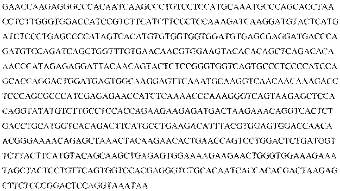
(Signal peptide)
(VL)
抗マウスPD-L1 scAb/huIL-15RαSu/muIgG2A Fc融合タンパク質のアミノ酸配列(シグナルペプチド配列を含む)は以下の通りである(配列番号:12):
(シグナルペプチド)

(VL)


(Signal peptide)
(VL)
一部の場合では、該リーダーペプチドが成熟ポリペプチドから開裂される。 In some cases, the leader peptide is cleaved from the mature polypeptide.
抗PD-L1 scAb/IL-15N72D及び抗PD-L1 scAb/IL-15RαSu/Fc配列を、以前(米国特許第8,507,222号、参照によって本明細書に組み込まれる)に記載のように発現ベクターにクローニングし、該発現ベクターをCHO細胞にトランスフェクションした。CHO細胞内での2つの構築物の共発現により、可溶性抗PD-L1 scAb/IL-15N72D:抗PD-L1 scAb/IL-15RαSu/Fc複合体の形成及び分泌が許容され、プロテインAクロマトグラフィーを用いてCHO細胞培養上清からそれを精製した。 The anti-PD-L1 scAb/IL-15N72D and anti-PD-L1 scAb/IL-15RαSu/Fc sequences were cloned into an expression vector as previously described (U.S. Patent No. 8,507,222, incorporated herein by reference), and the expression vector was transfected into CHO cells. Co-expression of the two constructs in CHO cells allowed the formation and secretion of a soluble anti-PD-L1 scAb/IL-15N72D:anti-PD-L1 scAb/IL-15RαSu/Fc complex, which was purified from CHO cell culture supernatants using Protein A chromatography.
精製された抗PD-L1 scAb/IL-15N72D:抗PD-L1 scAb/IL-15RαSu/Fcタンパク質複合体のSDS-PAGE分析が図3に示される。可溶性抗マウスPD-L1 scAb/huIL-15RαSu/muIgG2A及び抗マウスPD-L1 scAb/IL-15N72Dタンパク質に対応するバンドが、それぞれ~60kDa及び~40kDaで観察された。 SDS-PAGE analysis of purified anti-PD-L1 scAb/IL-15N72D:anti-PD-L1 scAb/IL-15RαSu/Fc protein complexes is shown in Figure 3. Bands corresponding to soluble anti-mouse PD-L1 scAb/huIL-15RαSu/muIgG2A and anti-mouse PD-L1 scAb/IL-15N72D proteins were observed at ~60 kDa and ~40 kDa, respectively.
実施例2:PD-L1 TxMの活性のインビトロ特性化
ELISAに基づく技術によりPD-L1 TxM複合体の形成が裏付けられた。図4Aでは、トランスフェクションされたCHO細胞に由来する細胞上清中の抗ヒトPD-L1 scAb/IL-15N72D:抗ヒトPD-L1 scAb/huIL-15RαSu/huIgG1 Fc融合タンパク質複合体を、捕捉抗体、抗ヒトIgG抗体(Jackson ImmunoResearch)及び検出抗体、ビオチン化抗ヒトIL-15抗体(BAM 247, R&D Systems)を有するhuIgG1/huIL-15に特異的なELISAを用いて検出した。
Example 2: In vitro characterization of PD-L1 TxM activity ELISA-based techniques confirmed the formation of PD-L1 TxM complexes. In Figure 4A, anti-human PD-L1 scAb/IL-15N72D:anti-human PD-L1 scAb/huIL-15RαSu/huIgG1 Fc fusion protein complexes in cell supernatants derived from transfected CHO cells were detected using an ELISA specific for huIgG1/huIL-15 with a capture antibody, anti-human IgG antibody (Jackson ImmunoResearch), and a detection antibody, biotinylated anti-human IL-15 antibody (BAM 247, R&D Systems).
マウス特異的PD-L1 TxMでは、捕捉抗体、affinipureロバ抗ネズミIgG(Jackson ImmunoResearch)又はヒト/類人猿IL15抗体(MAB647, R&D Systems)、及び検出抗体ホースラディッシュペルオキシダーゼ-affinipureロバ抗ネズミIgG(Jackson ImmunoResearch)又はビオチン化抗ヒトIL-15抗体(BAM 247, R&D Systems)をそれぞれ有するmIgG2a特異的又はhuIL15特異的ELISAを用いて、該融合タンパク質複合体を検出した(図4C)。陽性対照と比較して、精製されたマウス特異的PD-L1 TxMへの抗体反応性により該複合体の構造が立証された。 For mouse-specific PD-L1 TxM, the fusion protein complex was detected using mIgG2a-specific or huIL15-specific ELISA with capture antibodies, affinipure donkey anti-murine IgG (Jackson ImmunoResearch) or human/ape IL15 antibody (MAB647, R&D Systems), and detection antibodies, horseradish peroxidase-affinipure donkey anti-murine IgG (Jackson ImmunoResearch) or biotinylated anti-human IL-15 antibody (BAM 247, R&D Systems), respectively (Figure 4C). The structure of the complex was confirmed by antibody reactivity to purified mouse-specific PD-L1 TxM compared to a positive control.
腫瘍細胞においてこれらの融合タンパク質のPD-L1と結合する能力をまた調査した。MB231腫瘍細胞を保有する受容体を用いるフローサイトメトリーによって、ヒト特異的PD-L1 TxMの結合を評価した。これらの調査において、1×10⁵の細胞をPD-L1 TxM複合体で染色した。図5Aに示したように、APC共役マウス抗ヒトFc Ab(Biolegend)を用いて検出すると、フローサイトメトリー分析によりPD-L1 TxM複合体のMB231細胞との結合が明らかになった。図5Bでは、PD-L1 TxM複合体を用いてこの結合の特異性を試験し、市販のAPC共役抗ヒトPD-L1 Ab(Biolegend)を有するMB231細胞の染色を阻害した。同様に、フローサイトメトリー分析によりアベルマブに基づくPD-L1 TxM複合体のMB231腫瘍細胞との結合が明らかになった(図5C)。 The ability of these fusion proteins to bind PD-L1 on tumor cells was also investigated. Binding of human-specific PD-L1 TxM was assessed by flow cytometry using receptor bearing MB231 tumor cells. In these studies, 1×10 5 cells were stained with PD-L1 TxM conjugates. As shown in FIG. 5A, flow cytometric analysis revealed binding of PD-L1 TxM conjugates to MB231 cells when detected with APC-conjugated mouse anti-human Fc Ab (Biolegend). In FIG. 5B, the specificity of this binding was tested using PD-L1 TxM conjugates to inhibit staining of MB231 cells with a commercially available APC-conjugated anti-human PD-L1 Ab (Biolegend). Similarly, flow cytometric analysis revealed binding of avelumab-based PD-L1 TxM conjugates to MB231 tumor cells (FIG. 5C).
マウス特異的PD-L1 TxM複合体の結合を評価するために、PD-L1陽性5T33P骨髄腫及びMB49luc膀胱腫瘍細胞(5×10⁵細胞/試験)をPE又はBrilliant Violetで標識化した抗マウスPD-L1 Ab(100μL中2μg/試験)で初めに染色した(図6A及び図6B)。精製されたマウス特異的PD-L1 TxM複合体の追加によってこの結合の特異性を試験し、PD-L1リガンドと結合している抗体を阻害した。精製された抗mPD-L1 Ab(S1-PD-L1)を陽性対照として使用した。興味深いことに、該PD-L1 TxM複合体が腫瘍細胞上のPD-L1染色を、同量の抗PD-L1 Abよりもよく阻害することを見出した。このことをA20 B細胞リンパ腫細胞を用いる阻害調査においてさらに評価した。滴定分析により、PD-L1 TxMが抗PD-L1 Abの少なくとも5倍、腫瘍細胞表面に発現したPD-L1との相互作用を阻害するのに効果的であったことが示された(図7A及び図7B)。 To evaluate the binding of mouse-specific PD-L1 TxM conjugates, PD-L1 positive 5T33P myeloma and MB49luc bladder tumor cells ( 5x10⁵ cells/test) were first stained with PE or Brilliant Violet labeled anti-mouse PD-L1 Ab (2μg in 100μL/test) (Figure 6A and Figure 6B). The specificity of this binding was tested by the addition of purified mouse-specific PD-L1 TxM conjugates to inhibit antibody binding to the PD-L1 ligand. Purified anti-mPD-L1 Ab (S1-PD-L1) was used as a positive control. Interestingly, we found that the PD-L1 TxM conjugates inhibited PD-L1 staining on tumor cells better than the same amount of anti-PD-L1 Ab. This was further evaluated in an inhibition study using A20 B cell lymphoma cells. Titration analysis demonstrated that PD-L1 TxM was at least 5-fold more effective than anti-PD-L1 Ab in blocking interactions with PD-L1 expressed on the tumor cell surface (Figures 7A and 7B).
PD-L1 TxM複合体のIL-15免疫刺激性活性を評価するために、IL-15依存性32Dβ細胞(マウス造血細胞株)の増殖を評価した。増加したレベルのPD-L1 TxMを32Dβ細胞(104細胞/ウェル)に添加し、2日後にWST-1増殖試薬を用いて細胞増殖を決定した。図8に示すように、32Dβ細胞増殖における用量に依存的な増加はPD-L1 TxMによって介在され、該複合体の免疫刺激性活性を立証した。 To assess the IL-15 immunostimulatory activity of the PD-L1 TxM conjugates, IL-15-dependent proliferation of 32Dβ cells (a murine hematopoietic cell line) was evaluated. Increasing levels of PD-L1 TxM were added to 32Dβ cells ( 104 cells/well) and cell proliferation was determined after 2 days using WST-1 proliferation reagent. As shown in Figure 8, a dose-dependent increase in 32Dβ cell proliferation was mediated by PD-L1 TxM, demonstrating the immunostimulatory activity of the conjugates.
異なった形態のPD-L1 TxM複合体の特性及び活性を評価するためにさらなる調査を行った。PD-L1 scAb/IL-15N72D及び抗PD-L1 scAb/IL-15RαSu/Fcタンパク質を含む複合体は4つの抗PD-L1 scAb結合ドメイン(すなわち、四頭(4H))を有することが期待される一方、IL-15N72D及び抗PD-L1 scAb/IL-15RαSu/Fcタンパク質を含む複合体は2つの抗PD-L1 scAb結合ドメイン(すなわち、二頭(2H))を有することが期待される(図9A)。これらの複合体は、2H TxMと比較して4H TxMの標的細胞を結合する高い結合活性に基づく、異なる活性を有することが期待される。IL-15N72Dとのタンパク質融合は、IL-15の生物学的活性を低減することがまた示されている。それゆえに、4H TxMフォーマットは2H TxMと比較して低いIL-15活性を有することが期待される。高い(抗体様)標的化及び低い免疫刺激性活性が好まれる場合(すなわち4H TxMフォーマット)、又は低い標的化及び高い免疫刺激性活性(免疫サイトカイン)が好まれる場合には、これらの差異により利点が提供されることが期待される。 Further investigations were performed to evaluate the properties and activity of the different forms of PD-L1 TxM conjugates. The conjugates containing PD-L1 scAb/IL-15N72D and anti-PD-L1 scAb/IL-15RαSu/Fc protein are expected to have four anti-PD-L1 scAb binding domains (i.e., tetraheaded (4H)), while the conjugates containing IL-15N72D and anti-PD-L1 scAb/IL-15RαSu/Fc protein are expected to have two anti-PD-L1 scAb binding domains (i.e., bideave (2H)) (Figure 9A). These conjugates are expected to have different activities based on the higher avidity of 4H TxM to bind target cells compared to 2H TxM. Protein fusion with IL-15N72D has also been shown to reduce the biological activity of IL-15. Therefore, the 4H TxM format is expected to have lower IL-15 activity compared to 2H TxM. These differences are expected to provide advantages where high (antibody-like) targeting and low immunostimulatory activity are preferred (i.e., 4H TxM format), or low targeting and high immunostimulatory activity (immunocytokines) are preferred.
これらのフォーマットを評価するために、4Hマウス特異的PD-L1 TxM(2C2)及び2Hマウス特異的PD-L1 TxM(PDN3)を、抗マウスPD-L1 scAb/IL-15N72D及び抗マウスPD-L1 scAb-/IL-15RαSu/Fc発現ベクター又はIL-15N72D及び抗マウスPD-L1 scAb-/IL-15RαSu/Fc発現ベクターをそれぞれ用いてCHO細胞をトランスフェクションすることによって生成した。それからトランスフェクションされたCHO細胞上清からプロテインAクロマトグラフィーによって該TxM複合体を精製し、精製されたタンパク質を低減したSDS-PAGEによって評価した(図9B)。可溶性抗マウスPD-L1 scAb/huIL-15RαSu/muIgG2A、抗マウスPD-L1 scAb/IL-15N72D及びIL-15N72Dタンパク質と対応するバンドを、~60kDa、~40kDa及び~16kDaでそれぞれ観察した。さらに、精製した4H PD-L1 TxM(2C2)及び2H PD-L1 TxM(PDN3)複合体は、分析的サイズ排除クロマトグラフィー(SEC)によって分析した際に、単独のタンパク質ピークとして移動した(図9C及び図9D)。これらの結果により、2つの異なるPD-L1 TxM複合体を産生でき、期待される構造特性を有する可溶性タンパク質として精製できることが示される。 To evaluate these formats, 4H mouse-specific PD-L1 TxM (2C2) and 2H mouse-specific PD-L1 TxM (PDN3) were generated by transfecting CHO cells with anti-mouse PD-L1 scAb/IL-15N72D and anti-mouse PD-L1 scAb-/IL-15RαSu/Fc expression vector or IL-15N72D and anti-mouse PD-L1 scAb-/IL-15RαSu/Fc expression vector, respectively. The TxM complexes were then purified from transfected CHO cell supernatants by Protein A chromatography, and purified proteins were evaluated by reduced SDS-PAGE (Figure 9B). Bands corresponding to soluble anti-mouse PD-L1 scAb/huIL-15RαSu/muIgG2A, anti-mouse PD-L1 scAb/IL-15N72D, and IL-15N72D proteins were observed at ~60 kDa, ~40 kDa, and ~16 kDa, respectively. Furthermore, purified 4H PD-L1 TxM (2C2) and 2H PD-L1 TxM (PDN3) complexes migrated as single protein peaks when analyzed by analytical size exclusion chromatography (SEC) (Figures 9C and 9D). These results indicate that two distinct PD-L1 TxM complexes can be produced and purified as soluble proteins with expected structural characteristics.
上記に記載の調査と同様に、これらの融合タンパク質複合体の、免疫細胞のIL-2Rβ/γ及び腫瘍細胞のPD-L1と結合する能力を調査した。IL-2Rβ/γ陽性32Dβ細胞を4H PD-L1 TxM(2C2)、2H PD-L1 TxM(PDN3)又は対照のALT-803複合体でインキュベートした。洗浄段階に続いて、抗ヒトIL-15 Ab-PE(又はイソタイプ対照Ab)を添加し、フローサイトメトリーによって結合したTxM/ALT-803複合体を検出した。図10Aに示されるように、4H PD-L1 TxM(2C2)、2H PD-L1 TxM(PDN3)及びALT-803タンパク質は、対照と比較して32Dβ細胞の能力がある。PD-L1、PD-L1陽性5T33P骨髄腫細胞との結合を評価するために、Brilliant Violet 421(BV421)で標識化した抗マウスPD-L1 Ab(10F.9G2)で初めに染色した。精製した4H PD-L1 TxM(2C2)及び2H PD-L1 TxM(PDN3)複合体の追加によってこの結合の特異性を試験し、PD-L1リガンドと結合しているBV421抗体を阻害した(図10B及び図10C)。精製した抗mPD-L1 Ab(NJI6)を陽性対照として使用した。高い結合活性と一致して、1μgの4H PD-L1 TxMは6μgの2H PD-L1 TxMと同じくらい、抗PD-L1 Ab染色を阻害するのに効果的であった。これらの結果により、4H PD-L1及び2H PD-L1 TxM複合体がIL-2Rβ/γ及びPD-L1標的結合能力を保有することが裏付けられた。 Similar to the studies described above, we investigated the ability of these fusion protein complexes to bind to IL-2Rβ/γ on immune cells and PD-L1 on tumor cells. IL-2Rβ/γ-positive 32Dβ cells were incubated with 4H PD-L1 TxM (2C2), 2H PD-L1 TxM (PDN3) or control ALT-803 complexes. Following a washing step, anti-human IL-15 Ab-PE (or isotype control Ab) was added and bound TxM/ALT-803 complexes were detected by flow cytometry. As shown in Figure 10A, 4H PD-L1 TxM (2C2), 2H PD-L1 TxM (PDN3) and ALT-803 proteins are capable of binding to 32Dβ cells compared to the control. To assess binding to PD-L1, PD-L1-positive 5T33P myeloma cells were first stained with Brilliant Violet 421 (BV421)-labeled anti-mouse PD-L1 Ab (10F.9G2). The specificity of this binding was tested by the addition of purified 4H PD-L1 TxM (2C2) and 2H PD-L1 TxM (PDN3) conjugates to inhibit BV421 antibody binding to the PD-L1 ligand (Figure 10B and Figure 10C). Purified anti-mPD-L1 Ab (NJI6) was used as a positive control. Consistent with the high binding activity, 1 μg of 4H PD-L1 TxM was as effective as 6 μg of 2H PD-L1 TxM in inhibiting anti-PD-L1 Ab staining. These results confirm that 4H PD-L1 and 2H PD-L1 TxM complexes possess IL-2Rβ/γ and PD-L1 target binding ability.
上記に記載のように、マウス特異的4H PD-L1及び2H PD-L1 TxM複合体のIL-15免疫刺激性活性を、IL-15依存性32Dβ細胞の増殖に基づいて決定した。図11A及び図11Bに示されるように、32Dβ細胞増殖における用量依存性増加は4H PD-L1及び2H PD-L1 TxM複合体のどちらかによって介在されており、これらのTxMフォーマットの免疫刺激性活性を立証する。2H PD-L1 TxM複合体(PDN-3)は、ALT-803と比較するとIL-15生物活性においてわずかな減少を示す一方、4H PD-L1 TxM複合体(2C2)はIL-15生物活性において約30倍の減少を示す。このことは以前の結合ドメイン-IL-15N72D融合タンパク質の低いIL-15活性と一致する。 As described above, the IL-15 immunostimulatory activity of mouse-specific 4H PD-L1 and 2H PD-L1 TxM complexes was determined based on IL-15-dependent 32Dβ cell proliferation. As shown in Figures 11A and 11B, a dose-dependent increase in 32Dβ cell proliferation was mediated by either 4H PD-L1 or 2H PD-L1 TxM complexes, demonstrating the immunostimulatory activity of these TxM formats. The 2H PD-L1 TxM complex (PDN-3) exhibits a slight decrease in IL-15 bioactivity compared to ALT-803, while the 4H PD-L1 TxM complex (2C2) exhibits an approximately 30-fold decrease in IL-15 bioactivity. This is consistent with the low IL-15 activity of previous binding domain-IL-15N72D fusion proteins.
4Hヒト特異的PD-L1 TxM及び2Hヒト特異的PD-L1 TxM複合体で同様の調査を行った。抗ヒトPD-L1 scAb/IL-15N72D及び抗ヒトPD-L1 scAb-/IL-15RαSu/Fc発現ベクター又はIL-15N72D及び抗ヒトPD-L1 scAb/IL-15RαSu/Fc発現ベクターでCHO細胞をそれぞれトランスフェクションすることによりこれらのタンパク質を生成し、続いてプロテインAクロマトグラフィーを介して細胞培養上清から精製した。低減したSDS-PAGE分析により、精製した4Hヒト特異的PD-L1 TxM及び2Hヒト特異的PD-L1 TxM調製内に期待されたタンパク質バンドが裏付けられた(図12A)。同様に、分析的SECにより、精製した4Hヒト特異的PD-L1 TxM及び2Hヒト特異的PD-L1 TxM複合体が単独のタンパク質ピークとして移動することが示された(図12B及び図12C)。 Similar studies were performed with 4H human specific PD-L1 TxM and 2H human specific PD-L1 TxM conjugates. These proteins were produced by transfecting CHO cells with anti-human PD-L1 scAb/IL-15N72D and anti-human PD-L1 scAb-/IL-15RαSu/Fc expression vectors or IL-15N72D and anti-human PD-L1 scAb/IL-15RαSu/Fc expression vectors, respectively, and subsequently purified from cell culture supernatants via protein A chromatography. Reduced SDS-PAGE analysis confirmed the expected protein bands within the purified 4H human specific PD-L1 TxM and 2H human specific PD-L1 TxM preparations (Figure 12A). Similarly, analytical SEC showed that the purified 4H human-specific PD-L1 TxM and 2H human-specific PD-L1 TxM complexes migrated as single protein peaks (Figures 12B and 12C).
これらの融合タンパク質複合体の、腫瘍細胞のPD-L1と結合する能力を調査した。PD-L1陽性PC-3ヒト前立腺癌細胞を、10nMの精製したヒト特異的4H PD-L1 TxM、2H PD-L1 TxM又は対照2H抗CD20 scAb(2B8)TxM複合体の存在下、又は非存在下で、APCで標識化した抗マウスPD-L1Abで染色した(図13)。その結果により、ヒト特異的4H PD-L1及び2H PD-L1 TxM複合体が、ヒト腫瘍細胞と結合する抗PD-L1 Abを阻害する能力がある一方、対照のTxM複合体にはその能力がないことが示されている。以前の結果と一致して、4H PD-L1 TxM複合体は2H PD-L1 TxM複合体よりよい結合活性を示した。これらの結果により、ヒト特異的4H PD-L1及び2H PD-L1 TxM複合体が、ヒト腫瘍細胞に関してPD-L1標的結合活性を保有することが裏付けられる。
The ability of these fusion protein conjugates to bind to PD-L1 on tumor cells was investigated. PD-L1-positive PC-3 human prostate cancer cells were stained with APC-labeled anti-mouse PD-L1 Ab in the presence or absence of 10 nM purified human-specific 4H PD-L1 TxM, 2H PD-L1 TxM, or
上記に記載のように、ヒト特異的4H PD-L1及び2H PD-L1 TxM複合体のIL-15免疫刺激性活性を、IL-15依存性32Dβ細胞の増殖に基づいて決定した。図14A及び図14Bに示されるように、32Dβ細胞増殖における用量依存性増加が、4H PD-L1と2H PD-L1 TxM複合体のどちらかによって介在され、これらのTxMフォーマットの免疫刺激性活性を立証した。2H PD-L1 TxM複合体がALT-803と比較してIL-15生物活性においてわずかな減少を示す一方、4H PD-L1 TxM複合体はALT-803と比較してIL-15生物活性において約5倍の減少を示す。このことは以前の結合ドメイン-IL-15N72D融合タンパク質の低いIL-15活性と一致する。 As described above, the IL-15 immunostimulatory activity of human-specific 4H PD-L1 and 2H PD-L1 TxM conjugates was determined based on IL-15-dependent 32Dβ cell proliferation. As shown in Figures 14A and 14B, a dose-dependent increase in 32Dβ cell proliferation was mediated by either 4H PD-L1 or 2H PD-L1 TxM conjugates, demonstrating the immunostimulatory activity of these TxM formats. While the 2H PD-L1 TxM conjugates show a slight decrease in IL-15 bioactivity compared to ALT-803, the 4H PD-L1 TxM conjugates show an approximately 5-fold decrease in IL-15 bioactivity compared to ALT-803. This is consistent with the low IL-15 activity of previous binding domain-IL-15N72D fusion proteins.
実施例3:インビトロ及びインビボでのPD-L1 TxMの免疫刺激性及び抗腫瘍活性
PD-L1 TxMがインビボで免疫反応を刺激する能力をマウス内で評価した。C57BL/6マウスに200μlのPBS、ALT-803(0.4mg/kg、対照)、4Hマウス特異的PD-L1 TxM(200μg、2C2(T4M-mPD-L1))又は2Hマウス特異的PD-L1 TxM(200μg、PDN3(T2M-mPD-L1))を皮下注射した。処置から3日後、脾臓及びリンパ節を採取した。抗CD4、CD8、NK及びCD19Absを用いる免疫細胞サブセットの染色に続くフローサイトメトリーのために脾細胞及びリンパ球を調製した。図15Aに示されるように、ALT-803、2H PD-L1 TxM及び4H PD-L1 TxMでの処置はこれらのタンパク質の免疫刺激性活性と一致して脾臓重量の増加を誘発した。特に、2H PD-L1 TxM処置は、これらの複合体で観察されるIL-15活性における差異と一致して、4H PD-L1 TxMと比較して脾臓重量における重大な増加を誘発した。2H PD-L1 TxM及び4H PD-L1 TxMでの処置は、PBS対照群と比較して、マウスの脾臓及びリンパ節におけるCD8 T細胞及びNK細胞のパーセンテージの増加をまたもたらした(図15B及び図15C)。これらの免疫反応はこれらのTxM複合体のIL-15生物活性と一致している。
Example 3: Immunostimulatory and Antitumor Activity of PD-L1 TxMs In Vitro and In Vivo The ability of PD-L1 TxMs to stimulate immune responses in vivo was evaluated in mice. C57BL/6 mice were injected subcutaneously with 200 μl of PBS, ALT-803 (0.4 mg/kg, control), 4H mouse-specific PD-L1 TxM (200 μg, 2C2(T4M-mPD-L1)), or 2H mouse-specific PD-L1 TxM (200 μg, PDN3(T2M-mPD-L1)). Three days after treatment, spleens and lymph nodes were harvested. Splenocytes and lymphocytes were prepared for staining of immune cell subsets with anti-CD4, CD8, NK, and CD19 Abs followed by flow cytometry. As shown in Figure 15A, treatment with ALT-803, 2H PD-L1 TxM, and 4H PD-L1 TxM induced an increase in spleen weight, consistent with the immune stimulatory activity of these proteins. Notably, 2H PD-L1 TxM treatment induced a significant increase in spleen weight compared to 4H PD-L1 TxM, consistent with the differences in IL-15 activity observed with these complexes. Treatment with 2H PD-L1 TxM and 4H PD-L1 TxM also resulted in an increase in the percentage of CD8 T cells and NK cells in the spleen and lymph nodes of mice compared to the PBS control group (Figures 15B and 15C). These immune responses are consistent with the IL-15 bioactivity of these TxM complexes.
さらに、PD-L1 TxMの、腫瘍細胞に対して細胞傷害性を刺激する能力をインビボで評価した。PD-L1陽性細胞を製造者の指示に従ってCellTrace Violet(Invitrogen)で標識化し、免疫エフェクター細胞(すなわち脾細胞)と、R10培地(10%のウシ胎児血清を有するRPMI-1640)において10:1のエフェクター:5T33P骨髄腫標的の比率で、37℃かつ5%のCO2内で培養した。P-melマウス脾細胞の刺激によって3日間、抗CD3Ab(2C11:4μg/ml)を用いてエフェクター細胞を調製した。腫瘍細胞及びエフェクター細胞を4日間、マウス特異的PD-L1 TxMでインキュベートし、それからフローサイトメトリーによって分析し、標的細胞の生存を決定した。PBSは陰性対照として機能し、ALT-803(IL-15N72D:IL-15Rα/Fc)、抗PD-L1 Ab及びALT-803+抗PD-L1 Abは陽性対照として機能した。図16に示されるように、5T33P腫瘍細胞の有意な殺傷を、PBS処置と比較して2.1μgのPD-L1 TxMを含む群において見出した。 Additionally, the ability of PD-L1 TxM to stimulate cytotoxicity against tumor cells was evaluated in vivo. PD-L1 positive cells were labeled with CellTrace Violet (Invitrogen) according to the manufacturer's instructions and cultured with immune effector cells (i.e., splenocytes) at a 10:1 effector:5T33P myeloma target ratio in R10 medium (RPMI-1640 with 10% fetal bovine serum) at 37°C and 5% CO2 . Effector cells were prepared by stimulation of P-mel mouse splenocytes with anti-CD3 Ab (2C11: 4 μg/ml) for 3 days. Tumor and effector cells were incubated with mouse-specific PD-L1 TxM for 4 days and then analyzed by flow cytometry to determine target cell survival. PBS served as a negative control, and ALT-803 (IL-15N72D:IL-15Rα/Fc), anti-PD-L1 Ab, and ALT-803 + anti-PD-L1 Ab served as positive controls. As shown in Figure 16, significant killing of 5T33P tumor cells was found in the group containing 2.1 μg PD-L1 TxM compared to PBS treatment.
同様にインビトロでの抗腫瘍活性を、ヒト特異的2H PD-L1 TxM及び4H PD-L1 TxM複合体を用いて評価した。2人の異なるドナーからのヒトNK細胞を、Stemcell TechnologiesのNK細胞単離キットを用いて血液バフィーコートから精製し、エフェクター細胞として使用した。PD-L1陽性ヒト膵臓腫瘍細胞(SW1990)をCelltrace-violetで標識化し、標的細胞として使用した。ヒトNK細胞及びSW1990腫瘍細胞を5:1のE:T比率で、培地単独又は50nMの抗ヒトPD-L1 Ab(対照)、ヒト特異的2H PD-L1 TxM複合体又は4H PD-L1 TxM複合体を含む培地で混合した。40時間後、標的細胞の死滅のパーセントを、violetで標識化した標的細胞のヨウ化プロピジウム染色に基づくフローサイトメトリーによって評価した。図17に示されるように、ヒト特異的2H PD-L1 TxM又は4H PD-L1 TxM複合体のどちらかでインキュベートしたヒトNK細胞は、未処置のNK細胞又は抗ヒトPD-L1 Ab(すなわち伝統的なADCC)で処置されたNK細胞よりも、PD-L1陽性ヒト腫瘍細胞に対して重大な細胞傷害性を介在することができた。これらの結果は抗PD-L1 Absと比較してPD-L1 TxM複合体における免疫細胞介在性の標的化抗腫瘍活性の有意な改良を表している。 Similarly, in vitro antitumor activity was evaluated with human-specific 2H PD-L1 TxM and 4H PD-L1 TxM conjugates. Human NK cells from two different donors were purified from blood buffy coats using the NK cell isolation kit from Stemcell Technologies and used as effector cells. PD-L1 positive human pancreatic tumor cells (SW1990) were labeled with Celltrace-violet and used as target cells. Human NK cells and SW1990 tumor cells were mixed at an E:T ratio of 5:1 with medium alone or medium containing 50 nM anti-human PD-L1 Ab (control), human-specific 2H PD-L1 TxM conjugates or 4H PD-L1 TxM conjugates. After 40 hours, the percentage of target cell killing was evaluated by flow cytometry based on propidium iodide staining of violet-labeled target cells. As shown in Figure 17, human NK cells incubated with either human-specific 2H PD-L1 TxM or 4H PD-L1 TxM conjugates were able to mediate greater cytotoxicity against PD-L1 positive human tumor cells than untreated NK cells or NK cells treated with anti-human PD-L1 Abs (i.e., traditional ADCC). These results represent a significant improvement in immune cell-mediated targeted anti-tumor activity of PD-L1 TxM conjugates compared to anti-PD-L1 Abs.
同所性5T33P骨髄腫モデルを使用して、腫瘍を保有する動物におけるPD-L1 TxMの有効性を評価した。メスのC57BL/6NHsdマウス(4匹のマウス/群)に5T33P骨髄腫細胞(1×107/マウス)を0日目に静脈内注射した。低用量のPD-L1 TxM(0.11mg/kg、0.05mg/kgのALT-803に対するモル当量用量)又は高用量のPD-L1 TxM(52.5μg/用量、25μg/用量の抗PD-L1 Abに対するモル当量用量)を、それから7日目と14日目に皮下投与した。ALT-803(0.15mg/kg)及びALT-803(0.05mg/kg)+抗PD-L1 Ab(25μg/用量)は陽性対照として機能し、PBSは陰性対照として機能した。生存(又は後肢麻痺による病的状態)を研究終点として観測した。明らかに、高用量のPD-L1 TxM(52.5μg/マウス)処置はPBS処置と比較して腫瘍を保有するマウスの生存を長引かせることが見出された(図18)。この効果は比較可能なALT-803+抗PD-L1 Abの組み合わせの治療で観察されたものと等しかった。腫瘍を保有する動物のPD-L1 TxM処置に続く明らかな毒性は観察されなかった。
An orthotopic 5T33P myeloma model was used to evaluate the efficacy of PD-L1 TxM in tumor-bearing animals. Female C57BL/6NHsd mice (4 mice/group) were injected intravenously with 5T33P myeloma cells ( 1x10⁷ /mouse) on
さらに、同所性MB49luc腫瘍を保有するマウスにおけるPD-L1 TxM複合体の抗腫瘍活性を評価した。C57BL/6NHsdマウス(6マウス/群)の膀胱に、MB49luc膀胱腫瘍細胞(3×104/マウス)を0日目に膀胱内注入した。腫瘍を保有するマウスをマウス特異的2H PD-L1 TxM(2.8mg/kg)で7日目、11日目、14日目及び18日目に皮下で処置した。ALT-803処置(0.2mg/kg、subQ)及びALT-803(0.2mg/kg、subQ)+抗PD-L1 Ab(50μg/用量、subQ)が陽性対照として機能し、PBSが陰性対照として機能した。生存(又は病的状態)を研究終点として観測した。図19に示されるように、2H PD-L1 TxM処置がPBS処置と比較してMB49luc腫瘍を保有するマウスの生存を長引かせることを見出した。2H PD-L1 TxMの抗腫瘍効果はALT-803及びALT-803+抗PD-L1 Ab陽性対照で観察されたものと同じくらい良いか、それらより良かった。
Additionally, antitumor activity of PD-L1 TxM conjugates in mice bearing orthotopic MB49luc tumors was evaluated. The bladders of C57BL/6NHsd mice (6 mice/group) were intravesically instilled with MB49luc bladder tumor cells ( 3x10⁴ /mouse) on
実施例4:抗CTLA4結合ドメイン(CTLA4 TxM)及び抗PD-1結合ドメイン(PD-1 TxM)を含むIL-15に基づく融合タンパク質複合体の生成
実施例1~3に記載された融合タンパク質複合体と同様に、CTLA4及びPD-L1を認識する結合ドメインを含む本発明の融合タンパク質を生成した。具体的には、一本鎖抗CTLA4抗体とhuIL-15N72D及びIL-15RαSu/Fc鎖を結合する構築物を製造した。抗CTLA4一本鎖抗体(抗CTLA4scAb)配列はフレキシブルリンカー配列を介して結合した重鎖及び軽鎖V抗体ドメイン抗体のコーディング領域を含む。一本鎖抗体ドメインはVH-リンカー-VL又はVL-リンカー-VHフォーマットのどちらかに配置されていてもよい。一部の場合では、抗CTLA4scAbはIL-15N72D及び/又はIL-15RαSu/Fc鎖のC末端と結合している。他の場合では、抗CTLA4はIL-15N72D及び/又はIL-15RαSu/Fc鎖のN末端と結合している。これらの構築物において、マウス又はヒトCTLA4分子のいずれかに特異的な抗CTLA4scAbsを使用した。
Example 4: Generation of IL-15-based fusion protein complexes containing anti-CTLA4 binding domain (CTLA4 TxM) and anti-PD-1 binding domain (PD-1 TxM) Similar to the fusion protein complexes described in Examples 1-3, fusion proteins of the invention were generated that contain binding domains recognizing CTLA4 and PD-L1. Specifically, constructs were produced that combine single chain anti-CTLA4 antibodies with huIL-15N72D and IL-15RαSu/Fc chains. The anti-CTLA4 single chain antibody (anti-CTLA4scAb) sequence comprises heavy and light chain V antibody domain antibody coding regions linked via a flexible linker sequence. The single chain antibody domains may be arranged in either a VH-linker-VL or VL-linker-VH format. In some cases, the anti-CTLA4scAb is linked to the C-terminus of the IL-15N72D and/or IL-15RαSu/Fc chain. In other cases, anti-CTLA4 is linked to the N-terminus of IL-15N72D and/or IL-15RαSu/Fc chain. In these constructs, anti-CTLA4 scAbs specific for either the mouse or human CTLA4 molecule were used.
抗ヒトCTLA-4 scAb/huIL-15RαSu/huIgG1 Fc構築物の核酸配列(リーダー配列を含む)は以下の通りである(配列番号:13):
(シグナルペプチド)

(VL)


(Signal peptide)
(VL)
ヒトCTLA-4 scAb/huIL-15RαSu/huIgG1 Fcタンパク質のアミノ酸配列(リーダー配列を含む)は以下の通りである(配列番号:14):
(シグナルペプチド)

(VL)


(Signal peptide)
(VL)
一部の場合では、該リーダーペプチドが成熟ポリペプチドから開裂される。 In some cases, the leader peptide is cleaved from the mature polypeptide.
同様に、抗マウスCTLA-4 scAb/huIL-15RαSu/mIgG2a構築物の核酸配列(リーダー配列を含む)は以下の通りである(配列番号:15):
(シグナルペプチド)

(VL)


(Signal peptide)
(VL)
抗マウスCTLA-4 scAb/huIL-15RαSu/mIgG2a融合タンパク質のアミノ酸配列(リーダー配列を含む)は以下の通りである(配列番号:16):
(シグナルペプチド)

(VL)


(Signal peptide)
(VL)
一部の場合では、該リーダーペプチドが成熟ポリペプチドから開裂される。 In some cases, the leader peptide is cleaved from the mature polypeptide.
上記に示されるように、抗ヒト及びマウスCTLA4 scAbドメインをIL-15N72Dタンパク質との融合としてまた生成した。 As shown above, anti-human and mouse CTLA4 scAb domains were also generated as fusions with the IL-15N72D protein.
同様に、抗ヒトPD1 scAb/IL-15N72D構築物の核酸配列(シグナルペプチド配列及び終止コドンを含む)は以下の通りである(配列番号:17):
(シグナルペプチド)

(VL)


(Signal peptide)
(VL)
抗PD-L1 scAb-IL-15N72D融合タンパク質のアミノ酸配列(シグナルペプチド配列を含む)は以下の通りである(配列番号:18):
(シグナルペプチド)

(VL)


(Signal peptide)
(VL)
一部の場合では、該リーダーペプチドが成熟ポリペプチドから開裂される。上記に示されるように、抗ヒトPD-L1 scAbドメインをhuIL-15RαSu/Fc構築物の融合としてまた生成した。 In some cases, the leader peptide is cleaved from the mature polypeptide. As shown above, the anti-human PD-L1 scAb domain was also generated as a fusion of the huIL-15RαSu/Fc construct.
実施例1及び以前(米国特許第8,507,222号の実施例1及び2、参照によって本明細書に組み込まれる)に記載のように、該配列を発現ベクター内にクローニングし、該発現ベクターをCHO細胞内にトランスフェクションした。一部の場合では、同じ又は異なる(すなわち抗PD-L1及び抗CTLA4 scAb)結合ドメインを有するhuIL-15N72D及びhuIL-15RαSu/Fc融合タンパク質の両方をコードするベクターで該CHO細胞をトランスフェクションした。該融合タンパク質複合体を、CHO細胞培養上清からプロテインA親和性クロマトグラフィーを用いて上記に記載のように精製した。 The sequences were cloned into expression vectors and the expression vectors were transfected into CHO cells as described in Example 1 and previously (Examples 1 and 2 of U.S. Pat. No. 8,507,222, incorporated herein by reference). In some cases, the CHO cells were transfected with vectors encoding both huIL-15N72D and huIL-15RαSu/Fc fusion proteins with the same or different (i.e., anti-PD-L1 and anti-CTLA4 scAb) binding domains. The fusion protein complexes were purified from CHO cell culture supernatants using Protein A affinity chromatography as described above.
上記に記載の抗ヒトPD-L1 scAb/抗ヒトCTLA-4 scAb TxM複合体に加えて、抗PD-L1 scAb/抗ヒトCTLA-4 scAb TxM複合体を、抗ヒトPD-L1 scAb/IL-15N72D(配列番号:1)及び抗ヒトCTLA-4 scAb/huIL-15RαSu/huIgG1 Fc(配列番号:15)構築物を含む発現ベクターでCHO細胞を共トランスフェクションすることによって生成した。これらの融合タンパク質複合体を、CHO細胞培養上清からプロテインA親和性クロマトグラフィーを用いて上記に記載のように精製した。 In addition to the anti-human PD-L1 scAb/anti-human CTLA-4 scAb TxM complexes described above, anti-PD-L1 scAb/anti-human CTLA-4 scAb TxM complexes were generated by co-transfecting CHO cells with expression vectors containing the anti-human PD-L1 scAb/IL-15N72D (SEQ ID NO:1) and anti-human CTLA-4 scAb/huIL-15RαSu/huIgG1 Fc (SEQ ID NO:15) constructs. These fusion protein complexes were purified from CHO cell culture supernatants using Protein A affinity chromatography as described above.
実施例5:他の結合ドメインを含むIL-15に基づく融合タンパク質複合体の生成
実施例1~4に記載の融合タンパク質複合体と同様に、CD47、GITR、ssDNA及び他の疾患関連標的(すなわちCD20、CD19等)を認識する結合ドメインを含む、本発明の融合タンパク質複合体を生成した。
Example 5: Generation of IL-15-Based Fusion Protein Complexes Containing Other Binding Domains Similar to the fusion protein complexes described in Examples 1-4, fusion protein complexes of the invention were generated that contain binding domains recognizing CD47, GITR, ssDNA, and other disease-associated targets (i.e., CD20, CD19, etc.).
CD47は、マクロファージの抑制性受容体として機能するシグナル調節タンパク質α(SIRPα)と関与することによって免疫回避を促進する細胞表面分子である。この「私を食べないで(don’t eat me)シグナル」は、CD47及びSIRPαの相互作用を阻害することによって中断させることがあり、それゆえにマクロファージによる抗体依存性細胞食作用(ADCP)を回復させる。本発明のIL-15ドメインはNK細胞及びCD8+細胞を活性化させ拡大させる一方、それらの細胞傷害性活性を増加させる。十分に高い濃度で、本発明のFc領域はNK細胞及びマクロファージのFcγ受容体と、それぞれADCC又はADCPに相互作用してもよい。この例により、CD47/SIRPα経路、IL-15ドメインを通じたNK細胞及びCD8+細胞の活性化を阻害し、Fc介在性ADCC/ADCPを介した腫瘍のクリアランスを許容するナノボディのVh領域(NbVh;PNAS2016 113(19)E2646-E2654)を含む融合タンパク質複合体の生成及び初めの特性化が記載される。下記に詳細に記載のように、抗マウスCD47 NbVh/huIL-15N72D及び抗マウスCD47 NbVh/huIL-15RαSu/mIgG2a Fcを含むタンパク質複合体を生成した。 CD47 is a cell surface molecule that promotes immune evasion by engaging signal regulatory protein alpha (SIRPα) which functions as an inhibitory receptor on macrophages. This "don't eat me signal" can be disrupted by inhibiting the interaction of CD47 and SIRPα, thus restoring antibody-dependent cellular phagocytosis (ADCP) by macrophages. The IL-15 domains of the present invention activate and expand NK cells and CD8 + cells while increasing their cytotoxic activity. At sufficiently high concentrations, the Fc regions of the present invention may interact with Fcγ receptors on NK cells and macrophages, respectively, for ADCC or ADCP. This example describes the generation and initial characterization of fusion protein complexes containing the Vh region of a nanobody (NbVh; PNAS2016 113(19)E2646-E2654) that inhibits the CD47/SIRPα pathway, NK and CD8 + cell activation through the IL-15 domain, and allows tumor clearance via Fc-mediated ADCC/ADCP. Protein complexes containing anti-mouse CD47 NbVh/huIL-15N72D and anti-mouse CD47 NbVh/huIL-15RαSu/mIgG2a Fc were generated as described in detail below.
具体的には、抗マウスCD47 NbVhをhuIL-15N72D鎖と結合させる構築物を製造した。該抗マウスCD47 NbVh配列はアルパカナノボディの重鎖可変ドメインのコーディング領域を含む。該抗マウスCD47 NbVhはhuIL-15N72DのN末端と結合している。huIL-15N72DのN末端と結合した抗マウスCD47 NbVhを含む構築物の核酸配列及びタンパク質配列を下記に示した。 Specifically, a construct was produced that linked an anti-mouse CD47 NbVh to the huIL-15N72D chain. The anti-mouse CD47 NbVh sequence includes the coding region for the heavy chain variable domain of an alpaca nanobody. The anti-mouse CD47 NbVh is linked to the N-terminus of huIL-15N72D. The nucleic acid and protein sequences of the construct containing the anti-mouse CD47 NbVh linked to the N-terminus of huIL-15N72D are shown below.
抗マウスCD47 NbVh/huIL-15N72D構築物の核酸配列(シグナルペプチド配列を含む)は以下の通りである(配列番号:19):
(シグナルペプチド)


(Signal peptide)
抗マウスCD47 NbVh/IL-15N72D融合タンパク質のアミノ酸配列(シグナルペプチド配列を含む)は以下の通りである(配列番号:20):
(シグナルペプチド)


(Signal peptide)
抗マウスCD47 NbVh/huIL-15RαSu/mIgG2a Fc構築物の核酸配列(リーダー配列を含む)は以下の通りである(配列番号:21):
(シグナルペプチド)


(Signal peptide)
抗マウスCD47 NbVh/huIL-15RαSu/mIgG2a Fc融合タンパク質のアミノ酸配列(リーダー配列を含む)は以下の通りである(配列番号:22):
(シグナルペプチド)


(Signal peptide)
上記に示されるように、一部の場合では、該リーダーペプチドが成熟ポリペプチドから開裂される。 As noted above, in some cases the leader peptide is cleaved from the mature polypeptide.
ヒトCD47に特異的な抗体に由来する一本鎖抗体ドメインを用いて、同様の構築物を生成した。抗ヒトCD47 scAb/huIL-15N72D構築物の核酸配列(シグナルペプチド配列を含む)は以下の通りである(配列番号:23):
(シグナルペプチド)

(VL)


(Signal peptide)
(VL)
抗ヒトCD47 scAb/huIL-15N72D融合タンパク質のアミノ酸配列(シグナルペプチド配列を含む)は以下の通りである(配列番号:24):
(シグナルペプチド)

(VL)



(Signal peptide)
(VL)
抗ヒトCD47 scAb/huIL-15RαSu/hIgG1 Fc構築物の核酸配列(リーダー配列を含む)は以下の通りである(配列番号:25):
(シグナルペプチド)

(VL)


(Signal peptide)
(VL)
抗ヒトCD47 scAb/huIL-15RαSu/hIgG1 Fc融合タンパク質のアミノ酸配列(リーダー配列を含む)は以下の通りである(配列番号:26):
(シグナルペプチド)

(VL)


(Signal peptide)
(VL)
GITRリガンドとGITR間の相互作用が免疫細胞に刺激性シグナルを提供することが知られており、それゆえにGITRリガンド(GITRL)がIL-15の免疫刺激性活性と潜在的に協力して作用することができる免疫アゴニスト分子であることが知られている。それゆえに、ヒトGITRLとhuIL-15N72D鎖を結合する構築物を製造した。 The interaction between GITR ligand and GITR is known to provide stimulatory signals to immune cells, and therefore GITR ligand (GITRL) is known to be an immune agonist molecule that can potentially act in concert with the immunostimulatory activity of IL-15. Therefore, a construct combining human GITRL and the huIL-15N72D chain was produced.
ヒトGITRL/huIL-15N72D構築物の核酸配列(シグナルペプチド配列を含む)は以下の通りである(配列番号:27):
(シグナルペプチド)


(Signal peptide)
ヒトGITRL/IL-15N72D融合タンパク質のアミノ酸配列(シグナルペプチド配列を含む)は以下の通りである(配列番号:28):
(シグナルペプチド)


(Signal peptide)
ヒトGITRL/huIL-15RαSu/hIgG1 Fc構築物の核酸配列(リーダー配列を含む)は以下の通りである(配列番号:29):
(シグナルペプチド)


(Signal peptide)
ヒトGITRL/huIL-15RαSu/hIgG1 Fc融合タンパク質のアミノ酸配列(リーダー配列を含む)は以下の通りである(配列番号:30):
(シグナルペプチド)


(Signal peptide)
疾患細胞によって発現される抗原を標的化する結合ドメインを含む本発明の融合タンパク質複合体を生成することがまた可能である。かかる抗原は腫瘍細胞を含む疾患細胞によって放出された一本鎖DNA(ssDNA)を含むことができる。それゆえに、本発明の融合タンパク質複合体を、ssDNAを認識する一本鎖Abドメインを用いて生成した(Hu51-4抗体からのTNT scAb)。 It is also possible to generate fusion protein complexes of the invention that contain binding domains that target antigens expressed by diseased cells. Such antigens can include single-stranded DNA (ssDNA) released by diseased cells, including tumor cells. Therefore, fusion protein complexes of the invention were generated with single-stranded Ab domains that recognize ssDNA (TNT scAb from Hu51-4 antibody).
TNT scAb/huIL-15N72D構築物の核酸配列(シグナルペプチド配列を含む)は以下の通りである(配列番号:31):
(シグナルペプチド)

(VL)


(Signal peptide)
(VL)
TNT scAb/IL-15N72D融合タンパク質のアミノ酸配列(シグナルペプチド配列を含む)は以下の通りである(配列番号:32):
(シグナルペプチド)

(VL)


(Signal peptide)
(VL)
TNT/huIL-15RαSu/hIgG1 Fc構築物の核酸配列(リーダー配列を含む)は以下の通りである(配列番号:33):
(シグナルペプチド)

(VL)


(Signal peptide)
(VL)
TNT scAb/huIL-15RαSu/hIgG1 Fc融合タンパク質のアミノ酸配列(リーダー配列を含む)は以下の通りである(配列番号:34):
(シグナルペプチド)

(VL)


(Signal peptide)
(VL)
疾患細胞によって発現される他の抗原を標的化する結合ドメインを含む本発明の融合タンパク質複合体をまた生成することができる。かかる抗原は腫瘍細胞を含む疾患細胞に発現した組織因子若しくはCD33、又は免疫細胞に発現したチェックポイント阻害剤を含むことができる。 Fusion protein complexes of the invention can also be generated that contain binding domains that target other antigens expressed by diseased cells. Such antigens can include tissue factor or CD33 expressed on diseased cells, including tumor cells, or checkpoint inhibitors expressed on immune cells.
組織因子(TF)は複数の腫瘍細胞型で過剰発現することが報告されている膜貫通糖タンパク質である。重要なことに、増加したTF発現は、高められた転移の可能性に繋がるがん細胞のシグナリング、腫瘍細胞の移動及び減少したアポトーシスと関係している。それゆえに、TFの標的化はこのタンパク質を過剰発現する腫瘍細胞型に対する免疫療法戦略において有益であることがある。キメラ抗組織因子抗体(ALT-803)がかつて生成され、臨床的に試験されてきた。この抗体のヒト化された可変鎖(hOAT)がまた特性化された。それゆえに、ヒト組織因子を認識する一本鎖Abドメイン(hOAT scAb)を有する本発明の融合タンパク質複合体を生成した。 Tissue factor (TF) is a transmembrane glycoprotein that has been reported to be overexpressed in multiple tumor cell types. Importantly, increased TF expression is associated with cancer cell signaling, tumor cell migration, and decreased apoptosis leading to enhanced metastatic potential. Therefore, targeting TF may be beneficial in immunotherapeutic strategies against tumor cell types that overexpress this protein. A chimeric anti-tissue factor antibody (ALT-803) has been previously generated and clinically tested. A humanized variable chain (hOAT) of this antibody has also been characterized. Therefore, we have generated the fusion protein complex of the present invention with a single chain Ab domain (hOAT scAb) that recognizes human tissue factor.
hOAT scAb/huIL-15RαSu/hIgG1 Fc構築物の核酸配列(リーダー配列を含む)は以下の通りである(配列番号:35):
(シグナルペプチド)

(VL)


(Signal peptide)
(VL)
hOAT scAb/huIL-15RαSu/hIgG1 Fc融合タンパク質のアミノ酸配列(シグナルペプチド配列を含む)は以下の通りである(配列番号:36):
(シグナルペプチド)

(VL)


(Signal peptide)
(VL)
一部の場合では、該リーダーペプチドが成熟ポリペプチドから開裂される。 In some cases, the leader peptide is cleaved from the mature polypeptide.
hOAT scAb/huIL-15N72D融合タンパク質を発現する、同様の構築物をまた生成することができる。 A similar construct can also be generated that expresses the hOAT scAb/huIL-15N72D fusion protein.
実施例1及び以前(米国特許第8,507,222号の実施例1及び2、参照によって本明細書に組み込まれる)に記載のように、発現ベクターに該配列をクローニングし、該発現ベクターをCHO細胞にトランスフェクションした。一部の場合では、本発明と同じ又は異なる結合ドメインを有するhuIL-15N72D及びhuIL-15RαSu/Fc融合タンパク質の両方をコードするベクターを用いて該CHO細胞をトランスフェクションした。上記に記載のように、プロテインA親和性クロマトグラフィーを用いてCHO細胞培養上清から融合タンパク質複合体を精製した。 The sequences were cloned into an expression vector and the expression vector was transfected into CHO cells as described in Example 1 and previously (Examples 1 and 2 of U.S. Pat. No. 8,507,222, incorporated herein by reference). In some cases, the CHO cells were transfected with vectors encoding both huIL-15N72D and huIL-15RαSu/Fc fusion proteins with the same or different binding domains of the present invention. Fusion protein complexes were purified from CHO cell culture supernatants using Protein A affinity chromatography as described above.
本発明の融合タンパク質複合体を、CD33を認識する一本鎖Abドメイン(CD33 scAb)を用いてまた生成した。CD33 scAb/huIL-15N72D構築物の核酸配列(シグナルペプチド配列を含む)は以下の通りである(配列番号:37):
(シグナルペプチド)

(VL)


(Signal peptide)
(VL)
CD33 scAb/IL-15N72D融合タンパク質のアミノ酸配列(シグナルペプチド配列を含む)は以下の通りである(配列番号:38):
(シグナルペプチド)

(VL)



(Signal peptide)
(VL)
一部の場合では、該リーダーペプチドが成熟ポリペプチドから開裂される。 In some cases, the leader peptide is cleaved from the mature polypeptide.
CD33 scAb/huIL-15RαSu/hIgG1 Fc構築物の核酸配列(リーダー配列を含む)は以下の通りである(配列番号:39):
(シグナルペプチド)

(VL)


(Signal peptide)
(VL)
CD33 scAb/huIL-15RαSu/hIgG1 Fc融合タンパク質のアミノ酸配列(リーダー配列を含む)は以下の通りである(配列番号:40):
(シグナルペプチド)

(VL)


(Signal peptide)
(VL)
一部の場合では、該リーダーペプチドが成熟ポリペプチドから開裂される。 In some cases, the leader peptide is cleaved from the mature polypeptide.
該配列を実施例1及び以前(米国特許第8,507,222号の実施例1及び2、参照によって本明細書に組み込まれる)に記載のように発現ベクター内にクローニングし、該発現ベクターをCHO細胞内にトランスフェクションした。一部の場合では、本発明と同じ又は異なる結合ドメインを有するhuIL-15N72D及びhuIL-15RαSu/Fc融合タンパク質の両方をコードするベクターで該CHO細胞をトランスフェクションした。該融合タンパク質複合体をCHO細胞培養上清から上記に記載のようにプロテインA親和性クロマトグラフィーを用いて精製した。 The sequences were cloned into expression vectors as described in Example 1 and previously (Examples 1 and 2 of U.S. Pat. No. 8,507,222, incorporated herein by reference), and the expression vectors were transfected into CHO cells. In some cases, the CHO cells were transfected with vectors encoding both huIL-15N72D and huIL-15RαSu/Fc fusion proteins with the same or different binding domains of the invention. The fusion protein complexes were purified from CHO cell culture supernatants using Protein A affinity chromatography as described above.
細胞間接着分子1(ICAM-1)はイムノグロブリン上科の細胞表面糖タンパク質である。細胞表面上のICAM-1タンパク質発現のレベルが、多様な充実性腫瘍の転移能と正に相関していることが明らかにされた。リンパ球機能と関連した抗原1(LFA-1)はすべてのT細胞及びB細胞、マクロファージ、好中球並びにNK細胞上に見いだせる。これがICAM-1と、具体的にはその「Iドメイン」を通じて結合し、細胞間接着(免疫学的/細胞傷害性シナプス形成)又はローリング(管外遊出に先立って血流中の細胞の動作を遅くすること)を維持することが知られている。該Iドメインは単独で2の変異体、すなわちK287C及びK294Cの添加を有するICAM-1との高親和性結合を支持することができる。そのために、腫瘍を標的化し、huIL-15N72D:huIL-15RαSu複合体を介してエフェクター免疫分子の活性化及び局在化を容易にするために、変異体を有するLFA-1 Iドメインを含むTxMを創出した。 Intercellular adhesion molecule 1 (ICAM-1) is a cell surface glycoprotein of the immunoglobulin superfamily. It has been shown that the level of ICAM-1 protein expression on the cell surface positively correlates with the metastatic potential of various solid tumors. Lymphocyte function associated antigen 1 (LFA-1) is found on all T and B cells, macrophages, neutrophils, and NK cells. It is known to bind to ICAM-1, specifically through its "I domain", and maintain cell-cell adhesion (immunological/cytotoxic synapse formation) or rolling (slowing down the movement of cells in the bloodstream prior to extravasation). The I domain alone can support high affinity binding to ICAM-1 with the addition of two mutants, namely K287C and K294C. To this end, we created a TxM containing a mutated LFA-1 I domain to target tumors and facilitate activation and localization of effector immune molecules via the huIL-15N72D:huIL-15RαSu complex.
ヒトLFA-1 Iドメイン(K287C/K294C)/huIL-15RαSu/huIgG1 Fc構築物の核酸配列(リーダー配列を含む)は以下の通りである(配列番号:41):
(シグナルペプチド)


(Signal peptide)
成熟ヒトLFA-1 Iドメイン(K287C/K294C)/huIL-15RαSu/huIgG1 Fc融合タンパク質のアミノ酸配列(リーダー配列を含む)は以下の通りである(配列番号:42):
(シグナルペプチド)


(Signal peptide)
一部の場合では、該リーダーペプチドが成熟ポリペプチドから開裂される。 In some cases, the leader peptide is cleaved from the mature polypeptide.
ヒトLFA-1 Iドメイン(K287C/K294C)/huIL-15N72D融合タンパク質を発現する、同様の構築物を生成することができる。 A similar construct can be generated expressing the human LFA-1 I domain (K287C/K294C)/huIL-15N72D fusion protein.
実施例1及び以前(米国特許第8,507,222号の実施例1及び2、参照によって本明細書に組み込まれる)に記載のように該配列を発現ベクター内にクローニングし、該発現ベクターをCHO細胞内にトランスフェクションした。一部の場合では、本発明と同じ又は異なる結合ドメインを有するhuIL-15N72D及びhuIL-15RαSu/Fc融合タンパク質の両方をコードするベクターで該CHO細胞をトランスフェクションした。該融合タンパク質複合体を、プロテインA親和性クロマトグラフィーを用いて該CHO細胞培養上清から精製した。 The sequences were cloned into expression vectors as described in Example 1 and previously (Examples 1 and 2 of U.S. Pat. No. 8,507,222, incorporated herein by reference), and the expression vectors were transfected into CHO cells. In some cases, the CHO cells were transfected with vectors encoding both huIL-15N72D and huIL-15RαSu/Fc fusion proteins with the same or different binding domains of the present invention. The fusion protein complexes were purified from the CHO cell culture supernatants using Protein A affinity chromatography.
例えば、CHO細胞をhuIL-15N72D発現ベクターでトランスフェクションした。細胞をヒトLFA-1 Iドメイン(K287C/K294C)/huIL-15RαSu/huIgG1 Fc構築物)を発現するベクターでまたトランスフェクションした。CHO細胞内の2つの構築物の共発現により、可溶性huIL-15N72D:ヒトLFA-1 Iドメイン(K287C/K294C)/huIL-15RαSu/huIgG1 Fc複合体(2*hLFA1/TxMと称する)の形成と分泌を可能にした。 For example, CHO cells were transfected with a huIL-15N72D expression vector. The cells were also transfected with a vector expressing human LFA-1 I domain (K287C/K294C)/huIL-15RαSu/huIgG1 Fc construct. Co-expression of the two constructs in CHO cells allowed the formation and secretion of a soluble huIL-15N72D:human LFA-1 I domain (K287C/K294C)/huIL-15RαSu/huIgG1 Fc complex (referred to as 2*hLFA1/TxM).
Notch 1はI型膜貫通タンパク質群の構成員であり、複数の上皮細胞成長因子様(EGF)リピートからなる細胞外ドメインを含む構造的特性を共有している。その過剰発現は、いくつかの腫瘍型において、免疫療法のための魅力的な標的になる。デルタ様タンパク質4(DLL4)はNotch 1のいくつかのリガンドであり、高親和性を有することが示されている。それゆえに、DLL4の細胞外ドメイン(27~529位)を、TxM複合体の創出においてNotch 1の標的化に使用した。
hDLL4/huIL-15RαSu/huIgG1 Fc構築物の核酸配列(リーダー配列を含む)は以下の通りである(配列番号:43):
(シグナルペプチド)


(Signal peptide)
成熟hDLL4/huIL-15RαSu/huIgG1 Fc融合タンパク質のアミノ酸配列(リーダー配列を含む)は以下の通りである(配列番号:44):
(シグナルペプチド)


(Signal peptide)
一部の場合では、該リーダーペプチドが成熟ポリペプチドから開裂される。 In some cases, the leader peptide is cleaved from the mature polypeptide.
hDLL4ドメイン/huIL-15N72D融合タンパク質を発現する、同様の構築物を生成することができる。 Similar constructs can be generated that express the hDLL4 domain/huIL-15N72D fusion protein.
実施例1及び以前(米国特許第8,507,222号の実施例1及び2、参照によって本明細書に組み込まれる)に記載のように該配列を発現ベクター内にクローニングし、該発現ベクターをCHO細胞内にトランスフェクションした。一部の場合では、本発明と同じ又は異なる結合ドメインを有するhuIL-15N72D及びhuIL-15RαSu/Fc融合タンパク質の両方をコードするベクターで該CHO細胞をトランスフェクションした。上記に記載のように、プロテインA親和性クロマトグラフィーを用いて該融合タンパク質複合体をCHO細胞培養上清から精製した。 The sequences were cloned into expression vectors as described in Example 1 and previously (Examples 1 and 2 of U.S. Pat. No. 8,507,222, incorporated herein by reference), and the expression vectors were transfected into CHO cells. In some cases, the CHO cells were transfected with vectors encoding both huIL-15N72D and huIL-15RαSu/Fc fusion proteins with the same or different binding domains of the present invention. The fusion protein complexes were purified from CHO cell culture supernatants using Protein A affinity chromatography as described above.
例えば、CHO細胞内のhuIL-15N72D及びhDLL4/huIL-15RαSu/huIgG1 Fc発現ベクターの共発現によって、可溶性TxM複合体(2*hDLL4/TxMと称する)の形成と分泌を可能にした。 For example, coexpression of huIL-15N72D and hDLL4/huIL-15RαSu/huIgG1 Fc expression vectors in CHO cells allowed the formation and secretion of a soluble TxM complex (referred to as 2*hDLL4/TxM).
T細胞イムノグロブリン及びムチンドメイン含有-3(Tim-3)は、IFN-γ産生CD4+Tヘルパー1(Th1)及びCD8+T細胞傷害性1(Tc1)T細胞に見出される免疫チェックポイントである。それゆえに、がんの免疫療法にとって魅力的な標的である。それゆえに、本発明の融合タンパク質複合体を、ヒトTim-3を認識する一本鎖Abドメイン(haTIM3scFv)を用いて生成した。 T cell immunoglobulin and mucin domain-containing-3 (Tim-3) is an immune checkpoint found in IFN-γ-producing CD4 + T helper 1 (Th1) and CD8 + T cytotoxic 1 (Tc1) T cells and is therefore an attractive target for cancer immunotherapy. Therefore, a fusion protein complex of the present invention was generated with a single chain Ab domain (haTIM3scFv) that recognizes human Tim-3.
haTIM3scFv/huIL-15RαSu/huIgG1 Fc構築物の核酸配列(リーダー配列を含む)は以下の通りである(配列番号:45):
(シグナルペプチド)

(VL)


(Signal peptide)
(VL)
haTIM3scFv/huIL-15RαSu/huIgG1 Fc融合タンパク質のアミノ酸配列(リーダー配列を含む)は以下の通りである(配列番号:46):
(シグナルペプチド)

(VL)


(Signal peptide)
(VL)
一部の場合では、該リーダーペプチドが成熟ポリペプチドから開裂される。 In some cases, the leader peptide is cleaved from the mature polypeptide.
haTIM3scFv/huIL-15N72D融合タンパク質を発現する、同様の構築物を生成することができる。 A similar construct can be generated that expresses the haTIM3scFv/huIL-15N72D fusion protein.
実施例1及び以前(米国特許第8,507,222号の実施例1及び2、参照によって本明細書に組み込まれる)に記載のように該配列を発現ベクター内にクローニングし、該発現ベクターをCHO細胞内にトランスフェクションした。一部の場合では、本発明と同じ又は異なる結合ドメインを有するhuIL-15N72D及びhuIL-15RαSu/Fc融合タンパク質の両方をコードするベクターで該CHO細胞をトランスフェクションした。上記に記載のように、プロテインA親和性クロマトグラフィーを用いて該融合タンパク質複合体を該CHO細胞培養上清から精製した。 The sequences were cloned into expression vectors as described in Example 1 and previously (Examples 1 and 2 of U.S. Pat. No. 8,507,222, incorporated herein by reference), and the expression vectors were transfected into CHO cells. In some cases, the CHO cells were transfected with vectors encoding both huIL-15N72D and huIL-15RαSu/Fc fusion proteins with the same or different binding domains of the invention. The fusion protein complexes were purified from the CHO cell culture supernatants using Protein A affinity chromatography as described above.
例えば、huIL-15N72D発現ベクターでCHO細胞をトランスフェクションした。細胞をhaTIM3scFv/huIL-15RαSu/huIgG1 Fc構築物を発現するベクターでまたトランスフェクションした。CHO細胞内の2つの構築物の共発現により、可溶性huIL-15N72D:haTIM3scFv/huIL-15RαSu/huIgG1 Fc複合体(2*haTIM3/TxMと称する)の形成と分泌を可能にした。 For example, CHO cells were transfected with a huIL-15N72D expression vector. The cells were also transfected with a vector expressing the haTIM3scFv/huIL-15RαSu/huIgG1 Fc construct. Co-expression of the two constructs in CHO cells allowed the formation and secretion of a soluble huIL-15N72D:haTIM3scFv/huIL-15RαSu/huIgG1 Fc complex (referred to as 2*haTIM3/TxM).
腫瘍標的化分子に加えて、ウイルス感染した細胞を検出して、それに反した行動を取るTxM複合体を創出することができる。極めて強力な、広く中和するHIV特異的モノクローナル抗体(bNAbs)の近年の発見により、強力な治療剤の新しい種類が提供される。中和抗体がHIVエンベロープ(Env)を標的化し、インビボでのウイルス複製を効率的に抑圧できることが以前から知られている。このAb介在性の抑圧を、潜在的ウイルス複製を目覚めさせ、活性化したエフェクター細胞で(IL-15刺激を介して)殺傷する「キック・アンド・キル」アプローチと組み合わせるために、bNAbsの一本鎖抗体ドメイン(scFvs)を含むTxM複合体を産生した。bNAbs N6、2G12、VRC07及び10-1074に由来する4つの異なる抗HIV TxMの創出及び特性化を下記に記載した。 In addition to tumor targeting molecules, TxM conjugates can be created that detect and act against virally infected cells. The recent discovery of highly potent, broadly neutralizing HIV-specific monoclonal antibodies (bNAbs) provides a new class of powerful therapeutic agents. It has long been known that neutralizing antibodies can target the HIV envelope (Env) and efficiently suppress viral replication in vivo. To combine this Ab-mediated suppression with a "kick-and-kill" approach to awaken latent viral replication and kill it with activated effector cells (via IL-15 stimulation), TxM conjugates containing single-chain antibody domains (scFvs) of bNAbs were generated. The creation and characterization of four different anti-HIV TxMs derived from bNAbs N6, 2G12, VRC07 and 10-1074 are described below.
N6scFv/huIL-15RαSu/huIgG1 Fc構築物の核酸配列(リーダー配列を含む)は以下の通りである(配列番号:47):
(シグナルペプチド)

(VL)


(Signal peptide)
(VL)
2G12scFv/huIL-15RαSu/huIgG1 Fc構築物の核酸配列(リーダー配列を含む)は以下の通りである(配列番号:48):
(シグナルペプチド)

(VL)


(Signal peptide)
(VL)
VRC07(523)scFv/huIL-15RαSu/huIgG1 Fc構築物の核酸配列(リーダー配列を含む)は以下の通りである(配列番号:49):
(シグナルペプチド)

(VL)


(Signal peptide)
(VL)
10-1074scFv/huIL-15RαSu/huIgG1 Fc構築物の核酸配列(リーダー配列を含む)は以下の通りである(配列番号:50):
(シグナルペプチド)

(VL)


(Signal peptide)
(VL)
成熟N6scFv/huIL-15RαSu/huIgG1 Fc融合タンパク質のアミノ酸配列(リーダー配列を含む)は以下の通りである(配列番号:51):
(シグナルペプチド)

(VL)


(Signal peptide)
(VL)
成熟2G12scFv/huIL-15RαSu/huIgG1 Fc融合タンパク質のアミノ酸配列(リーダー配列を含む)は以下の通りである(配列番号:52):
(シグナルペプチド)

(VL)


(Signal peptide)
(VL)
成熟VRC07(523)scFv/huIL-15RαSu/huIgG1 Fc融合タンパク質のアミノ酸配列(リーダー配列を含む)は以下の通りである(配列番号:53):
(シグナルペプチド)

(VL)


(Signal peptide)
(VL)
成熟10-1074scFv/huIL-15RαSu/huIgG1 Fc融合タンパク質のアミノ酸配列(リーダー配列を含む)は以下の通りである(配列番号:54):
(シグナルペプチド)

(VL)


(Signal peptide)
(VL)
一部の場合では、該リーダーペプチドが成熟ポリペプチドから開裂される。 In some cases, the leader peptide is cleaved from the mature polypeptide.
上記に記載のbNAb scFv/huIL-15N72D融合タンパク質を発現する、同様の構築物を生成することができる。 Similar constructs can be generated that express the bNAb scFv/huIL-15N72D fusion protein described above.
実施例1及び以前(米国特許第8,507,222号の実施例1及び2、参照によって本明細書に組み込まれる)に記載のように該配列を発現ベクター内にクローニングし、該発現ベクターをCHO細胞内にトランスフェクションした。一部の場合では、本発明と同じ又は異なる結合ドメインを有するhuIL-15N72D及びhuIL-15RαSu/Fc融合タンパク質の両方をコードするベクターで該CHO細胞をトランスフェクションした。上記に記載のように、プロテインA親和性クロマトグラフィーを用いて該融合タンパク質複合体をCHO細胞培養上清から精製した。 The sequences were cloned into expression vectors as described in Example 1 and previously (Examples 1 and 2 of U.S. Pat. No. 8,507,222, incorporated herein by reference), and the expression vectors were transfected into CHO cells. In some cases, the CHO cells were transfected with vectors encoding both huIL-15N72D and huIL-15RαSu/Fc fusion proteins with the same or different binding domains of the present invention. The fusion protein complexes were purified from CHO cell culture supernatants using Protein A affinity chromatography as described above.
例えば、CHO細胞内のhuIL-15N72D及びN6scFv/huIL-15RαSu/huIgG1 Fc発現ベクターの共発現によって、2*hN6/TxMと称する可溶性TxM複合体の形成と分泌を可能にした。CHO細胞内のhuIL-15N72D及び2G12scFv/huIL-15RαSu/huIgG1 Fc発現ベクターの共発現によって、2*h2G12/TxMと称する可溶性TxM複合体の形成と分泌を可能にした。CHO細胞内のhuIL-15N72D及びVRC07(523)scFv/huIL-15RαSu/huIgG1 Fc発現ベクターの共発現によって、2*hVRC07(523)/TxMと称する可溶性TxM複合体の形成と分泌を可能にした。CHO細胞内のhuIL-15N72D及び10-1074scFv/huIL-15RαSu/huIgG1 Fc発現ベクターの共発現によって、2*h10-1074/TxMと称する可溶性TxM複合体の形成と分泌を可能にした。 For example, co-expression of huIL-15N72D and N6scFv/huIL-15RαSu/huIgG1 Fc expression vector in CHO cells allowed the formation and secretion of a soluble TxM complex designated 2*hN6/TxM. Co-expression of huIL-15N72D and 2G12scFv/huIL-15RαSu/huIgG1 Fc expression vector in CHO cells allowed the formation and secretion of a soluble TxM complex designated 2*h2G12/TxM. Co-expression of huIL-15N72D and VRC07(523)scFv/huIL-15RαSu/huIgG1 Fc expression vector in CHO cells allowed the formation and secretion of a soluble TxM complex designated 2*hVRC07(523)/TxM. Coexpression of huIL-15N72D and the 10-1074scFv/huIL-15RαSu/huIgG1 Fc expression vector in CHO cells allowed the formation and secretion of a soluble TxM complex, designated 2*h10-1074/TxM.
上記に示されるように、プロテインAクロマトグラフィー又は他の分離法(すなわちイオン交換、疎水性及び/又はサイズ排除クロマトグラフィー、並びに濾過方法)によって該TxM複合体をCHO細胞上清から精製することができる。さらに、精製したタンパク質はゲル、クロマトグラフィー及び他の分析方法によって特性化することができる。例えば、図20は2のscAb若しくは結合ドメイン(すなわち二頭(2H)のIL-15N72D:抗PD-L1 scAb/IL-15RαSu/Fc複合体)又は4のscAb若しくは結合ドメイン(すなわち四頭(4H)の抗PD-L1 scAb/IL-15N72D:抗PD-L1 scAb/IL-15RαSu/Fc複合体)又は異なる標的化ドメインの組み合わせ(すなわち腫瘍標的化ドメイン/抗PDL1 scAb TxM複合体)を含む多様なTxM複合体のサイズ排除クロマトグラフィー分析を示している。SECクロマトグラフは、プロテインAで精製したTxMタンパク質が初めはIL-15N72D:IL-15RαSu/Fc複合体と一致した移動パターンを有する大きなタンパク質ピークに含まれることを示している。 As shown above, the TxM complexes can be purified from the CHO cell supernatant by Protein A chromatography or other separation methods (i.e., ion exchange, hydrophobic and/or size exclusion chromatography, and filtration methods). The purified proteins can be further characterized by gel, chromatography, and other analytical methods. For example, FIG. 20 shows size exclusion chromatography analysis of various TxM complexes containing two scAbs or binding domains (i.e., biheaded (2H) IL-15N72D:anti-PD-L1 scAb/IL-15RαSu/Fc complex) or four scAbs or binding domains (i.e., quadruple headed (4H) anti-PD-L1 scAb/IL-15N72D:anti-PD-L1 scAb/IL-15RαSu/Fc complex) or combinations of different targeting domains (i.e., tumor targeting domain/anti-PDL1 scAb TxM complex). SEC chromatographs show that Protein A-purified TxM protein is initially contained within a large protein peak with a migration pattern consistent with the IL-15N72D:IL-15RαSu/Fc complex.
scAb又は結合ドメインを含む類似のTxM構築物を、他のCD抗原、サイトカイン又はケモカイン受容体又はリガンド、成長因子受容体又はリガンド、細胞間接着分子、MHC/MHC様分子、Fc受容体、Toll様受容体、NK受容体、TCRs、BCRs、陽性/陰性共刺激性受容体又はリガンド、死受容体又はリガンド、腫瘍関連抗原、ウイルスにコードされる又はバクテリアにコードされる抗原、及びバクテリアに特異的な抗原配列を用いて容易に生成することができる。特に関心を引くのは、CD4、CD19、CD20、CD21、CD22、CD23、CD25、CD30、CD33、CD38、CD40、CD44、CD51、CD52、CD70、CD74、CD80、CD123、CD152、CD147、CD221、EGFR、HER-2/neu、HER-1、HER-3、HER-4、CEA、OX40リガンド、cMet、組織因子、ネクチン4、PSA、PSMA、EGFL7、FGFR、IL-6受容体、IGF-1受容体、GD2、CA-125、EpCam、死受容体5、MUC1、VEGFR1、VEGFR2、PDGFR、Trail R2、葉酸受容体、アンジオポイエチン2、alphavbeta3インテグリン受容体、HLA-DR抗原及び本明細書に記載の他の疾患標的の抗原に対して疾患特異的に結合するドメイン(例えばscAbs)を有するTxMである。HIV、HCV、HBC、CMV、HTLV、HPV、EBV、RSV及び他のウイルスに由来するウイルス抗原に対する抗体ドメイン、特にHIVエンベロープスパイク及び/又はgp210及びgp41エピトープを認識するものが、また関心を引くものである。当該分野で知られている配列から、又は当該分野で知られている多様な源(すなわち脊椎動物の宿主又は細胞、組み合わせライブラリー、ランダム合成ライブラリー、コンピューターモデリング等)から単離されたデノボから、かかる抗体ドメインを生成することができる。
Similar TxM constructs containing scAbs or binding domains can be readily generated using other CD antigens, cytokine or chemokine receptors or ligands, growth factor receptors or ligands, intercellular adhesion molecules, MHC/MHC-like molecules, Fc receptors, Toll-like receptors, NK receptors, TCRs, BCRs, positive/negative costimulatory receptors or ligands, death receptors or ligands, tumor-associated antigens, virally- or bacterially-encoded antigens, and bacterial-specific antigen sequences. Of particular interest are CD4, CD19, CD20, CD21, CD22, CD23, CD25, CD30, CD33, CD38, CD40, CD44, CD51, CD52, CD70, CD74, CD80, CD123, CD152, CD147, CD221, EGFR, HER-2/neu, HER-1, HER-3, HER-4, CEA, OX40 ligand, cMet, tissue factor,
実施例6:他のTxMの活性の特性化
CTLA-4陽性免疫細胞を用いてCTLA-4 TxMの結合活性を評価した。マウス特異的CTLA-4 TxMの調査において、マウスのリンパ球におけるCTLA-4の発現は、まず抗CD3 Ab(2C11、4μg/ml)によって4日間にわたり誘発された。PE抗マウスCTLA-4抗体(クローンUC10-4B9)又はPEアルメニアンハムスターIgGアイソトープ対照での染色によってCTLA-4発現を評価した。図21Aに示されるように、フローサイトメトリー分析によりマウスCTLA-4が著しく誘発されたことが明らかになった。マウス特異的CTLA-4 TxM(100μl上清)の添加により、陽性対照抗mCTLA-4抗体(クローンHB304)に行ったように、抗マウスCTLA-4抗体結合を阻害することができた。ヒト特異的CTLA-4 TxMの調査のために、ヒトPBMC中のCTLA-4の発現が抗CD3 Ab(OKT3:4μg/ml)によって3日間にわたり誘発された。それからPE抗ヒトCTLA-4抗体(クローンBNI3、Biolegend)又はPEマウスIgG2a、κアイソタイプ対照で細胞を染色した。上記に記載の結果と一致して、ヒト特異的CTLA-4 TxM(CL-8-100ul)はヒト免疫細胞の表面上のCTLA-4を阻害することができた(図21B)。これらの結果はCTLA-4 TxM複合体の特異性を明らかにしている。
Example 6: Characterization of the activity of other TxMs The binding activity of CTLA-4 TxMs was evaluated using CTLA-4 positive immune cells. In investigating mouse-specific CTLA-4 TxMs, expression of CTLA-4 in mouse lymphocytes was first induced by anti-CD3 Ab (2C11, 4 μg/ml) for 4 days. CTLA-4 expression was assessed by staining with PE anti-mouse CTLA-4 antibody (clone UC10-4B9) or PE Armenian hamster IgG isotope control. As shown in FIG. 21A, flow cytometry analysis revealed that mouse CTLA-4 was significantly induced. Addition of mouse-specific CTLA-4 TxMs (100 μl supernatant) was able to inhibit anti-mouse CTLA-4 antibody binding, as did the positive control anti-mCTLA-4 antibody (clone HB304). To investigate human-specific CTLA-4 TxM, expression of CTLA-4 in human PBMC was induced by anti-CD3 Ab (OKT3: 4 μg/ml) for 3 days. Cells were then stained with PE anti-human CTLA-4 Ab (clone BNI3, Biolegend) or PE mouse IgG2a, kappa isotype control. Consistent with the results described above, human-specific CTLA-4 TxM (CL-8-100 ul) was able to inhibit CTLA-4 on the surface of human immune cells (Figure 21B). These results demonstrate the specificity of the CTLA-4 TxM complex.
同様に、PD-L1陽性5T33黒色腫腫瘍細胞(実施例2に記載の方法による)及びCTLA-4陽性免疫細胞上のマウス特異的PD-L1/CTLA-4 TxM複合体の結合活性を評価した。図22A及び図22Bに示されるように、PD-L1/CTLA-4 TxM(上清)は細胞表面上に発現したPD-L1及びCTLA-4両方との結合を阻害することができた。このことは複数特異的なTxM複合体が、結合した結合ドメインの各々の反応性を保持することをまた示している。 Similarly, the binding activity of mouse-specific PD-L1/CTLA-4 TxM complexes on PD-L1-positive 5T33 melanoma tumor cells (by the method described in Example 2) and CTLA-4-positive immune cells was evaluated. As shown in Figures 22A and 22B, PD-L1/CTLA-4 TxM (supernatant) was able to inhibit binding to both PD-L1 and CTLA-4 expressed on the cell surface, again indicating that the multispecific TxM complexes retain the reactivity of each of the combined binding domains.
CD47陽性細胞を用いてCD47 TxM構築物の直接結合を評価した。図23A及び図23Bに示されるように、マウス及びヒトに特異的なCD47 TxM複合体はマウスB16F10黒色腫及びヒトJurkat T細胞をそれぞれ染色することができた。これらの結果は、これらの複合体がCD47結合活性を保持していることを示している。 Direct binding of the CD47 TxM constructs was assessed using CD47-positive cells. As shown in Figures 23A and 23B, mouse- and human-specific CD47 TxM conjugates were able to stain mouse B16F10 melanoma and human Jurkat T cells, respectively. These results indicate that these conjugates retain CD47-binding activity.
一本鎖DNA ELISA方法(ALPCO ssDNA ELISAキット35-SSSHU-E01)を、TNT scAbドメインを含むTxM複合体の結合を評価するために使用した。簡潔には、TNT scAbドメインを含む精製したTxMタンパク質を逐次希釈し、100μLをヒト組み換え一本鎖DNAでコーティングされたELISAウェルに追加した。30分のインキュベーション後、該ウェルを洗浄し、100μLのHRP抗ヒトIgG抗体を添加した。さらなるインキュベーション及び洗浄段階後、結合したTxMタンパク質をTMB基質で検出した。ウェルの吸収度は450nMと読めた。図24A及び図24Bに示したように、TNT scAb TxM及びTNT scAb/抗ヒトPD-L1 scAb TxM複合体は、潜在的にTNT scAb/抗ヒトPD-L1 scAb TxM内の2H TNT scAbと比較してTNT scAb TxM内の4H TNT scAbの高い結合活性のために、TNT scAb/抗ヒトPD-L1 scAb TxM(10279pM)のものと比較して低いKd(188pM)を有するTNT scAb TxMと一本鎖DNAを結合することができた。 A single-stranded DNA ELISA method (ALPCO ssDNA ELISA kit 35-SSSHU-E01) was used to assess binding of TxM complexes containing the TNT scAb domain. Briefly, purified TxM protein containing the TNT scAb domain was serially diluted and 100 μL was added to ELISA wells coated with human recombinant single-stranded DNA. After a 30-minute incubation, the wells were washed and 100 μL of HRP anti-human IgG antibody was added. After further incubation and washing steps, bound TxM protein was detected with TMB substrate. The absorbance of the wells was read at 450 nM. As shown in Figures 24A and 24B, TNT scAb TxM and TNT scAb/anti-human PD-L1 scAb TxM complexes could bind single-stranded DNA with TNT scAb TxM having a lower Kd (188 pM) compared to that of TNT scAb/anti-human PD-L1 scAb TxM (10279 pM), potentially due to the higher binding activity of 4H TNT scAb in TNT scAb TxM compared to 2H TNT scAb in TNT scAb/anti-human PD-L1 scAb TxM.
TNT scAb TxM複合体の腫瘍細胞と結合する能力を、該細胞を固定し透過させることで腫瘍細胞のDNAをさらして、また評価した。初めの調査では、MB231乳癌細胞を1.5%のパラホルムアルデヒドでまず固定し、0.1%のサポニンで透過し、それから105の細胞(106細胞/mL)を30分間にわたり室温で、0.01~100nMのTNT scAb TxM、TNT scAb/抗ヒトPD-L1 scAb TxM又は2H抗ヒトPD-L1 scAb TxM(陰性対照)でインキュベートした。該細胞を洗浄し、抗ヒトIgG Fc-APCで染色し、それからフローサイトメトリーで分析した。図25Aは、異なるTxM濃度でのMB231細胞染色の平均蛍光強度(MFI)を示しており、透過した乳腫瘍細胞とのTNT scAb TxM及びTNT scAb/抗ヒトPD-L1 scAb TxMの特異的及び濃度依存性の結合を裏付けている。最小の結合は陰性対照のPD-L1 scAb TxM複合体に見られ、MB231細胞株上のPD-L1の低いレベルの発現と一致した。固定し透過したPD-L1陰性A549ヒト肺腫瘍細胞で類似の調査を行った。再度、その結果(図25B)により、透過した肺腫瘍細胞とのTNT scAb TxM及びTNT scAb/抗ヒトPD-L1 scAb TxMの特異的かつ濃度依存性の結合が裏付けられた。 The ability of TNT scAb TxM conjugates to bind tumor cells was also assessed by fixing and permeabilizing the cells to expose the tumor cell DNA. In initial studies, MB231 breast cancer cells were first fixed with 1.5% paraformaldehyde and permeabilized with 0.1% saponin, then 105 cells ( 106 cells/mL) were incubated for 30 minutes at room temperature with 0.01-100 nM TNT scAb TxM, TNT scAb/anti-human PD-L1 scAb TxM, or 2H anti-human PD-L1 scAb TxM (negative control). The cells were washed, stained with anti-human IgG Fc-APC, and then analyzed by flow cytometry. Figure 25A shows the mean fluorescence intensity (MFI) of MB231 cell staining at different TxM concentrations, confirming the specific and concentration-dependent binding of TNT scAb TxM and TNT scAb/anti-human PD-L1 scAb TxM to permeabilized breast tumor cells. Minimal binding was seen with the negative control PD-L1 scAb TxM conjugate, consistent with the low level expression of PD-L1 on the MB231 cell line. Similar studies were performed with fixed and permeabilized PD-L1 negative A549 human lung tumor cells. Again, the results (Figure 25B) confirmed the specific and concentration-dependent binding of TNT scAb TxM and TNT scAb/anti-human PD-L1 scAb TxM to permeabilized lung tumor cells.
さらに、hOAT scAb及び/又は抗ヒトPD-L1 scAbドメインを含むTxM複合体の、腫瘍細胞と結合する能力を評価した。SW1990ヒト膵癌細胞株は高いレベルのヒトTF及び低いレベルのPD-L1を発現する。この調査では、105のSW1990細胞(106細胞/mL)を30分間にわたり室温で、0.01~100nMの二頭(h2)のhOATscAb/TxM、抗ヒトPD-L1scAb/hOATscAb/TxM、h2*抗ヒトPD-L1scAb/TxM又は対照のhOAT Ab又は対照の抗ヒトPD-L1 Ab(アベルマブ)とインキュベートした。該細胞を洗浄し、抗ヒトIgG Fc-APCで染色し、それからフローサイトメトリーで分析した。図26は、異なるタンパク質濃度でのSW1990細胞染色の平均蛍光強度(MFI)を示している。その結果、hOAT scAb(h2*hOATscAb/TxM及び抗ヒトPD-L1scAb/hOATscAb/TxM)を含むTxM複合体が、対照のhOAT Abと類似した高いレベルの、SW1990腫瘍細胞上のヒトTFの染色を示すことが裏付けられた。抗ヒトPD-L1 scAb(h2*抗ヒトPD-L1scAb/TxM)を含むTxM複合体は、対照の抗ヒトPD-L1 Ab(アベルマブ)と類似して、SW1990腫瘍細胞上のヒトPD-L1の低いレベルの染色を示す。 Additionally, the ability of TxM conjugates containing hOAT scAb and/or anti-human PD-L1 scAb domains to bind tumor cells was evaluated. SW1990 human pancreatic cancer cell line expresses high levels of human TF and low levels of PD-L1. In this study, 10 5 SW1990 cells (10 6 cells/mL) were incubated for 30 min at room temperature with 0.01-100 nM of biheaded (h2) hOAT scAb/TxM, anti-human PD-L1 scAb/hOAT scAb/TxM, h2* anti-human PD-L1 scAb/TxM or control hOAT Ab or control anti-human PD-L1 Ab (avelumab). The cells were washed, stained with anti-human IgG Fc-APC and then analyzed by flow cytometry. Figure 26 shows the mean fluorescence intensity (MFI) of SW1990 cell staining at different protein concentrations. The results confirm that TxM conjugates containing hOAT scAb (h2*hOATscAb/TxM and anti-human PD-L1scAb/hOATscAb/TxM) show high levels of staining of human TF on SW1990 tumor cells similar to the control hOAT Ab. TxM conjugates containing anti-human PD-L1 scAb (h2*anti-human PD-L1scAb/TxM) show low levels of staining of human PD-L1 on SW1990 tumor cells similar to the control anti-human PD-L1 Ab (avelumab).
ELISAに基づく方法を使用して、huIL-15N72D:ヒトLFA-1 Iドメイン(K287C/K294C)/huIL-15RαSu/huIgG1 Fc複合体の形成を裏付けた。huIL-15N72D及びヒトLFA-1 Iドメイン(K287C/K294C)/huIL-15RαSu/huIgG1 Fc発現ベクターで共トランスフェクションしたCHO細胞からの培養上清内の結合活性を評価した。図27Aでは、捕捉抗体、抗ヒトIL-15抗体(R&D Systems)及び検出抗体、抗ヒトIgG抗体(Jackson ImmunoResearch)を有するhuIL15/huIgG1に特異的なELISAを用いて融合タンパク質複合体を検出した。この結合を、トランスフェクションされなかったCHO細胞を含む培地の上清のみを用いて対照サンプルと比較した。その結果により、2*hLFA1/TxMの産生及び適切な複合体の形成が示されている。 An ELISA-based method was used to confirm the formation of huIL-15N72D:human LFA-1 I domain (K287C/K294C)/huIL-15RαSu/huIgG1 Fc complex. Binding activity was assessed in culture supernatants from CHO cells co-transfected with huIL-15N72D and human LFA-1 I domain (K287C/K294C)/huIL-15RαSu/huIgG1 Fc expression vectors. In Figure 27A, fusion protein complexes were detected using an ELISA specific for huIL15/huIgG1 with a capture antibody, anti-human IL-15 antibody (R&D Systems), and a detection antibody, anti-human IgG antibody (Jackson ImmunoResearch). The binding was compared to a control sample using only culture supernatant containing untransfected CHO cells. The results show the production of 2*hLFA1/TxM and the formation of the appropriate complex.
その上、ICAM-1との2*hLFA1/TxM結合をELISAで評価した(図27B)。イムノプレートのウェルを1μgのヒトICAM-1-Fc(Biolegend)でコーティングした。洗浄段階後、2*hLFA1/TxMを含むCHO培養上清を該細胞に添加した。インキュベーション及びさらなる洗浄段階後、HRP共役抗ヒトIL-15抗体(R&D Systems)を用いて融合タンパク質複合体の結合を検出した。ABTSのインキュベーション後にウェルの吸収度は405nmと読めた。図27Bの結果により、この複合体がICAM-1を認識することが示される。 Moreover, 2*hLFA1/TxM binding to ICAM-1 was evaluated by ELISA (Figure 27B). The wells of an immunoplate were coated with 1 μg human ICAM-1-Fc (Biolegend). After a washing step, CHO culture supernatant containing 2*hLFA1/TxM was added to the cells. After incubation and further washing steps, binding of the fusion protein complex was detected using an HRP-conjugated anti-human IL-15 antibody (R&D Systems). After incubation with ABTS, the absorbance of the wells was read at 405 nm. The results in Figure 27B show that the complex recognizes ICAM-1.
類似のELISAに基づく方法により、トランスフェクションされたCHO細胞培養上清内のhuIL-15N72D:hDLL4/huIL-15RαSu/huIgG1 Fc複合体の形成が裏付けられた。図28では、捕捉抗体、抗ヒトIL-15抗体(R&D Systems)及び検出抗体、抗ヒトIgG抗体(Jackson ImmunoResearch)を有するhuIL15/huIgG1に特異的なELISAを用いて、培養上清内の融合タンパク質複合体を検出した。トランスフェクションされなかったCHO細胞を含む培地の上清のみを用いて、該サンプルを対照サンプルと比較した。その結果により、2*hDLL4/TxM複合体の産生及び適切な複合体形成が示される。 A similar ELISA-based method confirmed the formation of huIL-15N72D:hDLL4/huIL-15RαSu/huIgG1 Fc complexes in transfected CHO cell culture supernatants. In FIG. 28, a huIL15/huIgG1 specific ELISA with a capture antibody, anti-human IL-15 antibody (R&D Systems), and a detection antibody, anti-human IgG antibody (Jackson ImmunoResearch), was used to detect the fusion protein complexes in the culture supernatants. The samples were compared to control samples using only culture supernatants containing untransfected CHO cells. The results show the production of 2*hDLL4/TxM complexes and proper complex formation.
ELISAに基づく方法により、huIL-15N72D:haTIM3scFv/huIL-15RαSu/huIgG1 Fc複合体の形成がまた裏付けられた。図29では、捕捉抗体、抗ヒトIL-15抗体(R&D Systems)及び検出抗体、抗ヒトIgG抗体(Jackson ImmunoResearch)を有するhuIL15/huIgG1に特異的なELISAを用いて、トランスフェクションされたCHO培養上清内の融合タンパク質複合体を検出した。トランスフェクションされなかったCHO細胞を含む培地の上清のみを用いて、この結合を対照サンプルと比較した。その結果により、2*haTIM3/TxM複合体の産生及び適切な複合体形成が示される。 An ELISA-based method also confirmed the formation of the huIL-15N72D:haTIM3scFv/huIL-15RαSu/huIgG1 Fc complex. In FIG. 29, a huIL15/huIgG1 specific ELISA with a capture antibody, anti-human IL-15 antibody (R&D Systems) and a detection antibody, anti-human IgG antibody (Jackson ImmunoResearch), was used to detect the fusion protein complex in transfected CHO culture supernatant. This binding was compared to a control sample using only culture supernatant containing untransfected CHO cells. The results show the production of 2*haTIM3/TxM complex and proper complex formation.
bNAb scFv TxM発現ベクターで共トランスフェクションしたCHO細胞からの上清を使用して、該TxM複合体の発現及び結合能力を決定した。ELISAに基づく方法により、bNAb scFv TxM複合体の形成が裏付けられた。図30A及び図30Bでは、捕捉抗体、抗ヒトIL-15抗体(R&D Systems)及び検出抗体、抗ヒトIgG抗体(Jackson ImmunoResearch)を有するhuIL15/huIgG1に特異的なELISAを用いて、トランスフェクションされたCHO培養上清内の融合タンパク質複合体を検出した。陽性対照のTxMはhCD20を認識するものの1つである。その結果により、4つの異なるbNAb scFv TxM複合体の産生及び適した複合体形成が示される。 Supernatants from CHO cells co-transfected with bNAb scFv TxM expression vectors were used to determine the expression and binding capacity of the TxM complexes. An ELISA-based method confirmed the formation of bNAb scFv TxM complexes. In Figures 30A and 30B, fusion protein complexes were detected in transfected CHO culture supernatants using an ELISA specific for huIL15/huIgG1 with a capture antibody, anti-human IL-15 antibody (R&D Systems), and a detection antibody, anti-human IgG antibody (Jackson ImmunoResearch). The positive control TxM is one that recognizes hCD20. The results show the production of four different bNAb scFv TxM complexes and proper complex formation.
その上、HIVタンパク質標的(gp120(SF162.LS)及びgp140(SF162.LS))とのbNAb scFv TxM結合をELISAで評価した。この調査のために、イムノプレートのウェルを0.1μgのHIV gp120(SF162.LS)又はgp140(SF162.LS)(ProtTech, Inc.)でコーティングした。洗浄段階後、bNAb scFv TxMを含むCHO培養上清を該細胞に添加した。陰性対照のTxMはhCD20を認識するものの1つである。インキュベーション及びさらなる洗浄段階に続いて、HRP共役抗ヒトIgG抗体(Jackson ImmunoResearch)を用いて融合タンパク質複合体の結合を検出した。ABTSでのインキュベーション後のウェルの吸収度は405nmと読めた。図30Cから図30Fの結果により、bNAb scFv TxM複合体がHIVタンパク質標的を認識することが示される。 Moreover, bNAb scFv TxM binding to HIV protein targets (gp120 (SF162.LS) and gp140 (SF162.LS)) was assessed by ELISA. For this study, wells of an immunoplate were coated with 0.1 μg of HIV gp120 (SF162.LS) or gp140 (SF162.LS) (ProtTech, Inc.). After a washing step, CHO culture supernatant containing bNAb scFv TxM was added to the cells. The negative control TxM is one that recognizes hCD20. Following incubation and further washing steps, binding of the fusion protein complex was detected using an HRP-conjugated anti-human IgG antibody (Jackson ImmunoResearch). The absorbance of the wells after incubation with ABTS was read at 405 nm. The results in Figures 30C to 30F show that the bNAb scFv TxM complex recognizes an HIV protein target.
全体としてこれらの結果は、多様な免疫チェックポイント及びシグナル分子に特異的な結合ドメインを有するTxM複合体を生成できることを明らかにし、分子を標的化するための高められた結合活性を提供する。これらの複合体はIL-15免疫刺激性活性を示し、細胞の標的抗原に対して免疫介在性細胞傷害性を向かわせることができる。これらの複合体は動物腫瘍モデルにおいてまた高い有効性がある。 Overall, these results demonstrate that TxM complexes can be generated with specific binding domains for a variety of immune checkpoint and signaling molecules, providing enhanced avidity for targeting molecules. These complexes exhibit IL-15 immunostimulatory activity and can direct immune-mediated cytotoxicity against cellular target antigens. These complexes are also highly efficacious in animal tumor models.
実施例7:TxM複合体の免疫刺激性及び抗腫瘍活性
実施例2に示されるように、TxM複合体のIL-15免疫刺激性活性を、IL-2Rβ/γを保有する免疫細胞、例えば32Dβ細胞株の増殖に基づいて評価した。簡潔には、増加する濃度の精製したTxMタンパク質を、200μLのIMDM:10%のFBS培地内の32Dβ細胞(104細胞/ウェル)に添加し、細胞を3日間にわたり37℃でインキュベートした。4時間後、吸収度を(正常化のための600nmの参照波長で)570nmと測定し、代謝的に活性な細胞によるPrestoBlue、すなわちレザズリンに基づく溶液の減少に基づいて細胞増殖を決定した。TxM複合体のIL-15生物活性の半値有効濃度(EC50)を、吸収度とTxMタンパク質濃度間の関係に基づいてそれから決定した。表1は、本発明の結合ドメインを含む多様なTxM複合体のIL-15 EC50の値を示す。その結果により、2つのscAb/結合ドメイン(すなわち二頭(2H)の抗PD-L1 scAb/IL-15N72D:抗PD-L1 scAb/IL-15RαSu/Fc複合体)又は4つのscAb/結合ドメイン(すなわち四頭(4H)の抗PD-L1 scAb/IL-15N72D:抗PD-L1 scAb/IL-15RαSu/Fc複合体)又は異なる標的化ドメインの組み合わせ(すなわち腫瘍標的化ドメイン/抗PDL1scAb TxM複合体)を有するものを含む、多様な精製されたTxM複合体の免疫刺激性活性が裏付けられた。
Example 7: Immunostimulatory and Antitumor Activity of TxM Conjugates As shown in Example 2, the IL-15 immunostimulatory activity of TxM conjugates was evaluated based on the proliferation of immune cells bearing IL-2Rβ/γ, e.g., the 32Dβ cell line. Briefly, increasing concentrations of purified TxM protein were added to 32Dβ cells ( 104 cells/well) in 200 μL of IMDM:10% FBS medium and the cells were incubated at 37° C. for 3 days. After 4 hours, absorbance was measured at 570 nm (with a reference wavelength of 600 nm for normalization) and cell proliferation was determined based on the reduction of PrestoBlue, a resazurin-based solution, by metabolically active cells. The half-maximal effective concentration (EC50) of the IL-15 bioactivity of the TxM conjugates was then determined based on the relationship between absorbance and TxM protein concentration. Table 1 shows the IL-15 EC50 values of various TxM conjugates containing the binding domains of the invention. The results confirmed the immunostimulatory activity of a variety of purified TxM conjugates, including those with two scAb/binding domains (i.e., biheaded (2H) anti-PD-L1 scAb/IL-15N72D:anti-PD-L1 scAb/IL-15RαSu/Fc conjugate) or four scAb/binding domains (i.e., tetraheaded (4H) anti-PD-L1 scAb/IL-15N72D:anti-PD-L1 scAb/IL-15RαSu/Fc conjugate) or different targeting domain combinations (i.e., tumor targeting domain/anti-PDL1 scAb TxM conjugate).

hOAT scAb TxMの、腫瘍細胞に対する免疫細胞傷害性を刺激する能力をインビトロで評価した。ヒトNK細胞をStemcell TechnologiesによるNK細胞単離キットで血液バフィーコートから精製し、エフェクター細胞として使用した。TF陽性ヒト膵臓腫瘍細胞、すなわちSW1990をCelltrace-violetで標識化し、標的細胞として使用した。ヒトNK細胞及びSW1990腫瘍細胞を1:1のE:T比で、培地単独又は10nMのhOAT Ab(対照)又は2H hOAT scAb TxM複合体を含む培地で混合した。40時間後、標的細胞死のパーセントをvioletで標識化された標的細胞のヨウ化プロピジウム染色に基づくフローサイトメトリーで評価した。図31に示されるように、2H hOAT scAb TxM複合体でインキュベートしたヒトNK細胞は、未処置のNK細胞又はhOAT Abで処置されたNK細胞(すなわち従来のADCC)よりも、TF陽性ヒト腫瘍細胞に対して高い細胞傷害性を介在することができた。これらの結果により、抗TF Absと比較して、免疫細胞介在性の標的化した抗TF TxM複合体の抗腫瘍活性に有意な改良が示される。 The ability of hOAT scAb TxM to stimulate immune cytotoxicity against tumor cells was evaluated in vitro. Human NK cells were purified from blood buffy coat with an NK cell isolation kit by Stemcell Technologies and used as effector cells. TF-positive human pancreatic tumor cells, namely SW1990, were labeled with Celltrace-violet and used as target cells. Human NK cells and SW1990 tumor cells were mixed at an E:T ratio of 1:1 in medium alone or medium containing 10 nM hOAT Ab (control) or 2H hOAT scAb TxM complex. After 40 hours, the percentage of target cell death was evaluated by flow cytometry based on propidium iodide staining of violet-labeled target cells. As shown in FIG. 31, human NK cells incubated with 2H hOAT scAb TxM conjugates were able to mediate higher cytotoxicity against TF-positive human tumor cells than untreated NK cells or NK cells treated with hOAT Ab (i.e., conventional ADCC). These results indicate a significant improvement in the antitumor activity of immune cell-mediated targeted anti-TF TxM conjugates compared to anti-TF Abs.
TxM複合体の、T細胞活性のチェックポイント介在性阻害を克服する活性は以前に記載のインビトロアッセイで評価される(Steward, R, et al Cancer Immunol Res 2015 3(9)1052-1062)。例えば、単離したばかりの一次ヒトT細胞を抗CD3及び抗CD28でコーティングされたビーズと共に培養し、増加した免疫細胞増殖(BrDU取り込みによって測定)及びIFNγ放出(ELISAによって測定)を明らかにした。ビーズ上のPD-L1抗体の添加は、PD-L1/PD-1相互作用の阻害シグナルのために、T細胞増殖及びIFNγ放出を有意に低減させた。抗CD3、抗CD28、PD-L1にコーティングされたビーズ及びT細胞と関連した可溶性PD-L1 TxM又はPD-1 TxMの添加は、PD-L1/PD-1相互作用の阻害のためにT細胞増殖及びIFNγ放出を増加させる。抗CD3、抗CD28、抗CTLA-4にコーティングされたビーズ及びT細胞と関連してCTLA-4 TxMを用いる類似のアッセイは、CTLA-4 TxMの免疫チェックポイント阻害活性をまた明らかにする。 The activity of the TxM complexes to overcome checkpoint-mediated inhibition of T cell activity was assessed in a previously described in vitro assay (Steward, R, et al Cancer Immunol Res 2015 3(9)1052-1062). For example, freshly isolated primary human T cells were cultured with anti-CD3 and anti-CD28 coated beads and revealed increased immune cell proliferation (measured by BrDU incorporation) and IFNγ release (measured by ELISA). Addition of PD-L1 antibody on the beads significantly reduced T cell proliferation and IFNγ release due to an inhibitory signal of PD-L1/PD-1 interaction. Addition of soluble PD-L1 TxM or PD-1 TxM in association with anti-CD3, anti-CD28, PD-L1 coated beads and T cells increases T cell proliferation and IFNγ release due to inhibition of PD-L1/PD-1 interaction. Similar assays using CTLA-4 TxM in conjunction with anti-CD3, anti-CD28, and anti-CTLA-4 coated beads and T cells also demonstrate the immune checkpoint inhibitory activity of CTLA-4 TxM.
これらの複合体の抗腫瘍活性をマウスの異種移植片モデルにおいて、ヒト腫瘍細胞株及び患者由来の腫瘍細胞を用いて評価する(Morton, J. J., et al. Cancer Research 2016 doi: 10.1158/0008-5472)。市販のヒト化されたマウスモデル(すなわちHu-CD34 NSGTM, Jackson laboratory)を発展させ、ヒト腫瘍細胞株及び患者由来の腫瘍細胞に由来する腫瘍に対してのヒト免疫細胞反応に関する免疫療法の活性を評価した。例えば、PD-L1陽性の皮下ヒトMDA-MB-231乳癌腫瘍を保有するHu-CD34 NSGTMマウスを、PBS又は増加するレベルのPD-L1 TxM又はPD-1 TxM(すなわち2週間にわたり、1週間に2回の皮下投与)で処置し、腫瘍成長を評価する。腫瘍容積における用量依存性の減少は、PD-L1陽性ヒト腫瘍に対するPD-L1 TxM及び/又はPD-1 TxMの治療活性の証拠を提供する。充実性腫瘍マウスモデルが患者由来のPD-L1陽性腫瘍細胞(すなわちBR1126(TM00098)、LG1306(TM00302))を用いてまた使用可能である。BR1126腫瘍を保有するHu-CD34 NSGTMマウスのPD-L1 TxM及び/又はPD-L1 TxMの活性を、処置後の腫瘍成長又はマウス生存を判断して評価する。加えて、血液及び腫瘍ミクロ環境内のT細胞反応の処置依存性変化をこれらのモデルにおいて評価した。PD-L1 TxM及び/又はPD-1 TxM処置後の血液又は腫瘍中のT細胞のレベル又は活性(すなわちIFNγ陽性細胞)の増加により、腫瘍保有マウス内のこれらの複合体の免疫刺激性活性の証拠が提供される。併せて、これらの調査はインビボでのヒト腫瘍に対するPD-L1 TxM及び/又はPD-1 TxMの免疫細胞介在性活性を明らかにする。 The antitumor activity of these conjugates is evaluated in mouse xenograft models using human tumor cell lines and patient-derived tumor cells (Morton, JJ, et al. Cancer Research 2016 doi: 10.1158/0008-5472). A commercially available humanized mouse model (i.e., Hu-CD34 NSG ™ , Jackson laboratory) was developed to evaluate the activity of immunotherapies on human immune cell responses against tumors derived from human tumor cell lines and patient-derived tumor cells. For example, Hu-CD34 NSG ™ mice bearing PD-L1 positive subcutaneous human MDA-MB-231 breast cancer tumors are treated with PBS or increasing levels of PD-L1 TxM or PD-1 TxM (i.e., subcutaneous administration twice a week for 2 weeks) and tumor growth is evaluated. Dose-dependent reductions in tumor volume provide evidence of therapeutic activity of PD-L1 TxM and/or PD-1 TxM against PD-L1 positive human tumors. Solid tumor mouse models can also be used using patient-derived PD-L1 positive tumor cells (i.e., BR1126 (TM00098), LG1306 (TM00302)). Activity of PD-L1 TxM and/or PD-L1 TxM in Hu-CD34 NSG TM mice bearing BR1126 tumors is assessed by assessing tumor growth or mouse survival after treatment. In addition, treatment-dependent changes in T cell responses within the blood and tumor microenvironment were assessed in these models. Increases in the levels or activity of T cells (i.e., IFNγ positive cells) in the blood or tumor following PD-L1 TxM and/or PD-1 TxM treatment provide evidence of the immunostimulatory activity of these complexes in tumor-bearing mice. Together, these studies demonstrate immune cell-mediated activity of PD-L1 TxM and/or PD-1 TxM against human tumors in vivo.
他の実施形態
本発明はその詳細な記載と共に記載されている一方、前述の記載は説明を意図しており、本発明の範囲を限定することを意図しておらず、それは添付の請求項の範囲によって定義される。他の態様、便宜、修正が以下の請求項の範囲内に存在する。
While the invention has been described in conjunction with its detailed description, the foregoing description is intended to be illustrative and not to limit the scope of the invention, which is defined by the appended claims. Other aspects, advantages, and modifications are within the scope of the following claims.
本明細書に引用される特許文献及び科学文献は、当業者に入手可能である知識を証明する。本明細書に引用されるすべての米国特許及び公開公報又は非公開の米国特許出願は、参照によって組み込まれる。本明細書に引用されるすべての公開された外国特許及び特許出願は、参照によって組み込まれる。本明細書に引用されるアクセッション番号によって示されるGenbank及びNCBI寄託は、参照によって組み込まれる。本明細書に引用されるすべての他の公開文献、文書、原稿、及び科学文献は参照によって本明細書に組み込まれる。 The patent and scientific literature cited herein attests to knowledge available to those of skill in the art. All U.S. patents and published or unpublished U.S. patent applications cited herein are incorporated by reference. All published foreign patents and patent applications cited herein are incorporated by reference. Genbank and NCBI deposits identified by accession numbers cited herein are incorporated by reference. All other published literature, documents, manuscripts, and scientific literature cited herein are incorporated by reference.
本発明がその好ましい実施形態に関して詳細に示され記載されている一方で、添付の請求項によって包含される本発明の範囲から逸脱しない限り、形式上及び詳細において多様な変更がなされてもよいことが当業者には理解されよう。
また、本発明は以下の実施形態を含む。
[1]
少なくとも2の可溶性タンパク質を含む単離された可溶性融合タンパク質複合体であって、
第一の可溶性タンパク質がインターロイキン-15(IL-15)ポリペプチドドメインを含み、かつ第二の可溶性タンパク質がイムノグロブリンFcドメインと融合した可溶性のIL-15受容体αスシ結合ドメイン(IL-15RαSu)であって、
第一又は第二の可溶性タンパク質のうちの1つが疾患の抗原、免疫チェックポイント分子又は免疫シグナル分子と特異的に結合する結合ドメインをさらに含むものであって、
第一の可溶性タンパク質のIL-15ドメインが第二の可溶性タンパク質のIL-15RαSuドメインと結合して可溶性融合タンパク質複合体を形成する、
可溶性融合タンパク質複合体。
[2]
第一又は第二の可溶性タンパク質のうちの1つが疾患の抗原、免疫チェックポイント分子又は免疫シグナル分子と特異的に結合する第二の結合ドメインをさらに含む、[1]に記載の可溶性融合タンパク質複合体。
[3]
前記IL-15ポリペプチドがN72D変異体を含むIL-15多様体(IL-15N72D)である、[1]又は[2]に記載の可溶性融合タンパク質複合体。
[4]
前記結合ドメインが、ポリペプチドリンカー配列によってイムノグロブリン重鎖可変ドメインと共有結合したイムノグロブリン軽鎖可変ドメインを含む、[1]~[3]のいずれか1項に記載の可溶性融合タンパク質複合体。
[5]
前記結合ドメインが、プログラム細胞死リガンド(PD-L1)、プログラム細胞死1(PD-1)、細胞傷害性Tリンパ球に関連したタンパク質4(CTLA-4)、分化抗原群33(CD33)、分化抗原群47(CD47)、グルココルチコイドに誘発される腫瘍壊死因子受容体(TNFR)群に関する遺伝子(GITR)、リンパ球機能に関連した抗原1(LFA-1)、組織因子(TF)、デルタ様タンパク質4(DLL4)、一本鎖DNA又はT細胞イムノグロブリン及びムチンドメイン含有-3(Tim-3)を含む1又はそれ以上の分子と特異的に結合する、[1]~[4]のいずれか1項に記載の可溶性融合タンパク質複合体。
[6]
第一の可溶性タンパク質が配列番号:2、6、10、18、20、24、28、32又は38のうちの1つに示されたアミノ酸配列を含む、[1]~[5]のいずれか1項に記載の可溶性融合タンパク質複合体。
[7]
第二の可溶性タンパク質が配列番号:4、8、12、14、16、22、26、30、34、36、40、42、44、46、51、52、53又は54のうちの1つに示されたアミノ酸配列を含む、[1]~[5]のいずれか1項に記載の可溶性融合タンパク質複合体。
[8]
[1]~[7]のいずれか1項に記載の第二の可溶性融合タンパク質複合体と共有結合した[1]~[7]のいずれか1項に記載の第一の可溶性融合タンパク質複合体を含む、可溶性融合タンパク質複合体。
[9]
第一の可溶性融合タンパク質複合体が、第一の可溶性融合タンパク質複合体のFcドメインと第二の可溶性融合タンパク質複合体のFcドメインを結合するジスルフィド結合によって、第二の可溶性融合タンパク質複合体と共有結合される、[8]に記載の可溶性融合タンパク質複合体。
[10]
[6]に記載の第一の可溶性タンパク質をコードする核酸配列であって、前記核酸配列が配列番号:1、5、9、17、19、23、27、31又は37のうちの1つに示された配列を含む核酸配列。
[11]
前記核酸配列が前記可溶性タンパク質をコードする配列と作動可能に結合されたプロモーター、翻訳開始シグナル及びリーダー配列をさらに含む、[10]に記載の核酸配列。
[12]
[7]に記載の第二の可溶性タンパク質をコードする核酸配列であって、前記核酸配列が配列番号:3、7、11、13、15、21、25、29、33、35、39、41、43、45、47、48、49又は50のうちの1つに示された配列を含む核酸配列。
[13]
前記核酸配列が、可溶性タンパク質をコードする配列と作動可能に結合されたプロモーター、翻訳開始シグナル及びリーダー配列をさらに含む、[12]に記載の核酸配列。
[14]
[11]及び/又は[13]に記載の核酸配列を含むDNAベクター。
[15]
前記疾患の抗原が新生組織形成、感染症又は自己免疫性疾患と関連している、[1]~[9]のいずれか1項に記載の可溶性融合タンパク質複合体。
[16]
標的細胞を殺傷する方法であって:
a)複数の細胞を[1]に記載の可溶性融合タンパク質複合体に接触させ、そこで複数の細胞が[1]又は[2]に記載のIL-15ドメインによって認識されたIL-15受容体(IL-15R)鎖を保有する免疫細胞をさらに含むか、あるいは[1]又は[2]に記載の結合ドメインによって認識された疾患の抗原、チェックポイント又はシグナル分子、及び標的細胞を保有する免疫細胞をさらに含むこと;
b)IL-15R若しくはシグナル分子を介して、又はチェックポイント分子の阻害を介して、免疫細胞を活性化させること;
c)活性化免疫細胞によって標的細胞を殺傷すること;
を含む方法。
[17]
標的細胞を殺傷する方法であって:
a)複数の細胞を[2]に記載の可溶性融合タンパク質複合体に接触させ、そこで複数の細胞が[1]又は[2]に記載のFcドメインによって認識されたFc受容体鎖を保有する免疫細胞、及び[1]又は[2]に記載の抗原に特異的な結合ドメインによって認識された抗原を保有する標的細胞をさらに含むこと;
b)免疫細胞を結合し活性化させるのに十分な標的細胞上の抗原と免疫細胞上のFc受容体鎖間の特異的な結合複合体(架橋)を形成すること;
c)結合した活性化免疫細胞によって標的細胞を殺傷すること;
を含む方法。
[18]
標的細胞が腫瘍細胞、感染細胞又は自己免疫性疾患に関連した細胞である、[16]又は[17]に記載の方法。
[19]
結合ドメインが抗PD-L1抗体を含む、[16]又は[17]に記載の方法。
[20]
対象の免疫反応を刺激する方法であって、[1]~[9]又は[15]のいずれか1項に記載の可溶性融合タンパク質複合体の有効量を対象に投与することを含む方法。
[21]
治療を必要とする対象の新生組織形成、感染症又は自己免疫性疾患の治療方法であって、前記対象に[1]~[15]又は[17]のいずれか1項に記載の可溶性融合タンパク質複合体を含む医薬組成物の有効量を投与することを含み、それにより前記新生組織形成、感染症又は自己免疫性疾患を治療することを含む方法。
[22]
前記新生組織形成が、膠芽腫、前立腺癌、血液のがん、B細胞新生物、多発性骨髄腫、B細胞リンパ腫、B細胞非ホジキンリンパ腫、ホジキンリンパ腫、急性リンパ性白血病、急性骨髄性白血病、皮膚T細胞性リンパ腫、T細胞リンパ種、充実性腫瘍、尿路上皮/膀胱腫瘍、黒色腫、肺癌、腎細胞癌、乳癌、胃癌及び食道癌、前立腺癌、膵癌、結腸直腸癌、卵巣癌、非小細胞肺癌、並びに頭部及び頸部の扁平上皮癌からなる群から選択される、[21]に記載の方法。
[23]
前記有効量が約1~100μg/kg間の前記融合タンパク質複合体である、[21]又は[22]に記載の方法。
[24]
前記融合タンパク質複合体が少なくとも1週間に1回投与される、[21]又は[22]に記載の方法。
[25]
前記医薬組成物が全身、局所、静脈内、皮下又は腫瘍内に投与される、[21]~[24]のいずれか1項に記載の方法。
[26]
前記融合タンパク質複合体が対象の免疫反応を誘発する、[21]~[25]のいずれか1項に記載の方法。
[27]
前記融合タンパク質複合体が免疫細胞の増殖を増加させる、[21]~[26]のいずれか1項に記載の方法。
[28]
前記融合タンパク質複合体が、前記新生物形成、感染症又は自己免疫性疾患に関連した細胞に対する免疫細胞の反応を刺激する、[21]~[27]のいずれか1項に記載の方法。
[29]
配列番号:1、3、5、7、9、11、13、15、17、19、21、23、25、27、29、31、33、35、37、35、39、41、43、45、47、48、49又は50を含む核酸配列。
[30]
配列番号:2、4、6、8、9、10、12、14、16、18、20、22、24、26、28、30、32、34、36、38、40、42、44、46、51、52、53又は54を含むペプチド。
[31]
ヒト免疫不全ウイルス(HIV)感染を予防又は治療する方法であって、配列番号:51、52、53又は54を含む可溶性融合タンパク質複合体の治療上の有効量を含む組成物を対象に投与することを含む方法。
[32]
[1]~[9]又は[15]のいずれか1項に記載の単離された可溶性融合タンパク質複合体、[10]、[12]又は[29]のいずれか1項に記載の核酸配列、[14]に記載のDNAベクター、[30]に記載のペプチド配列を含む医薬組成物。
While the present invention has been shown and described in detail with respect to preferred embodiments thereof, it will be understood by those skilled in the art that various changes in form and detail may be made therein without departing from the scope of the invention as encompassed by the appended claims.
The present invention also includes the following embodiments.
[1]
1. An isolated soluble fusion protein complex comprising at least two soluble proteins,
the first soluble protein comprises an interleukin-15 (IL-15) polypeptide domain, and the second soluble protein is a soluble IL-15 receptor alpha sushi binding domain (IL-15RαSu) fused to an immunoglobulin Fc domain;
one of the first or second soluble proteins further comprises a binding domain that specifically binds to a disease antigen, an immune checkpoint molecule, or an immune signaling molecule;
the IL-15 domain of the first soluble protein binds to the IL-15Rα Su domain of the second soluble protein to form a soluble fusion protein complex;
Soluble fusion protein complexes.
[2]
The soluble fusion protein complex of [1], wherein one of the first or second soluble proteins further comprises a second binding domain that specifically binds to a disease antigen, an immune checkpoint molecule, or an immune signaling molecule.
[3]
The soluble fusion protein complex according to [1] or [2], wherein the IL-15 polypeptide is an IL-15 variant containing the N72D mutation (IL-15N72D).
[4]
The soluble fusion protein complex according to any one of [1] to [3], wherein the binding domain comprises an immunoglobulin light chain variable domain covalently linked to an immunoglobulin heavy chain variable domain by a polypeptide linker sequence.
[5]
The soluble fusion protein complex according to any one of [1] to [4], wherein the binding domain specifically binds to one or more molecules comprising programmed cell death ligand (PD-L1), programmed cell death 1 (PD-1), cytotoxic T-lymphocyte-associated protein 4 (CTLA-4), cluster of differentiation 33 (CD33), cluster of differentiation 47 (CD47), glucocorticoid-induced gene for tumor necrosis factor receptor (TNFR) (GITR), lymphocyte function-associated antigen 1 (LFA-1), tissue factor (TF), delta-like protein 4 (DLL4), single-stranded DNA, or T-cell immunoglobulin and mucin domain-containing-3 (Tim-3).
[6]
The soluble fusion protein complex according to any one of [1] to [5], wherein the first soluble protein comprises an amino acid sequence set forth in one of SEQ ID NOs: 2, 6, 10, 18, 20, 24, 28, 32, or 38.
[7]
The soluble fusion protein complex according to any one of [1] to [5], wherein the second soluble protein comprises an amino acid sequence set forth in one of SEQ ID NOs: 4, 8, 12, 14, 16, 22, 26, 30, 34, 36, 40, 42, 44, 46, 51, 52, 53, or 54.
[8]
A soluble fusion protein complex comprising a first soluble fusion protein complex according to any one of [1] to [7] covalently bound to a second soluble fusion protein complex according to any one of [1] to [7].
[9]
The soluble fusion protein complex of
[10]
A nucleic acid sequence encoding the first soluble protein according to [6], wherein the nucleic acid sequence comprises a sequence shown in one of SEQ ID NOs: 1, 5, 9, 17, 19, 23, 27, 31 or 37.
[11]
The nucleic acid sequence according to
[12]
A nucleic acid sequence encoding the second soluble protein according to [7], wherein the nucleic acid sequence comprises a sequence shown in one of SEQ ID NOs: 3, 7, 11, 13, 15, 21, 25, 29, 33, 35, 39, 41, 43, 45, 47, 48, 49 or 50.
[13]
The nucleic acid sequence according to claim 12, further comprising a promoter, a translation initiation signal and a leader sequence operably linked to the sequence encoding a soluble protein.
[14]
A DNA vector comprising the nucleic acid sequence according to [11] and/or [13].
[15]
The soluble fusion protein complex according to any one of [1] to [9], wherein the disease antigen is associated with neoplasia, infectious disease or autoimmune disease.
[16]
1. A method of killing a target cell, comprising:
a) contacting a plurality of cells with the soluble fusion protein complex of [1], wherein the plurality of cells further comprises an immune cell bearing an IL-15 receptor (IL-15R) chain recognized by the IL-15 domain of [1] or [2], or further comprises an immune cell bearing a disease antigen, checkpoint or signal molecule, and a target cell recognized by the binding domain of [1] or [2];
b) activating immune cells via IL-15R or signaling molecules, or via inhibition of checkpoint molecules;
c) killing of target cells by activated immune cells;
The method includes:
[17]
1. A method of killing a target cell, comprising:
a) contacting a plurality of cells with the soluble fusion protein complex of [2], wherein the plurality of cells further comprises an immune cell bearing an Fc receptor chain recognized by the Fc domain of [1] or [2], and a target cell bearing an antigen recognized by the antigen-specific binding domain of [1] or [2];
b) forming a specific binding complex (bridge) between the antigen on the target cell and the Fc receptor chain on the immune cell sufficient to bind and activate the immune cell;
c) killing of the target cells by the bound activated immune cells;
The method includes:
[18]
The method according to [16] or [17], wherein the target cell is a tumor cell, an infected cell, or a cell associated with an autoimmune disease.
[19]
The method according to [16] or [17], wherein the binding domain comprises an anti-PD-L1 antibody.
[20]
A method for stimulating an immune response in a subject, comprising administering to the subject an effective amount of a soluble fusion protein complex described in any one of [1] to [9] or [15].
[21]
A method for treating neoplasia, infectious disease or autoimmune disease in a subject in need of such treatment, comprising administering to the subject an effective amount of a pharmaceutical composition comprising a soluble fusion protein complex according to any one of [1] to [15] or [17], thereby treating the neoplasia, infectious disease or autoimmune disease.
[22]
22. The method of claim 21, wherein the neoplasia is selected from the group consisting of glioblastoma, prostate cancer, hematological cancer, B-cell neoplasm, multiple myeloma, B-cell lymphoma, B-cell non-Hodgkin's lymphoma, Hodgkin's lymphoma, acute lymphocytic leukemia, acute myeloid leukemia, cutaneous T-cell lymphoma, T-cell lymphoma, solid tumor, urothelial/bladder tumor, melanoma, lung cancer, renal cell carcinoma, breast cancer, gastric and esophageal cancer, prostate cancer, pancreatic cancer, colorectal cancer, ovarian cancer, non-small cell lung cancer, and squamous cell carcinoma of the head and neck.
[23]
The method according to [21] or [22], wherein the effective amount is between about 1 and 100 μg/kg of the fusion protein complex.
[24]
The method of any one of [21] to [22], wherein the fusion protein complex is administered at least once a week.
[25]
The method according to any one of [21] to [24], wherein the pharmaceutical composition is administered systemically, locally, intravenously, subcutaneously, or intratumorally.
[26]
The method according to any one of [21] to [25], wherein the fusion protein complex induces an immune response in a subject.
[27]
The method according to any one of [21] to [26], wherein the fusion protein complex increases proliferation of immune cells.
[28]
The method according to any one of [21] to [27], wherein the fusion protein complex stimulates an immune cell response against cells associated with the neoplasia, infection, or autoimmune disease.
[29]
A nucleic acid sequence comprising SEQ ID NO: 1, 3, 5, 7, 9, 11, 13, 15, 17, 19, 21, 23, 25, 27, 29, 31, 33, 35, 37, 35, 39, 41, 43, 45, 47, 48, 49 or 50.
[30]
A peptide comprising SEQ ID NO: 2, 4, 6, 8, 9, 10, 12, 14, 16, 18, 20, 22, 24, 26, 28, 30, 32, 34, 36, 38, 40, 42, 44, 46, 51, 52, 53 or 54.
[31]
A method for preventing or treating human immunodeficiency virus (HIV) infection, comprising administering to a subject a composition comprising a therapeutically effective amount of a soluble fusion protein complex comprising SEQ ID NO:51, 52, 53 or 54.
[32]
A pharmaceutical composition comprising the isolated soluble fusion protein complex according to any one of [1] to [9] or [15], the nucleic acid sequence according to any one of [10], [12] or [29], the DNA vector according to [14], or the peptide sequence according to [30].
Claims (4)
第一の可溶性タンパク質がインターロイキン-15(IL-15)ポリペプチドドメインを含み、かつ第二の可溶性タンパク質がイムノグロブリンFcドメインと融合した可溶性のIL-15受容体αスシ結合ドメイン(IL-15RαSu)であって、
第一の可溶性タンパク質が結合ドメインを含み、免疫チェックポイント分子又は免疫シグナル分子と特異的に結合し、配列番号2または配列番号6に示されるアミノ酸配列と実質的に同一であって、
第一の可溶性タンパク質のIL-15ポリペプチドドメインが第二の可溶性タンパク質のIL-15RαSuドメインと結合して可溶性融合タンパク質複合体を形成する、
可溶性融合タンパク質複合体。 1. An isolated soluble fusion protein complex comprising at least two soluble proteins,
the first soluble protein comprises an interleukin-15 (IL-15) polypeptide domain, and the second soluble protein is a soluble IL-15 receptor alpha sushi binding domain (IL-15RαSu) fused to an immunoglobulin Fc domain;
The first soluble protein comprises a binding domain, specifically binds to an immune checkpoint molecule or an immune signaling molecule, and is substantially identical to the amino acid sequence set forth in SEQ ID NO:2 or SEQ ID NO:6;
the IL-15 polypeptide domain of the first soluble protein binds to the IL-15RαSu domain of the second soluble protein to form a soluble fusion protein complex;
Soluble fusion protein complexes.
Applications Claiming Priority (5)
| Application Number | Priority Date | Filing Date | Title |
|---|---|---|---|
| US201662411216P | 2016-10-21 | 2016-10-21 | |
| US62/411,216 | 2016-10-21 | ||
| US201762513964P | 2017-06-01 | 2017-06-01 | |
| US62/513,964 | 2017-06-01 | ||
| PCT/US2017/057757 WO2018075989A1 (en) | 2016-10-21 | 2017-10-21 | Multimeric il-15-based molecules |
Publications (3)
| Publication Number | Publication Date |
|---|---|
| JP2019533449A JP2019533449A (en) | 2019-11-21 |
| JP2019533449A5 JP2019533449A5 (en) | 2020-10-22 |
| JP7492336B2 true JP7492336B2 (en) | 2024-05-29 |
Family
ID=62019612
Family Applications (1)
| Application Number | Title | Priority Date | Filing Date |
|---|---|---|---|
| JP2019521081A Active JP7492336B2 (en) | 2016-10-21 | 2017-10-21 | Multimeric IL-15-Based Molecules |
Country Status (12)
| Country | Link |
|---|---|
| US (3) | US20180200366A1 (en) |
| EP (2) | EP4613776A3 (en) |
| JP (1) | JP7492336B2 (en) |
| KR (2) | KR20190091264A (en) |
| CN (2) | CN118562016A (en) |
| AU (1) | AU2017345791B2 (en) |
| BR (1) | BR112019007920A2 (en) |
| CA (2) | CA3200275A1 (en) |
| IL (1) | IL266100B2 (en) |
| MX (1) | MX2019004681A (en) |
| SG (1) | SG11201903306SA (en) |
| WO (1) | WO2018075989A1 (en) |
Families Citing this family (66)
| Publication number | Priority date | Publication date | Assignee | Title |
|---|---|---|---|---|
| CN101743249B (en) | 2007-05-11 | 2017-08-08 | 阿尔托生物科学有限公司 | Fusion molecules and IL‑15 variants |
| KR102762243B1 (en) | 2014-06-30 | 2025-02-05 | 알토 바이오사이언스 엘엘씨 | Il-15-based molecules and methods of use thereof |
| JP2019519516A (en) | 2016-05-18 | 2019-07-11 | モデルナティーエックス, インコーポレイテッド | MRNA combination therapy for the treatment of cancer |
| CN109715196A (en) | 2016-06-13 | 2019-05-03 | 转矩医疗股份有限公司 | Compositions and methods for promoting immune cell function |
| MY203000A (en) | 2016-10-14 | 2024-06-01 | Xencor Inc | Il15/il15r� heterodimeric fc-fusion proteins |
| EP4613776A3 (en) | 2016-10-21 | 2025-11-26 | Altor BioScience Corporation | Multimeric il-15-based molecules |
| KR102867975B1 (en) | 2017-03-06 | 2025-10-14 | 알토 바이오사이언스 엘엘씨 | IL-15-based Fusions to IL-12and IL-18 |
| KR102698752B1 (en) * | 2017-03-20 | 2024-08-26 | 캔서 테라퓨틱스 래버러토리즈 인코포레이티드 | Humanized anti-nuclear antibodies for targeting necrosis in cancer therapy |
| WO2019006472A1 (en) | 2017-06-30 | 2019-01-03 | Xencor, Inc. | Targeted heterodimeric fc fusion proteins containing il-15/il-15ra and antigen binding domains |
| JP2020533971A (en) | 2017-08-28 | 2020-11-26 | アルター・バイオサイエンス・コーポレーション | A fusion of IL-15 to IL-7 and IL-21 |
| CA3074826A1 (en) | 2017-09-05 | 2019-03-14 | Torque Therapeutics, Inc. | Therapeutic protein compositions and methods of making and using the same |
| EA202092044A1 (en) * | 2018-03-14 | 2021-03-03 | Дзе Юнайтед Стейтс Оф Америка, Эз Репрезентед Бай Дзе Секретари, Департмент Оф Хелс Энд Хьюман Сёрвисез | CHIMERIC ANTIGEN RECEPTORS AGAINST CD33 AND THEIR APPLICATION |
| MA52289A (en) | 2018-04-18 | 2021-02-24 | Xencor Inc | FC HETERODIMERUS IL-15 / IL-15RA FUSION PROTEINS AND THEIR USES |
| EP3781598A1 (en) | 2018-04-18 | 2021-02-24 | Xencor, Inc. | Tim-3 targeted heterodimeric fusion proteins containing il-15/il-15ra fc-fusion proteins and tim-3 antigen binding domains |
| SG11202010163QA (en) | 2018-04-18 | 2020-11-27 | Xencor Inc | Pd-1 targeted heterodimeric fusion proteins containing il-15/il-15ra fc-fusion proteins and pd-1 antigen binding domains and uses thereof |
| WO2019204646A1 (en) | 2018-04-18 | 2019-10-24 | Xencor, Inc. | Lag-3 targeted heterodimeric fusion proteins containing il-15/il-15ra fc-fusion proteins and lag-3 antigen binding domains |
| MX2020013621A (en) | 2018-06-19 | 2021-03-25 | Nantcell Inc | COMPOSITIONS AND METHODS OF HIV TREATMENT. |
| WO2020023713A1 (en) * | 2018-07-26 | 2020-01-30 | Nantbio, Inc. | Tri-cytokine txm compositions and methods |
| CN120365435A (en) * | 2018-08-30 | 2025-07-25 | 免疫生物公司 | Single-chain chimeric polypeptides and multi-chain chimeric polypeptides and uses thereof |
| EP3843788A1 (en) | 2018-08-30 | 2021-07-07 | HCW Biologics, Inc. | Single-chain chimeric polypeptides and uses thereof |
| CN112996805A (en) * | 2018-08-30 | 2021-06-18 | Hcw生物科技公司 | Multi-chain chimeric polypeptides and uses thereof |
| EP3847242A4 (en) | 2018-09-07 | 2022-06-08 | NantBio, Inc. | Targeted il-12 treatments and methods to stimulate hank and nk92mi cells |
| US12331320B2 (en) | 2018-10-10 | 2025-06-17 | The Research Foundation For The State University Of New York | Genome edited cancer cell vaccines |
| AU2019359475A1 (en) | 2018-10-12 | 2021-05-20 | Xencor, Inc. | PD-1 targeted IL-15/IL-15Ralpha Fc fusion proteins and uses in combination therapies thereof |
| KR102533540B1 (en) | 2018-10-31 | 2023-05-17 | 난트퀘스트, 인크. | ELIMINATION OF PD-L1-POSITIVE MALIGNANCIES BY PD-L1 CHIMERIC ANTIGEN RECEPTOR-EXPRESSING NK CELLS |
| CN113438961A (en) | 2018-12-20 | 2021-09-24 | Xencor股份有限公司 | Targeting heterodimeric Fc fusion proteins containing IL-15/IL-15R α and NKG2D antigen binding domains |
| AU2020206269A1 (en) * | 2019-01-11 | 2021-08-05 | Memorial Sloan Kettering Cancer Center | Multimerization of IL-15/IL-15R-alpha-Fc complexes to enhance immunotherapy |
| CN114040978B (en) * | 2019-05-31 | 2025-10-17 | 希望之城公司 | T cells modified with CD 33-targeted chimeric antigen receptor for the treatment of CD 33-positive malignancies |
| JP7713393B2 (en) | 2019-06-21 | 2025-07-25 | イミュニティーバイオ インコーポレイテッド | Multi-chain chimeric polypeptides and uses thereof |
| US20220289838A1 (en) * | 2019-08-19 | 2022-09-15 | Nantong Yichen Biopharma. Co. Ltd. | Immunocytokine, Preparation for the Same, and Uses Thereof |
| US20210061871A1 (en) * | 2019-08-29 | 2021-03-04 | Nantbio, Inc. | Modified N-810 and Methods Therefor |
| RU2753282C2 (en) * | 2019-09-19 | 2021-08-12 | Закрытое Акционерное Общество "Биокад" | IMMUNOCYTOKIN INCLUDING A HETERODIMERIC PROTEIN COMPLEX BASED ON ОСНОВЕ IL-15/IL-15Rα AND ITS APPLICATION |
| EP4041757A4 (en) * | 2019-09-26 | 2024-04-17 | Regents of the University of Minnesota | Nk engager compounds that bind viral antigens and methods of use |
| TW202128757A (en) | 2019-10-11 | 2021-08-01 | 美商建南德克公司 | Pd-1 targeted il-15/il-15ralpha fc fusion proteins with improved properties |
| JP2023502712A (en) * | 2019-11-21 | 2023-01-25 | インサーム(インスティテュ ナシオナル ドゥ ラ サンテ エ ドゥ ラ ルシェルシェ メディカル) | Novel PD-1-targeted immunotherapy with anti-PD-1/IL-15 immunocytokines |
| US20210188933A1 (en) * | 2019-12-18 | 2021-06-24 | Nantcell, Inc | Methods of treating pancytopenia |
| US20210244821A1 (en) * | 2020-02-05 | 2021-08-12 | Novartis Ag | Cho cell expressed het il-15 |
| KR20220140535A (en) | 2020-02-11 | 2022-10-18 | 에이치씨더블유 바이올로직스, 인크. | Chromatographic resins and uses thereof |
| EP4103599A1 (en) | 2020-02-11 | 2022-12-21 | HCW Biologics, Inc. | Methods of activating regulatory t cells |
| US12115191B2 (en) | 2020-02-11 | 2024-10-15 | Immunitybio, Inc. | Methods of treating age-related and inflammatory diseases |
| EP4143231A1 (en) | 2020-04-29 | 2023-03-08 | HCW Biologics, Inc. | Anti-cd26 proteins and uses thereof |
| US12024545B2 (en) | 2020-06-01 | 2024-07-02 | HCW Biologics, Inc. | Methods of treating aging-related disorders |
| JP2023532279A (en) * | 2020-06-26 | 2023-07-27 | ソレント・セラピューティクス・インコーポレイテッド | Oncolytic Herpes Simplex Virus (HSV) Expressing Immunomodulatory Fusion Proteins |
| US12006507B2 (en) | 2020-07-22 | 2024-06-11 | Nantcell, Inc. | Electroporation with active compensation |
| KR102863510B1 (en) * | 2020-08-03 | 2025-09-23 | 난트셀, 인크. | Drug-Specific Pharmacokinetic Assay for IL-15 Superagonists Cross-Reference to Related Applications |
| WO2022042576A1 (en) * | 2020-08-27 | 2022-03-03 | 盛禾(中国)生物制药有限公司 | Multifunctional fusion protein and use thereof |
| WO2022090203A1 (en) * | 2020-10-26 | 2022-05-05 | Cytune Pharma | IL-2/IL-15Rβү AGONIST FOR TREATING SQUAMOUS CELL CARCINOMA |
| EP4232068A1 (en) * | 2020-10-26 | 2023-08-30 | Cytune Pharma | IL-2/IL-15RBetaGamma AGONIST FOR TREATING NON-MELANOMA SKIN CANCER |
| WO2022140701A1 (en) * | 2020-12-24 | 2022-06-30 | Xencor, Inc. | Icos targeted heterodimeric fusion proteins containing il-15/il-15ra fc-fusion proteins and icos antigen binding domains |
| CN112795543B (en) * | 2021-02-04 | 2022-09-27 | 华中农业大学 | Hybridoma cell strain, monoclonal antibody secreted by hybridoma cell strain and resisting grass carp IL-15R alpha and application of monoclonal antibody |
| JP2024506292A (en) * | 2021-02-05 | 2024-02-13 | サルブリス バイオセラピューティクス,インコーポレイティド | IL-15 fusion protein and its production and usage methods |
| CN114917329B (en) * | 2021-02-11 | 2023-07-21 | 兰州大学第二医院 | Combination of anti-CD 87 antibody and anti-PD 1 antibody for treating gastric cancer |
| CN113106068A (en) * | 2021-03-26 | 2021-07-13 | 深圳市先康达生命科学有限公司 | Immune cell of autocrine IL-15 and anti-PD1 fusion protein |
| WO2022260849A1 (en) | 2021-06-09 | 2022-12-15 | Nant Holdings Ip, Llc | Methods and systems for producing a protein of interest in a plant |
| WO2023049832A1 (en) * | 2021-09-23 | 2023-03-30 | Sagittarius Bio, Inc. | Il-12 and il-15 polypeptides |
| US20230151095A1 (en) | 2021-11-12 | 2023-05-18 | Xencor, Inc. | Bispecific antibodies that bind to b7h3 and nkg2d |
| EP4507711A1 (en) | 2022-04-13 | 2025-02-19 | ImmunityBio, Inc. | Multi-chain chimeric polypeptide for use in the treatment of treating neuroinflammatory disorder |
| CN116217735A (en) * | 2022-08-03 | 2023-06-06 | 深圳市先康达生命科学有限公司 | Fusion protein combining autocrine IL-15 and anti-TIM3 and application thereof |
| CN115806628A (en) * | 2022-08-03 | 2023-03-17 | 深圳市先康达生命科学有限公司 | A fusion protein combining autocrine IL-15 and anti-TIGIT and its application |
| CN115806629A (en) * | 2022-08-03 | 2023-03-17 | 深圳市先康达生命科学有限公司 | A fusion protein combining autocrine IL-15 and anti-CTLA4 and its application |
| WO2024102636A1 (en) | 2022-11-07 | 2024-05-16 | Xencor, Inc. | Bispecific antibodies that bind to b7h3 and mica/b |
| CN116789837B (en) * | 2023-03-24 | 2024-02-23 | 山西纳安生物科技股份有限公司 | An anti-tissue factor humanized antibody and its prepared antibody-conjugated drug and its application |
| CN116574192A (en) * | 2023-03-30 | 2023-08-11 | 上海妙聚生物科技有限公司 | A conditionally released and activated cytokine fusion protein and its preparation and application |
| WO2024263698A1 (en) * | 2023-06-22 | 2024-12-26 | Memorial Sloan-Kettering Cancer Center | Anti-pd-l1 immunoglobulin-related compositions comprising il-15-il-15ra fusion polypeptides and uses thereof |
| WO2025085672A1 (en) | 2023-10-17 | 2025-04-24 | Xencor, Inc. | Bispecific antibodies that bind to nkp46 and mica/b |
| WO2025119308A1 (en) * | 2023-12-08 | 2025-06-12 | 百奥泰生物制药股份有限公司 | Fusion protein and use thereof |
Citations (8)
| Publication number | Priority date | Publication date | Assignee | Title |
|---|---|---|---|---|
| JP2008545397A (en) | 2005-05-17 | 2008-12-18 | ユニバーシティ オブ コネチカット | Compositions and methods for immunomodulation in organisms |
| JP2010527919A (en) | 2007-05-11 | 2010-08-19 | アルター・バイオサイエンス・コーポレーション | Fusion molecules and IL-15 variants |
| JP2010531878A (en) | 2007-06-27 | 2010-09-30 | マリン ポリマー テクノロジーズ,インコーポレーテッド | Complex of IL-15 and IL-15Rα and use thereof |
| WO2013163427A1 (en) | 2012-04-25 | 2013-10-31 | The United States Of America, As Represented By The Secretary, Department Of Health & Human Services | Antibodies to treat hiv-1 infection |
| JP2013541335A (en) | 2010-09-21 | 2013-11-14 | アルター・バイオサイエンス・コーポレーション | Multimeric IL-15 soluble fusion molecule and methods for its production and use |
| JP2015521592A (en) | 2012-06-18 | 2015-07-30 | ノバルティス アーゲー | Stabilized gp120 |
| JP2015534982A (en) | 2012-10-18 | 2015-12-07 | ザ ロックフェラー ユニバーシティー | Broad neutralizing anti-HIV antibody |
| WO2016154003A1 (en) | 2015-03-20 | 2016-09-29 | The United States Of America, As Represented By The Secretary, Department Of Health & Human Services | Neutralizing antibodies to gp120 and their use |
Family Cites Families (56)
| Publication number | Priority date | Publication date | Assignee | Title |
|---|---|---|---|---|
| US1975A (en) | 1841-02-12 | Manner of constructing corn-shellers | ||
| JPS5291285A (en) | 1976-01-23 | 1977-08-01 | Hitachi Ltd | Escalator |
| JPS5841623B2 (en) | 1978-12-25 | 1983-09-13 | 株式会社東芝 | Mass spectrometer ion detector |
| JPS5718916A (en) | 1980-07-11 | 1982-01-30 | Iseki Agricult Mach | Shaking table sorter of automatic feeding type thresher |
| JPS5770185A (en) | 1980-10-21 | 1982-04-30 | Kazuo Makino | Preparation of blended oil for coal liquefaction |
| JPS57144287A (en) | 1981-03-02 | 1982-09-06 | Toray Ind Inc | Cyclopentabenzofuran derivative |
| US5116964A (en) | 1989-02-23 | 1992-05-26 | Genentech, Inc. | Hybrid immunoglobulins |
| US5314995A (en) | 1990-01-22 | 1994-05-24 | Oncogen | Therapeutic interleukin-2-antibody based fusion proteins |
| AU5098393A (en) | 1992-08-14 | 1994-03-15 | Government Of The United States Of America, As Represented By The Secretary Of The Department Of Health And Human Services, The | Recombinant toxin with increased half-life |
| US5747654A (en) | 1993-06-14 | 1998-05-05 | The United States Of America As Represented By The Department Of Health And Human Services | Recombinant disulfide-stabilized polypeptide fragments having binding specificity |
| AU680909B2 (en) | 1994-04-06 | 1997-08-14 | Immunex Corporation | Interleukin-15 |
| WO1995027722A1 (en) | 1994-04-06 | 1995-10-19 | Immunex Corporation | Interleukin-15 |
| US5541087A (en) | 1994-09-14 | 1996-07-30 | Fuji Immunopharmaceuticals Corporation | Expression and export technology of proteins as immunofusins |
| US7008624B1 (en) | 1995-02-22 | 2006-03-07 | Immunex Corporation | Antagonists of interleukin-15 |
| US5795966A (en) | 1995-02-22 | 1998-08-18 | Immunex Corp | Antagonists of interleukin-15 |
| US6096871A (en) | 1995-04-14 | 2000-08-01 | Genentech, Inc. | Polypeptides altered to contain an epitope from the Fc region of an IgG molecule for increased half-life |
| US5534592A (en) | 1995-09-22 | 1996-07-09 | The Goodyear Tire & Rubber Company | High performance blend for tire treads |
| JP4046354B2 (en) | 1996-03-18 | 2008-02-13 | ボード オブ リージェンツ,ザ ユニバーシティ オブ テキサス システム | Immunoglobulin-like domain with increased half-life |
| WO1997041232A1 (en) | 1996-04-26 | 1997-11-06 | Beth Israel Deaconess Medical Center | Antagonists of interleukin-15 |
| ATE229342T1 (en) | 1997-02-21 | 2002-12-15 | Vlaams Interuniv Inst Biotech | USE OF INTERLEUKIN-15 |
| EP1284747A2 (en) | 2000-05-12 | 2003-02-26 | Beth Israel Deaconess Medical Center, Inc. | Compositions and methods for achieving immune suppression |
| AU7524601A (en) | 2000-06-05 | 2001-12-17 | Sunol Molecular Corp | T cell receptor fusions and conjugates and methods of use thereof |
| EP1349873B1 (en) | 2000-09-14 | 2009-04-01 | Beth Israel Deaconess Medical Center, Inc. | Modulation of il-2- and il-15-mediated t cell responses |
| US20030180888A1 (en) | 2000-11-03 | 2003-09-25 | Millennium Pharmaceuticals, Inc. | CD2000 and CD2001 molecules, and uses thereof |
| AU2003210060B2 (en) | 2002-03-22 | 2010-02-25 | Aprogen, Inc. | Humanized antibody and process for preparing same |
| WO2004028339A2 (en) | 2002-09-27 | 2004-04-08 | Brigham And Women's Hospital, Inc. | Treatment of patients with multiple sclerosis based on gene expression changes in central nervous system tissues |
| JP4892335B2 (en) | 2003-02-26 | 2012-03-07 | ゲンマブ エー/エス | Human antibody specific for interleukin-15 (IL-15) |
| DE10324708A1 (en) | 2003-05-30 | 2004-12-16 | Ltn Servotechnik Gmbh | Slip ring element and method for its production |
| CN1233822C (en) | 2003-09-05 | 2005-12-28 | 中国科学技术大学 | Interleukin-15 gene modified natural killing cell strain and its preparation method |
| KR101131297B1 (en) | 2003-11-10 | 2012-03-30 | 알토 바이오사이언스 코포레이션 | Soluble tcr molecules and methods of use |
| ES2367027T3 (en) | 2004-02-27 | 2011-10-27 | Inserm (Institut National De La Santé Et De La Recherche Medicale) | IL-15 BINDING SITE FOR IL-15RALFA AND SPECIFIC IL-15 MUTANTS THAT HAVE AGONIST / ANTAGONIST ACTIVITY. |
| EP1586585A1 (en) | 2004-04-14 | 2005-10-19 | F. Hoffmann-La Roche Ag | Expression system for the production of IL-15/Fc fusion proteins and their use |
| CN100334112C (en) | 2004-10-15 | 2007-08-29 | 上海海欣生物技术有限公司 | Interfusion protein of human interleukin 15 and Fe |
| EP1814580B1 (en) | 2004-11-24 | 2016-08-10 | Fred Hutchinson Cancer Research Center | Methods of using il-21 for adoptive immunotherapy and identification of tumor antigens |
| WO2006063974A2 (en) | 2004-12-13 | 2006-06-22 | Cytos Biotechnology Ag | Il-15 antigen arrays and uses thereof |
| EP1777294A1 (en) | 2005-10-20 | 2007-04-25 | Institut National De La Sante Et De La Recherche Medicale (Inserm) | IL-15Ralpha sushi domain as a selective and potent enhancer of IL-15 action through IL-15Rbeta/gamma, and hyperagonist (IL15Ralpha sushi -IL15) fusion proteins |
| ES2352561T3 (en) | 2006-06-30 | 2011-02-21 | Conaris Research Institute Ag | IMPROVED SGP DIMMERS 130FC. |
| US7965180B2 (en) | 2006-09-28 | 2011-06-21 | Semiconductor Energy Laboratory Co., Ltd. | Wireless sensor device |
| SI2856876T1 (en) | 2007-03-30 | 2018-04-30 | Memorial Sloan-Kettering Cancer Center | Constituent expression of costimulatory ligands on indirectly transmitted T lymphocytes |
| AU2013273643C1 (en) | 2007-05-11 | 2016-08-11 | Altor Bioscience Corporation | Fusion molecules and il-15 variants |
| AU2008279550B2 (en) | 2007-06-21 | 2012-08-09 | Angelica Therapeutics, Inc. | Modified toxins |
| US7837588B2 (en) | 2007-07-20 | 2010-11-23 | American Axle & Manufacturing, Inc. | Pre-load mechanism for helical gear differential |
| US20110070191A1 (en) | 2008-03-19 | 2011-03-24 | Wong Hing C | T cell receptor fusions and conjugates and methods of use there of |
| US11053299B2 (en) | 2010-09-21 | 2021-07-06 | Immunity Bio, Inc. | Superkine |
| EP2537933A1 (en) | 2011-06-24 | 2012-12-26 | Institut National de la Santé et de la Recherche Médicale (INSERM) | An IL-15 and IL-15Ralpha sushi domain based immunocytokines |
| US20130302274A1 (en) | 2011-11-25 | 2013-11-14 | Roche Glycart Ag | Combination therapy |
| US9682143B2 (en) | 2012-08-14 | 2017-06-20 | Ibc Pharmaceuticals, Inc. | Combination therapy for inducing immune response to disease |
| WO2014028776A1 (en) | 2012-08-15 | 2014-02-20 | Zyngenia, Inc. | Monovalent and multivalent multispecific complexes and uses thereof |
| US20150359853A1 (en) | 2012-10-24 | 2015-12-17 | Admune Therapeutics Llc | Il-15r alpha forms, cells expressing il-15r alpha forms, and therapeutic uses of il-15r alpha and il-15/il-15r alpha complexes |
| PL3094351T3 (en) * | 2014-01-15 | 2022-06-27 | Kadmon Corporation, Llc | Immunomodulatory agents |
| KR102762243B1 (en) | 2014-06-30 | 2025-02-05 | 알토 바이오사이언스 엘엘씨 | Il-15-based molecules and methods of use thereof |
| CN104672325A (en) | 2015-03-11 | 2015-06-03 | 福建农林大学 | Method for preparing phycocyanin from fresh spirulina |
| JP2018532729A (en) | 2015-09-25 | 2018-11-08 | アルター・バイオサイエンス・コーポレーション | Interleukin-15 superagonist significantly enhances graft versus tumor activity |
| US10865230B2 (en) | 2016-05-27 | 2020-12-15 | Altor Bioscience, Llc | Construction and characterization of multimeric IL-15-based molecules with CD3 binding domains |
| EP4613776A3 (en) | 2016-10-21 | 2025-11-26 | Altor BioScience Corporation | Multimeric il-15-based molecules |
| US11168138B2 (en) | 2018-03-26 | 2021-11-09 | Altor Bioscience, Llc | Anti-PDL1, IL-15 and TGF-beta receptor combination molecules |
-
2017
- 2017-10-21 EP EP25187905.2A patent/EP4613776A3/en active Pending
- 2017-10-21 JP JP2019521081A patent/JP7492336B2/en active Active
- 2017-10-21 CN CN202410633109.2A patent/CN118562016A/en active Pending
- 2017-10-21 KR KR1020197014404A patent/KR20190091264A/en not_active Ceased
- 2017-10-21 CA CA3200275A patent/CA3200275A1/en active Pending
- 2017-10-21 KR KR1020217015704A patent/KR102392142B1/en active Active
- 2017-10-21 CN CN201780079905.4A patent/CN110799528B/en active Active
- 2017-10-21 BR BR112019007920-3A patent/BR112019007920A2/en unknown
- 2017-10-21 SG SG11201903306SA patent/SG11201903306SA/en unknown
- 2017-10-21 MX MX2019004681A patent/MX2019004681A/en unknown
- 2017-10-21 EP EP17861811.2A patent/EP3529263B1/en active Active
- 2017-10-21 US US15/789,985 patent/US20180200366A1/en not_active Abandoned
- 2017-10-21 WO PCT/US2017/057757 patent/WO2018075989A1/en not_active Ceased
- 2017-10-21 AU AU2017345791A patent/AU2017345791B2/en active Active
- 2017-10-21 CA CA3041310A patent/CA3041310C/en active Active
-
2019
- 2019-04-17 IL IL266100A patent/IL266100B2/en unknown
-
2020
- 2020-01-24 US US16/752,606 patent/US11369679B2/en active Active
-
2021
- 2021-01-29 US US17/163,239 patent/US11318201B2/en active Active
Patent Citations (8)
| Publication number | Priority date | Publication date | Assignee | Title |
|---|---|---|---|---|
| JP2008545397A (en) | 2005-05-17 | 2008-12-18 | ユニバーシティ オブ コネチカット | Compositions and methods for immunomodulation in organisms |
| JP2010527919A (en) | 2007-05-11 | 2010-08-19 | アルター・バイオサイエンス・コーポレーション | Fusion molecules and IL-15 variants |
| JP2010531878A (en) | 2007-06-27 | 2010-09-30 | マリン ポリマー テクノロジーズ,インコーポレーテッド | Complex of IL-15 and IL-15Rα and use thereof |
| JP2013541335A (en) | 2010-09-21 | 2013-11-14 | アルター・バイオサイエンス・コーポレーション | Multimeric IL-15 soluble fusion molecule and methods for its production and use |
| WO2013163427A1 (en) | 2012-04-25 | 2013-10-31 | The United States Of America, As Represented By The Secretary, Department Of Health & Human Services | Antibodies to treat hiv-1 infection |
| JP2015521592A (en) | 2012-06-18 | 2015-07-30 | ノバルティス アーゲー | Stabilized gp120 |
| JP2015534982A (en) | 2012-10-18 | 2015-12-07 | ザ ロックフェラー ユニバーシティー | Broad neutralizing anti-HIV antibody |
| WO2016154003A1 (en) | 2015-03-20 | 2016-09-29 | The United States Of America, As Represented By The Secretary, Department Of Health & Human Services | Neutralizing antibodies to gp120 and their use |
Non-Patent Citations (5)
| Title |
|---|
| J. Biol. Chem., 2006, Vol.281, No.3, pp.1612-1619 |
| J. Biol. Chem., Published 2016.09.20, Vol.291, No.46, pp.23869-23881 |
| J. Clin. Oncol., 2016, Vol.4, No.15 suppl, p.5533 |
| J. Immunol., 2008, Vol.180, pp.2099-2106 |
| Nature, 2014, Vol.515, pp.558-562 |
Also Published As
| Publication number | Publication date |
|---|---|
| EP3529263B1 (en) | 2025-08-06 |
| US20180200366A1 (en) | 2018-07-19 |
| KR20210062749A (en) | 2021-05-31 |
| US20200261575A1 (en) | 2020-08-20 |
| US11318201B2 (en) | 2022-05-03 |
| EP4613776A2 (en) | 2025-09-10 |
| IL266100B2 (en) | 2023-02-01 |
| CA3041310C (en) | 2023-09-05 |
| EP4613776A3 (en) | 2025-11-26 |
| JP2019533449A (en) | 2019-11-21 |
| IL266100A (en) | 2019-06-30 |
| US11369679B2 (en) | 2022-06-28 |
| CN118562016A (en) | 2024-08-30 |
| SG11201903306SA (en) | 2019-05-30 |
| CA3200275A1 (en) | 2018-04-26 |
| KR20190091264A (en) | 2019-08-05 |
| WO2018075989A1 (en) | 2018-04-26 |
| MX2019004681A (en) | 2019-10-07 |
| US20210196821A1 (en) | 2021-07-01 |
| CN110799528B (en) | 2024-06-14 |
| CN110799528A (en) | 2020-02-14 |
| EP3529263A1 (en) | 2019-08-28 |
| KR102392142B1 (en) | 2022-04-28 |
| AU2017345791A1 (en) | 2019-05-09 |
| CA3041310A1 (en) | 2018-04-26 |
| AU2017345791B2 (en) | 2020-10-22 |
| BR112019007920A2 (en) | 2019-10-08 |
| EP3529263A4 (en) | 2020-07-08 |
| IL266100B (en) | 2022-10-01 |
Similar Documents
| Publication | Publication Date | Title |
|---|---|---|
| US11318201B2 (en) | Multimeric IL-15-based molecules | |
| JP7720950B2 (en) | IL-15-based fusions to IL-12 and IL-18 | |
| US11168138B2 (en) | Anti-PDL1, IL-15 and TGF-beta receptor combination molecules | |
| EP3464352B1 (en) | Construction and characterization of multimeric il-15-based molecules with cd3 binding domains | |
| CA3025343C (en) | Construction and characterization of multimeric il-15-based molecules with cd3 binding domains | |
| HK40014316B (en) | Multimeric il-15-based molecules | |
| HK40014316A (en) | Multimeric il-15-based molecules | |
| HK40007105B (en) | Construction and characterization of multimeric il-15-based molecules with cd3 binding domains | |
| HK40007105A (en) | Construction and characterization of multimeric il-15-based molecules with cd3 binding domains |
Legal Events
| Date | Code | Title | Description |
|---|---|---|---|
| A521 | Request for written amendment filed |
Free format text: JAPANESE INTERMEDIATE CODE: A523 Effective date: 20200910 |
|
| A621 | Written request for application examination |
Free format text: JAPANESE INTERMEDIATE CODE: A621 Effective date: 20200910 |
|
| A131 | Notification of reasons for refusal |
Free format text: JAPANESE INTERMEDIATE CODE: A131 Effective date: 20210927 |
|
| A521 | Request for written amendment filed |
Free format text: JAPANESE INTERMEDIATE CODE: A523 Effective date: 20211220 |
|
| A02 | Decision of refusal |
Free format text: JAPANESE INTERMEDIATE CODE: A02 Effective date: 20220405 |
|
| A521 | Request for written amendment filed |
Free format text: JAPANESE INTERMEDIATE CODE: A523 Effective date: 20220801 |
|
| C60 | Trial request (containing other claim documents, opposition documents) |
Free format text: JAPANESE INTERMEDIATE CODE: C60 Effective date: 20220801 |
|
| C11 | Written invitation by the commissioner to file amendments |
Free format text: JAPANESE INTERMEDIATE CODE: C11 Effective date: 20220816 |
|
| A911 | Transfer to examiner for re-examination before appeal (zenchi) |
Free format text: JAPANESE INTERMEDIATE CODE: A911 Effective date: 20220921 |
|
| C21 | Notice of transfer of a case for reconsideration by examiners before appeal proceedings |
Free format text: JAPANESE INTERMEDIATE CODE: C21 Effective date: 20220926 |
|
| A912 | Re-examination (zenchi) completed and case transferred to appeal board |
Free format text: JAPANESE INTERMEDIATE CODE: A912 Effective date: 20221014 |
|
| C211 | Notice of termination of reconsideration by examiners before appeal proceedings |
Free format text: JAPANESE INTERMEDIATE CODE: C211 Effective date: 20221020 |
|
| A521 | Request for written amendment filed |
Free format text: JAPANESE INTERMEDIATE CODE: A523 Effective date: 20240226 |
|
| A61 | First payment of annual fees (during grant procedure) |
Free format text: JAPANESE INTERMEDIATE CODE: A61 Effective date: 20240517 |
|
| R150 | Certificate of patent or registration of utility model |
Ref document number: 7492336 Country of ref document: JP Free format text: JAPANESE INTERMEDIATE CODE: R150 |