JP5619604B2 - Blood analyzer, blood analysis method, hemolyzing agent and staining agent - Google Patents
Blood analyzer, blood analysis method, hemolyzing agent and staining agent Download PDFInfo
- Publication number
- JP5619604B2 JP5619604B2 JP2010511054A JP2010511054A JP5619604B2 JP 5619604 B2 JP5619604 B2 JP 5619604B2 JP 2010511054 A JP2010511054 A JP 2010511054A JP 2010511054 A JP2010511054 A JP 2010511054A JP 5619604 B2 JP5619604 B2 JP 5619604B2
- Authority
- JP
- Japan
- Prior art keywords
- measurement sample
- sample
- blood
- measurement
- hemolytic agent
- Prior art date
- Legal status (The legal status is an assumption and is not a legal conclusion. Google has not performed a legal analysis and makes no representation as to the accuracy of the status listed.)
- Expired - Fee Related
Links
Images


Classifications
-
- C—CHEMISTRY; METALLURGY
- C12—BIOCHEMISTRY; BEER; SPIRITS; WINE; VINEGAR; MICROBIOLOGY; ENZYMOLOGY; MUTATION OR GENETIC ENGINEERING
- C12Q—MEASURING OR TESTING PROCESSES INVOLVING ENZYMES, NUCLEIC ACIDS OR MICROORGANISMS; COMPOSITIONS OR TEST PAPERS THEREFOR; PROCESSES OF PREPARING SUCH COMPOSITIONS; CONDITION-RESPONSIVE CONTROL IN MICROBIOLOGICAL OR ENZYMOLOGICAL PROCESSES
- C12Q1/00—Measuring or testing processes involving enzymes, nucleic acids or microorganisms; Compositions therefor; Processes of preparing such compositions
- C12Q1/68—Measuring or testing processes involving enzymes, nucleic acids or microorganisms; Compositions therefor; Processes of preparing such compositions involving nucleic acids
-
- G—PHYSICS
- G01—MEASURING; TESTING
- G01N—INVESTIGATING OR ANALYSING MATERIALS BY DETERMINING THEIR CHEMICAL OR PHYSICAL PROPERTIES
- G01N33/00—Investigating or analysing materials by specific methods not covered by groups G01N1/00 - G01N31/00
- G01N33/48—Biological material, e.g. blood, urine; Haemocytometers
- G01N33/50—Chemical analysis of biological material, e.g. blood, urine; Testing involving biospecific ligand binding methods; Immunological testing
- G01N33/5005—Chemical analysis of biological material, e.g. blood, urine; Testing involving biospecific ligand binding methods; Immunological testing involving human or animal cells
- G01N33/5094—Chemical analysis of biological material, e.g. blood, urine; Testing involving biospecific ligand binding methods; Immunological testing involving human or animal cells for blood cell populations
-
- G—PHYSICS
- G01—MEASURING; TESTING
- G01N—INVESTIGATING OR ANALYSING MATERIALS BY DETERMINING THEIR CHEMICAL OR PHYSICAL PROPERTIES
- G01N33/00—Investigating or analysing materials by specific methods not covered by groups G01N1/00 - G01N31/00
- G01N33/48—Biological material, e.g. blood, urine; Haemocytometers
- G01N33/50—Chemical analysis of biological material, e.g. blood, urine; Testing involving biospecific ligand binding methods; Immunological testing
- G01N33/53—Immunoassay; Biospecific binding assay; Materials therefor
- G01N33/569—Immunoassay; Biospecific binding assay; Materials therefor for microorganisms, e.g. protozoa, bacteria, viruses
- G01N33/56905—Protozoa
-
- G—PHYSICS
- G01—MEASURING; TESTING
- G01N—INVESTIGATING OR ANALYSING MATERIALS BY DETERMINING THEIR CHEMICAL OR PHYSICAL PROPERTIES
- G01N2333/00—Assays involving biological materials from specific organisms or of a specific nature
- G01N2333/435—Assays involving biological materials from specific organisms or of a specific nature from animals; from humans
- G01N2333/44—Assays involving biological materials from specific organisms or of a specific nature from animals; from humans from protozoa
- G01N2333/445—Plasmodium
-
- Y—GENERAL TAGGING OF NEW TECHNOLOGICAL DEVELOPMENTS; GENERAL TAGGING OF CROSS-SECTIONAL TECHNOLOGIES SPANNING OVER SEVERAL SECTIONS OF THE IPC; TECHNICAL SUBJECTS COVERED BY FORMER USPC CROSS-REFERENCE ART COLLECTIONS [XRACs] AND DIGESTS
- Y02—TECHNOLOGIES OR APPLICATIONS FOR MITIGATION OR ADAPTATION AGAINST CLIMATE CHANGE
- Y02A—TECHNOLOGIES FOR ADAPTATION TO CLIMATE CHANGE
- Y02A50/00—TECHNOLOGIES FOR ADAPTATION TO CLIMATE CHANGE in human health protection, e.g. against extreme weather
- Y02A50/30—Against vector-borne diseases, e.g. mosquito-borne, fly-borne, tick-borne or waterborne diseases whose impact is exacerbated by climate change
Landscapes
- Health & Medical Sciences (AREA)
- Life Sciences & Earth Sciences (AREA)
- Engineering & Computer Science (AREA)
- Immunology (AREA)
- Chemical & Material Sciences (AREA)
- Molecular Biology (AREA)
- Biomedical Technology (AREA)
- Urology & Nephrology (AREA)
- Hematology (AREA)
- Cell Biology (AREA)
- Physics & Mathematics (AREA)
- Biotechnology (AREA)
- Microbiology (AREA)
- Analytical Chemistry (AREA)
- Biochemistry (AREA)
- General Health & Medical Sciences (AREA)
- Tropical Medicine & Parasitology (AREA)
- Food Science & Technology (AREA)
- Medicinal Chemistry (AREA)
- General Physics & Mathematics (AREA)
- Pathology (AREA)
- Organic Chemistry (AREA)
- Ecology (AREA)
- Virology (AREA)
- Proteomics, Peptides & Aminoacids (AREA)
- Wood Science & Technology (AREA)
- Zoology (AREA)
- Biophysics (AREA)
- Bioinformatics & Cheminformatics (AREA)
- General Engineering & Computer Science (AREA)
- Genetics & Genomics (AREA)
- Investigating Or Analysing Biological Materials (AREA)
Description
本発明は、血液分析装置、血液分析方法、溶血剤および染色剤に関し、特に、白血球を分類し、マラリア感染赤血球を検出する血液分析装置、血液分析方法、ならびに、この血液分析方法に用いる溶血剤および染色剤に関する。 The present invention relates to a blood analyzer, a blood analysis method, a hemolytic agent, and a staining agent, and in particular, a blood analysis device that classifies white blood cells and detects malaria-infected red blood cells, a blood analysis method, and a hemolytic agent used in this blood analysis method And dyeing agents.
従来、白血球を分類する血液分析装置が知られている。このような血液分析装置は、たとえば、特開2006−292738号公報に開示されている。また、従来、マラリア感染赤血球を検出する血液分析装置も知られている。このような血液分析装置は、たとえば、特開2006−304774号公報に開示されている。 Conventionally, blood analyzers that classify white blood cells are known. Such a blood analyzer is disclosed in, for example, Japanese Patent Application Laid-Open No. 2006-292738. Conventionally, blood analyzers that detect malaria-infected erythrocytes are also known. Such a blood analyzer is disclosed in, for example, Japanese Patent Application Laid-Open No. 2006-304774.
上記特開2006−292738号公報に記載の血液分析装置は、白血球分類用の専用の試薬を用いて、フローサイトメータ(光情報生成部)により散乱光および蛍光を測定し、測定試料中の白血球を4つに分類するように構成されている。 The blood analyzer described in JP-A-2006-292738 measures scattered light and fluorescence with a flow cytometer (optical information generation unit) using a reagent dedicated for white blood cell classification, and white blood cells in a measurement sample Are classified into four.
上記特開2006−304774号公報に記載の血液分析装置は、マラリア感染赤血球検出用の専用の試薬を用いて、フローサイトメータ(光情報生成部)により散乱光および蛍光を測定し、測定試料中のマラリア感染赤血球を検出するように構成されている。 The blood analyzer described in JP-A-2006-304774 uses a dedicated reagent for detecting malaria-infected erythrocytes, measures scattered light and fluorescence with a flow cytometer (light information generation unit), and in a measurement sample. Is configured to detect malaria-infected erythrocytes.
また、近年、白血球分類およびマラリア感染赤血球検出の両方を行うことが可能な血液分析装置が望まれている。 In recent years, a blood analyzer capable of performing both white blood cell classification and malaria-infected red blood cell detection has been desired.
しかしながら、上記特開2006−292738号公報の血液分析装置では、白血球分類用の専用の試薬を用いて白血球分類を行うとともに、上記特開2006−304774号公報の血液分析装置では、白血球分類用試薬とは組成の異なるマラリア感染赤血球検出用の専用の試薬を用いてマラリア感染赤血球検出を行うので、上記特開2006−292738号公報の血液分析装置と上記特開2006−304774号公報の血液分析装置とを組み合わせて、白血球分類およびマラリア感染赤血球検出の両方を行うことが可能な血液分析装置を得た場合でも、白血球分類およびマラリア感染赤血球検出の両方を行うために組成の異なる2種類の試薬を別々に開発する必要があり、その結果、ユーザに負担がかかるという問題点がある。 However, the blood analyzer disclosed in JP-A-2006-292738 performs white blood cell classification using a reagent dedicated for white blood cell classification, and the blood analyzer disclosed in JP-A-2006-304774 discloses a reagent for white blood cell classification. Since the detection of malaria-infected erythrocytes is performed using a dedicated reagent for detection of malaria-infected erythrocytes having a different composition from that of the blood analysis apparatus described above, the blood analysis apparatus disclosed in JP-A-2006-292738 and the blood analysis apparatus disclosed in JP-A-2006-304774 are disclosed. Even if a blood analyzer capable of performing both leukocyte classification and malaria-infected erythrocyte detection is obtained in combination, two types of reagents with different compositions are used to perform both leukocyte classification and malaria-infected erythrocyte detection. There is a problem that it is necessary to develop separately and as a result, a burden is placed on the user.
この発明は、上記のような課題を解決するためになされたものであり、この発明の1つの目的は、試薬開発に起因するユーザへの負担を軽減しながら、測定試料中の白血球を4つに分類し、かつ、マラリア感染赤血球を検出することが可能な血液分析装置、血液分析方法、ならびに、この血液分析方法に用いる溶血剤および染色剤を提供することである。 The present invention has been made to solve the above-described problems, and one object of the present invention is to reduce four burdens of leukocytes in a measurement sample while reducing the burden on the user due to reagent development. And a blood analyzer capable of detecting malaria-infected erythrocytes, a blood analysis method, and a hemolyzing agent and a staining agent used in the blood analysis method.
上記目的を達成するために、この発明の第1の局面による血液分析装置は、血液試料と溶血剤とを含む第1測定試料と、前記血液試料と前記溶血剤と同じ溶血剤と染色剤とを含む第2測定試料とを調製することが可能な試料調製部と、前記第1測定試料から、第1蛍光情報と、少なくとも2種類の第1散乱光情報とを生成するとともに、前記第2測定試料から、第2蛍光情報と第2散乱光情報とを生成することが可能な光情報生成部と、前記第1測定試料に含まれる白血球を分析する場合、前記光情報生成部により生成された前記第1蛍光情報と前記2種類の第1散乱光情報とに基づいて、前記第1測定試料中の白血球を、少なくとも、単球と、好中球と、好酸球と、他の集団との4つに第1分類し、前記第2測定試料に含まれるマラリア感染赤血球を分析する場合、前記光情報生成部により生成さ
れた前記第2蛍光情報と前記第2散乱光情報とに基づいて、前記第2測定試料中の血球を、マラリア感染赤血球とマラリア感染赤血球以外の集団とに分類する制御部とを備え、前記第2測定試料における前記溶血剤の希釈倍率は、前記第1測定試料における前記溶血剤の希釈倍率と異なる。
To achieve the above object, a blood analyzer according to a first aspect of the present invention includes a first measurement sample including a blood sample and a hemolytic agent, a hemolytic agent and a staining agent that are the same as the blood sample and the hemolytic agent, and A first preparation unit capable of preparing a second measurement sample including the first fluorescence information and at least two types of first scattered light information from the first measurement sample, and the second measurement sample. When analyzing a white blood cell contained in the first measurement sample and an optical information generation unit capable of generating the second fluorescence information and the second scattered light information from the measurement sample, the optical information generation unit generates the second fluorescence information and the second scattered light information. Based on the first fluorescence information and the two types of first scattered light information, leukocytes in the first measurement sample are at least monocytes, neutrophils, eosinophils, and other populations. Firstly classified into four categories, and malaria sensation contained in the second measurement sample When analyzing red blood cells, based on the second fluorescence information and the second scattered light information generated by the optical information generation unit, blood cells in the second measurement sample are classified as malaria-infected red blood cells and malaria-infected red blood cells. and a control unit which classifies the to and collective, dilution of the hemolytic agent in said second measurement sample, Ru different from the dilution ratio of the hemolytic agent in said first measurement sample.
上記第1の局面による血液分析装置において、好ましくは、前記第1測定試料に含まれる白血球を分析する場合、前記光情報生成部は、前記試料調製部によって調製された前記第1測定試料から前記第1蛍光情報と少なくとも2種類の前記第1散乱光情報とを生成し、前記第2測定試料に含まれるマラリア感染赤血球を分析する場合、前記光情報生成部は、前記試料調製部によって調製された前記第2測定試料から前記第2蛍光情報と前記第2散乱光情報とを生成する。
上記第1の局面による血液分析装置において、好ましくは、試料調製部は、前記血液試料と前記溶血剤とを含む第3測定試料をさらに調製することが可能であり、前記血液分析装置は、前記第3測定試料から、試料の電気情報を生成する電気情報生成部をさらに備え、前記制御部は、前記第1及び第3測定試料に含まれる白血球を分析する場合、前記電気情報生成部により生成された電気情報に基づいて、前記第3測定試料中の白血球を少なくともリンパ球とリンパ球以外の集団とに第2分類するとともに、前記第1分類および前記第2分類の分類結果に基づいて、前記測定試料中の白血球を、少なくとも、リンパ球と、好塩基球と、単球と、好中球と、好酸球との5つに分類するように構成されている。
In the blood analyzer according to the first aspect, preferably, when analyzing leukocytes contained in the first measurement sample, the optical information generation unit is configured to extract the first measurement sample prepared by the sample preparation unit from the first measurement sample. When generating first fluorescence information and at least two types of first scattered light information and analyzing malaria-infected erythrocytes contained in the second measurement sample, the optical information generation unit is prepared by the sample preparation unit. The second fluorescence information and the second scattered light information are generated from the second measurement sample.
In the blood analysis device according to the first aspect, preferably, the sample preparation unit can further prepare a third measurement sample including the blood sample and the hemolytic agent , and the blood analysis device includes: An electrical information generating unit that generates electrical information of the sample from the third measurement sample is further provided, and the control unit is generated by the electrical information generation unit when analyzing the white blood cells contained in the first and third measurement samples. Based on the electrical information that has been performed, the white blood cells in the third measurement sample is secondly classified into at least lymphocytes and a group other than lymphocytes, and based on the classification results of the first classification and the second classification, The white blood cells in the measurement sample are classified into at least five types: lymphocytes, basophils, monocytes, neutrophils, and eosinophils.
この場合、好ましくは、第3測定試料から、試料の透過光情報または散乱光情報の少なくとも一方を生成する第2光情報生成部をさらに備え、制御部は、第2光情報生成部により生成された透過光情報または散乱光情報の少なくとも一方に基づいて、第3測定試料中のヘモグロビン濃度を取得するように構成されている。 In this case, preferably, the apparatus further includes a second optical information generation unit that generates at least one of transmitted light information or scattered light information of the sample from the third measurement sample, and the control unit is generated by the second optical information generation unit. The hemoglobin concentration in the third measurement sample is obtained based on at least one of the transmitted light information and the scattered light information.
上記第1の局面による血液分析装置において、好ましくは、第2測定試料における溶血剤の希釈倍率は、第1測定試料における溶血剤の希釈倍率よりも小さい。 In the blood analyzer according to the first aspect , the dilution factor of the hemolytic agent in the second measurement sample is preferably smaller than the dilution factor of the hemolytic agent in the first measurement sample.
上記第1の局面による血液分析装置において、好ましくは、試料調製部は、血液試料、所定の試薬容器に収容された溶血剤、および、所定量の希釈液を混合することにより第1測定試料を調製し、血液試料、所定の試薬容器に収容された溶血剤、および、所定量よりも少ない量の希釈液を混合することにより第2測定試料を調製するように構成されている。 In the blood analyzer according to the first aspect described above, preferably, the sample preparation unit mixes the blood sample, the hemolytic agent contained in a predetermined reagent container, and a predetermined amount of the diluted solution to prepare the first measurement sample. The second measurement sample is prepared by mixing and mixing the blood sample, the hemolytic agent accommodated in a predetermined reagent container, and a diluent less than the predetermined amount.
この場合、好ましくは、試料調製部は、溶血剤と希釈液とを混合した状態で、血液試料を混合することにより第2測定試料を調製するように構成されている。 In this case, preferably, the sample preparation unit is configured to prepare the second measurement sample by mixing the blood sample in a state where the hemolytic agent and the diluent are mixed.
上記第1の局面による血液分析装置において、好ましくは、試料調製部は、少なくとも血液試料、および、第1測定試料に用いられる溶血剤が収容された第1試薬容器とは異なる第2試薬容器に収容された溶血剤を混合することにより第2測定試料を調製するように構成されている。 In the blood analyzer according to the first aspect described above, preferably, the sample preparation unit is provided in a second reagent container different from the first reagent container containing at least a blood sample and a hemolytic agent used for the first measurement sample. A second measurement sample is prepared by mixing the stored hemolytic agent.
上記第1の局面による血液分析装置において、好ましくは、溶血剤は、2種類のカチオン性界面活性剤を含む。 In the blood analyzer according to the first aspect, preferably, the hemolytic agent includes two kinds of cationic surfactants.
上記第1の局面による血液分析装置において、染色剤は、以下の式で示される蛍光色素と、ノニオン界面活性剤とを含んでいてもよい。 In the blood analyzer according to the first aspect, the staining agent may include a fluorescent dye represented by the following formula and a nonionic surfactant.

この発明の第2の局面による血液分析方法は、血液試料と所定の希釈倍率の溶血剤とを含む第1測定試料を調製するステップと、血液試料と前記溶血剤と同じ溶血剤と染色剤とを含み、この溶血剤の希釈倍率は前記溶血剤の前記所定の希釈倍率と異なる第2測定試料を調製するステップと、第1測定試料から、第1蛍光情報と、少なくとも2種類の第1散乱光情報とを生成するステップと、第2測定試料から、第2蛍光情報と第2散乱光情報とを生成するステップと、第1測定試料から生成された第1蛍光情報と2種類の第1散乱光情報とに基づいて、第1測定試料中の白血球を、少なくとも、単球と、好中球と、好酸球と、他の集団との4つに分類するステップと、第2測定試料から生成された第2蛍光情報と第2散乱光情報とに基づいて、第2測定試料中の血球を、マラリア感染赤血球とマラリア感染赤血球以外の集団とに分類するステップとを備える。 How blood analysis according to a second aspect of the invention includes the steps of preparing a first measurement sample containing a blood sample and a predetermined dilution of the hemolytic agent, and the same hemolytic agent and a staining agent with the blood sample and the hemolytic agent only including the steps dilution ratio of the hemolytic agent to prepare the second measurement sample is different from the predetermined dilution ratio of the hemolytic agent, a first measurement sample, a first fluorescent information, the at least two first Generating scattered light information; generating second fluorescence information and second scattered light information from the second measurement sample; first fluorescence information generated from the first measurement sample; A step of classifying the white blood cells in the first measurement sample into at least four types of monocytes, neutrophils, eosinophils, and other populations based on the one scattered light information; Based on the second fluorescence information and the second scattered light information generated from the sample The blood cells in the second measurement sample, and a step of classifying into a population of non-malaria-infected erythrocytes and malaria infected erythrocytes.
以下、本発明の実施形態を図面に基づいて説明する。 Hereinafter, embodiments of the present invention will be described with reference to the drawings.
まず、図1〜図11を参照して、本発明の一実施形態による血液分析装置1の構成について説明する。
First, with reference to FIGS. 1-11, the structure of the
本実施形態による血液分析装置1は、図1に示すように、血液検査に使用される装置であり、測定ユニット2と、データ処理ユニット3とによって主として構成されている。また、血液分析装置1は、たとえば、病院または病理検査施設などの医療機関の施設内に設置されている。また、血液分析装置1では、測定ユニット2により血液試料中に含まれる成分について所定の測定を行い、この測定データをデータ処理ユニット3で受信して分析処理を行っている。そして、測定ユニット2とデータ処理ユニット3とは、互いにデータ通信可能なように、データ伝送ケーブル3aにより接続されている。なお、測定ユニット2とデータ処理ユニット3とは、データ伝送ケーブル3aにより直接接続される構成であってもよいし、たとえば、電話回線を使用した専用回線、LANまたはインターネットなどの通信ネットワークを介して接続されていてもよい。
As shown in FIG. 1, the
測定ユニット2は、図2に示すように、試料供給部4と、WBC分類測定部5と、DC測定部6と、HGB測定部7と、制御部8と、通信部9とを含んでいる。また、図3に示すように、測定ユニット2の正面右下部分には、血液試料を収容した採血管20をセット可能に構成された採血管セット部2aが設けられている。この採血管セット部2aは、その近傍に設けられたボタンスイッチ2bをユーザが押下することにより、手前方向に迫り出すように構成されている。ユーザは、採血管セット部2aが迫り出した状態で採血管20をセットすることが可能である。そして、採血管20をセットした後、ユーザが再度ボタンスイッチ2bを押下することにより、採血管セット部2aは測定ユニット2の内部に戻されるように構成されている。
As shown in FIG. 2, the
測定ユニット2の内部には、図4および図5に示すように、測定試料を吸引するピペット21、および、血液試料と試薬とを混合調製するためのチャンバ22、23(図5参照)などが設けられている。ピペット21は、上下方向に延びた管状に形成されており、その先端は鋭く尖っている。また、ピペット21は、図示しないシリンジポンプに連結されており、このシリンジポンプの動作によって液体を所定量だけ吸引するとともに、吐出することが可能なように構成されている。また、ピペット21は、移動機構に接続されており、上下方向および前後方向にそれぞれ移動可能に構成されている。また、ピペット21は、採血管20を密閉するゴム製のキャップ20aに、鋭利な先端を穿刺することにより、採血管20に収容された血液試料を吸引するように構成されている。また、ピペット21は、血液試料を吸引した後、移動機構により所定の位置まで移動され、チャンバ22および23内に血液試料を供給するように構成されている。
As shown in FIGS. 4 and 5, the
試料供給部4は、チャンバ22および23、複数の電磁弁、ダイヤフラムポンプなどを有する流体ユニットである。チャンバ22は、赤血球、血小板の測定、およびヘモグロビン濃度の測定に用いられる測定試料を調製するために設けられている。また、チャンバ23は、白血球の測定に用いられる測定試料を調製するために設けられている。また、試料供給部4により構成される流体ユニットには、試薬容器が接続されている。具体的には、希釈液を収容するための希釈液容器24、溶血剤100を収容するための溶血剤容器25およびマラリア検出用の測定試料に用いられる染色液を収容するための染色液容器26が流体ユニットに接続されている。これにより、希釈液および溶血剤100をチャンバ22に供給することが可能であるとともに、希釈液、溶血剤100および染色液をチャンバ23に供給することが可能である。
The sample supply unit 4 is a fluid
WBC分類測定部5は、光学式のフローサイトメータであり、半導体レーザ光を用いたフローサイトメトリー法により、白血球分類検出およびマラリア感染赤血球検出(以下、マラリア検出という)を行うために設けられている。また、WBC分類測定部5は、測定試料の液流を形成するフローセル51(図6参照)を有している。フローセル51は、透光性を有する石英、ガラス、合成樹脂などの材料によって管状に構成されており、その内部が測定試料およびシース液(希釈液)が通流する流路となっている。このフローセル51には、内部空間が他の部分よりも細く絞り込まれたオリフィス51aが設けられている。また、オリフィス51aの入口付近は二重管構造となっており、その内側管部分は試料ノズル51bとなっている。また、試料ノズル51bの外側の空間はシース液(希釈液)が通流する流路51cであり、シース液(希釈液)は、流路51cを通流し、オリフィス51aに導入される。このようにフローセル51に供給されたシース液(希釈液)は、試料ノズル51bから吐出された測定試料を取り囲むように流れる。そして、オリフィス51aによって測定試料の流れが細く絞り込まれ、測定試料に含まれる白血球、赤血球などの粒子がシース液(希釈液)に取り囲まれて1つずつオリフィス51aを通過する。
The WBC
また、WBC分類測定部5には、半導体レーザ光源52が、フローセル51のオリフィス51aへ向けてレーザ光を出射するように配置されている。この半導体レーザ光源52は、青紫色半導体レーザ素子52aを有し、波長が約405nmの青紫色レーザ光を出射することが可能なように構成されている。また、半導体レーザ光源52とフローセル51との間には、複数のレンズからなる照射レンズ系53が配置されている。この照射レンズ系53によって、半導体レーザ光源52から出射された平行ビームがビームスポットに集束されるようになっている。また、半導体レーザ光源52から直線的に延びた光軸上には、フローセル51を挟んで照射レンズ系53に対向するように、ビームストッパ54aが設けられており、ビームストッパ54aは、半導体レーザ光源52からの直接光を遮光するように構成されている。
In the WBC
また、ビームストッパ54aのさらに光軸下流側には、フォトダイオード54が配置されている。フォトダイオード54は、フローセル51を流れる測定試料により生じるレーザ光の散乱光を受光するように構成されている。具体的には、半導体レーザ光源52から直線的に延びた光軸に沿って進行する光のうち、半導体レーザ光源52の直接光はビームストッパ54aによって遮断されるので、フォトダイオード54は、概ね光軸方向に沿って進行する散乱光(以下、前方散乱光という)のみを受光するように構成されている。また、フォトダイオード54は、フローセル51から発せられた前方散乱光を光電変換し、これによって生じた電気信号(以下、前方散乱光信号という)をアンプ54bに伝達するように構成されている。そして、アンプ54bは、伝達された前方散乱光信号を増幅し、制御部8に出力するように構成されている。
A
また、フローセル51の側方であって、半導体レーザ光源52からフォトダイオード54へ直線的に延びる光軸に対して直交する方向には、側方集光レンズ55が配置されており、この側方集光レンズ55は、フローセル51内を通過する血球にレーザ光を照射したときに発生する側方光(前記光軸に対して交差する方向へ出射される光)を集光するように構成されている。側方集光レンズ55の下流側にはダイクロイックミラー56が設けられており、ダイクロイックミラー56は、側方集光レンズ55から送られる信号光を散乱光成分と蛍光成分とに分けるように構成されている。ダイクロイックミラー56の側方(側方集光レンズ55とダイクロイックミラー56とを結ぶ光軸方向に交差する方向)には、側方散乱光受光用のフォトダイオード57が設けられており、ダイクロイックミラー56の光軸下流側には、光学フィルタ58aおよびアバランシェフォトダイオード58が設けられている。また、フォトダイオード57は、ダイクロイックミラー56で分けられた側方散乱光成分を光電変換し、これによって生じた電気信号(以下、側方散乱光信号という)をアンプ57aに伝達するように構成されている。そして、アンプ57aは、伝達された側方散乱光信号を増幅し、制御部8に出力するように構成されている。
A
また、アバランシェフォトダイオード58は、光学フィルタ58aにより波長選択された後の側方蛍光成分を光電変換し、これによって生じた電気信号(側方蛍光信号)をアンプ58bに伝達するように構成されている。そして、アンプ58bは、伝達された側方蛍光信号を増幅し、制御部8に出力するように構成されている。
The
DC測定部6は、シースフローDC検出法により、赤血球数(RBC)および血小板数(PLT)を測定することが可能なように構成されている。また、DC測定部6は、赤血球パルス波高値検出法により、ヘマトクリット値(HCT)を算出するための測定データも得ることが可能に構成されている。さらに、DC測定部6は、リンパ球比率を算出するための白血球数(WBC)検出にも用いられる。また、DC測定部6は、フローセルを有しており、このフローセルにチャンバ22から測定試料が移送されるようになっている。たとえば、赤血球数および血小板数の測定を行う場合には、図8に示すように、チャンバ22において血液試料と希釈液とが混合調製された測定試料が、シース液(希釈液)とともに試料供給部4からフローセルに移送される。そして、フローセル内では、測定試料がシース液(希釈液)によって取り囲まれた状態の液流が形成される。
The
HGB測定部7は、メトヘモグロビン法により、血色素量(HGB)を測定するように構成されている。HGB測定部7は、図9に示すように、希釈試料を収容するセルを有しており、このセルにチャンバ22から測定試料が移送されるようになっている。そして、HGB測定部7は、波長が約555nmの光を照射する発光ダイオードを有しており、上記セル中の測定試料に発光ダイオードからの光を照射することによって、その吸光度を測定するように構成されている。なお、ヘモグロビンの測定を行う場合には、チャンバ22において、血液試料、希釈液および溶血剤100が混合され測定試料が調製される。
The
制御部8は、CPU、ROM、RAMなどから構成されており、測定ユニット2の各部の動作制御を行うように構成されている。
The
通信部9は、たとえば、RS−232Cインタフェース、USBインタフェース、Ethernet(登録商標)インタフェースであり、データ処理ユニット3との間でデータの送受信を行うことが可能なように構成されている。
The communication unit 9 is, for example, an RS-232C interface, a USB interface, or an Ethernet (registered trademark) interface, and is configured to be able to transmit and receive data to and from the
データ処理ユニット3は、図2に示すように、CPU31、ROM32、RAM33、ハードディスク34、通信インタフェース35、キーボードおよびマウスなどの入力部36、およびディスプレイ装置37を備えるコンピュータによって構成されている。データ処理ユニット3のハードディスク34には、オペレーティングシステムと、測定ユニット2から受信した測定データを分析処理するためのアプリケーションプログラムがインストールされている。
As shown in FIG. 2, the
ここで、本実施形態では、データ処理ユニット3のCPU31は、このアプリケーションプログラムを実行することにより、測定データを分析処理し、白血球数(WBC)、赤血球数(RBC)、血色素量(HGB)、ヘマトクリット値(HCT)、平均赤血球容積(MCV)、平均赤血球血色素量(MCH)、平均赤血球血色素濃度(MCHC)、血小板数(PLT)を算出するように構成されている。さらに、CPU31は、前方散乱光信号、側方散乱光信号、側方蛍光信号を用いてスキャッタグラムを作成して、白血球を好中球(Neut)、リンパ球、単球(Mono)、好酸球(EO)、好塩基球(BASO)の5つに分類するように構成されている。
Here, in the present embodiment, the
通信インタフェース35は、たとえば、RS−232Cインタフェース、USBインタフェース、Ethernet(登録商標)インタフェースであり、測定ユニット2との間でデータの送受信を行うことが可能に構成されている。
The
また、本実施形態における溶血剤100は、図10に示すように、2種類のカチオン性界面活性剤(ラウリルトリメチルアンモニウムクロライド;34.1mM、ステアリルトリメチルアンモニウムクロライド;1.7mM)を含み、かつ、標識物質を含んでいない。また、この溶血剤100は、血液中のヘモグロビンをメトヘモグロビンへと転化する性質を有している。また、溶血剤100は、pHを約5〜約7に保つためにリン酸緩衝液を含んでいる。これにより、マラリア原虫を赤血球内部に保持した状態で、後述する蛍光色素が細胞膜を透過できるように、赤血球の細胞膜を部分的に溶解することが可能となる。また、後述するように、各測定に用いられる各測定試料は、それぞれ、溶血剤100の希釈倍率、および、血液試料の希釈倍率が異なっている。このように2種類のカチオン性界面活性剤を用いることによって、組成が異なる2種類以上の溶血剤を用いることなく希釈倍率を変えるだけで、測定試料中の白血球を4つに分類し、かつ、マラリア感染赤血球の検出を行うことが可能である。
In addition, as shown in FIG. 10, the
また、本実施形態では、染色液は、図11に示す化学式の構造を有する蛍光色素(たとえば、Invitrogen社のヘキスト34580)と、実質的に赤血球の細胞膜を溶解するノニオン界面活性剤群のうちの1つとを含有している。この蛍光色素は、具体的には、DNA選択的蛍光色素であり、好ましくは、DNA選択的ビスベンズイミド系蛍光色素がよい。なお、DNA選択的蛍光色素とは、RNAよりもDNAを強く染色する蛍光色素であり、DNA選択的ビスベンズイミド系蛍光色素とは、骨格がビスイミド系のものである。このように、DNA選択的蛍光色素を用いることによって、核のない赤血球は染色されず、マラリア原虫のDNAが染色されるので、上記したフローサイトメータにより得られるスキャッタグラム(図17参照)から、容易に、内部にマラリア原虫を有するマラリア感染赤血球とマラリア感染赤血球以外の集団とを分類することが可能である。なお、この蛍光色素は、半導体レーザ光源52aから出射される青紫色レーザ光(波長が約405nm)により励起可能である。
Further, in this embodiment, the staining solution is a fluorescent dye (for example, Invitrogen's Hoechst 34580) having the chemical structure shown in FIG. 11 and a nonionic surfactant group that substantially dissolves the cell membrane of erythrocytes. Contains one. The fluorescent dye is specifically a DNA selective fluorescent dye, and preferably a DNA selective bisbenzimide fluorescent dye. The DNA-selective fluorescent dye is a fluorescent dye that stains DNA more strongly than RNA, and the DNA-selective bisbenzimide fluorescent dye has a skeleton of bisimide. Thus, by using a DNA-selective fluorescent dye, nucleated red blood cells are not stained, but malaria parasite DNA is stained. From the scattergram (see FIG. 17) obtained by the flow cytometer described above, It is possible to easily classify malaria-infected erythrocytes having malaria parasites and groups other than malaria-infected erythrocytes. This fluorescent dye can be excited by blue-violet laser light (wavelength is about 405 nm) emitted from the semiconductor
次に、図12〜図17を参照して、本発明の一実施形態による血液分析装置1における試料分析処理について説明する。
Next, with reference to FIGS. 12 to 17, a sample analysis process in the
まず、血液分析装置1が起動されると、アプリケーションプログラムなどの初期化が行われた後、ステップS1において、データ処理ユニット3のCPU31により、ユーザからの測定開始指示があったか否かが判断され、指示があるまでこの判断が繰り返される。そして、測定開始指示があった場合には、ステップS2において、データ処理ユニット3から測定ユニット2に測定開始指示信号が送信される。
First, when the
そして、ステップS21において、測定ユニット2の制御部8により、測定開始指示信号が受信されたか否かが判断され、受信するまでこの判断が繰り返される。測定ユニット2が測定開始指示信号を受信すると、ステップS22において、ピペット21により、採血管セット部2aにセットされた採血管20から血液試料が吸引される。
In step S21, the
そして、ステップS23において、試料供給部4により、RBC/PLT測定試料(以下、第4測定試料という)が調製される。具体的には、図13に示すように、希釈液容器24から所定量(たとえば、2.0mL)の希釈液、および、ピペット21により採血管20から吸引された所定量(たとえば、6μL)の血液試料がチャンバ22に供給され、攪拌される。これにより、所定量(たとえば、2.0mL)の第4測定試料が調製される。その後、ステップS24において、チャンバ22内の第4測定試料の一部(たとえば、1mL)が、シース液(希釈液)とともにDC測定部6に移送されるとともに、DC測定部6により、第4測定試料のRBCおよびPLT検出が行われる。
In step S23, the sample supply unit 4 prepares an RBC / PLT measurement sample (hereinafter referred to as a fourth measurement sample). Specifically, as shown in FIG. 13, a predetermined amount (for example, 2.0 mL) of diluent from the
そして、ステップS25において、試料供給部4により、WBC(DC検出用)・HGB測定試料(以下、第3測定試料という)が調製される。具体的には、図13に示すように、所定量(たとえば、1mL)の第4測定試料が残存するチャンバ22に、溶血剤容器25から所定量(たとえば、0.5mL)の溶血剤100が供給され、攪拌される。すなわち、チャンバ22において血液試料と希釈液とが混合された後、溶血剤100が混合されて第3測定試料が調製される。これにより、溶血剤100が3倍に希釈(溶血剤/希釈液=1/2)され、血液試料が500倍に希釈された第3測定試料が調製される。また、これにより、赤血球が溶血されるとともに、ヘモグロビンがメトヘモグロビンへと転化される。その後、ステップS26において、チャンバ22内の第3測定試料がDC測定部6に移送されて、第3測定試料のWBC測定が行われる。また、ステップS27において、第3測定試料がHGB測定部7に移送されて、第3測定試料のHGB検出が行われる。
In step S25, the sample supply unit 4 prepares a WBC (for DC detection) / HGB measurement sample (hereinafter referred to as a third measurement sample). Specifically, as shown in FIG. 13, a predetermined amount (for example, 0.5 mL) of the
そして、ステップS28において、試料供給部4により、WBC(分類用)測定試料(以下、第1測定試料という)が調製される。具体的には、上記した第3測定試料に含まれるのと同じ溶血剤100が25倍に希釈(溶血剤/希釈液=1/24)された所定量(たとえば、1mL)の希釈溶血剤、および、採血管20から吸引された所定量(たとえば、10μL)の血液試料がチャンバ23に供給され、攪拌される。これにより、血液試料が100倍に希釈された第1測定試料が調製される。その後、ステップS29において、チャンバ23内の第1測定試料が、シース液(希釈液)とともにWBC分類測定部5に移送されるとともに、WBC分類測定部5により、第1測定試料のWBC検出が行われる。
In step S28, the sample supply unit 4 prepares a WBC (for classification) measurement sample (hereinafter referred to as a first measurement sample). Specifically, a predetermined amount (for example, 1 mL) of a diluted hemolytic agent obtained by diluting the same
ここで、本実施形態では、ステップS30において、試料供給部4により、マラリア測定試料(以下、第2測定試料という)が調製される。具体的には、上記した第1測定試料に含まれるのと同じ溶血剤100が9倍に希釈(溶血剤/希釈液=1/8)された所定量(たとえば、1mL)の希釈溶血剤に、採血管20から吸引された所定量(たとえば、10μL)の血液試料、および、染色液容器26から所定量(たとえば、10μL)の染色液がチャンバ23に供給され、攪拌される。すなわち、チャンバ23において溶血剤100と希釈液とが混合された状態で、血液試料および染色液が混合される。これにより、溶血剤100は希釈液で希釈された状態で血液試料に混合されるので、血液試料が所望の濃度よりも高い濃度の溶血剤と混合されるのを抑制することが可能である。そして、血液試料が100倍に希釈された第2測定試料が調製される。このように、第2測定試料における溶血剤100の希釈倍率(9倍)を、第1測定試料における溶血剤100の希釈倍率(25倍)よりも小さくすることによって、測定試料中の赤血球を適度に溶血することができるので、精度よくマラリア感染赤血球の検出を行うことが可能となる。また、これにより、溶血剤容器25に収容された共通の溶血剤100を用いて、第1測定試料および第2測定試料の両方を調製することが可能となる。その後、ステップS31において、チャンバ23内の第2測定試料が、シース液(希釈液)とともにWBC分類測定部5に移送されるとともに、WBC分類測定部5により、第2測定試料のマラリア検出が行われる。そして、ステップS32において、各検出部において測定された測定データが、測定ユニット2からデータ処理ユニット3に送信される。
Here, in this embodiment, a malaria measurement sample (hereinafter referred to as a second measurement sample) is prepared by the sample supply unit 4 in step S30. Specifically, a predetermined amount (for example, 1 mL) of a diluted hemolytic agent in which the same
データ処理ユニット3では、ステップS3において、測定ユニット2が送信した測定データが受信されたか否かが判断され、受信するまでこの判断が繰り返される。そして、測定データを受信すると、ステップS4において、CPU31により、ステップS26で測定されたWBC検出による測定データに基づいて、白血球数(WBC)が算出される。また、ステップS5において、CPU31により、WBC検出による測定データに基づいて、図14に示すように、白血球の粒度分布図が作成され、白血球数(WBC)に対するリンパ球比率が算出される。なお、リンパ球は、粒度分布図において、左から1つ目の山(集団)として現れる。
In step S3, the
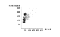
次に、ステップS6において、CPU31により、ステップS29で測定されたWBC分類検出による測定データに基づいて、白血球がリンパ球と好塩基球との集団、単球および顆粒球(好中球と好酸球との集団)の3つに分類される。具体的には、CPU31は、前方散乱光信号および側方散乱光信号を用いて、図15に示すように、スキャッタグラムを作成し、このスキャッタグラムから、リンパ球と好塩基球との集団、単球および顆粒球(好中球と好酸球との集団)のそれぞれの白血球数(WBC)に対する比率を算出する。ここで、図22には、実際に被験者から採取した血液試料を、本実施形態における溶血剤(図10参照)を用いて測定し、その結果得られた前方散乱光信号および側方散乱光信号によるスキャッタグラムを示す。図22に示すように、実際の測定結果においても、スキャッタグラム上で、白血球をリンパ球と好塩基球との集団、単球および顆粒球(好中球と好酸球との集団)の3つに分類することが可能であることが分かる。
Next, in step S6, based on the measurement data by WBC classification detection measured in step S29 by the
そして、ステップS7において、CPU31により、WBC分類検出による測定データに基づいて、白血球が好酸球および好酸球以外の集団の2つに分類される。具体的には、CPU31は、前方散乱光信号および側方蛍光信号を用いて、図16に示すように、スキャッタグラムを作成し、このスキャッタグラムから、白血球数(WBC)に対する好酸球比率を算出する。この側方蛍光信号は、半導体レーザ光源52から出射された青紫色レーザ光(波長が約405nm)により励起された白血球の自家蛍光に基づくものであり、好酸球は、白血球の好酸球以外の集団よりも強い蛍光強度を有している。なお、CPU31は、側方散乱光信号および側方蛍光信号によるスキャッタグラムから、白血球数(WBC)に対する好酸球比率を算出することも可能である。ここで、図23には、実際に被験者から採取した血液試料を、本実施形態における溶血剤(図10参照)を用いて測定し、その結果得られた前方散乱光信号および側方蛍光信号によるスキャッタグラムを示す。図23に示すように、実際の測定結果においても、スキャッタグラム上で、好酸球と好酸球以外の集団とに分類することが可能であることが分かる。
In step S7, the
その後、ステップS8において、CPU31により、ステップS6で算出された顆粒球(好中球と好酸球との集団)比率から、ステップS7で算出された好酸球比率を差し引くことによって、白血球数(WBC)に対する好中球比率が算出される。これにより、白血球がリンパ球と好塩基球との集団、単球、好中球および好酸球の4つに分類される。そして、ステップS9において、CPU31により、リンパ球と好塩基球との集団の比率から、ステップS5で算出されたリンパ球比率を差し引くことによって、白血球数(WBC)に対する好塩基球比率が算出される。これにより、白血球がリンパ球、好塩基球、単球、好中球および好酸球の5つに分類される。
Thereafter, in step S8, the
また、本実施形態では、ステップS10において、CPU31により、ステップS31で測定されたマラリア検出による測定データに基づいて、マラリア感染赤血球がマラリア感染赤血球以外の集団から分類される。具体的には、CPU31は、前方散乱光信号および側方蛍光信号を用いて、図17に示すように、スキャッタグラムを作成し、このスキャッタグラムから、マラリア感染赤血球をマラリア感染赤血球以外の集団から分類する。より具体的には、図17のスキャッタグラムにおいて、マラリアに感染していない赤血球は蛍光強度の小さい領域に現れる一方、マラリア感染赤血球は比較的蛍光強度の大きい領域に現れる。また、白血球は、そのサイズおよびDNA量に起因して、蛍光強度および散乱光強度の両方が大きい領域に現れる。これにより、マラリア感染の有無を判断することが可能となる。
In this embodiment, in step S10, the
次に、ステップS11において、CPU31により、ステップS24で測定されたRBC/PLT検出による測定データに基づいて、赤血球数(RBC)、血小板数(PLT)およびヘマトクリット値(HCT)が算出される。
Next, in step S11, the
そして、ステップS12において、CPU31により、ステップS27で測定されたHGB検出による測定データに基づいて、血色素量(HGB)が算出される。すなわち、SLSヘモグロビン法を用いたHGB検出により得られた吸光度に基づいて、ヘモグロビン濃度が算出される。これにより、白血球を5つ(好中球、リンパ球、単球、好酸球および好塩基球)に分類するために用いる測定試料と同じ第3測定試料を用いて、ヘモグロビン濃度を取得可能である。
In step S12, the
その後、ステップS13において、上記のように算出された赤血球数(RBC)、ヘマトクリット値(HCT)および血色素量(HGB)から、CPU31により、平均赤血球容積(MCV)、平均赤血球血色素量(MCH)および平均赤血球血色素濃度(MCHC)が算出される。各値の算出式をそれぞれ以下の式(1)〜(3)に示す。
Thereafter, in step S13, from the red blood cell count (RBC), hematocrit value (HCT) and hemoglobin amount (HGB) calculated as described above, the
MCV=(HCT/RBC)×1000・・・・・(1)
上記式(1)において、MCVは平均赤血球容積(fL)、HCTはヘマトクリット値(%)、RBCは赤血球数(×104/μL)をそれぞれ表す。MCV = (HCT / RBC) × 1000 (1)
In the above formula (1), MCV represents the mean red blood cell volume (fL), HCT represents the hematocrit value (%), and RBC represents the number of red blood cells (× 10 4 / μL).
MCH=(HGB/RBC)×1000・・・・・(2)
上記式(2)において、MCHは平均赤血球血色素量(pg)、HGBは血色素量(g/dL)、RBCは赤血球数(×104/μL)をそれぞれ表す。MCH = (HGB / RBC) × 1000 (2)
In the above formula (2), MCH represents the average amount of red blood cell pigment (pg), HGB represents the amount of hemoglobin (g / dL), and RBC represents the number of red blood cells (× 10 4 / μL).
MCHC=(HGB/HCT)×100・・・・・(3)
上記式(3)において、MCHCは平均赤血球血色素濃度(g/dL)、HGBは血色素量(g/dL)、HCTはヘマトクリット値(%)をそれぞれ表す。MCHC = (HGB / HCT) × 100 (3)
In the above formula (3), MCHC represents the average erythrocyte hemoglobin concentration (g / dL), HGB represents the hemoglobin amount (g / dL), and HCT represents the hematocrit value (%).
そして、ステップS14において、上記のように算出された、白血球数(WBC)、赤血球数(RBC)、血色素量(HGB)、ヘマトクリット値(HCT)、平均赤血球容積(MCV)、平均赤血球血色素量(MCH)、平均赤血球血色素濃度(MCHC)、血小板数(PLT)の算出結果がディスプレイ装置37に出力される。さらに、白血球数(WBC)に対する好中球比率、リンパ球比率、単球比率、好酸球比率および好塩基球比率がディスプレイ装置37に出力されるとともに、マラリア検出の結果も出力される。なお、白血球数(WBC)に対する各血球比率に加えて、白血球数(WBC)および各血球比率に基づいて算出された好中球数、リンパ球数、単球数、好酸球数および好塩基球数が出力される。
In step S14, the white blood cell count (WBC), red blood cell count (RBC), hemoglobin amount (HGB), hematocrit value (HCT), average red blood cell volume (MCV), average red blood cell hemoglobin amount (calculated as described above) MCH), average erythrocyte hemoglobin concentration (MCHC), and platelet count (PLT) are output to the
その後、ステップS15において、ユーザからのシャットダウン指示の有無が判断され、指示がない場合には、ステップS1に移行される。シャットダウン指示があった場合には、血液分析装置1における試料分析処理のデータ処理ユニット3の動作が終了される。また、測定ユニット2側では、ステップS32で測定データをデータ処理ユニット3に送信した後、ステップS33において、ユーザからのシャットダウン指示があったか否かが判断され、指示がない場合には、ステップS21に移行される。シャットダウン指示があった場合には、血液分析装置1における試料分析処理の測定ユニット2の動作が終了される。
Thereafter, in step S15, it is determined whether or not there is a shutdown instruction from the user. If there is no instruction, the process proceeds to step S1. When there is a shutdown instruction, the operation of the
本実施形態では、上記のように、WBC分類測定部5により血液試料および溶血剤100を含む第1測定試料から生成された側方蛍光信号、前方散乱光信号および側方散乱光信号に基づいて、第1測定試料中の白血球を、少なくとも、単球と、好中球と、好酸球と、他の集団との4つに分類し、WBC分類測定部5により血液試料、前記溶血剤100と同じ溶血剤100、および染色剤を含む第2測定試料から生成された側方蛍光信号および前方散乱光信号に基づいて、第2測定試料中の血球をマラリア感染赤血球とマラリア感染赤血球以外の集団とに分類するCPU31を設けることによって、白血球を4つに分類するための溶血剤とマラリア感染赤血球を検出するための溶血剤とを共通化することができるので、白血球分類およびマラリア感染赤血球検出を行うために組成の異なる2種類の試薬(溶血剤)を開発する必要がない。これにより、試薬の開発に起因するユーザへの負担を軽減しながら、測定試料中の白血球を4つに分類し、かつ、マラリア感染赤血球の検出を行うことができる。
In the present embodiment, as described above, based on the side fluorescence signal, the forward scattered light signal, and the side scattered light signal generated from the first measurement sample including the blood sample and the
また、本実施形態では、DC測定部6により得られた測定データに基づいて、第3測定試料中の白血球をリンパ球とリンパ球以外の集団とに分類するとともに、この分類結果と上記した白血球の4分類の分類結果とに基づいて、測定試料中の白血球を、少なくとも、リンパ球と、好塩基球と、単球と、好中球と、好酸球との5つに分類するようにCPU31を構成することによって、白血球分類およびマラリア感染赤血球検出を行うための溶血剤と同じ溶血剤100を用いて、白血球をリンパ球とリンパ球以外の集団とに分類することができるので、組成の異なる溶血剤を別途開発することなく、測定試料中の白血球を5つに分類することができる。
In the present embodiment, the white blood cells in the third measurement sample are classified into lymphocytes and a group other than lymphocytes based on the measurement data obtained by the
また、本実施形態による血液分析方法では、上記のように、WBC分類測定部5により血液試料および溶血剤100を含む第1測定試料から生成された側方蛍光信号、前方散乱光信号および側方散乱光信号に基づいて、第1測定試料中の白血球を、少なくとも、単球と、好中球と、好酸球と、他の集団との4つに分類するステップと、WBC分類測定部5により血液試料、前記溶血剤100と同じ溶血剤100、および染色剤を含む第2測定試料から生成された側方蛍光信号および前方散乱光信号に基づいて、第2測定試料中の血球をマラリア感染赤血球とマラリア感染赤血球以外の集団とに分類するステップとを設けることによって、白血球を4つに分類するための溶血剤とマラリア感染赤血球を検出するための溶血剤とを共通化することができるので、白血球分類およびマラリア感染赤血球検出を行うために組成の異なる2種類の試薬(溶血剤)を開発する必要がない。これにより、試薬の開発に起因するユーザへの負担を軽減しながら、測定試料中の白血球を4つに分類し、かつ、マラリア感染赤血球の検出を行うことができる。
Further, in the blood analysis method according to the present embodiment, as described above, the side fluorescent signal, the forward scattered light signal, and the side light generated from the first measurement sample including the blood sample and the
また、本実施形態による溶血剤を、上記のように、WBC分類測定部5により血液試料および溶血剤100を含む第1測定試料から生成された側方蛍光信号、前方散乱光信号および側方散乱光信号に基づいて、第1測定試料中の白血球を、少なくとも、単球と、好中球と、好酸球と、他の集団との4つに分類するステップと、WBC分類測定部5により血液試料、前記溶血剤100と同じ溶血剤100、および染色剤を含む第2測定試料から生成された側方蛍光信号および前方散乱光信号に基づいて、第2測定試料中の血球をマラリア感染赤血球とマラリア感染赤血球以外の集団とに分類するステップとを備えた血液分析方法に用いることによって、白血球を4つに分類するための溶血剤とマラリア感染赤血球を検出するための溶血剤とを共通化することができるので、白血球分類およびマラリア感染赤血球検出を行うために組成の異なる2種類の試薬(溶血剤)を開発する必要がない。これにより、試薬の開発に起因するユーザへの負担を軽減しながら、測定試料中の白血球を4つに分類し、かつ、マラリア感染赤血球の検出を行うことができる。
In addition, as described above, the hemolytic agent according to the present embodiment is converted from the side fluorescence signal, the forward scattered light signal, and the side scattering generated from the WBC
また、本実施形態による染色剤を、上記のように、WBC分類測定部5により血液試料および溶血剤100を含む第1測定試料から生成された側方蛍光信号、前方散乱光信号および側方散乱光信号に基づいて、第1測定試料中の白血球を、少なくとも、単球と、好中球と、好酸球と、他の集団との4つに分類するステップと、WBC分類測定部5により血液試料、前記溶血剤100と同じ溶血剤100、および染色剤を含む第2測定試料から生成された側方蛍光信号および前方散乱光信号に基づいて、第2測定試料中の血球をマラリア感染赤血球とマラリア感染赤血球以外の集団とに分類するステップとを備えた血液分析方法に用いることによって、白血球を4つに分類するための溶血剤とマラリア感染赤血球を検出するための溶血剤とを共通化することができるので、白血球分類およびマラリア感染赤血球検出を行うために組成の異なる2種類の試薬(溶血剤)を開発する必要がない。これにより、試薬の開発に起因するユーザへの負担を軽減しながら、測定試料中の白血球を4つに分類し、かつ、マラリア感染赤血球の検出を行うことができる。
Further, as described above, the staining agent according to the present embodiment is obtained by using the side fluorescence signal, the forward scattered light signal, and the side scattering generated from the first measurement sample including the blood sample and the
(実施例)
次に、目視により得られたマラリア感染率と、上記実施形態に記載の方法(試薬は、上記実施形態に記載の試薬と同様のものを使用した)に基づいて得られたマラリア感染率との関係を以下の表1に示す。なお、目視による測定と上記実施形態に記載の方法による測定とは、複数の血液試料について、同じ検体について行った。(Example)
Next, the malaria infection rate obtained by visual observation and the malaria infection rate obtained based on the method described in the above embodiment (the same reagent as the reagent described in the above embodiment was used). The relationship is shown in Table 1 below. The visual measurement and the measurement by the method described in the above embodiment were performed on the same specimen for a plurality of blood samples.
表1に示すように、目視により得られたマラリア感染率と、上記実施形態に記載の方法に基づいて得られたマラリア感染率とは略一致しており、上記実施形態に記載の方法によれば、マラリア感染赤血球を精度よく検出できることを確認することができた。 As shown in Table 1, the malaria infection rate obtained by visual observation and the malaria infection rate obtained based on the method described in the above embodiment are substantially the same, and according to the method described in the above embodiment. It was confirmed that malaria-infected erythrocytes could be detected with high accuracy.
なお、目視によるマラリア感染率は、以下の式(4)により算出した。 In addition, the malaria infection rate by visual observation was computed by the following formula | equation (4).
マラリア感染率(%)=X/Y×100・・・・・(4)
上記式(4)において、Xは目視によりカウントされた所定数(=Y)の赤血球のうち、マラリアに感染していると決定された赤血球の数、Yは上記所定数をそれぞれ表す。なお、表1において、Yは、サンプル1および2については約10,000、サンプル3については約20,000、サンプル4については約30,000である。Malaria infection rate (%) = X / Y × 100 (4)
In the above formula (4), X represents the number of red blood cells determined to be infected with malaria among the predetermined number (= Y) of red blood cells counted visually, and Y represents the predetermined number. In Table 1, Y is about 10,000 for
また、上記実施形態に記載の方法に基づいて得られたマラリア感染率は、以下の式(5)により算出した。 Moreover, the malaria infection rate obtained based on the method as described in the said embodiment was computed by the following formula | equation (5).
マラリア感染率(%)=(6)/(7)×100・・・・・(5)
上記式(5)において、(6)はA×B/C、(7)は同じ血液試料を多項目自動血球分析装置XE−2100(シスメックス株式会社製)で測定して得られた赤血球数をそれぞれ表す。なお、上記(6)のAは図17のマラリア領域内の血球数、Bは同じ血液試料を多項目自動血球分析装置XE−2100で測定して得られた白血球数、Cは図17の白血球領域内の血球数である。Malaria infection rate (%) = (6) / (7) × 100 (5)
In the above formula (5), (6) is A × B / C, (7) is the number of red blood cells obtained by measuring the same blood sample with a multi-item automatic blood cell analyzer XE-2100 (manufactured by Sysmex Corporation). Represent each. A in (6) above is the number of blood cells in the malaria region of FIG. 17, B is the number of white blood cells obtained by measuring the same blood sample with the multi-item automatic blood cell analyzer XE-2100, and C is the white blood cell of FIG. The number of blood cells in the region.
なお、今回開示された実施形態は、全ての点で例示であって制限的なものではないと考えられるべきである。本発明の範囲は、上記した実施形態の説明ではなく特許請求の範囲によって示され、さらに特許請求の範囲と均等の意味および範囲内での全ての変更が含まれる。 In addition, it should be thought that embodiment disclosed this time is an illustration and restrictive at no points. The scope of the present invention is shown not by the above description of the embodiments but by the scope of claims for patent, and further includes meanings equivalent to the scope of claims for patent and all modifications within the scope.
たとえば、上記実施形態では、WBC検出、HGB検出、WBC分類検出およびマラリア検出に共通に用いられる溶血剤を収容する1つの、試薬容器としての溶血剤容器を試料供給部に接続する例を示したが、本発明はこれに限らず、各検出に用いる溶血剤をそれぞれ別々に収容するように4つの溶血剤容器を試料供給部に接続してもよいし、上記4つの検出のうち、いずれかに用いる溶血剤を共通の溶血剤容器に収容し、2つまたは3つの溶血剤容器を試料供給部に接続してもよい。また、5つ以上の溶血剤容器を試料供給部に接続してもよい。この際、各溶血剤容器に収容する溶血剤を、それぞれ予め所定の希釈倍率に希釈しておけば、各検出に用いる測定試料を調製する際に、溶血剤を所望の希釈倍率に希釈するための工程を別途設ける必要がない。 For example, in the above embodiment, an example is shown in which a hemolytic agent container as a reagent container that accommodates a hemolytic agent commonly used for WBC detection, HGB detection, WBC classification detection, and malaria detection is connected to the sample supply unit. However, the present invention is not limited to this, and four hemolytic agent containers may be connected to the sample supply unit so as to accommodate the hemolytic agent used for each detection separately, and any one of the above four detections. The hemolytic agent used in the above may be accommodated in a common hemolytic agent container, and two or three hemolytic agent containers may be connected to the sample supply unit. Further, five or more hemolytic agent containers may be connected to the sample supply unit. At this time, if the hemolytic agent accommodated in each hemolytic agent container is previously diluted to a predetermined dilution rate, the hemolytic agent is diluted to a desired dilution rate when preparing a measurement sample used for each detection. There is no need to provide a separate process.
また、上記実施形態では、溶血剤の一例として、標識物質を含まない溶血剤を示したが、本発明はこれに限らず、溶血剤が標識物質を含んでいてもよい。 Moreover, in the said embodiment, although the hemolytic agent which does not contain a labeling substance was shown as an example of the hemolytic agent, this invention is not limited to this, The hemolytic agent may contain the labeling substance.
また、上記実施形態では、WBC検出およびHGB検出に、WBC分類検出に用いる溶血剤と同じ溶血剤を用いる例を示したが、本発明はこれに限らず、WBC検出、HGB検出およびWBC分類検出にそれぞれ別々の専用の溶血剤を用いてもよい。 In the above embodiment, an example in which the same hemolytic agent as that used for WBC classification detection is used for WBC detection and HGB detection is shown. However, the present invention is not limited to this, and WBC detection, HGB detection, and WBC classification detection are used. Separate hemolytic agents may be used.
また、上記実施形態では、試料分析処理において、早いものからRBC/PLT検出、WBC検出、HGB検出、WBC分類検出およびマラリア検出の順序で各検出処理を行う例を示したが、本発明はこれに限らず、試料分析処理において、上記以外の順序で各検出処理を行ってもよい。また、試料分析処理における、白血球分類処理、マラリア分類処理、赤血球数・血小板数算出処理、および、血色素量算出処理の順序も適宜変更可能である。 In the above-described embodiment, an example is shown in which each detection process is performed in the order of RBC / PLT detection, WBC detection, HGB detection, WBC classification detection, and malaria detection from the earliest in the sample analysis process. In addition, in the sample analysis process, each detection process may be performed in a sequence other than the above. In addition, the order of the white blood cell classification process, the malaria classification process, the red blood cell count / platelet count calculation process, and the hemoglobin amount calculation process in the sample analysis process can be appropriately changed.
また、上記実施形態では、青紫色半導体レーザ素子を有する半導体レーザ光源を設ける例を示したが、本発明はこれに限らず、青色半導体レーザ素子またはアルゴンレーザ素子など、青紫色半導体レーザ素子以外のレーザ素子を有する光源を設けてもよい。 Moreover, although the example which provides the semiconductor laser light source which has a blue-violet semiconductor laser element was shown in the said embodiment, this invention is not limited to this, Other than a blue-violet semiconductor laser element, such as a blue semiconductor laser element or an argon laser element A light source having a laser element may be provided.
また、上記実施形態では、第2測定試料における溶血剤を9倍に希釈する構成の例を示したが、本発明はこれに限られない。また、第2測定試料における溶血剤は、9倍以上12倍以下に希釈することが好ましい。 Moreover, although the example of the structure which dilutes the hemolytic agent in a 2nd measurement sample 9 times was shown in the said embodiment, this invention is not limited to this. Further, the hemolytic agent in the second measurement sample is preferably diluted 9 times or more and 12 times or less.
また、上記実施形態では、溶血剤の一例として、アルキルトリメチルアンモニウム塩であって、アルキル基の炭素数が12以上18以下であるカチオン性界面活性剤(ラウリルトリメチルアンモニウムクロライド;34.1mM、ステアリルトリメチルアンモニウムクロライド;1.7mM)を含む溶血剤を示したが、本発明はこれに限らず、カチオン性界面活性剤(上記実施形態では、ラウリルトリメチルアンモニウムクロライドとステアリルトリメチルアンモニウムクロライドの合計)のWBC(分類用)測定試料における濃度が、0.62mM以上2.15mM以下であれば、上記以外の濃度のカチオン性界面活性剤を含む溶血剤であってもよい。なお、上記実施形態では、溶血剤を25倍希釈して測定試料を調製しているので、カチオン性界面活性剤のWBC(分類用)測定試料における濃度が0.62mMになるときのカチオン性界面活性剤の溶血剤における濃度は15.5mMであり、カチオン性界面活性剤のWBC(分類用)測定試料における濃度が2.15mMになるときのカチオン性界面活性剤の溶血剤における濃度は53.75mMである。また、上記の溶血剤に代えて、アルキル基の炭素数が8以上10以下であるカチオン性界面活性剤を用いれば、カチオン性界面活性剤のWBC(分類用)測定試料における濃度が2.15mM以上であっても測定可能である。
Moreover, in the said embodiment, as an example of a hemolytic agent, it is an alkyl trimethyl ammonium salt, Comprising: Cationic surfactant (lauryl trimethyl ammonium chloride; 34.1 mM, stearyl trimethyl whose alkyl group has 12 to 18 carbon atoms) A hemolytic agent containing ammonium chloride (1.7 mM) was shown, but the present invention is not limited to this, and a WBC (a total of lauryltrimethylammonium chloride and stearyltrimethylammonium chloride in the above embodiment) is not limited thereto. If the concentration in the measurement sample is 0.62 mM or more and 2.15 mM or less, it may be a hemolytic agent containing a cationic surfactant having a concentration other than the above. In the above embodiment, since the measurement sample is prepared by diluting the
ここで、本発明の一実施形態による血液分析装置において、溶血剤におけるカチオン性界面活性剤の濃度を変動させた場合の実験結果について説明する。実験では、溶血剤におけるカチオン性界面活性剤の濃度が微量ずつ異なる複数の実験結果を得たが、ここでは、代表して、カチオン性界面活性剤のWBC(分類用)測定試料における濃度が2.15mMである溶血剤を用いた場合、および、カチオン性界面活性剤のWBC(分類用)測定試料における濃度が0.62mMである溶血剤を用いた場合の2つの実験結果について説明する。 Here, an experimental result when the concentration of the cationic surfactant in the hemolytic agent is varied in the blood analyzer according to the embodiment of the present invention will be described. In the experiment, a plurality of experimental results were obtained in which the concentration of the cationic surfactant in the hemolytic agent varies by a small amount. Here, representatively, the concentration of the cationic surfactant in the WBC (for classification) measurement sample is 2 Two experimental results in the case of using a hemolytic agent having a concentration of 15 mM and in the case of using a hemolytic agent having a concentration of 0.62 mM in the WBC (for classification) measurement sample of the cationic surfactant will be described.
図18および図20に示すように、スキャッタグラム上で、白血球をリンパ球と好塩基球との集団、単球および顆粒球(好中球と好酸球との集団)の3つに分類することが可能である。また、図19および図21に示すように、スキャッタグラム上で、好酸球と好酸球以外の集団とに分類することが可能である。また、これらの分類結果から白血球を、リンパ球と好塩基球との集団、単球、好中球および好酸球の4つに分類することが可能である。したがって、カチオン性界面活性剤のWBC(分類用)測定試料における濃度が0.62mM以上2.15mM以下の範囲においては、白血球を4分類することが可能であると考えられる。 As shown in FIGS. 18 and 20, on the scattergram, leukocytes are classified into three groups: a lymphocyte and basophil group, a monocyte, and a granulocyte (neutrophil and eosinophil group). It is possible. Further, as shown in FIGS. 19 and 21, it is possible to classify into eosinophils and groups other than eosinophils on the scattergram. From these classification results, it is possible to classify leukocytes into four groups: a group of lymphocytes and basophils, monocytes, neutrophils and eosinophils. Therefore, it is considered that leukocytes can be classified into four categories when the concentration of the cationic surfactant in the WBC (for classification) measurement sample is in the range of 0.62 mM to 2.15 mM.
Claims (11)
前記第1測定試料から、第1蛍光情報と、少なくとも2種類の第1散乱光情報とを生成するとともに、前記第2測定試料から、第2蛍光情報と第2散乱光情報とを生成することが可能な光情報生成部と、
前記第1測定試料に含まれる白血球を分析する場合、前記光情報生成部により生成された前記第1蛍光情報と前記2種類の第1散乱光情報とに基づいて、前記第1測定試料中の白血球を、少なくとも、単球と、好中球と、好酸球と、他の集団との4つに第1分類し、前記第2測定試料に含まれるマラリア感染赤血球を分析する場合、前記光情報生成部により生成された前記第2蛍光情報と前記第2散乱光情報とに基づいて、前記第2測定試料中の血球を、マラリア感染赤血球とマラリア感染赤血球以外の集団とに分類する制御部とを備え、
前記第2測定試料における前記溶血剤の希釈倍率は、前記第1測定試料における前記溶血剤の希釈倍率と異なる、血液分析装置。 A sample preparation unit capable of preparing a first measurement sample including a blood sample and a hemolytic agent, and a second measurement sample including the blood sample and the same hemolytic agent and staining agent as the hemolytic agent;
Generating first fluorescence information and at least two types of first scattered light information from the first measurement sample, and generating second fluorescence information and second scattered light information from the second measurement sample; An optical information generation unit capable of
When analyzing white blood cells contained in the first measurement sample, based on the first fluorescence information generated by the light information generation unit and the two types of first scattered light information, When the white blood cells are first classified into at least four types of monocytes, neutrophils, eosinophils, and other populations, and the malaria-infected red blood cells contained in the second measurement sample are analyzed, A control unit that classifies blood cells in the second measurement sample into malaria-infected erythrocytes and groups other than malaria-infected erythrocytes based on the second fluorescence information and the second scattered light information generated by the information generation unit It equipped with a door,
The dilution ratio of the hemolytic agent in the second measurement sample, that different from the dilution ratio of the hemolytic agent in said first measurement sample, a blood analyzer.
前記第2測定試料に含まれるマラリア感染赤血球を分析する場合、前記光情報生成部は、前記試料調製部によって調製された前記第2測定試料から前記第2蛍光情報と前記第2散乱光情報とを生成する、請求項1の血液分析装置。 When analyzing the white blood cells contained in the first measurement sample, the optical information generation unit includes the first fluorescence information and at least two types of the first scattered light from the first measurement sample prepared by the sample preparation unit. Generating information and
When analyzing malaria-infected erythrocytes contained in the second measurement sample, the optical information generation unit includes the second fluorescence information and the second scattered light information from the second measurement sample prepared by the sample preparation unit. The blood analyzer according to claim 1, wherein
前記血液分析装置は、前記第3測定試料から、試料の電気情報を生成する電気情報生成部をさらに備え、
前記制御部は、前記第1及び第3測定試料に含まれる白血球を分析する場合、前記電気情報生成部により生成された電気情報に基づいて、前記第3測定試料中の白血球を少なくともリンパ球とリンパ球以外の集団とに第2分類するとともに、前記第1分類および前記第2分類の分類結果に基づいて、前記測定試料中の白血球を、少なくとも、リンパ球と、好塩基球と、単球と、好中球と、好酸球との5つに分類するように構成されている、請求項1または2に記載の血液分析装置。 The sample preparation unit can further prepare a third measurement sample including the blood sample and the hemolytic agent,
The blood analyzer further includes an electrical information generation unit that generates electrical information of the sample from the third measurement sample,
When analyzing the white blood cells contained in the first and third measurement samples, the control unit determines at least the white blood cells in the third measurement sample as lymphocytes based on the electrical information generated by the electrical information generation unit. Secondly classified into a group other than lymphocytes, and based on the classification results of the first classification and the second classification, leukocytes in the measurement sample are at least lymphocytes, basophils, and monocytes The blood analyzer according to claim 1, wherein the blood analyzer is configured to be classified into five types: neutrophils and eosinophils.
前記制御部は、前記第2光情報生成部により生成された前記透過光情報または前記散乱光情報の少なくとも一方に基づいて、前記第3測定試料中のヘモグロビン濃度を取得するように構成されている、請求項3に記載の血液分析装置。 A second optical information generation unit that generates at least one of transmitted light information or scattered light information of the sample from the third measurement sample;
The control unit is configured to acquire a hemoglobin concentration in the third measurement sample based on at least one of the transmitted light information or the scattered light information generated by the second light information generation unit. The blood analyzer according to claim 3.
血液試料と前記溶血剤と同じ溶血剤と染色剤とを含み、この溶血剤の希釈倍率は前記溶血剤の前記所定の希釈倍率と異なる第2測定試料を調製するステップと、
前記第1測定試料から、第1蛍光情報と、少なくとも2種類の第1散乱光情報とを生成するステップと、
前記第2測定試料から、第2蛍光情報と第2散乱光情報とを生成するステップと、前記第1測定試料から生成された前記第1蛍光情報と前記2種類の第1散乱光情報とに基づいて、前記第1測定試料中の白血球を、少なくとも、単球と、好中球と、好酸球と、他の集団との4つに分類するステップと、
前記第2測定試料から生成された前記第2蛍光情報と前記第2散乱光情報とに基づいて、前記第2測定試料中の血球を、マラリア感染赤血球とマラリア感染赤血球以外の集団とに分類するステップとを備える、血液分析方法。 Preparing a first measurement sample including a blood sample and a hemolyzing agent at a predetermined dilution rate ;
A step saw including the same hemolytic agent as the blood sample and the hemolytic agent and staining agents, dilution ratio of the hemolytic agent to prepare the second measurement sample is different from the predetermined dilution ratio of the hemolytic agent,
Generating first fluorescence information and at least two types of first scattered light information from the first measurement sample;
The step of generating second fluorescence information and second scattered light information from the second measurement sample, the first fluorescence information generated from the first measurement sample and the two types of first scattered light information And classifying the leukocytes in the first measurement sample into at least four of monocytes, neutrophils, eosinophils, and other populations;
Based on the second fluorescence information and the second scattered light information generated from the second measurement sample, blood cells in the second measurement sample are classified into a group other than malaria-infected red blood cells and malaria-infected red blood cells. A blood analysis method comprising the steps of:
Applications Claiming Priority (3)
| Application Number | Priority Date | Filing Date | Title |
|---|---|---|---|
| JP2008123403 | 2008-05-09 | ||
| JP2008123403 | 2008-05-09 | ||
| PCT/JP2009/058327 WO2009136573A1 (en) | 2008-05-09 | 2009-04-28 | Blood analyzer, blood analysis method, hemolytic agent and staining agent |
Publications (2)
| Publication Number | Publication Date |
|---|---|
| JPWO2009136573A1 JPWO2009136573A1 (en) | 2011-09-08 |
| JP5619604B2 true JP5619604B2 (en) | 2014-11-05 |
Family
ID=41264628
Family Applications (1)
| Application Number | Title | Priority Date | Filing Date |
|---|---|---|---|
| JP2010511054A Expired - Fee Related JP5619604B2 (en) | 2008-05-09 | 2009-04-28 | Blood analyzer, blood analysis method, hemolyzing agent and staining agent |
Country Status (5)
| Country | Link |
|---|---|
| US (2) | US8920726B2 (en) |
| EP (2) | EP2293062B1 (en) |
| JP (1) | JP5619604B2 (en) |
| CN (2) | CN104297463A (en) |
| WO (1) | WO2009136573A1 (en) |
Families Citing this family (36)
| Publication number | Priority date | Publication date | Assignee | Title |
|---|---|---|---|---|
| GB2494653A (en) * | 2011-09-13 | 2013-03-20 | Orreco Ltd | Apparatus for blood analysis |
| CN103091286B (en) * | 2011-10-31 | 2016-08-17 | 深圳迈瑞生物医疗电子股份有限公司 | The erythrocytic recognition methods of Infected With Plasmodium and device |
| SE537208C2 (en) | 2012-12-03 | 2015-03-03 | Tommy Forsell | Blood analyzer for malaria analysis |
| JP6364426B2 (en) | 2012-12-31 | 2018-07-25 | ベックマン コールター, インコーポレイテッド | Immature platelet counting system and method |
| JP2015021938A (en) | 2013-07-23 | 2015-02-02 | シスメックス株式会社 | Analyte analysis device, disease monitoring system and data management method of analyte analysis device |
| WO2015029032A1 (en) | 2013-08-26 | 2015-03-05 | Parasight Ltd. | Digital microscopy systems, methods and computer program products |
| JP6383216B2 (en) * | 2014-08-08 | 2018-08-29 | シスメックス株式会社 | Blood analysis method, blood analysis apparatus and program |
| WO2016043683A1 (en) * | 2014-09-18 | 2016-03-24 | Ciftci Ihsan Hakki | A blood count device |
| JP6352750B2 (en) | 2014-09-26 | 2018-07-04 | シスメックス株式会社 | Blood analyzer and blood analysis method |
| WO2016130964A1 (en) | 2015-02-13 | 2016-08-18 | Abbott Laboratories | Decapping and capping apparatus, systems and methods for use in diagnostic analyzers |
| JP6661278B2 (en) | 2015-03-27 | 2020-03-11 | シスメックス株式会社 | Blood analyzer and blood analysis method |
| US10634660B2 (en) * | 2015-03-31 | 2020-04-28 | Sony Corporation | Electrical characteristic measurement device, electrical characteristic measurement method, and blood condition analysis system |
| JP6635720B2 (en) | 2015-08-31 | 2020-01-29 | シスメックス株式会社 | Blood analyzer and blood analysis method |
| JP6621620B2 (en) * | 2015-08-31 | 2019-12-18 | シスメックス株式会社 | Blood analysis method, staining solution and blood analyzer used therefor |
| US10488644B2 (en) * | 2015-09-17 | 2019-11-26 | S.D. Sight Diagnostics Ltd. | Methods and apparatus for detecting an entity in a bodily sample |
| JP6871729B2 (en) * | 2016-12-06 | 2021-05-12 | シスメックス株式会社 | Detection method and blood analyzer for red blood cells infected with Plasmodium |
| CN120801147A (en) * | 2018-08-16 | 2025-10-17 | 上海宜晟生物科技有限公司 | Cellular analysis of body fluids, particularly blood |
| CN114174826A (en) * | 2019-09-19 | 2022-03-11 | 深圳迈瑞动物医疗科技有限公司 | Animal blood cell analysis method, analyzer and storage medium |
| AU2020370212B2 (en) | 2019-10-22 | 2025-09-25 | S.D. Sight Diagnostics Ltd | Accounting for errors in optical measurements |
| CN110579613A (en) * | 2019-10-28 | 2019-12-17 | 深圳开立生物医疗科技股份有限公司 | Blood analyzer |
| CN114787625B (en) | 2019-12-12 | 2026-01-13 | 思迪赛特诊断有限公司 | Detection of platelets in a blood sample |
| BR112022011312A2 (en) | 2019-12-12 | 2022-08-23 | S D Sight Diagnostics Ltd | ANALYSIS OF AN ANALYTE DISPOSED IN A MEDIUM |
| JP7425597B2 (en) * | 2019-12-25 | 2024-01-31 | シスメックス株式会社 | Blood analysis method |
| US12332265B2 (en) | 2019-12-27 | 2025-06-17 | Beckman Coulter, Inc. | Sample preparation instrument |
| WO2021179177A1 (en) * | 2020-03-10 | 2021-09-16 | 深圳迈瑞生物医疗电子股份有限公司 | Blood analyzer, blood analysis method, and computer readable storage medium |
| CN111521834B (en) * | 2020-03-20 | 2023-03-24 | 佛山市顺德区德维医疗科技有限公司 | Reagent for XN series full-automatic modular blood body fluid analyzer |
| JP7583401B2 (en) | 2020-04-13 | 2024-11-14 | 国立大学法人大阪大学 | Hematology Analyzer |
| CN113702267B (en) * | 2020-05-22 | 2025-08-12 | 深圳迈瑞动物医疗科技股份有限公司 | Method for detecting hemoglobin concentration and blood cell analyzer |
| CN113959911B (en) * | 2020-07-20 | 2024-06-18 | 深圳迈瑞生物医疗电子股份有限公司 | Detection method, reagent and application of antiplatelet aggregation interference |
| CN114252386A (en) * | 2020-09-23 | 2022-03-29 | 深圳迈瑞生物医疗电子股份有限公司 | Sample detection method and sample analyzer |
| US20240103000A1 (en) | 2020-09-29 | 2024-03-28 | Universidade Do Minho | Automatic device for non-invasive malaria diagnosis through optical reflectance techniques, methods and uses thereof |
| WO2022115982A1 (en) | 2020-12-01 | 2022-06-09 | 深圳迈瑞生物医疗电子股份有限公司 | Sample analysis method, sample analyzer, and computer-readable storage medium |
| CN115201154A (en) * | 2021-04-08 | 2022-10-18 | 深圳迈瑞生物医疗电子股份有限公司 | Method for detecting blood sample and sample analyzer |
| CN115219392A (en) * | 2021-04-14 | 2022-10-21 | 深圳迈瑞生物医疗电子股份有限公司 | Sample analyzer and sample analysis method |
| CN118185751B (en) * | 2024-04-07 | 2024-12-10 | 重庆大学 | Leucocyte classification detection system and method |
| CN121898960A (en) * | 2024-10-21 | 2026-04-21 | 希森美康株式会社 | Subject measurement device |
Citations (5)
| Publication number | Priority date | Publication date | Assignee | Title |
|---|---|---|---|---|
| JP2004141150A (en) * | 2002-10-04 | 2004-05-20 | Sysmex Corp | Method for detecting plasmodium, device for detecting the same and reagent kit for the same |
| JP2006292738A (en) * | 2005-03-17 | 2006-10-26 | Sysmex Corp | Sample analyzer, sample analysis method, and blood analyzer |
| JP2006304774A (en) * | 2005-03-29 | 2006-11-09 | Sysmex Corp | Method for detecting malaria-infected erythrocytes, detection reagent and erythrocyte membrane partial lysis reagent used therefor |
| JP2007024844A (en) * | 2005-07-21 | 2007-02-01 | Sysmex Corp | Method and system for hemanalysis |
| JP2007525674A (en) * | 2004-02-27 | 2007-09-06 | ベックマン コールター,インコーポレイティド | How to detect malaria parasites and other parasitic infections |
Family Cites Families (13)
| Publication number | Priority date | Publication date | Assignee | Title |
|---|---|---|---|---|
| JPH05322882A (en) * | 1992-05-21 | 1993-12-07 | Hitachi Ltd | Blood analyzer |
| JP3830613B2 (en) | 1997-04-18 | 2006-10-04 | シスメックス株式会社 | Reagent for measuring leukocyte and hemoglobin concentration in blood |
| US6210969B1 (en) | 1999-04-28 | 2001-04-03 | Coulter International Corp. | Composition and method for differentiation of basophil and eosinophil subpopulations of leukocytes in blood |
| US7553453B2 (en) * | 2000-06-02 | 2009-06-30 | Honeywell International Inc. | Assay implementation in a microfluidic format |
| EP1406088A3 (en) * | 2002-10-04 | 2004-04-14 | Sysmex Corporation | Reagent kits and methods for detecting malaria parasites by two-steps surfactant-mediated hemolysis |
| JP2005333868A (en) * | 2004-05-26 | 2005-12-08 | Sysmex Corp | Method and device for measuring malaria parasite |
| CN1834659A (en) * | 2005-03-17 | 2006-09-20 | 希森美康株式会社 | Sample analysis device, sample analysis method, and blood analysis device |
| US9243993B2 (en) * | 2005-03-17 | 2016-01-26 | Sysmex Corporation | Sample analyzer and sample analyzing method |
| EP1715345B1 (en) * | 2005-03-29 | 2013-12-04 | Sysmex Corporation | A reagent for partially lysing a cell membrane of a red blood cell, a reagent for detecting malaria infected red blood cells, and a sample analyzing method for detecting malaria infected red blood cells |
| JP2006313151A (en) * | 2005-04-07 | 2006-11-16 | Sysmex Corp | Blood analyzer, sample analyzer and flow cytometer |
| JP4964446B2 (en) * | 2005-09-14 | 2012-06-27 | シスメックス株式会社 | Analytical apparatus and sample information processing program |
| JP4745030B2 (en) * | 2005-11-15 | 2011-08-10 | シスメックス株式会社 | Blood analyzer |
| EP2012120B1 (en) | 2006-04-21 | 2014-03-12 | Nihon Kohden Corporation | Method of classifying particles and apparatus therefor |
-
2009
- 2009-04-28 EP EP09742697.7A patent/EP2293062B1/en not_active Not-in-force
- 2009-04-28 JP JP2010511054A patent/JP5619604B2/en not_active Expired - Fee Related
- 2009-04-28 WO PCT/JP2009/058327 patent/WO2009136573A1/en not_active Ceased
- 2009-04-28 CN CN201410546643.6A patent/CN104297463A/en active Pending
- 2009-04-28 EP EP14198316.3A patent/EP2889620B1/en not_active Not-in-force
- 2009-04-28 CN CN200980116617.7A patent/CN102016573B/en not_active Expired - Fee Related
-
2010
- 2010-11-09 US US12/942,752 patent/US8920726B2/en active Active
-
2014
- 2014-11-03 US US14/530,973 patent/US9328375B2/en active Active
Patent Citations (5)
| Publication number | Priority date | Publication date | Assignee | Title |
|---|---|---|---|---|
| JP2004141150A (en) * | 2002-10-04 | 2004-05-20 | Sysmex Corp | Method for detecting plasmodium, device for detecting the same and reagent kit for the same |
| JP2007525674A (en) * | 2004-02-27 | 2007-09-06 | ベックマン コールター,インコーポレイティド | How to detect malaria parasites and other parasitic infections |
| JP2006292738A (en) * | 2005-03-17 | 2006-10-26 | Sysmex Corp | Sample analyzer, sample analysis method, and blood analyzer |
| JP2006304774A (en) * | 2005-03-29 | 2006-11-09 | Sysmex Corp | Method for detecting malaria-infected erythrocytes, detection reagent and erythrocyte membrane partial lysis reagent used therefor |
| JP2007024844A (en) * | 2005-07-21 | 2007-02-01 | Sysmex Corp | Method and system for hemanalysis |
Also Published As
| Publication number | Publication date |
|---|---|
| US20110053212A1 (en) | 2011-03-03 |
| EP2293062A1 (en) | 2011-03-09 |
| WO2009136573A1 (en) | 2009-11-12 |
| EP2293062B1 (en) | 2015-01-28 |
| EP2889620B1 (en) | 2017-10-11 |
| CN102016573A (en) | 2011-04-13 |
| CN102016573B (en) | 2014-11-12 |
| EP2889620A1 (en) | 2015-07-01 |
| EP2293062A4 (en) | 2011-05-25 |
| CN104297463A (en) | 2015-01-21 |
| US8920726B2 (en) | 2014-12-30 |
| US20150050643A1 (en) | 2015-02-19 |
| US9328375B2 (en) | 2016-05-03 |
| JPWO2009136573A1 (en) | 2011-09-08 |
Similar Documents
| Publication | Publication Date | Title |
|---|---|---|
| JP5619604B2 (en) | Blood analyzer, blood analysis method, hemolyzing agent and staining agent | |
| JP5627634B2 (en) | Blood analyzer and blood analysis method | |
| JP5670052B2 (en) | Blood analyzer, blood analysis method and computer program | |
| CN105891090B (en) | Blood cell analyzer, body fluid analysis method, and control system therefor | |
| JP4745030B2 (en) | Blood analyzer | |
| JP2006313151A (en) | Blood analyzer, sample analyzer and flow cytometer | |
| CN114450589A (en) | Method for analyzing red blood cells in blood sample and blood analysis system | |
| JP5441466B2 (en) | Veterinary blood cell analyzer | |
| JP6621620B2 (en) | Blood analysis method, staining solution and blood analyzer used therefor | |
| US20260110618A1 (en) | Sample measurement apparatus | |
| JP7425597B2 (en) | Blood analysis method | |
| WO2026070928A1 (en) | Measurement device | |
| JP2025010969A (en) | Method for detecting platelet aggregation | |
| JP2026059654A (en) | Measuring apparatus, reagents, and analytical methods |
Legal Events
| Date | Code | Title | Description |
|---|---|---|---|
| A521 | Request for written amendment filed |
Free format text: JAPANESE INTERMEDIATE CODE: A523 Effective date: 20120417 |
|
| A621 | Written request for application examination |
Free format text: JAPANESE INTERMEDIATE CODE: A621 Effective date: 20120417 |
|
| A131 | Notification of reasons for refusal |
Free format text: JAPANESE INTERMEDIATE CODE: A131 Effective date: 20131217 |
|
| A521 | Request for written amendment filed |
Free format text: JAPANESE INTERMEDIATE CODE: A523 Effective date: 20140203 |
|
| TRDD | Decision of grant or rejection written | ||
| A01 | Written decision to grant a patent or to grant a registration (utility model) |
Free format text: JAPANESE INTERMEDIATE CODE: A01 Effective date: 20140819 |
|
| A61 | First payment of annual fees (during grant procedure) |
Free format text: JAPANESE INTERMEDIATE CODE: A61 Effective date: 20140917 |
|
| R150 | Certificate of patent or registration of utility model |
Ref document number: 5619604 Country of ref document: JP Free format text: JAPANESE INTERMEDIATE CODE: R150 |
|
| R250 | Receipt of annual fees |
Free format text: JAPANESE INTERMEDIATE CODE: R250 |
|
| R250 | Receipt of annual fees |
Free format text: JAPANESE INTERMEDIATE CODE: R250 |
|
| R250 | Receipt of annual fees |
Free format text: JAPANESE INTERMEDIATE CODE: R250 |
|
| LAPS | Cancellation because of no payment of annual fees |