JP4733893B2 - Pseudomonas aeruginosa antigen - Google Patents
Pseudomonas aeruginosa antigen Download PDFInfo
- Publication number
- JP4733893B2 JP4733893B2 JP2001542538A JP2001542538A JP4733893B2 JP 4733893 B2 JP4733893 B2 JP 4733893B2 JP 2001542538 A JP2001542538 A JP 2001542538A JP 2001542538 A JP2001542538 A JP 2001542538A JP 4733893 B2 JP4733893 B2 JP 4733893B2
- Authority
- JP
- Japan
- Prior art keywords
- protein
- pseudomonas aeruginosa
- sds
- protein according
- minutes
- Prior art date
- Legal status (The legal status is an assumption and is not a legal conclusion. Google has not performed a legal analysis and makes no representation as to the accuracy of the status listed.)
- Expired - Fee Related
Links
Images


Classifications
-
- C—CHEMISTRY; METALLURGY
- C07—ORGANIC CHEMISTRY
- C07K—PEPTIDES
- C07K14/00—Peptides having more than 20 amino acids; Gastrins; Somatostatins; Melanotropins; Derivatives thereof
- C07K14/195—Peptides having more than 20 amino acids; Gastrins; Somatostatins; Melanotropins; Derivatives thereof from bacteria
- C07K14/21—Peptides having more than 20 amino acids; Gastrins; Somatostatins; Melanotropins; Derivatives thereof from bacteria from Pseudomonadaceae (F)
-
- A—HUMAN NECESSITIES
- A61—MEDICAL OR VETERINARY SCIENCE; HYGIENE
- A61P—SPECIFIC THERAPEUTIC ACTIVITY OF CHEMICAL COMPOUNDS OR MEDICINAL PREPARATIONS
- A61P31/00—Antiinfectives, i.e. antibiotics, antiseptics, chemotherapeutics
- A61P31/04—Antibacterial agents
-
- A—HUMAN NECESSITIES
- A61—MEDICAL OR VETERINARY SCIENCE; HYGIENE
- A61K—PREPARATIONS FOR MEDICAL, DENTAL OR TOILETRY PURPOSES
- A61K38/00—Medicinal preparations containing peptides
-
- A—HUMAN NECESSITIES
- A61—MEDICAL OR VETERINARY SCIENCE; HYGIENE
- A61K—PREPARATIONS FOR MEDICAL, DENTAL OR TOILETRY PURPOSES
- A61K39/00—Medicinal preparations containing antigens or antibodies
-
- Y—GENERAL TAGGING OF NEW TECHNOLOGICAL DEVELOPMENTS; GENERAL TAGGING OF CROSS-SECTIONAL TECHNOLOGIES SPANNING OVER SEVERAL SECTIONS OF THE IPC; TECHNICAL SUBJECTS COVERED BY FORMER USPC CROSS-REFERENCE ART COLLECTIONS [XRACs] AND DIGESTS
- Y10—TECHNICAL SUBJECTS COVERED BY FORMER USPC
- Y10S—TECHNICAL SUBJECTS COVERED BY FORMER USPC CROSS-REFERENCE ART COLLECTIONS [XRACs] AND DIGESTS
- Y10S530/00—Chemistry: natural resins or derivatives; peptides or proteins; lignins or reaction products thereof
- Y10S530/806—Antigenic peptides or proteins
-
- Y—GENERAL TAGGING OF NEW TECHNOLOGICAL DEVELOPMENTS; GENERAL TAGGING OF CROSS-SECTIONAL TECHNOLOGIES SPANNING OVER SEVERAL SECTIONS OF THE IPC; TECHNICAL SUBJECTS COVERED BY FORMER USPC CROSS-REFERENCE ART COLLECTIONS [XRACs] AND DIGESTS
- Y10—TECHNICAL SUBJECTS COVERED BY FORMER USPC
- Y10S—TECHNICAL SUBJECTS COVERED BY FORMER USPC CROSS-REFERENCE ART COLLECTIONS [XRACs] AND DIGESTS
- Y10S530/00—Chemistry: natural resins or derivatives; peptides or proteins; lignins or reaction products thereof
- Y10S530/82—Proteins from microorganisms
- Y10S530/825—Bacteria
Landscapes
- Chemical & Material Sciences (AREA)
- Health & Medical Sciences (AREA)
- Organic Chemistry (AREA)
- General Health & Medical Sciences (AREA)
- Medicinal Chemistry (AREA)
- Life Sciences & Earth Sciences (AREA)
- Gastroenterology & Hepatology (AREA)
- Proteomics, Peptides & Aminoacids (AREA)
- Molecular Biology (AREA)
- Genetics & Genomics (AREA)
- Biophysics (AREA)
- Biochemistry (AREA)
- Pharmacology & Pharmacy (AREA)
- Nuclear Medicine, Radiotherapy & Molecular Imaging (AREA)
- Veterinary Medicine (AREA)
- Communicable Diseases (AREA)
- Animal Behavior & Ethology (AREA)
- Chemical Kinetics & Catalysis (AREA)
- Oncology (AREA)
- Public Health (AREA)
- General Chemical & Material Sciences (AREA)
- Peptides Or Proteins (AREA)
- Medicines Containing Antibodies Or Antigens For Use As Internal Diagnostic Agents (AREA)
- Preparation Of Compounds By Using Micro-Organisms (AREA)
- Investigating Or Analysing Biological Materials (AREA)
- Micro-Organisms Or Cultivation Processes Thereof (AREA)
Description
【0001】
本出願は、シュードモナス・エルジノサ(以下、P.エルジノサ)由来の抗原蛋白質およびこれらの蛋白質の医療における使用、とくにP.エルジノサ感染の処置、予防および診断における使用に関するものである。
【0002】
P.エルジノサはグラム陰性の好気性の運動能力のある細菌である。それは、環境中に遍在する細胞外日和見病原体であって、免疫不全者においてかなりの罹患率、死亡率をもたらす。それに感染することは、嚢胞性線維症、熱傷、慢性気管支炎、気管支拡張症および癌をもつ患者において、とくに重大事である。
【0003】
P.エルジノサのゲノムは最近配列が決定されており、そのプロジェクトの詳細はインターネットに出ている(http://www.pseudomonas.com)。しかしながら、本発明の優先日には、その情報は完全ではなく、確認されてもいなかった。この情報は今や完全であり、確認されている。
【0004】
免疫応答の確認、ワクチン候補および診断検査に適当な成分の探求は、P.エルジノサの外膜成分に焦点を当ててきた。P.エルジノサの外膜は、リポ多糖内毒素(エンドトキシン)を含む毒素や、リン脂質および外膜蛋白質(OMPs(outer membrane proteins))を含んでいる。
【0005】
それら種々のP.エルジノサ外膜蛋白質(OMPs)には、アルファベット命名システムが適用されている。数種の蛋白質がこの体系によって特性を記述されているが、いくつかのものの発現は一過性であるにすぎず、栄養の利用可能性、培養条件および抗生物質の存在に高度に依存する。現在のところ、F、H2およびIと命名された3つの主要なOMPsが、P.エルジノサのすべての系統において、抗原として共通していると認められており、高いコピー数で発現される。
【0006】
本発明者らは、OMPs及び細胞質蛋白質の両者の均一な標品を単離するべく、種々の蛋白質精製法を採用してきた。液体カラムクロマトグラフィーおよびゲル電気泳動段階に改良を加えて、ツヴィッタジェント(Zwittergent)抽出法を用いて、数種の蛋白質が単離、同定され、ワクチンとしての可能性が評価されている。それら蛋白質は、それらの分子量によって表示され、それらの同一性はアミノ末端の配列によって確認された。
【0007】
本発明者らは、あるP.エルジノサ標本から蛋白質を単離し、同定した。これらの蛋白質を、Pa13、Pa20(ACP)、Pa40(アミダーゼ)、Pa45およびPa80と名付ける。Pa13、Pa20(ACP)、Pa40(アミダーゼ)、Pa45およびPa80について、アミノ末端の配列が得られている。配列分析に続いて、得られたデータを、BLAST(ベーシック・ローカル・アラインメント・サーチ・ツール、国立生物工学情報センター、合衆国メリーランド州ベセスダ、Altschulら、Nucleic Acids Research、25 3389−3402(1997))を用いての検索に付した。Pa20は、シュードモナス・シリンガエ(P.syringae)およびシュードモナス・エルジノサ(P.エルジノサ)からのある蛋白質と相同性を有していたので、ACPに属するもとの考えられた。Pa40は、既知のP.エルジノサの脂肪族アミダーゼと相同性を有していた。この検索では、Pa13、Pa45およびPa80と名付けた蛋白質は見出されなかった。
【0009】
本発明によれば、シュードモナス・エルジノサ由来の抗原性蛋白質が提供され、特に、還元条件下でSDS PAGEによって求めた分子量が約45kDaであり、
M R A E L N Q G L I D F L K A
Met Arg Ala Glu Leu Asn Gln Gly Leu Ile Asp Phe Leu Lys Ala (配列番号2)
なるアミノ末端配列を有する蛋白質が提供される。
【0011】
当業者ならば、本発明の蛋白質またはポリペプチドの相同体または誘導体も本発明の枠内(即ち、抗原性/免疫原性の物質として)で利用できるであろうことを認識するであろう。例えば、一つの又はそれ以上の付加、欠失、置換又はそのようなものを含む蛋白質またはポリペプチドは本発明に包含される。更に加えて、あるアミノ酸を類似「タイプ」の別のアミノ酸によって置換することも可能である。例えば、ある疎水性アミノ酸を別の疎水性アミノ酸で置換することである。アミノ酸配列を比較するためには、CLUSTALプログラムなどのプログラムを使用することができる。このプログラムはアミノ酸配列を比較し、いずれかの配列の適切な位置にスペースを挿入することによって最上のアライメント(alignment) を見出す。ある最上のアライメントについて、同一性または相似性(同一性に加えてアミノ酸タイプの維持)を計算することが可能である。BLASTxなどのプログラムは、最長範囲の類似配列を整列させ、その適合状態に値を割り当てる。かくして、各々に異なるスコア(点数)を有するいくつかの類似領域が見出される場合の比較を得ることができる。本発明では、両タイプの分析が考えられる。
【0012】
本発明の目的では、上記アルゴリズムの一つを用いたときに、かなりの数の構成アミノ酸が相同性を示すならば、ある蛋白質配列は別の蛋白質配列と実質的に相同であるとみなすことができる。アミノ酸の少なくとも40%、50%、60%、70%、80%、90%、さらには99%が相同であればよく、この順に好ましさが増す。
【0013】
本明細書において、蛋白質の分子量は、還元条件下のSDS−PAGEによって測定している。当業者ならば理解するであろうが、この方法によって得られる分子量の値の正確さは、約±10%にすぎない。
【0014】
ここで論じるように、本発明の蛋白質は、抗原性物質として有用である。かかる物質は、「抗原性」及び/又は「免疫原性」でありうる。一般に、「抗原性」は、当該蛋白質が抗体を産生させるために使用できるものであるか、または実際に生体内で免疫応答を惹起できることを意味するために採用される。「免疫原性」は、当該蛋白質が生体内で防御免疫応答を惹起できることを意味するために用いられる。従って、後者の場合には、該蛋白質は、抗体応答を生じさせうるだけでなく、さらに非抗体ベースの免疫応答をも生じさせうる可能性がある。
【0015】
本発明による蛋白質は、P.エルジノサからの抽出によって得ることができ、従って、本発明の更なる側面では、以下の工程を包含する単離・精製された蛋白質の調製方法が提供される。
【0016】
(a)P.エルジノサの培養物を調製し、それらを適切な条件下で増殖させ、それらを採取し、続いて、遠心分離により洗浄して、洗浄ずみの細胞ペレットを生じさせる工程
【0017】
(b)洗浄ずみの細胞を適当な緩衝液に再懸濁させ、続いて細胞を破壊する工程
【0018】
(c)遠心分離で細胞破砕片を除去して、可溶性の細胞蛋白質を含有する上清を得る工程
【0019】
(d)得られた溶液を陰イオン交換クロマトグラフィーに付し、塩化ナトリウム勾配で溶出させ、個々のピークに対応する画分をプールする工程
【0020】
(e)28mm(内径)カラム中で重合させた4%T−0.36%Cアクリルアミド−N,N−メチレンビスアクリルアミドスタッキングゲルを備えた10%T−1.42%Cアクリルアミド−N,N−メチレンビスアクリルアミド分離用ゲルを用いてのSDS−PAGEにより、蛋白質画分を精製する、泳動は1%(w/v)SDS、0.025Mトリス、0.2Mグリシン緩衝液pH8.3を用い、上部及び下部チャンバー中、40mA、10Wで14時間行い、pH8.3の0.025Mトリス緩衝液を用いて溶出させ、1ml/分の流速で6mlずつの画分を集める工程
【0021】
(f)分子量45kDaの蛋白質を含有する画分を選び、選んだ画分から蛋白質を単離する工程。
【0022】
別法として、適当なDNAを発現させて、それらの蛋白質を調製してもよい。
【0023】
それゆえ、本発明のさらなる側面において、本発明による蛋白質をコードする組換えまたは単離核酸もしくはそれに相補的な核酸が提供される。
【0024】
発現のためには、その核酸(DNAであってよい)をベクター(プラスミド、コスミドまたはファージであってよい)に挿入すればよい。このベクターを宿主生物(原核生物であっても真核生物であってもよい)のゲノム中に組込む。
【0025】
その核酸または核酸含有ベクターは、処置されるべき被検体内で本発明の蛋白質を発現するのに適したものであってもよい。すなわち、それは、DNAワクチンの形であってもよい。DNAワクチンを調製するのに適した方法および薬剤は、当業者にはよく知られているところである。
【0026】
前述の蛋白質とともに、本発明者らは、さらなるP.エルジノサ蛋白質を単離・精製した。これらの蛋白質のいくつかは、抗原性であり、従って、P.エルジノサ感染の処置または予防のための方法に有用であると思われる。その方法とはかかる処置の必要な患者に、それら蛋白質のいずれかまたはそれの抗原性画分を有効量投与することからなる。
【0027】
それらの蛋白質は、P.エルジノサ感染の診断にも有用である。
【0028】
それゆえ、本発明のさらなる側面では、医療において、特にP.エルジノサ感染の処置、予防又は診断において、使用するための蛋白質または該蛋白質をコードする核酸が提供され、該蛋白質はP.エルジノサ由来のものであり、還元条件下でSDS PAGEによって求めた分子量が約45kDaであり、
M R A E L N Q G L I D F L K A
Met Arg Ala Glu Leu Asn Gln Gly Leu Ile Asp Phe Leu Lys Ala (配列番号2)
なるアミノ末端配列を有する蛋白質、または該蛋白質の構成アミノ酸配列に対してアミノ酸の少なくとも90%が相同のアミノ酸配列を有すると共に該蛋白質の抗原性を保持した相同体である。
【0042】
P.エルジノサ感染の処置または予防に加えて、本発明の蛋白質は、かかる感染の診断にも有用である。かくして、本発明のさらなる側面は、
【0043】
(a)上に定義した抗原性蛋白質またはその相同体を試験すべき試料と接触させ;
【0044】
(b)P.エルジノサに対する抗体の存在を検出すること
からなるP.エルジノサの検出方法を提供する。
【0045】
本発明の更なる一側面においては、P.エルジノサ感染の処置、予防または診断としての薬剤を調製するに際しての蛋白質又はその蛋白質をコードする核酸の用途が提供され、該蛋白質はP.エルジノサ由来のものであって、還元条件下でSDS PAGEによって求めた分子量が約45kDaであり、
M R A E L N Q G L I D F L K A
Met Arg Ala Glu Leu Asn Gln Gly Leu Ile Asp Phe Leu Lys Ala (配列番号2)
なるアミノ末端配列を有する蛋白質、または該蛋白質の構成アミノ酸配列に対してアミノ酸の少なくとも90%が相同のアミノ酸配列を有すると共に該蛋白質の抗原性を保持した相同体である。
【0051】
すでに述べたように、本発明者らによって単離された蛋白質は抗原性であることが示されており、従って、それら及び/又はそれらの断片は、P.エルジノサ感染の処置または診断のためのワクチンまたは薬剤として使用できる。
【0052】
かくして、本発明は、上に定義した蛋白質またはその相同体を医薬として許容できる賦形剤とともに含有してなる医薬組成物をも提供する。
【0053】
この組成物はワクチン組成物であってもよく、その場合、それはアジュバントをも含んでいてよい。当該技術において周知のアジュバントの例としては、水酸化アルミニウムなどの無機ゲル又は不完全フロイントアジュバントなどの油中水型乳剤が挙げられる。他の有用なアジュバントは当業者には周知である。
【0054】
上述の通り、本発明者らによって単離された蛋白質は抗原性であり、それゆえ、本発明は、上に定義した蛋白質またはその相同体に特異的に結合する抗体をも提供する。該抗体はモノクローナル抗体であっても、ポリクローナル抗体であってもよい。モノクローナルおよびポリクローナル両抗体の調製手法は当業者には周知である。
【0055】
抗原性または免疫原性の蛋白質またはポリペプチドのスクリーニングによってエピトープ領域、すなわち蛋白質またはポリペプチドの抗原性または免疫原性に寄与している領域を確認できることは、周知である。当業者に周知の諸方法を用いて、断片及び/又は相同体及び/又は誘導体の抗原性をテストすることができる。従って、本発明の断片は、かかるエピトープ領域の一つ以上を包含しているはずであり、またはそれらの抗原性/免疫原性を保持するのに充分なだけ係る領域に類似しているはずである。例えば、本発明の断片の場合には、同一性の度合いは恐らく重要ではないであろう。それらがここに記載した蛋白質またはポリペプチド、相同体または誘導体の特定の部分と100%同一でありうるのだから。必要不可欠なのは、繰り返すが、当該断片がそれを誘導するもととなった蛋白質の抗原性/免疫原性を保持していることである。
【0056】
該蛋白質は、経腸、たとえば経口、経鼻、口腔内、局所または経肛門投与あるいは非経口投与を含む種々の経路によって投与すればよい。
【0057】
前記組成物がとる形態およびそれが含有する賦形剤は、当然のことながら、選択した投与経路に依存する。例えば、経口処方は、シロップ剤、エリキシル剤、錠剤の形態であってよく、錠剤は、胃内での分解から蛋白質を保護するために腸溶性のコーティングが施されていてもよい。経鼻または経皮処方は、通常、それぞれ噴霧剤または貼付剤である。注射用処方は、蒸留水またはその他の医薬として許容しうる溶媒または懸濁剤中の溶液または懸濁液であってよい。
【0058】
さらなる一面では、本発明は、ここで定義した蛋白質の有効量を被験者に投与する段階を包含するところのP.エルジノサに対する被験者に予防接種を行う方法を提供する。
【0059】
ワクチン接種の場合の本発明の蛋白質の適切な用量は、非経口的に投与するとき、約5〜100マイクログラムであろう。しかし、経鼻および経口投与の場合には、この用量は、処方、アジュバント、患者のプロフィールなどにもよるが、10〜100倍になるであろう。
【0060】
当業者ならば理解するであろうが、上記用途のいずれにおいても、全長蛋白質にかえて、それら蛋白質の上記抗原性断片を使用することができる。
【0061】
本発明の各面の好ましい特徴は、各々の面について相通じるものである。本明細書中で述べた先行文献を、法の許す限りにおいて引用によりここに挿入する。
【0062】
以下の実施例および図面により、本発明をさらに詳細に説明する。
【0063】
実施例1−P.エルジノサからの蛋白質の単離
菌株
精製のため、免疫化と相同試験のために、ムコイド型(粘液状)P.エルジノサ単離株385、血清型2を用いた。この菌株は、もともとは、嚢胞性線維症の慢性罹患患者から単離されたものである。細菌株は、10%グリセリン(v/v)を添加したニュートリエント・ブロス(オキソイド・ユニパス(株)、英国ハンプシャー州ベイシングストーク)中、−85℃で貯蔵した。
【0064】
細菌培養条件
各々の抽出に、ニュートリエント寒天平板培地(オキソイド・ユニパス(株)、英国ハンプシャー州ベイシングストーク)200枚を使用した。ローン(lawn)培養用寒天平板に細菌を画線接種し、37℃で一夜インキュベートした。細菌をかきとって集め、遠心分離(12000xg、14分間、4℃、ベックマン遠心分離機)によって3回洗った。各々の遠心分離工程に続いて、細菌ペレットを確保し、つぎに新鮮な無菌のリン酸緩衝生理食塩水(PBS)中に再懸濁させた。
【0065】
蛋白質精製
洗った細菌ペレットを20mlの1M酢酸ナトリウムおよび1mMβ―メルカプトエタノール(pH4)中に再懸濁させた。懸濁液を室温で45分間攪拌したのち、80mlの500mM塩化カルシウム、5%w/vツヴィッタジェント(Zwittergent )3―14TM(カルビオケム、オーストラリア、NS、アレキサンドリア)を加えた。これを室温で90分間攪拌状態においたのち、20%(v/v)無水エタノールを加えた。溶液を4℃で2時間放置した。この懸濁液を、17000xgで、4℃において15分間遠心分離して、上清を最終濃度80%(v/v)エタノールに調整した。この溶液を4℃で一夜保持した。溶液を、17000xgで、4℃において25分間遠心分離し、蛋白質ペレットを30mlの緩衝液Z(5%ツヴィッタジェント3―14TM(w/v)、50mMトリスおよび0.01M EDTA,pH8.0)に再懸濁させ、室温で45分間インキュベートした。溶液を、12000xgで、4℃において15分間遠心分離し、上清を確保し、蒸留水に対して4℃で一夜透析した。粗製蛋白質抽出液を−70℃で凍結し、凍結乾燥した。
【0066】
陰イオン交換クロマトグラフィー
バイオ・スケール(Bio-scale)Q2TMおよびQ5TM(バイオ・ラド(Bio-Rad))を用いて、陰イオン交換クロマトグラフィーを行った。それらのカラムは、負に荷電した蛋白質の結合を促進するために、強塩基性第四級アンモニウム基(-N+(CH3)3)で誘導体化したMP10保持体マトリックスであった。カラムを20mlの緩衝液A(20mMトリス−HCl、pH8.5)により、流速2ml/分で平衡化した。凍結乾燥した粗製蛋白質抽出物を<5mg/ml(Q2)または<20mg/ml(Q5)の濃度になるよう緩衝液Aに懸濁させた。緩衝化塩化ナトリウム(緩衝液B、20mMトリス、pH8.5、500mM塩化ナトリウム)を濃度を増しながら、流速1ml/分で導入して、蛋白質をカラムから溶出させた。ピーク頂部の画分をそれぞれ一夜透析し、−70℃で凍結し、凍結乾燥して、SDS−PAGE分析に付した。異なるピークに相当する画分を別々にプールして、さらに精製した。
【0067】
SDS−分取(preparative)用ポリアクリルアミドゲル電気泳動を用いての精製
部分精製した蛋白質を、分取用ポリアクリルアミドゲル電気泳動を用いてさらに精製した。陰イオン交換カラムからのプールした画分を凍結乾燥し、極小量の蒸留水に再懸濁させ、さらに4倍量のSDS還元性緩衝液(62.5mMトリス、pH6.8;10%v/vグリセリン;2%w/vSDS;5%v/vβ―メルカプトエタノール;1.2x10−3%w/vブロモフェノールブルー)に溶解させた。この調製物を37℃で少なくとも30分間インキュベートしたのち、電気泳動カラムのスタッキングゲルに装填した。
【0068】
分取用SDS−PAGEは、28mm(内径)カラム中で重合させた4%T−0.36%Cアクリルアミド−N,N−メチレンビスアクリルアミドスタッキングゲル10mlを備えた10%T−1.42%Cアクリルアミド−N,N−メチレンビスアクリルアミド分離用ゲル20mlを含むバイオ−ラドの491プレプ・セルを用いて実施した。スタッキングおよび分離用のゲルは、30%/2.67w/vアクリルアミド/ビス−アクリルアミド単量体貯蔵液(バイオ−ラド)から調製した。上部及び下部チャンバーで、1%(w/v)SDS、0.025Mトリス、0.2Mグリシン緩衝液pH8.3を用いて蛋白質を移動させた。泳動条件は、40mA、10W、14時間であった。0.025Mトリス、pH8.3を用いて蛋白質を溶出させ、流速1ml/分で6mlずつの画分を集めた。集めた画分を−70℃で凍結し、凍結乾燥し、5画分ごとにSDS−PAGEを用いて分析した。同じ蛋白質を含有する確認ずみの一連の画分をつぎにプールした。
【0069】
電気溶出
いくつかの精製では、分取用ゲル電気泳動に代えて電気溶出を用いた。プロテアンIITMxiセル(バイオ−ラド)および垂直スラブ電気泳動セットアップを用いての電気泳動により、粗製蛋白質または部分精製画分を分離した。電気泳動は、16cm(w)x16cm(h)、1.0mm(d)のユニット中で重合させた10%T−1.42%Cアクリルアミド−N,N−メチレンビスアクリルアミド分離用ゲルと4%T−0.36%Cアクリルアミド−N,N−メチレンビスアクリルアミドスタッキングゲルを用いて行った。スタッキングゲルを通しての泳動の間は16mA、分離用ゲル中での分離には24mAで電気泳動を行った。
【0070】
スラブゲルからの蛋白質バンドの溶出は、Gel EluterTM(バイオ−ラド)全体を用いて達成した。溶出は、ゲルの厚みを横切る方向に、250mAで45分間実施した。狭いチェンバー内に溶出した蛋白質を真空下に12mmx75mmのチューブに集めた。
【0071】
SDSの除去
電気泳動条件下で単離した蛋白質はSDSを含有しており、これをその後に除去した。これは、濃縮した蛋白質画分に燐酸カリウムを20mM濃度になるよう添加することを含んでいた。試料を60分間氷の上に置いておき、沈殿したSDSを、6000xg、4℃で20分間遠心分離して除去した。試料を透析により脱塩した。
【0072】
画分分析
液体カラムクロマトグラフィーおよび分取用ゲル電気泳動または電気溶出の両者からの画分の蛋白質含量を、分析用SDS―PAGEおよび銀またはクーマシー染色によって評価した。特定溶出画分中の関心の対象である蛋白質の存在は、分析用染色SDS−PAGEゲルによって確認した。均一な(単一の)バンドのみを含有する画分をプールし、Pierce Micro BCATM蛋白質アッセイ試薬およびPierceTMアルブミン標準(ラボラトリーサプライズ、オーストラリア、NSW、マリックヴィル)を用いて、蛋白質濃度を求めた。
【0073】
分析用SDS−PAGE
ラエムリ(Laemmli)が記載しているところに準じて、SDS−PAGEを行って、関心の対象である蛋白質の存在について画分を分析した。10μlの画分試料を、SDSおよびジチオスレイトール(DTT)を含有する等量の試料緩衝液に加え、5分間煮沸した。BioradTMシステムを用い、ミニゲルで、10−15%の勾配の電気泳動を行った。分子量マーカー(ファルマシア)を同じゲル上で泳動させて、蛋白質の分子量を求めた。
【0074】
SDSの除去
この方法は、濃縮した蛋白質試料に燐酸カリウムを20mM濃度になるよう添加することを含んでいた。つぎに、試料を60分間氷の上に置いておき、沈殿したSDSを、6000g、4℃で20分間遠心分離して除去し、蒸留水に対する透析を用いた。
【0075】
ポリアクリルアミドゲルの染色
分析的SDS−PAGEに続いて、クーマシー染色または銀染色を行った。400mlのメタノール、100mlの酢酸および50mlの脱イオン水に溶解させた1.0gのクーマシーブルーR−250(バイオラドTM)からなるクーマシー染色液中で、ゲルを60分間染色した。400mlのメタノール、100mlの酢酸および500mlの脱イオン水からなる脱色液中で、脱色を行った。
銀染色は、30%メタノール、10%酢酸およびホルムアルデヒド中で四時間ゲルを固定することを含んでいた。50%エタノールで洗ったのち、0.8mMチオ硫酸ナトリウムで1分間、ゲルの前処理を行った。3回水で洗ったのち、銀染色液中で20分間インキュベートした。さらに2回水で洗ったのち、現像液(18%炭酸ナトリウム、ホルムアルデヒドおよびチオ硫酸ナトリウム)中でのインキュベーションを行った。50%メタノールおよび12%酢酸の溶液で反応を停止させた。
【0076】
蛋白質濃度の測定
蛋白質濃度は、Pierce Micro BCA蛋白質アッセイ試薬およびPierceアルブミン標準(ラボラトリーサプライズ、オーストラリア、NSW、マリックヴィル)を用いて、求めた。
【0077】
リポ多糖の測定
SDS−PAGEゲルの銀染色およびE−TOXATEカブトガニ溶解物試験(シグマ、合衆国ミズーリ州セントルイス)によるアッセイの両者によって、リポ多糖(LPS)の存在を評価した。
【0078】
結果
【0079】
図1は、P.エルジノサのツヴィッタジェント抽出物の陰イオン交換クロマトグラフィーによる溶出プロフィルを示している。7つのピークが明瞭に見られる。最初の5つのピークを個々にプールして、SDS−PAGEで分析し、それら蛋白質の分子量を求めた(図2)。実施例4で論じる免疫化研究のために、分取用電気泳動によって蛋白質をさらに精製した。図3は、免疫化研究に用いた精製蛋白質のSDS−PAGE分析を示している。
【0080】
実施例2
Pa80の精製
免疫化研究に用いるのに十分な量の蛋白質を単離するための精製プロトコールを、P.エルジノサからの蛋白質のために開発した。ツヴィッタジェント界面活性剤抽出、液体カラムクロマトグラフィーおよび分取用SDS−PAGEからなる3工程プロセスを、分離のために利用した。抗原Pa80をP.エルジノサ385(血清型2)から精製した。SDS−PAGEにより、精製した試料の均一性が確認された。Pa80(80kDa)を単離し、内毒素汚染がないこと、従ってそれのワクチンとしての可能性を検討するのに適していることが確認した。
【0081】
材料および方法
P.エルジノサ株385(血清型2)の菌体を用いて、免疫化のためのPa80を精製した。細菌を一夜培養し、集菌し、洗い、先に記載したようにして(実施例1)、界面活性剤で抽出可能な粗製の試料を得た。凍結乾燥した粗製の抽出物を、極小量の蒸留水およびSDS−還元性緩衝液に再懸濁させた。Pa80の精製は、陰イオン交換クロマトグラフィーおよび分取用SDS−PAGEによって行った。
Pa80特異的抗血清を用いて、ウェスタンブロットおよびドットブロットを行った。ウェスタンブロットのためには、P.エルジノサ株385の細胞溶解物を、分析用SDS−PAGEにより、10−20%勾配のポリアクリルアミドゲルを用いての電気泳動によって分離し、ニトロセルロース膜(BioRadTM)に半乾式転移装置(BioRadTM)を用いて転移させた。トリス緩衝生理食塩水(TBS)中の1%v/vスキムミルク中で60分間ブロックしたのち、膜を、プールした免疫血清の1/1000希釈液とともに、室温で90分間インキュベートした。ついで、膜をTBSおよび0.05%のトゥイーン20(TTBS)で洗い、西洋ワサビペルオキシダーゼ(HRP)結合抗ラットIgGの1/1000希釈液中で90分間インキュベートした。HRP現像試薬(BioRadTM)を用いてブロットを現像し、GS570濃度計(BioRadTM)を用いてスキャンした。Pa385(血清型2)細胞溶解物のドットブロットを直接ニトロセルロース膜に適用した。その後の工程はウェスタンブロットの通りであった。
【0082】
結果
ツヴィッタジェント抽出によるPa80の精製
この研究により、Pa80特異的抗血清を用いて、細胞溶解物内のPa80の免疫認識が確認された。(図4および図5)
【0083】
陰イオン交換クロマトグラフィーを用いてのPa80の精製
ツヴィッタジェント抽出液からの粗製蛋白質を、陰イオン交換クロマトグラフィーを用いてさらに分離した。バイオスケールQ2(BioRadTM)は、複合抽出物を概ね7つの個々の蛋白質ピークに分離した(図1)。Pa80は、第五ピーク中にあると思われたので、SDS−PAGEで分子量により確認した(図2)。陰イオンカラムからのPa80の抽出条件は、80%緩衝液A(20mMトリス、pH8.5)および20%緩衝液B(1.5M NaCl、20mMトリス、pH8.5)、UV(AU)0.228、伝導率(mS/cm)0.674であった。
【0084】
分取用SDS−PAGEを用いてのPa80の精製
BioRad Prepセル491型およびプロテアンII分取用SDS−PAGEの両者を用いて、Pa80含有画分から変性条件下に蛋白質をさらに精製した。
銀染色したゲルを点検して、LPS汚染の有無を評価したところ、無しと考えられた。E−TOXATEカブトガニ溶解物試験を用いての定量では、すべての標品について検出レベル(限度0.015EU/ml)未満であることが見出された。
【0085】
【表1】
考察
Pa80の同定
Pa80は、ツヴィッタジェント抽出と陰イオン交換クロマトグラフィーとの組合せを用いて均質性となるまで精製されている。この蛋白質は、免疫化ラットからの血清によって認識され、肺への生きたP.エルジノサのチャレンジに対して保護作用があることが示される(実施例4参照)。この蛋白質は、P.エルジノサ感染に対する免疫化のために以前には試験されていなかった抗原である。
【0087】
実施例3−精製蛋白質の配列決定
オーストラリア国立大学分子構造・機能センター生体分子資源施設および英国リバプール大学において、Pa80について、NH2-末端および内部の両アミノ酸配列の分析が行われた。配列決定は、SDS−PAGE適合性のS−2−カルボキシアミドエチル化法を用いて行った。蛋白質のこのアルキル化反応は、10%グリセリン(v/v)、5%(w/v)SDS、0.25MトリスHCl、100mM 1,4−ジチオスレイトール(1,4−DTT)の溶液、pH8.3中で行った。蛋白質を、最初にこの混合物を90℃で15分間インキュベートすることにより還元した。つぎに、試料を37℃に冷却し、アクリルアミドを最終濃度3Mまで加え、混合物を、アルゴン気流中、遮光して30〜60分間インキュベートした。SDS還元性緩衝液を加え、試料をSDS−PAGEに付し、クーマシー染色によって蛋白質を可視化し、ゲルから切出した。配列分析に続いて、得られたデータをBLAST(ベーシック・ローカル・アラインメント・サーチ・ツール、国立生物工学情報センター、合衆国メリーランド州ベセスダ)検索に付した。
【0088】
Pa80の場合、試料をSDS−PAGEゲルで泳動させ、PVDF上にウェスタンブロットした。バンドを切出し、直接シークエンサーにかけた。N末端分析は、エドマン分解によって行った。
【0089】
蛋白質抗原の精製およびそれらの確認
上記のように、P.エルジノサ385から蛋白質を精製した。蛋白質の純度は、SDS−PAGE(図3)およびアミノ酸配列決定によって評価したが、ともに成功裏に均質な蛋白質標品を同定確認した。蛋白質標品中のLPSレベルの評価では、E−TOXATEキットのカブトガニ試験により評価したところでは、検出可能なレベルの内毒素は示されなかった(試験の検出限界は0.015内毒素単位/mlであった)。
【0090】
N末端アミノ酸配列分析に続いて、Genbankの配列およびPA01データベース中のインターネットに掲示されている配列の電子データベース検索を「Entrez」を用いて行い、蛋白質を同定しようとした(表2参照)。Pa13のN−末端配列は、AETIVNTTKA(配列番号1)であると決定された。この10残基の配列は、P.エルジノサに相当する配列が見つからず、この蛋白質が未だ配列決定されていないことが示唆された。
【0091】
Pa20のN末端配列は、STIEERVKKIVAEQL(配列番号4)であると決定された。これは、P.エルジノサ由来のアシルキャリヤー蛋白質(DDBJ/EMBL/GenBank受入れ番号O54439)およびシュードモナス・シリンガエ由来のそれ(DDBJ/EMBL/GenBank受入れ番号P80923)と15アミノ酸が重複して100%の同一性を示した。
【0092】
Pa40のN末端配列は、MRHGDISSSNDTVG(配列番号5)であると決定された。これは、P.エルジノサ由来のアミダーゼ(DDBJ/EMBL/GenBank受入れ番号P11436およびM27612)と14アミノ酸が重複して100%の同一性を示した。
【0093】
Pa45のN末端配列は、MRAELNQGLIDFLKA(配列番号4)であると決定された。
【0094】
Pa80のN末端配列は、MSEQNNEQRSQAA(配列番号3)であると決定された。
【0095】
【表2】
実施例4−ラットモデルにおける免疫化に続いての細菌クリアランス
雄性DA系SPFラットに、実験第1日にパイエル板内(IPP)免疫を行い、第14日に気管内(IT)追加免疫を行い、第21日に生きた細菌による攻撃感染(チャレンジ)を行った。動物は麻酔によって鎮静化した。正中腹部切開によって小腸を暴露し、27ゲージの針を用いて、各パイエル板に抗原を漿膜下注射した。免疫用蛋白質は、1ml当り100または200μg(それぞれラット一匹あたり接種材料の総量5または10μgに相当)の蛋白質を1:1比の不完全フロイントアジュバント(IFA)と燐酸緩衝化生理食塩水(PBS)とに乳化させて調製した。IPP免疫から14日後に、50μlのPBS中の5または10μgの蛋白質を動物に追加IT投与した。IPP免疫から21日後に、動物に、生きたP.エルジノサ(細菌数5x10⁸ CFU)を4時間にわたりチャレンジさせた。細菌は、栄養寒天平板上で5%CO2 中37℃で一夜培養してから、回収し、洗浄し、PBSに所要濃度になるよう懸濁させた。IT追加免疫用の蛋白質またはチャレンジ用の細菌を、経口的に気管に挿入したカニューレを介して鎮静化ラットの肺に導入した。細菌によるチャレンジから4時間後に、動物を安楽死させた。血液を集め、血清のアリコートを−20℃で保存して、抗体分析に備えた。肺を、5x2mlのPBSで洗い流しにより洗浄し、プールした洗浄液(BAL)を細菌数について評価した。肺洗浄に続いて、肺を切除し、ホモジナイズして、細菌数について評価した。実験は、実施例1で単離した蛋白質のすべてを用いて実施した。しかし、結果は、保護効果を示した蛋白質についてのみ示す。
【0097】
結果―肺クリアランスに対する免疫の効果
表3は、一次IPPおよび二次IT免疫のための用量および処置した動物の数(N)を示している。表4は、免疫ラットおよび対照ラットで得られた細菌クリアランスレベルを示している。結果は、細菌負荷は減少したが統計的有意には至らなかったPa20(ACP)免疫群における回収率を除いて、BALおよび肺ホモジネートの両者におけるクリアランスレベルの有意な差を示している。
【0098】
【表3】
【表4】
表4において、攻撃感染から4時間後のP.エルジノサのCFU(log10)として示した値は、生きたP.エルジノサによる肺への同種攻撃感染から4時間後のラットのBAL液または肺ホモジネートの平均±標準誤差である。
【0101】
Pa40(アミダーゼ)、Pa45およびPa80のBAL値は、非免疫群とは、p<0.01で有意差がある。
【0102】
Pa13のBAL値およびPa13、Pa20(CAP)、Pa40(アミダーゼ)、Pa45およびPa80の肺ホモジネート値は、非免疫および単回免疫群とは、p<0.05で有意差がある。
【0103】
実施例5−フローサイトメトリー
P.エルジノサ菌株385を、ニュートリエント・ブロス中、対数期の中間まで増殖させ、1000xgで、4℃において10分間遠心分離して集菌した。つぎに、細菌を、1:50希釈(PBS中)して、非免疫血清、免疫血清またはPBSとともに、37℃で1時間インキュベートした。細胞を遠心分離し、上清を除き、細菌を、抗ラットIgGに結合させたフルオレセインイソチオシアネートの1:50PBS希釈液200μL中に懸濁させた。37℃で30分間のインキュベーションののち、1.8mlのPBSを加えて、細菌細胞をフローサイトメトリー(コールターZL−MCL)により分析した。計20000の細胞を計数し、前方散乱、側方散乱および蛍光について対数モードの計器状態で、データを得た。
【0104】
結果
非免疫ラット血清を対照として用いたが、P.エルジノサへの非特異的結合は示さなかった(図6の曲線1)。蛋白質免疫ラットからの抗血清の結合の程度は、図6の曲線2に示されている。結合度は、グラフにおける蛍光曲線の右へのシフトの程度によって求める。かくして、抗Pa13は表面結合を示さなかった。一方、Pa20(ACP)、Pa40(アミダーゼ)、Pa45およびPa80に対する抗血清は、P.エルジノサに対する有意の表面結合を示した。
【0105】
実施例6−Pa40およびPa45遺伝子のクローニング
P.エルジノサPa385の染色体DNAの精製
P.エルジノサを、実施例1に記載の通りにして培養した。細菌をPBS30ml中で洗い、4000rpmで10分間の遠心分離によって回収し、この操作を反復した。洗浄したペレットを、10mlの50mMトリスHClおよび0.4mlの0.4M EDTA中に再懸濁させ、37℃で20分間インキュベートとした。つぎに、0.4mlの20mgリゾチーム/mlを加え、混合物を37℃で10分間さらにインキュベートし、540μgのプロテアーゼK、0.4mlの10%w/vSDSおよび10μlの10mgリボヌクレアーゼA/mlを加えた。混合物をつぎに37℃で2時間または懸濁液が澄明になるまでインキュベートした。最初の抽出のために、10mMトリスHClおよび1mM EDTAで飽和させたフェノール8mlを37℃で30秒間混合し、DNA相を抽出した。第2の抽出のため、DNA相を、フェノール/クロロホルム/イソアミルアルコール(25:24:1)が5mlはいった第2のチューブに移し、氷上で30秒間混合した。混合物をつぎに8000rpmで、4℃において15分間遠心分離した。第3の抽出のため、DNA相を、クロロホルム/イソアミルアルコール(24:1)が5mlはいったチューブへ移し、8000rpmで、4℃において15分間遠心分離した。第四の抽出には、第3の抽出工程を繰返した。2倍容の冷無水エタノールを用いて、DNAを沈殿させた。巻き取ったDNAを70%v/vエタノールに浸漬し、1−2mlのTE緩衝液に懸濁させて、4℃で保存した。
【0106】
オリゴヌクレオチドの設計
リーディングフレームへの正確な指向(オリエンテーション)を確保するために、位置指定クローニングを選択した。GC含量を極大化して、オリゴヌクレオチドをGIBCO BRLカスタムプライマー類(ライフ・テクノロジーズ、メリーランド州ロックヴィル)から得て、つぎのように設計した:
Pa40BF、アミダーゼ遺伝子の始まりをコードする5′オリゴヌクレオチド。配列(5′から3′へ向けて)GGC GGA TCC CGT CAC GGC GAT ATT TCC AGC AGC A。
このオリゴヌクレオチドの長さは34量体であり、BamHI制限部位に挿入した。カップリング効率は99%と推定され、GC含量は61%であった。
Pa40HR、アミダーゼ遺伝子の3′末端をコードする3′オリゴヌクレオチド。配列(5′から3′へ向けて)GGC AAG CTT GGC CTC CTT CTC CAG TCC CTC。このオリゴヌクレオチドの長さは30量体であり、HindIII制限部位に挿入した。カップリング効率は99%と推定され、GC含量は63%であった。Pa45BF、Pa45遺伝子の始まりをコードする5′オリゴヌクレオチド。配列(5′から3′へ向けて)GGC GGA TTC CGC GCA GAA CTC AAC CAG GGC CTG。このオリゴヌクレオチドの長さは33量体であり、BamHI制限部位に挿入した。カップリング効率は99%と推定され、GC含量は67%であった。
Pa45HR、Pa45遺伝子の3′末端をコードする3′オリゴヌクレオチド。配列(5′から3′へ向けて)GGC AAG CTT GGG CAG CTC GCT GCT GGC GTA GAA。このオリゴヌクレオチドの長さは33量体であり、HindIII制限部位に挿入した。カップリング効率は99%と推定され、GC含量は63%であった。
【0107】
ポリメラーゼ連鎖反応(PCR)
PCR反応混合物は、反応混合物全体で50μl中、1ngのPaDNA、15pmolの(各)プライマー、7.5μMの(各)dNTPおよび1UのTaqDNAポリメラーゼ(キアゲン)からなっていた。増幅に用いた条件は、サイクル1[94℃−3分間、55℃―10秒間、72℃―15秒間]x1サイクル;サイクル2[94℃―10秒間、50℃―10秒間、72℃―15秒間]x35サイクル;サイクル3[72℃―5分間、25℃−1分間]x1サイクルで、コーベットFTS4000サーマルシークエンサー(Corbett Researc、オーストラリア、NSW、シドニー)を使用した。PCR生成物のサイズおよび非特異的生成物は、1%寒天ゲルでの電気泳動により評価した。
【0108】
Pa40およびPa45遺伝子のクローニング
PCRに続いて、使用されなかったdNTPを、シリカマトリックス法(プロジェン・インダストリーズ)を用いて除去し、精製物をアガロースゲルで可視化して、回収をチェックした。PCR産物をpGEM T−easyベクター系(プロメガ、合衆国)に連結(ライゲート)した。挿入断片とベクターとのライゲーションは、T4DNAリガーゼを用いて、製造者が推奨している標準的条件のもとで実施した。試料を4℃で一夜放置した。
【0109】
連結したDNAを、CaCl2法によって形質転換に用いた。JM109コンピテント細胞を氷の上で5分間かけて融解させ、900μlの0.1M CaCl2を加えた。各200μlのアリコートに、1μlのDNAを加えた。挿入断片のない陽性対照プラスミドおよびDNAの代わりに水を用いた陰性対照を実施した。細胞を氷上で30分間インキュベートしたのち、45℃で45秒間熱ショックを加えた。細胞を1mlアリコートのLB培地に入れて回収し、37℃に置き、150rpmで1時間振盪した。形質転換混合物の1:100および1:10希釈物(LBブロスで希釈)ならびに未希釈の形質転換混合物を、アンピシリン100μg/mlを含有するLB寒天平板で培養した。プラスミド1μg当りの形質転換体の数を求めた。
【0110】
小培養の迅速スクリーニング
形質転換体の個々のコロニーを選択し、50μg/mlのアンピシリン、X−GalおよびIPTGを含有するLBブロス5ml中に入れ、37℃、180rpmで、一夜インキュベートした。ベクターを線状化後アガロースゲル上で可視化して、白いコロニーで挿入断片をチェックした。
【0111】
DNA配列の決定
組換えプラスミドからの二本鎖DNAは、NSW(ニュー・サウス・ウェールズ)大学で配列決定された。配列決定のための製品は、pUC/M13前進または逆戻りプライマーおよびビッグ・ダイ・ターミネーター混合物を用い、3.2pmolのプライマーと100ngの鋳型を滅菌水で体積10μlとして、調製した。カーベット・FTS4000サーマルシークエンサーで、サイクル系列[96℃―10秒間、50℃―5秒間、60℃―4分間]x25サイクルを実施した。配列分析には、DNAストライダー(Strider)1.3を用いた。
【0112】
結果
Pa40の配列および交差株の存在の証明
P.エルジノサ385由来のPa40の配列解析を図7に示す。決定された核酸配列のN末端領域のアミノ酸翻訳は、N末端アミノ酸配列決定で得られた配列番号5のアミノ酸と相同である。他のP.エルジノサ株、また他の血清型中にPa40遺伝子が存在するかどうかを、特定のプライマー:Pa40BFおよびPa40HRを用いて評価した。結果は、試験したすべての菌株および血清型に正確なサイズのPCR産物を証明している(下記表5参照)。
【0113】
Pa45の配列および交差株の存在の証明
P.エルジノサ385由来のPa45の配列解析を図8に示す。決定された核酸配列のN末端領域のアミノ酸翻訳は、N末端アミノ酸配列決定で得られた配列番号2のアミノ酸と相同である。他のP.エルジノサ株、また他の血清型中にPa40遺伝子が存在するかどうかを、特定のプライマー:Pa45BFおよびPa45HRを用いて評価した。結果は、試験したほとんどの菌株および血清型に正確なサイズのPCR産物を証明している(下記表5参照)。血清型Pa373、血清型12、血清型14および血清型17の場合には、PCR産物が得られなかった。
【0114】
【表5】
【図面の簡単な説明】
【図1】 粗製のツヴィッタジェント抽出液から陰イオン交換クロマトグラフィーによるP.エルジノサ蛋白質の溶出プロフィルを示す。
【図2】 P.エルジノサの粗製ツヴィッタジェント抽出液の陰イオン交換クロマトグラフィーからのピーク画分のSDS―PAGE分析を示す。
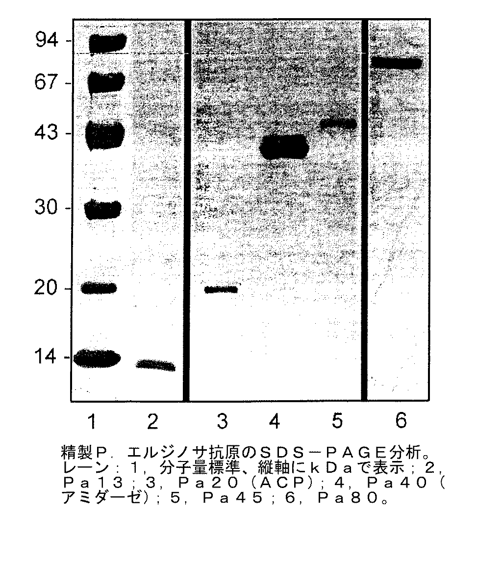
【図3】 精製P.エルジノサ抗原のSDS−PAGE分析の結果を示す。試料は4−15ポリアクリルアミドゲル上で分析し、クーマシー染色した。レーン:1−分子量標準(左の値の単位はkDa);2−Pa13;3−Pa20(ACP);4−Pa40(アミダーゼ);5−Pa45;6−Pa80。
【図4】 抗原特異的抗血清によるP.エルジノサ(菌株385)細胞中のPa80の認識を証明するウェスタンブロットを示す。
【図5】 抗原特異的抗血清によるPa80の認識を証明するP.エルジノサ385(血清型2)のドットブロットを示す。
【図6】 P.エルジノサに結合する抗原特異的抗体(IgG)を検出するためのカウント(縦軸)とlog蛍光(横軸)のグラフを示す。曲線1は、非免疫血清によるネガティブ結合を示し、曲線2は、蛋白質抗血清による結合に続く蛍光の検出を示す。
A:抗Pa13;B:抗Pa20(ACP);C:抗Pa40(アミダーゼ);D:抗Pa45;E:抗Pa80。
【図7】 Pa40の場合のDNAと翻訳されたアミノ酸配列を示す。
【図8】 Pa45の場合のDNAと翻訳されたアミノ酸配列を示す。[0001]
The present application relates to antigenic proteins derived from P. aeruginosa (hereinafter referred to as P. aeruginosa) and the use of these proteins in medicine, particularly P. aeruginosa. It relates to use in the treatment, prevention and diagnosis of Elginosa infections.
[0002]
P. Erginosa is a gram-negative aerobic motor bacterium. It is an ubiquitous extracellular opportunistic pathogen in the environment and causes considerable morbidity and mortality in immunocompromised individuals. Infecting it is particularly important in patients with cystic fibrosis, burns, chronic bronchitis, bronchiectasis and cancer.
[0003]
P. The Elginosa genome has recently been sequenced and details of the project are available on the Internet (http://www.pseudomonas.com). However, on the priority date of the present invention, the information was not complete and was not confirmed. This information is now complete and confirmed.
[0004]
The search for suitable components for confirmation of immune responses, vaccine candidates and diagnostic tests is described in The focus has been on the outer membrane components of Erjinosa. P. The outer membrane of Erjinosa contains toxins including lipopolysaccharide endotoxin (endotoxin), phospholipids and outer membrane proteins (OMPs).
[0005]
These various P.P. An alphabetic naming system has been applied to Erjinosa outer membrane proteins (OMPs). Several proteins have been characterized by this system, but the expression of some is only transient and highly dependent on nutrient availability, culture conditions and the presence of antibiotics. At present, the three major OMPs designated F, H2 and I are P.I. It is recognized as a common antigen in all strains of Erjinosa and is expressed at high copy number.
[0006]
The inventors have employed various protein purification methods to isolate uniform preparations of both OMPs and cytoplasmic proteins. With improvements to the liquid column chromatography and gel electrophoresis steps, several proteins have been isolated and identified using the Zwittergent extraction method to evaluate their potential as vaccines. The proteins were displayed by their molecular weight, and their identity was confirmed by the amino terminal sequence.
[0007]
The inventors have found that some P.I. Proteins were isolated and identified from Erginosa specimens. These proteins are named Pa13, Pa20 (ACP), Pa40 (amidase), Pa45 and Pa80. For Pa13, Pa20 (ACP), Pa40 (amidase), Pa45 and Pa80, amino terminal sequences have been obtained. Following sequence analysis, the data obtained was analyzed using BLAST (Basic Local Alignment Search Tool, National Center for Biotechnology Information, Bethesda, Maryland, USA, Altschul et al., Nucleic Acids Research,25 3389-3402 (1997)). Pa20 was considered to belong to ACP because it had homology to certain proteins from P. syringae and P. eruginosa. Pa40 is a known P.P. It has homology with the Erjinosa aliphatic amidase. This search did not find proteins named Pa13, Pa45, and Pa80.
[0009]
According to the present invention, Pseudomonas aeruginosa-derivedAntigenic proteinIn particular, the molecular weight determined by SDS PAGE under reducing conditions is about 45 kDa,
M R A E L N Q G L I D F L K A
Met Arg Ala Glu Leu Asn Gln Gly Leu Ile Asp Phe Leu Lys Ala (SEQ ID NO: 2)
Has an amino terminal sequenceproteinIs provided.
[0011]
One skilled in the art will recognize that homologues or derivatives of the protein or polypeptide of the present invention may also be utilized within the framework of the present invention (ie, as an antigenic / immunogenic substance). For example, a protein or polypeptide comprising one or more additions, deletions, substitutions or the like is encompassed by the present invention. In addition, one amino acid can be replaced by another amino acid of similar “type”. For example, substituting one hydrophobic amino acid with another hydrophobic amino acid. To compare amino acid sequences, a program such as the CLUSTAL program can be used. This program compares amino acid sequences and finds the best alignment by inserting spaces at appropriate positions in either sequence. For some top alignments, identity or similarity (maintaining amino acid type in addition to identity) can be calculated. A program such as BLASTx aligns the longest range of similar sequences and assigns a value to the fit state. Thus, a comparison can be obtained when several similar regions are found, each having a different score (score). In the present invention, both types of analysis are conceivable.
[0012]
For the purposes of the present invention, a protein sequence can be considered substantially homologous to another protein sequence if a significant number of constituent amino acids show homology when using one of the above algorithms. it can. It is sufficient that at least 40%, 50%, 60%, 70%, 80%, 90% and even 99% of amino acids are homologous, and the preference increases in this order.
[0013]
In this specification, the molecular weight of the protein is measured by SDS-PAGE under reducing conditions. As those skilled in the art will appreciate, the accuracy of the molecular weight values obtained by this method is only about ± 10%.
[0014]
As discussed herein, the protein of the present invention is useful as an antigenic substance. Such substances can be “antigenic” and / or “immunogenic”. In general, “antigenic” is employed to mean that the protein can be used to produce antibodies or can actually elicit an immune response in vivo. “Immunogenic” is used to mean that the protein can elicit a protective immune response in vivo. Thus, in the latter case, the protein may not only generate an antibody response, but may also generate a non-antibody based immune response.
[0015]
According to the inventionThe protein is P.I. Thus, a further aspect of the present invention provides a method for preparing an isolated and purified protein comprising the following steps:.
[0016]
(A) P.I. Prepare a culture of Erjinosa, grow them under appropriate conditions, harvest them and subsequently wash by centrifugation to give a washed cell pelletProcess
[0017]
(B) Resuspend washed cells in an appropriate buffer followed by cell destructionProcess
[0018]
(C) Cell debris is removed by centrifugation to obtain a supernatant containing soluble cell proteinsProcess
[0019]
(D) The resulting solution is subjected to anion exchange chromatography, eluting with a sodium chloride gradient, and fractions corresponding to individual peaks are pooled.Process
[0020]
(E) 10% T-1.42% C acrylamide-N, N with 4% T-0.36% C acrylamide-N, N-methylenebisacrylamide stacking gel polymerized in a 28 mm (inner diameter) column. -The protein fraction is purified by SDS-PAGE using a gel for separation of methylenebisacrylamide. The electrophoresis uses 1% (w / v) SDS, 0.025 M Tris, 0.2 M glycine buffer pH 8.3. Elute with 0.025M Tris buffer at pH 8.3, collect in 6 ml fractions at a flow rate of 1 ml / min, in the upper and lower chambers at 40 mA, 10 W for 14 hours.Process
[0021]
(F) Molecular weight45 kDaSelect a fraction containing the protein of and isolate the protein from the selected fractionProcess.
[0022]
Alternatively, suitable proteins may be prepared by expressing appropriate DNA.
[0023]
Therefore, in a further aspect of the present invention, the present inventionbyA recombinant or isolated nucleic acid encoding a protein or a nucleic acid complementary thereto is provided.
[0024]
For expression, the nucleic acid (which may be DNA) may be inserted into a vector (which may be a plasmid, cosmid or phage). This vector is integrated into the genome of the host organism, which may be prokaryotic or eukaryotic.
[0025]
The nucleic acid or nucleic acid-containing vector may be suitable for expressing the protein of the invention in the subject to be treated. That is, it may be in the form of a DNA vaccine. Suitable methods and agents for preparing DNA vaccines are well known to those skilled in the art.
[0026]
Along with the aforementioned proteins, the inventors have further described P.I. Erginosa protein was isolated and purified. Some of these proteins are antigenic and are therefore It would be useful in a method for the treatment or prevention of Elginosa infection. The method comprises administering to a patient in need of such treatment an effective amount of any of these proteins or an antigenic fraction thereof.
[0027]
These proteins are described in P.I. It is also useful for diagnosing Erginosa infection.
[0028]
Therefore, in a further aspect of the present invention, in medicine, especially P.I. Provided is a protein or a nucleic acid encoding the protein for use in the treatment, prevention or diagnosis of Elginosa infection. Derived from Erzinosa,The molecular weight determined by SDS PAGE under reducing conditions is about 45 kDa,
MRAELNQGLIDFLKA
Met Arg Ala Glu Leu Asn Gln Gly Leu Ile Asp Phe Leu Lys Ala (SEQ ID NO: 2)
A protein having an amino terminal sequence, or a homolog having at least 90% of the amino acid sequence homologous to the constituent amino acid sequence of the protein and retaining the antigenicity of the protein.
[0042]
P. In addition to treating or preventing Elginosa infections, the proteins of the present invention are also useful for the diagnosis of such infections. Thus, a further aspect of the present invention is
[0043]
(A) Antigenic protein as defined aboveOr its homologueIn contact with the sample to be tested;
[0044]
(B) P.I. Detecting the presence of antibodies against Erginosa
P. Erginosa'sDetection methodI will provide a.
[0045]
In a further aspect of the present invention, P.I. Provided is the use of a protein or a nucleic acid encoding the protein in the preparation of a medicament for the treatment, prevention or diagnosis of Elginosa infection. Derived from Erzinosa,The molecular weight determined by SDS PAGE under reducing conditions is about 45 kDa,
MRAELNQGLIDFLKA
Met Arg Ala Glu Leu Asn Gln Gly Leu Ile Asp Phe Leu Lys Ala (SEQ ID NO: 2)
A protein having an amino terminal sequence, or a homolog having at least 90% of the amino acid sequence homologous to the constituent amino acid sequence of the protein and retaining the antigenicity of the protein.
[0051]
As already mentioned, the proteins isolated by the inventors have been shown to be antigenic, and thus they and / or their fragments are P. a. It can be used as a vaccine or drug for the treatment or diagnosis of Elginosa infection.
[0052]
Thus, the present invention provides a protein as defined aboveOr its homologueIs also provided with a pharmaceutically acceptable excipient.
[0053]
The composition may be a vaccine composition, in which case it may also contain an adjuvant. Examples of adjuvants well known in the art include inorganic gels such as aluminum hydroxide or water-in-oil emulsions such as incomplete Freund's adjuvant. Other useful adjuvants are well known to those skilled in the art.
[0054]
As mentioned above, the protein isolated by the inventors is antigenic and therefore the present invention is a protein as defined above.Or its homologueAntibodies that specifically bind to are also provided. The antibody may be a monoclonal antibody or a polyclonal antibody. Techniques for preparing both monoclonal and polyclonal antibodies are well known to those skilled in the art.
[0055]
It is well known that an epitope region, that is, a region contributing to the antigenicity or immunogenicity of a protein or polypeptide can be confirmed by screening an antigenic or immunogenic protein or polypeptide. Methods that are well known to those skilled in the art can be used to test the antigenicity of fragments and / or homologues and / or derivatives. Thus, fragments of the invention should include one or more of such epitope regions, or be similar to such regions sufficient to retain their antigenicity / immunogenicity. is there. For example, in the case of fragments of the present invention, the degree of identity is probably not important. Because they can be 100% identical to specific portions of the proteins or polypeptides, homologues or derivatives described herein. It is essential that, again, the fragment retains the antigenicity / immunogenicity of the protein from which it was derived.
[0056]
The protein may be administered by various routes including enteral, such as oral, nasal, buccal, topical or anal administration or parenteral administration.
[0057]
The form taken by the composition and the excipients it contains will, of course, depend on the route of administration chosen. For example, oral formulations may be in the form of syrups, elixirs, tablets, and tablets may be enteric coated to protect the protein from degradation in the stomach. Nasal or transdermal formulations are usually sprays or patches, respectively. Injectable formulations may be solutions or suspensions in distilled water or other pharmaceutically acceptable solvents or suspensions.
[0058]
In a further aspect, the present invention includes a step of administering to a subject an effective amount of a protein as defined herein. A method of immunizing subjects against Erginosa is provided.
[0059]
A suitable dose of the protein of the invention for vaccination will be about 5-100 micrograms when administered parenterally. However, in the case of nasal and oral administration, this dose will be 10-100 times depending on the formulation, adjuvant, patient profile, etc.
[0060]
As will be appreciated by those skilled in the art, in any of the above applications, the antigenic fragments of the proteins can be used in place of the full-length proteins.
[0061]
Preferred features of each aspect of the invention are common to each face. Prior references mentioned herein are hereby incorporated by reference to the extent permitted by law.
[0062]
The following examples and figures illustrate the invention in more detail.
[0063]
Example 1-P. Isolation of proteins from Erjinosa.
Strain
For purification, mucoid type (mucous) P. cerevisiae P. Elginosa isolate 385,
[0064]
Bacterial culture conditions
For each extraction, 200 nutrient agar plates (Oxoid Unipass Co., Ltd., Basingstoke, Hampshire, UK) were used. Bacteria were streaked on a lawn agar plates and incubated overnight at 37 ° C. Bacteria were scraped and collected and washed three times by centrifugation (12000 × g, 14 minutes, 4 ° C., Beckman centrifuge). Following each centrifugation step, the bacterial pellet was secured and then resuspended in fresh sterile phosphate buffered saline (PBS).
[0065]
Protein purification
The washed bacterial pellet was resuspended in 20 ml 1 M sodium acetate and 1 mM β-mercaptoethanol (pH 4). The suspension is stirred for 45 minutes at room temperature and then 80 ml of 500 mM calcium chloride, 5% w / v Zwittergent 3-14TM(Calbiochem, Australia, NS, Alexandria) was added. This was allowed to stir at room temperature for 90 minutes and then 20% (v / v) absolute ethanol was added. The solution was left at 4 ° C. for 2 hours. The suspension was centrifuged at 17000 × g for 15 minutes at 4 ° C. and the supernatant was adjusted to a final concentration of 80% (v / v) ethanol. This solution was kept at 4 ° C. overnight. The solution was centrifuged at 17000 × g for 25 minutes at 4 ° C., and the protein pellet was washed with 30 ml of buffer Z (5% Zwittergent 3-14TM(W / v), 50 mM Tris and 0.01 M EDTA, pH 8.0) and incubated for 45 minutes at room temperature. The solution was centrifuged at 12000 × g for 15 minutes at 4 ° C., the supernatant was secured and dialyzed overnight at 4 ° C. against distilled water. The crude protein extract was frozen at -70 ° C and lyophilized.
[0066]
Anion exchange chromatography
Bio-scale Q2TMAnd Q5TMAnion exchange chromatography was performed using (Bio-Rad). These columns are strongly basic quaternary ammonium groups (-N to promote the binding of negatively charged proteins.+(CH3)3) Was derivatized with MP10 carrier matrix. The column was equilibrated with 20 ml buffer A (20 mM Tris-HCl, pH 8.5) at a flow rate of 2 ml / min. The lyophilized crude protein extract was suspended in buffer A to a concentration of <5 mg / ml (Q2) or <20 mg / ml (Q5). Buffered sodium chloride (buffer B, 20 mM Tris, pH 8.5, 500 mM sodium chloride) was introduced at increasing flow rates at a flow rate of 1 ml / min to elute the protein from the column. Each peak-top fraction was dialyzed overnight, frozen at -70 ° C., lyophilized and subjected to SDS-PAGE analysis. Fractions corresponding to different peaks were pooled separately and further purified.
[0067]
Purification using SDS-preparative polyacrylamide gel electrophoresis
The partially purified protein was further purified using preparative polyacrylamide gel electrophoresis. Pooled fractions from the anion exchange column were lyophilized, resuspended in a minimal amount of distilled water, and further 4 volumes of SDS reducing buffer (62.5 mM Tris, pH 6.8; 10% v / v Glycerol; 2% w / v SDS; 5% v / v β-mercaptoethanol; 1.2 × 10⁻³% W / v bromophenol blue). This preparation was incubated at 37 ° C. for at least 30 minutes before being loaded onto an electrophoresis column stacking gel.
[0068]
Preparative SDS-PAGE is 10% T-1.42% with 10 ml of 4% T-0.36% C acrylamide-N, N-methylenebisacrylamide stacking gel polymerized in a 28 mm (inner diameter) column. This was carried out using a Bio-Rad 491 prep cell containing 20 ml of C acrylamide-N, N-methylenebisacrylamide separation gel. Stacking and separation gels were prepared from a 30% / 2.67 w / v acrylamide / bis-acrylamide monomer stock (Bio-Rad). Proteins were transferred in the upper and lower chambers using 1% (w / v) SDS, 0.025 M Tris, 0.2 M glycine buffer pH 8.3. The electrophoresis conditions were 40 mA, 10 W, and 14 hours. Protein was eluted using 0.025 M Tris, pH 8.3, and 6 ml fractions were collected at a flow rate of 1 ml / min. The collected fractions were frozen at −70 ° C., lyophilized and analyzed every 5 fractions using SDS-PAGE. A series of confirmed fractions containing the same protein were then pooled.
[0069]
Electroelution
Some purifications used electroelution instead of preparative gel electrophoresis. Protean IITMCrude protein or partially purified fractions were separated by electrophoresis using an xi cell (Bio-Rad) and vertical slab electrophoresis setup. Electrophoresis was performed with a 10% T-1.42% C acrylamide-N, N-methylenebisacrylamide separation gel and 4% polymerized in 16 cm (w) × 16 cm (h), 1.0 mm (d) units. T-0.36% C acrylamide-N, N-methylenebisacrylamide stacking gel was used. Electrophoresis was performed at 16 mA during electrophoresis through the stacking gel and at 24 mA for separation in the separation gel.
[0070]
The elution of the protein band from the slab gel was performed using Gel Eluter.TM(Bio-Rad) was used as a whole. Elution was performed at 250 mA for 45 minutes across the gel thickness. The protein eluted in the narrow chamber was collected in a 12 mm x 75 mm tube under vacuum.
[0071]
SDS removal
The protein isolated under electrophoresis conditions contained SDS, which was subsequently removed. This involved adding potassium phosphate to the concentrated protein fraction to a concentration of 20 mM. The sample was left on ice for 60 minutes and the precipitated SDS was removed by centrifugation at 6000 × g, 4 ° C. for 20 minutes. The sample was desalted by dialysis.
[0072]
Fraction analysis
The protein content of fractions from both liquid column chromatography and preparative gel electrophoresis or electroelution was assessed by analytical SDS-PAGE and silver or Coomassie staining. The presence of the protein of interest in the specific elution fraction was confirmed by analytical staining SDS-PAGE gel. Fractions containing only a uniform (single) band are pooled and Pierce Micro BCATMProtein assay reagents and PierceTMProtein concentrations were determined using albumin standards (Laboratory Surprise, Australia, NSW, Marrickville).
[0073]
SDS-PAGE for analysis
SDS-PAGE was performed and fractions analyzed for the presence of the protein of interest, as described by Laemmli. 10 μl fraction sample was added to an equal volume of sample buffer containing SDS and dithiothreitol (DTT) and boiled for 5 minutes. BioradTMThe system was used for electrophoresis on a 10-15% gradient on a minigel. Molecular weight markers (Pharmacia) were run on the same gel to determine the molecular weight of the protein.
[0074]
SDS removal
This method involved adding potassium phosphate to a concentrated protein sample to a concentration of 20 mM. The sample was then placed on ice for 60 minutes and the precipitated SDS was removed by centrifugation at 6000 g for 20 minutes at 4 ° C. and dialysis against distilled water was used.
[0075]
Staining of polyacrylamide gel
Analytical SDS-PAGE was followed by Coomassie staining or silver staining. 1.0 g Coomassie Blue R-250 (BioRad) dissolved in 400 ml methanol, 100 ml acetic acid and 50 ml deionized water.TMThe gel was stained for 60 minutes in a Coomassie staining solution. Decolorization was performed in a decolorization solution consisting of 400 ml methanol, 100 ml acetic acid and 500 ml deionized water.
Silver staining involved fixing the gel in 30% methanol, 10% acetic acid and formaldehyde for 4 hours. After washing with 50% ethanol, the gel was pretreated with 0.8 mM sodium thiosulfate for 1 minute. After washing 3 times with water, it was incubated in silver stain for 20 minutes. After further washing with water twice, incubation in a developer (18% sodium carbonate, formaldehyde and sodium thiosulfate) was performed. The reaction was quenched with a solution of 50% methanol and 12% acetic acid.
[0076]
Measurement of protein concentration
Protein concentration was determined using Pierce Micro BCA protein assay reagents and Pierce albumin standards (Laboratory Surprise, Australia, NSW, Marrickville).
[0077]
Lipopolysaccharide measurement
The presence of lipopolysaccharide (LPS) was assessed by both silver staining of SDS-PAGE gels and assay by E-TOXATE horseshoe crab lysate test (Sigma, St. Louis, MO, USA).
[0078]
result
[0079]
FIG. Figure 5 shows the elution profile of Erzinosa zwittergent extract by anion exchange chromatography. Seven peaks are clearly seen. The first five peaks were individually pooled and analyzed by SDS-PAGE to determine the molecular weight of these proteins (FIG. 2). For the immunization studies discussed in Example 4, the protein was further purified by preparative electrophoresis. FIG. 3 shows SDS-PAGE analysis of the purified protein used in the immunization study.
[0080]
Example 2
Purification of Pa80
A purification protocol for isolating sufficient amounts of protein for use in immunization studies is described in P.I. Developed for proteins from Erginosa. A three-step process consisting of Zwittergent surfactant extraction, liquid column chromatography and preparative SDS-PAGE was utilized for the separation. Antigen Pa80 was changed to P. a. Purified from Erjinosa 385 (serotype 2). SDS-PAGE confirmed the homogeneity of the purified sample. Pa80 (80 kDa) was isolated and confirmed to be free of endotoxin contamination and therefore suitable for studying its potential as a vaccine.
[0081]
Materials and methods
P. Pa80 for immunization was purified using cells of Erjinosa strain 385 (serotype 2). Bacteria were cultured overnight, harvested, washed and a crude sample extractable with a surfactant was obtained as described above (Example 1). The lyophilized crude extract was resuspended in a minimal amount of distilled water and SDS-reducing buffer. Purification of Pa80 was performed by anion exchange chromatography and preparative SDS-PAGE.
Western and dot blots were performed using Pa80 specific antisera. For Western blot, P.I. Cell lysates of Erjinosa strain 385 were separated by electrophoresis on a 10-20% gradient polyacrylamide gel by analytical SDS-PAGE and analyzed with a nitrocellulose membrane (BioRadTM) Semi-dry transfer device (BioRad)TM). After blocking for 60 minutes in 1% v / v skim milk in Tris-buffered saline (TBS), the membrane was incubated with a 1/1000 dilution of pooled immune serum for 90 minutes at room temperature. The membrane was then washed with TBS and 0.05% Tween 20 (TTBS) and incubated for 90 minutes in a 1/1000 dilution of horseradish peroxidase (HRP) -conjugated anti-rat IgG. HRP developer (BioRadTM) Using GS570 densitometer (BioRad)TM). A dot blot of Pa385 (serotype 2) cell lysate was applied directly to a nitrocellulose membrane. Subsequent steps were as in Western blot.
[0082]
result
Purification of Pa80 by zwittergent extraction
This study confirmed the immune recognition of Pa80 in cell lysates using Pa80-specific antisera. (FIGS. 4 and 5)
[0083]
Purification of Pa80 using anion exchange chromatography
The crude protein from the zwittergent extract was further separated using anion exchange chromatography. Bioscale Q2 (BioRadTM) Separated the composite extract into approximately seven individual protein peaks (FIG. 1). Since Pa80 seemed to be in the fifth peak, it was confirmed by SDS-PAGE by molecular weight (FIG. 2). The extraction conditions of Pa80 from the anion column were 80% buffer A (20 mM Tris, pH 8.5) and 20% buffer B (1.5 M NaCl, 20 mM Tris, pH 8.5), UV (AU) 0. It was 228 and conductivity (mS / cm) was 0.674.
[0084]
Purification of Pa80 using preparative SDS-PAGE
Using both BioRad Prep cell type 491 and SDS-PAGE for Protean II preparative purification, the protein was further purified from the Pa80-containing fraction under denaturing conditions.
The silver-stained gel was inspected and evaluated for the presence of LPS contamination. Quantification using the E-TOXATE horseshoe crab lysate test was found to be below the level of detection (limit 0.015 EU / ml) for all preparations.
[0085]
[Table 1]
Consideration
Identification of Pa80
Pa80 has been purified to homogeneity using a combination of zwittergent extraction and anion exchange chromatography. This protein is recognized by sera from immunized rats and live P. pneumoniae into the lungs. It is shown to protect against Erjinosa challenge (see Example 4). This protein is P. aureus. Antigen not previously tested for immunization against Erginosa infection.
[0087]
Example 3-Sequencing of purified protein
National University of Australia Center for Molecular Structure and Function Biomolecular Resource Facility and University of Liverpool, UK2-Both terminal and internal amino acid sequences were analyzed. Sequencing was performed using the SDS-PAGE compatible S-2-carboxyamidoethylation method. This alkylation reaction of protein consists of a solution of 10% glycerin (v / v), 5% (w / v) SDS, 0.25M Tris HCl, 100
[0088]
In the case of Pa80, the sample was run on an SDS-PAGE gel and Western blotted on PVDF. The band was cut out and applied directly to the sequencer. N-terminal analysis was performed by Edman degradation.
[0089]
Purification of protein antigens and their confirmation
As described above, P.I. Protein was purified from Erjinosa 385. The purity of the protein was evaluated by SDS-PAGE (FIG. 3) and amino acid sequencing, both successfully identifying and confirming a homogeneous protein preparation. Evaluation of LPS levels in protein preparations showed no detectable levels of endotoxin as assessed by the horseshoe crab test of the E-TOXATE kit (the detection limit of the test was 0.015 endotoxin units / ml). Met).
[0090]
Following N-terminal amino acid sequence analysis, electronic database searches of Genbank sequences and sequences posted on the Internet in the PA01 database were performed using “Entrez” to identify proteins (see Table 2). The N-terminal sequence of Pa13 was determined to be AETIVNTTKA (SEQ ID NO: 1). This 10-residue sequence is described in P.I. The sequence corresponding to Erginosa was not found, suggesting that the protein has not been sequenced yet.
[0091]
The N-terminal sequence of Pa20 was determined to be STIERVKKIVAEQL (SEQ ID NO: 4). This is because P.A. An acyl carrier protein from Erginosa (DDBJ / EMBL / GenBank accession number O54439) and that from Pseudomonas syringae (DDBJ / EMBL / GenBank accession number P80923) showed 100% identity with 15 amino acids overlapping.
[0092]
The N-terminal sequence of Pa40 was determined to be MRHGDISSNDTVG (SEQ ID NO: 5). This is because P.A. Elginosa-derived amidase (DDBJ / EMBL / GenBank accession numbers P11436 and M27612) overlapped with 14 amino acids and showed 100% identity.
[0093]
The N-terminal sequence of Pa45 was determined to be MRAELNQGLIDFLKA (SEQ ID NO: 4).
[0094]
The N-terminal sequence of Pa80 was determined to be MSEQNNEQRSQAA (SEQ ID NO: 3).
[0095]
[Table 2]
Example 4-Bacterial clearance following immunization in a rat model
Male DA-based SPF rats were immunized with Peyer's patch (IPP) on
[0097]
Results-Effect of immunity on lung clearance
Table 3 shows the dose for primary IPP and secondary IT immunization and the number of animals treated (N). Table 4 shows the bacterial clearance levels obtained in immunized and control rats. The results show a significant difference in clearance levels in both BAL and lung homogenates, with the exception of recovery in the Pa20 (ACP) immunized group, where bacterial load was reduced but not statistically significant.
[0098]
[Table 3]
[Table 4]
In Table 4, P.P. 4 hours after the attack infection. The value shown as the CFU of Elginosa (log 10) is the living P. Mean ± standard error of rat BAL fluid or
[0101]
The BAL values for Pa40 (amidase), Pa45 and Pa80 are significantly different from the non-immunized group at p <0.01.
[0102]
The BAL value of Pa13 and the lung homogenate values of Pa13, Pa20 (CAP), Pa40 (amidase), Pa45 and Pa80 are significantly different from the non-immunized and single immunized groups at p <0.05.
[0103]
Example 5-Flow cytometry
P. Erginosa strain 385 was grown to mid-log phase in Nutrient broth and harvested by centrifugation at 1000 xg for 10 minutes at 4 ° C. The bacteria were then diluted 1:50 (in PBS) and incubated with non-immune serum, immune serum or PBS for 1 hour at 37 ° C. Cells were centrifuged, the supernatant removed, and the bacteria suspended in 200 μL of a 1:50 PBS dilution of fluorescein isothiocyanate conjugated to anti-rat IgG. After 30 minutes incubation at 37 ° C., 1.8 ml PBS was added and the bacterial cells were analyzed by flow cytometry (Coulter ZL-MCL). A total of 20000 cells were counted and data were obtained in forward mode, side scatter and fluorescence in the instrument mode in logarithmic mode.
[0104]
result
Non-immune rat serum was used as a control. No non-specific binding to Erginosa was shown (
[0105]
Example 6-Cloning of Pa40 and Pa45 genes
P. Purification of chromosomal DNA of Erjinosa Pa385
P. Erginosa was cultured as described in Example 1. Bacteria were washed in 30 ml PBS and collected by centrifugation at 4000 rpm for 10 minutes and this procedure was repeated. The washed pellet was resuspended in 10 ml 50 mM Tris HCl and 0.4 ml 0.4 M EDTA and incubated at 37 ° C. for 20 minutes. Next, 0.4 ml of 20 mg lysozyme / ml was added, the mixture was further incubated at 37 ° C. for 10 minutes, and 540 μg protease K, 0.4
[0106]
Oligonucleotide design
Site-directed cloning was chosen to ensure accurate orientation to the reading frame. With maximized GC content, oligonucleotides were obtained from GIBCO BRL custom primers (Life Technologies, Rockville, Md.) And designed as follows:
Pa40BF, 5 'oligonucleotide encoding the beginning of the amidase gene. Sequence (from 5 'to 3') GGC GGA TCC CGT CAC GGC GAT ATT TCC AGC AGC A.
This oligonucleotide was 34-mer in length and inserted into the BamHI restriction site. The coupling efficiency was estimated to be 99% and the GC content was 61%.
Pa40HR, a 3 ′ oligonucleotide encoding the 3 ′ end of the amidase gene. Sequence (from 5 'to 3') GGC AAG CTT GGC CTC CTT CTC CAG TCC CTC. This oligonucleotide was 30-mer in length and inserted into the HindIII restriction site. The coupling efficiency was estimated to be 99% and the GC content was 63%. Pa45BF, 5 'oligonucleotide encoding the beginning of the Pa45 gene. Sequence (from 5 'to 3') GGC GGA TTC CGC GCA GAA CTC AAC CAG GGC CTG. This oligonucleotide was 33-mer in length and was inserted into the BamHI restriction site. The coupling efficiency was estimated to be 99% and the GC content was 67%.
Pa45HR, a 3 ′ oligonucleotide encoding the 3 ′ end of the Pa45 gene. Sequence (from 5 'to 3') GGC AAG CTT GGG CAG CTC GCT GCT GGC GTA GAA. The oligonucleotide was 33-mer in length and inserted into the HindIII restriction site. The coupling efficiency was estimated to be 99% and the GC content was 63%.
[0107]
Polymerase chain reaction (PCR)
The PCR reaction mixture consisted of 1 ng PaDNA, 15 pmol (each) primer, 7.5 μM (each) dNTP and 1 U Taq DNA polymerase (Qiagen) in 50 μl of the total reaction mixture. The conditions used for amplification were: cycle 1 [94 ° C.-3 minutes, 55 ° C.-10 seconds, 72 ° C.-15 seconds] × 1 cycle; cycle 2 [94 ° C.-10 seconds, 50 ° C.-10 seconds, 72 ° C.-15 Second] x 35 cycles; cycle 3 [72 ° C-5 minutes, 25 ° C-1 minute] x 1 cycle, using a Corvette FTS4000 thermal sequencer (Corbett Research, Australia, NSW, Sydney). PCR product size and non-specific product were assessed by electrophoresis on a 1% agar gel.
[0108]
Cloning of Pa40 and Pa45 genes
Following PCR, unused dNTPs were removed using the silica matrix method (Progen Industries) and the purified product was visualized on an agarose gel to check recovery. The PCR product was ligated into the pGEM T-easy vector system (Promega, USA). Ligation of the insert with the vector was performed using T4 DNA ligase under standard conditions recommended by the manufacturer. The sample was left overnight at 4 ° C.
[0109]
The ligated DNA was used for transformation by the CaCl2 method. JM109 competent cells were thawed on ice for 5 minutes and 900 μl of 0.1
[0110]
Rapid screening of small cultures
Individual colonies of transformants were selected and placed in 5 ml LB broth containing 50 μg / ml ampicillin, X-Gal and IPTG and incubated overnight at 37 ° C. and 180 rpm. The vector was linearized and visualized on an agarose gel and the insert was checked with white colonies.
[0111]
Determination of DNA sequence
Double-stranded DNA from the recombinant plasmid was sequenced at the University of NSW (New South Wales). Products for sequencing were prepared using pUC / M13 forward or reverse primer and Big Die Terminator mix, 3.2 pmol primer and 100 ng template in 10 μl volume with sterile water. A cycle sequence [96 ° C.-10 seconds, 50 ° C.-5 seconds, 60 ° C.-4 minutes] × 25 cycles was carried out with a Carbet FTS4000 thermal sequencer. For sequence analysis, DNA Strider 1.3 was used.
[0112]
result
Proof of existence of Pa40 sequence and cross-strain
P. The sequence analysis of Pa40 derived from Erjinosa 385 is shown in FIG. The amino acid translation of the N-terminal region of the determined nucleic acid sequence is homologous to the amino acid of SEQ ID NO: 5 obtained by N-terminal amino acid sequencing. Other P.P. The presence of the Pa40 gene in the Erjinosa strain and other serotypes was evaluated using specific primers: Pa40BF and Pa40HR. The results demonstrate correct size PCR products for all strains and serotypes tested (see Table 5 below).
[0113]
Proof of the presence of Pa45 sequence and cross-strain
P. The sequence analysis of Pa45 derived from Erjinosa 385 is shown in FIG. The amino acid translation of the N-terminal region of the determined nucleic acid sequence is homologous to the amino acid of SEQ ID NO: 2 obtained by N-terminal amino acid sequencing. Other P.P. The presence of the Pa40 gene in Erjinosa strains and other serotypes was evaluated using specific primers: Pa45BF and Pa45HR. The results demonstrate a correctly sized PCR product for most strains and serotypes tested (see Table 5 below). In the case of serotype Pa373,
[0114]
[Table 5]
[Brief description of the drawings]
BRIEF DESCRIPTION OF THE DRAWINGS FIG. 1 shows P.A. The elution profile of Erjinosa protein is shown.
FIG. Figure 6 shows SDS-PAGE analysis of peak fractions from anion exchange chromatography of Erzinosa crude zwittergent extract.
FIG. 3 The result of the SDS-PAGE analysis of Erjinosa antigen is shown. Samples were analyzed on 4-15 polyacrylamide gels and Coomassie stained. Lane: 1-molecular weight standard (unit of left value is kDa); 2-Pa13; 3-Pa20 (ACP); 4-Pa40 (amidase); 5-Pa45; 6-Pa80.
FIG. 4 shows the results of P. elegans with antigen-specific antiserum. Shown is a Western blot demonstrating recognition of Pa80 in Erginosa (strain 385) cells.
FIG. 5 shows P. cerevisiae recognition of Pa80 by antigen specific antisera. A dot blot of Erjinosa 385 (serotype 2) is shown.
FIG. A graph of count (vertical axis) and log fluorescence (horizontal axis) for detecting an antigen-specific antibody (IgG) binding to Erjinosa is shown.
A: anti-Pa13; B: anti-Pa20 (ACP); C: anti-Pa40 (amidase); D: anti-Pa45; E: anti-Pa80.
FIG. 7 shows the DNA and translated amino acid sequence for Pa40.
FIG. 8 shows the DNA and translated amino acid sequence for Pa45.
Claims (10)
M R A E L N Q G L I D F L K A
Met Arg Ala Glu Leu Asn Gln Gly Leu Ile Asp Phe Leu Lys Ala (配列番号2)
なるアミノ末端配列を有する蛋白質。Pseudomonas aeruginosa-derived antigenic protein having a molecular weight of about 45 kDa determined by SDS PAGE under reducing conditions;
MRAELNQGLIDFLKA
Met Arg Ala Glu Leu Asn Gln Gly Leu Ile Asp Phe Leu Lys Ala (SEQ ID NO: 2)
A protein having an amino terminal sequence.
(b)シュードモナス・エルジノサに対する抗体の存在を検出すること
を特徴とするシュードモナス・エルジノサの検出方法。(A) contacting the protein of claim 2 with a sample to be tested;
(B) A method for detecting Pseudomonas aeruginosa, comprising detecting the presence of an antibody against Pseudomonas aeruginosa.
Applications Claiming Priority (3)
| Application Number | Priority Date | Filing Date | Title |
|---|---|---|---|
| GB9928676.7 | 1999-12-03 | ||
| GBGB9928676.7A GB9928676D0 (en) | 1999-12-03 | 1999-12-03 | Pseudomonas aeruginosa antigens |
| PCT/GB2000/004625 WO2001040473A2 (en) | 1999-12-03 | 2000-12-04 | Pseudomonas aeruginosa antigens |
Publications (2)
| Publication Number | Publication Date |
|---|---|
| JP2003515342A JP2003515342A (en) | 2003-05-07 |
| JP4733893B2 true JP4733893B2 (en) | 2011-07-27 |
Family
ID=10865705
Family Applications (1)
| Application Number | Title | Priority Date | Filing Date |
|---|---|---|---|
| JP2001542538A Expired - Fee Related JP4733893B2 (en) | 1999-12-03 | 2000-12-04 | Pseudomonas aeruginosa antigen |
Country Status (8)
| Country | Link |
|---|---|
| US (1) | US7517533B2 (en) |
| EP (1) | EP1240330A2 (en) |
| JP (1) | JP4733893B2 (en) |
| CN (2) | CN1409764A (en) |
| AU (1) | AU1541401A (en) |
| CA (1) | CA2392906A1 (en) |
| GB (1) | GB9928676D0 (en) |
| WO (1) | WO2001040473A2 (en) |
Families Citing this family (13)
| Publication number | Priority date | Publication date | Assignee | Title |
|---|---|---|---|---|
| NZ521531A (en) | 2000-02-28 | 2005-12-23 | Chiron S | Heterologous expression of neisserial proteins |
| AU2002340683C1 (en) * | 2001-11-13 | 2009-11-12 | Id Biomedical Corporation Of Quebec | Polypeptides of pseudomonas aeruginosa |
| EP1790659B1 (en) | 2001-11-13 | 2013-07-31 | ID Biomedical Corporation | Polypeptides of pseudomonas aeruginosa |
| CA2571673A1 (en) * | 2004-06-28 | 2006-01-05 | Proteome Systems Intellectual Property Pty Ltd | Novel methods of diagnosis of treatment of p. aeruginosa infection and reagents therefor |
| US20080193912A1 (en) * | 2004-08-03 | 2008-08-14 | Yiu-Lian Fong | Compositions and Methods for Preparation of Nucleic Acids from Microbial Samples |
| EP2612148B1 (en) | 2010-09-04 | 2019-06-12 | GlaxoSmithKline Biologicals SA | Bactericidal antibody assays to assess immunogenicity and potency of meningococcal capsular saccharide vaccines |
| US10598666B2 (en) | 2012-03-08 | 2020-03-24 | Glaxosmithkline Biologicals Sa | In vitro potency assay for protein-based meningococcal vaccines |
| CN102626516A (en) * | 2012-04-05 | 2012-08-08 | 青岛农业大学 | Pseudomonas aeruginosa and propolis inactivated vaccine for minks and preparation process |
| CN104297380B (en) * | 2014-11-05 | 2015-12-30 | 中国烟草总公司郑州烟草研究院 | A kind of LC-MS/MS detects the method for IPP, GPP, FPP and GGPP in fresh tobacco leaves |
| CN105732817B (en) * | 2016-03-02 | 2019-11-08 | 中国人民解放军第三军医大学 | A kind of Pseudomonas aeruginosa recombinant protein Vac33 and its preparation method and application |
| CN105622734B (en) * | 2016-03-02 | 2019-06-07 | 中国人民解放军第三军医大学 | The purification process of pseudomonas aeruginosa vaccine recombinant protein Vac14 |
| CN105732778A (en) * | 2016-03-02 | 2016-07-06 | 中国人民解放军第三军医大学 | Pseudomonas aeruginosa recombinant protein OprL as well as preparation method and application thereof |
| CN105622733A (en) * | 2016-03-02 | 2016-06-01 | 中国人民解放军第三军医大学 | Method for purifying pseudomonas aeruginosa vaccine recombinant protein Vac 9 |
Family Cites Families (5)
| Publication number | Priority date | Publication date | Assignee | Title |
|---|---|---|---|---|
| US5554372A (en) * | 1986-09-22 | 1996-09-10 | Emory University | Methods and vaccines comprising surface-active copolymers |
| GB9701489D0 (en) * | 1997-01-24 | 1997-03-12 | Auspharm Int Ltd | Antigen |
| US6248551B1 (en) * | 1997-03-28 | 2001-06-19 | Institut Pasteur | Helicobacter aliphatic amidase AmiE polypeptides, and DNA sequences encoding those polypeptides |
| US6551795B1 (en) * | 1998-02-18 | 2003-04-22 | Genome Therapeutics Corporation | Nucleic acid and amino acid sequences relating to pseudomonas aeruginosa for diagnostics and therapeutics |
| DK1282702T3 (en) * | 2000-05-10 | 2007-04-02 | Sanofi Pasteur Ltd | Immunogenic polypeptides encoded by KAGE minigens and uses thereof |
-
1999
- 1999-12-03 GB GBGB9928676.7A patent/GB9928676D0/en not_active Ceased
-
2000
- 2000-12-04 CN CN00817159A patent/CN1409764A/en active Pending
- 2000-12-04 US US10/148,414 patent/US7517533B2/en not_active Expired - Fee Related
- 2000-12-04 JP JP2001542538A patent/JP4733893B2/en not_active Expired - Fee Related
- 2000-12-04 CN CN200910176300A patent/CN101851279A/en active Pending
- 2000-12-04 CA CA002392906A patent/CA2392906A1/en not_active Abandoned
- 2000-12-04 WO PCT/GB2000/004625 patent/WO2001040473A2/en not_active Ceased
- 2000-12-04 EP EP00977781A patent/EP1240330A2/en not_active Withdrawn
- 2000-12-04 AU AU15414/01A patent/AU1541401A/en not_active Abandoned
Also Published As
| Publication number | Publication date |
|---|---|
| JP2003515342A (en) | 2003-05-07 |
| EP1240330A2 (en) | 2002-09-18 |
| WO2001040473A2 (en) | 2001-06-07 |
| US20040071713A1 (en) | 2004-04-15 |
| US7517533B2 (en) | 2009-04-14 |
| AU1541401A (en) | 2001-06-12 |
| GB9928676D0 (en) | 2000-02-02 |
| WO2001040473A3 (en) | 2002-01-31 |
| CA2392906A1 (en) | 2001-06-07 |
| CN101851279A (en) | 2010-10-06 |
| CN1409764A (en) | 2003-04-09 |
Similar Documents
| Publication | Publication Date | Title |
|---|---|---|
| JP4733893B2 (en) | Pseudomonas aeruginosa antigen | |
| KR19980703099A (en) | Surface protein of Neisseria meningitidis resistant to proteinase VII | |
| JP2009034107A (en) | Omp26 antigen from haemophilus influenzae | |
| JPH10504444A (en) | Vaccine against Moraxella catarrhalis | |
| CN110642927A (en) | Application of protein in preparation of medicine for preventing cryptococcus pyogenes infection | |
| AU2005206291B2 (en) | Lawsonia intracellularis subunit vaccines | |
| US20160052974A1 (en) | Novel genes and proteins of brachyspira hyodysenteriae and uses thereof | |
| CN116063548A (en) | A fusion protein KP-Ag1 and its application as Klebsiella pneumoniae vaccine antigen | |
| US8703432B2 (en) | Recombinant Treponema spp. proteins for use in vaccine, antibodies against said proteins, and diagnostic and therapeutic methods including the same | |
| El-Adhami et al. | Characterization of the gene encoding a 26-kilodalton protein (OMP26) from nontypeable Haemophilus influenzae and immune responses to the recombinant protein | |
| EP3498847A1 (en) | Vaccine composition for preventing porcine mycoplasma infection, comprising recombinant protein | |
| Ross et al. | Characterization of two outer membrane protein antigens of Porphyromonas gingivalis that are protective in a murine lesion model | |
| RS54963B1 (en) | STEREEPTOCOCCUS SUIS POLYPEPTIDES AND THE POLYNUCLEOTIDS THAT CODE THEM AND THEIR USE IN VACCINES AND DIAGNOSTICS | |
| Cullen et al. | Characterization of a locus encoding four paralogous outer membrane lipoproteins of Brachyspira hyodysenteriae | |
| CN101981052A (en) | Compositions, methods and kits | |
| JP2009029825A (en) | antigen | |
| US20090068218A1 (en) | Antigens for Vaccination Against and Detection of Mycoplasma Suis | |
| CN119343364A (en) | Leptospirosis vaccine | |
| WO2025219610A1 (en) | A vaccine for the treatment or prevention of pseudomonas aeruginosa infection in a subject. | |
| SG176520A1 (en) | Burkholderia pseudomallei outer membrane protein a | |
| JP2002512528A (en) | Compound encoding protective M-like protein of Streptococcus equi and its assay | |
| Couturier | Characterization of the cag type IV secretion system and unique filaments of Helicobacter pylori |
Legal Events
| Date | Code | Title | Description |
|---|---|---|---|
| A521 | Request for written amendment filed |
Free format text: JAPANESE INTERMEDIATE CODE: A523 Effective date: 20061116 |
|
| A621 | Written request for application examination |
Free format text: JAPANESE INTERMEDIATE CODE: A621 Effective date: 20071203 |
|
| A711 | Notification of change in applicant |
Free format text: JAPANESE INTERMEDIATE CODE: A711 Effective date: 20091112 |
|
| A521 | Request for written amendment filed |
Free format text: JAPANESE INTERMEDIATE CODE: A821 Effective date: 20091112 |
|
| A131 | Notification of reasons for refusal |
Free format text: JAPANESE INTERMEDIATE CODE: A131 Effective date: 20100210 |
|
| A601 | Written request for extension of time |
Free format text: JAPANESE INTERMEDIATE CODE: A601 Effective date: 20100510 |
|
| A601 | Written request for extension of time |
Free format text: JAPANESE INTERMEDIATE CODE: A601 Effective date: 20100610 |
|
| A602 | Written permission of extension of time |
Free format text: JAPANESE INTERMEDIATE CODE: A602 Effective date: 20100614 |
|
| A601 | Written request for extension of time |
Free format text: JAPANESE INTERMEDIATE CODE: A601 Effective date: 20100709 |
|
| A602 | Written permission of extension of time |
Free format text: JAPANESE INTERMEDIATE CODE: A602 Effective date: 20100726 |
|
| A521 | Request for written amendment filed |
Free format text: JAPANESE INTERMEDIATE CODE: A523 Effective date: 20100810 |
|
| A131 | Notification of reasons for refusal |
Free format text: JAPANESE INTERMEDIATE CODE: A131 Effective date: 20100908 |
|
| A601 | Written request for extension of time |
Free format text: JAPANESE INTERMEDIATE CODE: A601 Effective date: 20101208 |
|
| A602 | Written permission of extension of time |
Free format text: JAPANESE INTERMEDIATE CODE: A602 Effective date: 20101221 |
|
| A601 | Written request for extension of time |
Free format text: JAPANESE INTERMEDIATE CODE: A601 Effective date: 20110107 |
|
| A602 | Written permission of extension of time |
Free format text: JAPANESE INTERMEDIATE CODE: A602 Effective date: 20110120 |
|
| A601 | Written request for extension of time |
Free format text: JAPANESE INTERMEDIATE CODE: A601 Effective date: 20110208 |
|
| A602 | Written permission of extension of time |
Free format text: JAPANESE INTERMEDIATE CODE: A602 Effective date: 20110216 |
|
| A521 | Request for written amendment filed |
Free format text: JAPANESE INTERMEDIATE CODE: A523 Effective date: 20110307 |
|
| A01 | Written decision to grant a patent or to grant a registration (utility model) |
Free format text: JAPANESE INTERMEDIATE CODE: A01 Effective date: 20110330 |
|
| A01 | Written decision to grant a patent or to grant a registration (utility model) |
Free format text: JAPANESE INTERMEDIATE CODE: A01 |
|
| A61 | First payment of annual fees (during grant procedure) |
Free format text: JAPANESE INTERMEDIATE CODE: A61 Effective date: 20110425 |
|
| FPAY | Renewal fee payment (event date is renewal date of database) |
Free format text: PAYMENT UNTIL: 20140428 Year of fee payment: 3 |
|
| R150 | Certificate of patent or registration of utility model |
Free format text: JAPANESE INTERMEDIATE CODE: R150 |
|
| LAPS | Cancellation because of no payment of annual fees |