JP2022110036A - Methods of functional brain circuit analysis - Google Patents
Methods of functional brain circuit analysis Download PDFInfo
- Publication number
- JP2022110036A JP2022110036A JP2022076708A JP2022076708A JP2022110036A JP 2022110036 A JP2022110036 A JP 2022110036A JP 2022076708 A JP2022076708 A JP 2022076708A JP 2022076708 A JP2022076708 A JP 2022076708A JP 2022110036 A JP2022110036 A JP 2022110036A
- Authority
- JP
- Japan
- Prior art keywords
- amino acid
- seq
- region
- protein
- acid sequence
- Prior art date
- Legal status (The legal status is an assumption and is not a legal conclusion. Google has not performed a legal analysis and makes no representation as to the accuracy of the status listed.)
- Granted
Links
Images


Classifications
-
- A—HUMAN NECESSITIES
- A61—MEDICAL OR VETERINARY SCIENCE; HYGIENE
- A61B—DIAGNOSIS; SURGERY; IDENTIFICATION
- A61B5/00—Measuring for diagnostic purposes; Identification of persons
- A61B5/24—Detecting, measuring or recording bioelectric or biomagnetic signals of the body or parts thereof
- A61B5/316—Modalities, i.e. specific diagnostic methods
- A61B5/369—Electroencephalography [EEG]
- A61B5/377—Electroencephalography [EEG] using evoked responses
- A61B5/378—Visual stimuli
-
- A—HUMAN NECESSITIES
- A61—MEDICAL OR VETERINARY SCIENCE; HYGIENE
- A61B—DIAGNOSIS; SURGERY; IDENTIFICATION
- A61B5/00—Measuring for diagnostic purposes; Identification of persons
- A61B5/0059—Measuring for diagnostic purposes; Identification of persons using light, e.g. diagnosis by transillumination, diascopy, fluorescence
-
- A—HUMAN NECESSITIES
- A61—MEDICAL OR VETERINARY SCIENCE; HYGIENE
- A61B—DIAGNOSIS; SURGERY; IDENTIFICATION
- A61B5/00—Measuring for diagnostic purposes; Identification of persons
- A61B5/05—Detecting, measuring or recording for diagnosis by means of electric currents or magnetic fields; Measuring using microwaves or radio waves
- A61B5/055—Detecting, measuring or recording for diagnosis by means of electric currents or magnetic fields; Measuring using microwaves or radio waves involving electronic [EMR] or nuclear [NMR] magnetic resonance, e.g. magnetic resonance imaging
-
- A—HUMAN NECESSITIES
- A61—MEDICAL OR VETERINARY SCIENCE; HYGIENE
- A61B—DIAGNOSIS; SURGERY; IDENTIFICATION
- A61B5/00—Measuring for diagnostic purposes; Identification of persons
- A61B5/24—Detecting, measuring or recording bioelectric or biomagnetic signals of the body or parts thereof
- A61B5/316—Modalities, i.e. specific diagnostic methods
- A61B5/369—Electroencephalography [EEG]
- A61B5/372—Analysis of electroencephalograms
-
- A—HUMAN NECESSITIES
- A61—MEDICAL OR VETERINARY SCIENCE; HYGIENE
- A61B—DIAGNOSIS; SURGERY; IDENTIFICATION
- A61B5/00—Measuring for diagnostic purposes; Identification of persons
- A61B5/40—Detecting, measuring or recording for evaluating the nervous system
- A61B5/4058—Detecting, measuring or recording for evaluating the nervous system for evaluating the central nervous system
- A61B5/4064—Evaluating the brain
-
- A—HUMAN NECESSITIES
- A61—MEDICAL OR VETERINARY SCIENCE; HYGIENE
- A61N—ELECTROTHERAPY; MAGNETOTHERAPY; RADIATION THERAPY; ULTRASOUND THERAPY
- A61N5/00—Radiation therapy
- A61N5/06—Radiation therapy using light
- A61N5/0613—Apparatus adapted for a specific treatment
- A61N5/0622—Optical stimulation for exciting neural tissue
-
- A—HUMAN NECESSITIES
- A61—MEDICAL OR VETERINARY SCIENCE; HYGIENE
- A61B—DIAGNOSIS; SURGERY; IDENTIFICATION
- A61B2503/00—Evaluating a particular growth phase or type of persons or animals
- A61B2503/42—Evaluating a particular growth phase or type of persons or animals for laboratory research
Landscapes
- Health & Medical Sciences (AREA)
- Life Sciences & Earth Sciences (AREA)
- Engineering & Computer Science (AREA)
- Biomedical Technology (AREA)
- Animal Behavior & Ethology (AREA)
- Biophysics (AREA)
- Pathology (AREA)
- Physics & Mathematics (AREA)
- Veterinary Medicine (AREA)
- Public Health (AREA)
- General Health & Medical Sciences (AREA)
- Heart & Thoracic Surgery (AREA)
- Molecular Biology (AREA)
- Medical Informatics (AREA)
- Surgery (AREA)
- Nuclear Medicine, Radiotherapy & Molecular Imaging (AREA)
- Neurology (AREA)
- Psychology (AREA)
- Radiology & Medical Imaging (AREA)
- Neurosurgery (AREA)
- Psychiatry (AREA)
- High Energy & Nuclear Physics (AREA)
- Physiology (AREA)
- Magnetic Resonance Imaging Apparatus (AREA)
- Measurement And Recording Of Electrical Phenomena And Electrical Characteristics Of The Living Body (AREA)
- Medicines Containing Antibodies Or Antigens For Use As Internal Diagnostic Agents (AREA)
- Measurement Of The Respiration, Hearing Ability, Form, And Blood Characteristics Of Living Organisms (AREA)
- Radiation-Therapy Devices (AREA)
Abstract

【課題】生体の脳回路を解析する方法を提供する。
【解決手段】方法は、光遺伝学を使用して個体の脳の第1領域を刺激して、脳の異なる領域の機能的磁気共鳴画像法(fMRI)と組み合わせて、脳の第1領域と第2領域との個々のニューロン間の動的機能的結合を判定するステップを含み得る。この方法は、脳の第3領域を同定するステップをさらに含み得て、第3領域のニューロンは第1領域と第2領域との間の動的機能的結合に介在する。
【選択図】図1A
A method for analyzing a brain circuit of a living body is provided.
The method uses optogenetics to stimulate a first region of the brain of an individual to combine with functional magnetic resonance imaging (fMRI) of different regions of the brain to achieve the first region of the brain. Determining dynamic functional connections between individual neurons with the second region may be included. The method may further include identifying a third region of the brain, wherein neurons of the third region mediate dynamic functional connections between the first and second regions.
[Selection drawing] Fig. 1A
Description
神経組織の機能研究は、定義済み刺激を用いて1つ以上のニューロンを刺激するステップと、この1つ以上のニューロンの出力反応を測定するステップとを含む。ニューロンを刺激する方法および出力反応を測定する方法は、光遺伝学、機能的磁気共鳴画像法(fMRI)、電気生理学、脳波検査法(EEG)、および行動モニタリングを含む。 A functional study of neural tissue involves stimulating one or more neurons with a defined stimulus and measuring the output response of the one or more neurons. Methods of stimulating neurons and measuring output responses include optogenetics, functional magnetic resonance imaging (fMRI), electrophysiology, electroencephalography (EEG), and behavioral monitoring.
光遺伝学は光活性化ポリペプチド(チャネルとポンプ)を使用して、光依存的方法で光活性化ポリペプチドを発現するニューロン活動を変調する。 Optogenetics uses light-activated polypeptides (channels and pumps) to modulate the activity of neurons expressing light-activated polypeptides in a light-dependent manner.
血液酸素濃度依存性(BOLD)fMRIは、非侵襲性全脳イメージングに関して広く普及している技術である。BOLDシグナルは、神経活動に続いて脳血流(CBF)、脳血液量(CBV)、および脳酸素消費量(CMRO2 )の複合的変化を反映する。 Blood oxygenation dependent (BOLD) fMRI is a popular technique for non-invasive whole-brain imaging. BOLD signals reflect complex changes in cerebral blood flow (CBF), cerebral blood volume (CBV), and cerebral oxygen consumption (CMRO 2 ) following neural activity.
本開示は、生体の脳回路を解析する方法を有する。本開示の方法は、光遺伝学を使用して個体の脳の第1領域を刺激して、脳の異なる領域の機能的磁気共鳴画像法(fMRI)と組み合わせて、脳の第1領域と第2領域との個々のニューロン間の動的機能的結合を判定するステップを含み得る。この方法は、脳の第3領域を同定するステップをさらに含み得て、第3領域のニューロンは第1領域と第2領域との間の動的機能的結合に介在する。 The present disclosure comprises a method of analyzing brain circuits in living organisms. The methods of the present disclosure use optogenetics to stimulate a first region of the brain in an individual to combine functional magnetic resonance imaging (fMRI) of different regions of the brain to obtain Determining dynamic functional connections between individual neurons with the two regions may be included. The method may further include identifying a third region of the brain, wherein neurons of the third region mediate dynamic functional connections between the first and second regions.
本開示では用語「ポリペプチド」、「ペプチド」、および「タンパク質」は、任意の長さのアミノ酸ポリマーを指すために互換的に使用される。ポリマーは直線形であり得て、修飾アミノ酸を有し得て、および非アミノ酸によって離断され得る。用語はまた、例えばジスルフィド結合形成、グリコシル化、脂質化、アセチル化、燐酸化、またはラベリング成分との接合などの任意の他の操作など、修飾されたアミノ酸ポリマーを包含する。本開示に言う用語「アミノ酸」とは、天然および/または非天然、または合成のアミノ酸を指し、グリシン、およびDまたはL光学異性体の両方、およびアミノ酸アナログとペプチドミメティクスを含む。 The terms "polypeptide," "peptide," and "protein" are used interchangeably in this disclosure to refer to amino acid polymers of any length. The polymer can be linear, can have modified amino acids, and can be interrupted by non-amino acids. The term also encompasses amino acid polymers that have been modified, such as disulfide bond formation, glycosylation, lipidation, acetylation, phosphorylation, or any other manipulation such as conjugation with a labeling moiety. As used in this disclosure, the term "amino acid" refers to natural and/or unnatural or synthetic amino acids and includes glycine and both the D or L optical isomers and amino acid analogs and peptidomimetics.
用語「遺伝子改変」とは、異種核酸(例えば細胞に外因性の核酸)の細胞への導入に続いて細胞内で誘起された永続的または一時的な遺伝子変化を指す。遺伝子変化(「改変」)は、異種核酸をホスト細胞のゲノムに埋め込むことで、または一時的または安定的に染色体外因子として異種核酸を維持することで達成され得る。ここで細胞が真核細胞の場合、永続的遺伝子変化は核酸を細胞のゲノムに導入することで達成され得る。遺伝子改変の好適な方法として、ウィルス感染、トランスフェクション、結合、原形質融合、電気穿孔法、パーティクルガン技術、リン酸カルシウム沈殿、直接マイクロインジェクション、および同種のものを含む。 The term "genetic modification" refers to permanent or temporary genetic alterations induced within a cell following introduction into the cell of heterologous nucleic acid (eg, nucleic acid exogenous to the cell). Genetic alteration (“modification”) can be accomplished by embedding the heterologous nucleic acid into the genome of the host cell or by maintaining the heterologous nucleic acid as an extrachromosomal element, either transiently or stably. Here, where the cell is a eukaryotic cell, a permanent genetic change can be achieved by introducing nucleic acid into the genome of the cell. Suitable methods of genetic modification include viral infection, transfection, conjugation, protoplast fusion, electroporation, particle gun technology, calcium phosphate precipitation, direct microinjection, and the like.
「複数」とは、少なくとも2つのメンバーを含む。特定の場合において、複数は少なくとも10個、少なくとも100個、少なくとも1000個、少なくとも10,000個、少なくとも100,000個、少なくとも106 個、少なくとも107 個、少なくとも108 個、または少なくとも109 個、またはそれ以上のメンバーを含み得る。 A "plurality" includes at least two members. In certain cases, the plurality is at least 10, at least 100, at least 1000, at least 10,000, at least 100,000, at least 106 , at least 107 , at least 108 , or at least 109 It may contain one or more members.
本開示に言う「実質的」とは、関連する基本機能を変更することがない変更を許容しうる任意の定量的発現量の改変に適用され得る。 "Substantially" in the context of this disclosure can be applied to any modification of quantitative expression levels that can tolerate changes that do not alter the underlying function involved.
本開示に言う「個体」とは、本開示に記載の方法および技術に従う任意の好適な動物であり得て、いくつかの場合では個体は、哺乳動物、鳥類、は虫類、両生類などを含む脊椎動物であり得る。個体は、例えばヒト、マウス、ラット、ネコ、イヌ、ブタ、ウマ、ウシ、サル、非人類霊長類など、任意の好適な哺乳動物であり得る。 As used in this disclosure, an "individual" can be any suitable animal subject to the methods and techniques described in this disclosure, and in some cases the individual is a vertebrate, including mammals, birds, reptiles, amphibians, etc. can be The individual can be any suitable mammal, such as humans, mice, rats, cats, dogs, pigs, horses, cows, monkeys, non-human primates, and the like.
本開示に言う「セット」とは、1つ以上の因子を含み得る。 A "set" as referred to in this disclosure may include one or more factors.
本開示に言う「機能的」とは、生理学的に適切な、つまり生きている有機体に通常発生する処理を実施するために適切な、処理を記述するために使用され得る。処理は、基底の生理的に適切な処理を代表する、または直接または間接にその処理の読み取りである、測定された現象であり得る。 "Functional" in the context of this disclosure can be used to describe processes that are physiologically appropriate, ie, suitable for performing processes that normally occur in living organisms. A treatment can be a measured phenomenon that represents an underlying physiologically relevant treatment or that is a direct or indirect readout of that treatment.
本開示に言う「結合」とは、例えば細胞(ニューロンを含む)、組織領域(脳領域など)、組織、臓器など、2つの異なるエンティティ間の構造的および/または機能的な関係を指し得る。2つの脳領域間の機能的関係は、2つの領域の間の直接および/または間接の構造的結合(例えばシナプス結合)によって達成され得る。 As used in this disclosure, "connection" can refer to a structural and/or functional relationship between two different entities, such as cells (including neurons), tissue regions (such as brain regions), tissues, organs, and the like. A functional relationship between two brain regions can be achieved through direct and/or indirect structural connections (eg, synaptic connections) between the two regions.
本開示に言う「神経活動」とは、ニューロンの電気的活動(例えばニューロンの膜電位の変化)、ならびに1つ以上のニューロンの電気的活動の間接的指標を指し得る。したがって神経活動は、電場電位の変化、細胞内イオン濃度(例えば細胞内カルシウム濃度)の変化、および例えば機能的磁気共鳴画像法において血液酸素濃度依存性(BOLD)シグナルによって測定される場合のように、ニューロンの電気的活動によって誘起された磁気共鳴の変化を指し得る。 As used in this disclosure, "neural activity" can refer to neuronal electrical activity (eg, changes in neuronal membrane potential), as well as indirect indicators of one or more neuronal electrical activity. Neuronal activity is thus measured by changes in field potentials, changes in intracellular ion concentrations (e.g., intracellular calcium concentrations), and blood oxygenation dependent (BOLD) signals, e.g., in functional magnetic resonance imaging. , may refer to changes in magnetic resonance induced by neuronal electrical activity.
本開示に言う「動的」とは、時間次元において変化する処理を記述するために適用され得る。 "Dynamic" in the context of this disclosure may be applied to describe processes that change in the time dimension.
本開示に言う「定量的」とは、大きさによって規定される数値的特性、または大きさに関連する数値的特性を指す、または入力パターンが変化することに伴って出力が変化するシステム(例えば脳回路)を記述するための数値的特性を指す。 As used in this disclosure, "quantitative" refers to a numerical property defined by or related to magnitude, or a system whose output changes with changing input patterns (e.g. It refers to numerical properties for describing brain circuits).
本開示に言う「定性的」とは、数量の大きさによって規定されない特性を指し得る。例えば定性的判定は、有・無またはオン・オフの結果を判定するような判定を含み得る。 "Qualitative," as used in this disclosure, may refer to properties that are not defined by the magnitude of a quantity. For example, qualitative determinations may include determinations such as determining yes/no or on/off results.
本開示に言う「内部」とは、身体の外面の外側から視覚的または光学的にアクセスできない身体の任意の部分を指す。特定の場合において、この部分は身体の0.1mm以上の深さの表面下であり得る。 As used in this disclosure, "internal" refers to any part of the body that is not visually or optically accessible from outside the exterior surface of the body. In certain cases, this portion may be below the surface of the body at a depth of 0.1 mm or more.
本発明をさらに詳細に記述する前に、本発明は、説明されている、もちろん変更し得る特定の実施形態に限定されないことが理解されるべきである。また本開示で使用されている用語は特定の実施形態を説明するためにのみ使用されており、および本発明の範囲は特許請求の範囲によってのみ制限されるので、用語に制限する意図はないと理解されるべきである。 Before describing the invention in further detail, it is to be understood that this invention is not limited to the particular embodiments described, which may, of course, vary. Also, no limitation is intended to the terms, as the terminology used in this disclosure is used only to describe particular embodiments and the scope of the present invention is limited only by the claims. should be understood.
値範囲が与えられている場合、その間にある値は、文脈上明らかに別段の規定がない限り、下限値の単位の1/10までその範囲の上下限の間に入り、およびその記載範囲内の他の任意の記載値またはその間にある値は本発明内に含まれると理解されるべきである。このような小範囲の上下限は別途小範囲に含まれ得るが、およびまた本発明内に含まれ、記載範囲において特別に除外された限度に従う。記載範囲が限度の一方または両方を含む場合、その含まれる限度の一方または両方を除外する範囲もまた本発明に含まれる。 When a range of values is given, values lying in between fall between the upper and lower limits of the range to 1/10th of the unit of the lower value, and within the stated range, unless the context clearly dictates otherwise. Any other recited value of or values therebetween should be understood to be included within the invention. The upper and lower limits of such smaller ranges may be included in other smaller ranges, and are also encompassed within the invention, subject to any specifically excluded limit in the stated range. Where the stated range includes one or both of the limits, ranges excluding either or both of those included limits are also included in the invention.
別途規定がない限り、本開示で使用されている技術的および科学的用語はすべて、本発明が属する分野の当業者に一般に理解されているものと同じ意味を持つ。本開示に記載のものと同様または同等の方法または材料はまた、本発明の実施またはテストのために使用され得るが、ここでは好ましい方法および材料が記述される。本開示で言及されたすべての公開公報は、その公開公報が関連して引用された方法および/または材料を開示し、説明するために、参照によって本開示に組み込まれる。 Unless defined otherwise, all technical and scientific terms used in this disclosure have the same meaning as commonly understood by one of ordinary skill in the art to which this invention belongs. Although methods or materials similar or equivalent to those described in this disclosure can also be used to practice or test the present invention, preferred methods and materials are described herein. All publications mentioned in this disclosure are incorporated by reference into this disclosure to disclose and describe the methods and/or materials in connection with which the publications are cited.
本明細書および特許請求の範囲に言う単数形「a」、「an」、および「the」は、文脈上明らかに別段の規定がない限り、複数の指示対象を含むということに留意されねばならない。したがって、例えば「1つのニューロン」への参照は、そのような複数のニューロンを含み、および「光活性化ポリペプチド」への参照は、1つ以上の光活性化ポリペプチドおよび当業者には既知のその均等物などへの参照を含む。さらに特許請求の範囲は、任意のオプション要素を除外するように起草され得ることに留意されたい。従って本明細書は、請求項要素の列挙に関連して、または「否定的」制限の使用に関連して、「もっぱら」、「のみ」、および同種の用語などのそのような排他的用語を使用するための先行詞の役割を果すことを目的とする。 It should be noted that the singular forms "a," "an," and "the" as used in this specification and claims include plural referents unless the context clearly dictates otherwise. . Thus, for example, reference to "a neuron" includes a plurality of such neurons, and reference to "a light-activated polypeptide" includes one or more light-activated polypeptides and molecules known to those of skill in the art. including references to their equivalents, etc. It is further noted that the claims may be drafted to exclude any optional element. Accordingly, the specification will not use such exclusive terms, such as “exclusively,” “only,” and like terms, in connection with the recitation of claim elements or in connection with the use of “negative” limitations. Intended to serve as an antecedent for use.
明確にするためにいうと、別々の実施形態という文脈で記述されている本発明の特定の諸特徴も、また単一の実施形態において組み合わせて提供され得ることが認識されよう。逆に簡潔にいうと、単一の実施形態という文脈で記述されている本発明の様々な特徴もまた、個別に、または任意の好適な下位組み合わせにおいて提供され得る。本発明に係る実施形態のすべての組み合わせは、本発明によって明確に包含されており、および各々およびすべての組み合わせが個別に明示的にここで開示されたかのように、ここに開示される。加えて、様々な実施形態およびその要素のすべての下位組み合わせもまた、本発明によって明確に包含されており、および各々およびすべての下位組み合わせが個別に明示的にここで開示されたかのように、ここに開示される。 For clarity, it will be appreciated that specific features of the invention that are described in the context of separate embodiments can also be provided in combination in a single embodiment. Conversely, for brevity, various features of the invention that are described in the context of a single embodiment can also be provided separately or in any suitable subcombination. All combinations of embodiments in accordance with the invention are expressly encompassed by the invention and disclosed herein as if each and every combination were expressly disclosed herein individually. In addition, all subcombinations of the various embodiments and elements thereof are also expressly encompassed by the present invention, and all subcombinations herein as if each and every subcombination were individually and expressly disclosed herein. disclosed in
本開示で言及された公開公報は、それがもっぱら本出願の出願日に先立つ開示であるために提供される。このことは、本発明が先願発明によるかかる公開に先行する資格がないということを承認したと解釈してはならない。さらに、記載の公示期日は実際の公示日と異なり得るが、これは別途確認される必要がある。 The publications referred to in this disclosure are provided solely for their disclosure prior to the filing date of the present application. This should not be construed as an admission that the present invention is not entitled to antedate such publication by virtue of prior invention. Further, the dates of publication provided may be different from the actual publication dates, which may need to be independently confirmed.
本開示は、生体の脳回路を解析する方法を提供する。本開示の方法は、光遺伝学を使用して個体の脳の第1領域を刺激して、脳の異なる領域の機能的磁気共鳴画像法(fMRI)スキャニングと組み合わせて、脳の第1領域と第2領域との個々のニューロン間の動的機能的結合を判定するステップを含み得る。この方法は、脳の第3領域を同定するステップをさらに含み得て、この第3領域のニューロンは、電気生理学的記録法と組み合わせて光遺伝学的刺激を使用して、第1領域と第2領域との間の動的機能的結合に介在する。本方法を任意の好適な脳回路に適用することで、例えば1領域の特異的に活性化されたニューロンと、活性化されたニューロンによって動的に調節された第2領域のニューロンと、動的調節に介在する第3領域のニューロンとの間の機能的結合など、脳の異なる領域間の機能的結合を細胞レベルで明らかにし得る。 The present disclosure provides a method of analyzing brain circuits in living organisms. The methods of the present disclosure use optogenetics to stimulate a first region of the brain in an individual to combine with functional magnetic resonance imaging (fMRI) scanning of different regions of the brain to Determining dynamic functional connections between individual neurons with the second region may be included. The method may further include identifying a third region of the brain, wherein neurons in the third region are differentiated from the first region and the first region using optogenetic stimulation in combination with electrophysiological recordings. It mediates dynamic functional connections between the two regions. By applying the method to any suitable brain circuit, for example, one region of differentially activated neurons, a second region of neurons dynamically modulated by the activated neurons, and a dynamic Functional connections between different regions of the brain can be revealed at the cellular level, such as functional connections between neurons in the third region that mediate regulation.
本開示の方法は、好適なニューロン刺激と、必要に応じて神経活動測定プロトコルとの任意の数の組み合わせを使用して、異なる脳領域間の機能的結合を判定し得る。好適なプロトコルとして、電気生理学、神経活動の光誘起変調、脳波検査法(EEG)記録法、機能イメージング、および行動解析を含む。電気生理学は、単極、多電極、および/または電場電位記録法を含み得る。神経活動の光誘起変調は、本開示で詳しく説明したように任意の好適な光遺伝学方法を含み得る。機能イメージングは、fMRIと、遺伝的エンコードインジケータ(例えばカルシウムインジケータ、電位インジケータ、など)を使用する任意の機能イメージングプロトコルとを含み得る。行動解析は、覚醒、記憶(水迷路アッセイなど)、コンディショニング(恐怖条件付けなど)、感覚反応(例えば視覚的、体性感覚的、聴覚的、味覚的、および/または嗅覚的キューに対する反応)に関する行動アッセイなど、任意の好適な行動アッセイを含み得る。 The disclosed methods may use any number of combinations of suitable neuronal stimulation and, optionally, neural activity measurement protocols to determine functional connectivity between different brain regions. Suitable protocols include electrophysiology, light-induced modulation of neural activity, electroencephalography (EEG) recordings, functional imaging, and behavioral analysis. Electrophysiology can include monopolar, multi-electrode, and/or field electrography. Light-induced modulation of neural activity may involve any suitable optogenetic method, as detailed in this disclosure. Functional imaging can include fMRI and any functional imaging protocol that uses genetically encoded indicators (eg, calcium indicators, electrical potential indicators, etc.). Behavioral analyzes include behavior related to arousal, memory (such as the water maze assay), conditioning (such as fear conditioning), and sensory responses (such as responses to visual, somatosensory, auditory, gustatory, and/or olfactory cues). Any suitable behavioral assay may be included, such as an assay.
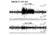
fMRIなどのいくつかのプロトコルは、神経活動の非侵襲性で脳全体の測定代表値を提供する。電気生理学などのいくつかのプロトコルは、細胞解像度と神経活動の迅速な指標、ならびに細胞解像度と神経活動の迅速な制御を提供する。光遺伝学などのいくつかのプロトコルは、定義済みニューロン群において、空間的に標的とされ、時間的に規定された活動電位発火の制御を提供する。アッセイの適切な組み合わせは、機能的脳回路の解剖に使用され得る。いくつかの場合、この組み合わせは以下の組み合わせ、光遺伝学とfMRI、光遺伝学と電気生理学、光遺伝学とEEG、光遺伝学と行動解析を含む。例えばEEGと行動解析、fMRIと電気生理学、電気生理学と行動解析など、任意の他の好適な組み合わせもまた、使用され得る。 Some protocols, such as fMRI, provide non-invasive, representative whole-brain measurements of neural activity. Some protocols, such as electrophysiology, provide rapid indication of cellular resolution and neural activity, as well as rapid control of cellular resolution and neural activity. Several protocols, such as optogenetics, provide spatially targeted and temporally defined control of action potential firing in defined neuronal populations. Appropriate combinations of assays can be used to dissect functional brain circuits. In some cases, this combination includes a combination of optogenetics and fMRI, optogenetics and electrophysiology, optogenetics and EEG, optogenetics and behavioral analysis. Any other suitable combination may also be used, for example EEG and behavioral analysis, fMRI and electrophysiology, electrophysiology and behavioral analysis.
ここに開示された方法は、前述のようにニューロン刺激と活動測定プロトコルの1つ以上との異なる組み合わせを使用することで、1つの生きた個体(例えばマウスまたはラット1匹、ヒト1人、1つの非ヒト哺乳動物)において異なる脳領域間の因果関係の解明に適する。したがっていくつかの実施形態において、脳の機能回路は単一動物において、光遺伝学とfMRI、光遺伝学と電気生理学、光遺伝学とEEG、および光遺伝学と行動解析の1つ以上の組み合わせを使用してアッセイされる。いくつかの実施形態において、脳の機能回路は単一動物において、光遺伝学とfMRI、光遺伝学と電気生理学、光遺伝学とEEG、および光遺伝学と行動解析のすべてを使用してアッセイされる。単一動物においてアッセイの異なる組み合わせを実施する順序は、任意の好適な順序であり得る。いくつかの場合ではアッセイの組み合わせは、光遺伝学とfMRI、光遺伝学とEEG/光遺伝学と行動解析、および光遺伝学と電気生理学の順序で実施されるが、ここでペア「光遺伝学とEEG」と「光遺伝学と行動解析」とは、任意の順序で実施され得る。プロトコルの他の組み合わせは、プロトコルと光遺伝学の組み合わせのうち任意のものの前後の任意の好適なポイントで実施され得る。 The methods disclosed herein use different combinations of neuronal stimulation and one or more activity measurement protocols, as described above, to study single living individuals (e.g., one mouse or rat, one human, one suitable for elucidating causal relationships between different brain regions in two non-human mammals). Thus, in some embodiments, functional brain circuits are combined in one or more of optogenetics and fMRI, optogenetics and electrophysiology, optogenetics and EEG, and optogenetics and behavioral analysis in a single animal. is assayed using In some embodiments, functional brain circuits are assayed in a single animal using optogenetics and fMRI, optogenetics and electrophysiology, optogenetics and EEG, and optogenetics and behavioral analysis. be done. The order of performing the different combinations of assays in a single animal can be any suitable order. In some cases, assay combinations are performed in the order optogenetics and fMRI, optogenetics and EEG/optogenetics and behavioral analysis, and optogenetics and electrophysiology, where paired "optogenetics" Physics and EEG" and "Optogenetics and Behavioral Analysis" can be performed in any order. Other combinations of protocols may be performed at any suitable point before or after any of the protocol and optogenetics combinations.
概して本方法の具現例は、脳の第1領域においてニューロンの定義済みセットの光遺伝学的刺激の組み合わせを使用するステップと、fMRIを用いて脳をスキャンすることで全脳レベルでの反応を測定して、脳の第1領域と第2領域との間の動的機能的結合を判定するステップとを含み得る。したがって第1領域のニューロンは、例えば光活性化イオンチャネルなど、光活性化ポリペプチドを含むように改変され得て、光活性化ポリペプチドは、適切な波長、照射量、および照射強度の光刺激を用いて第1領域を刺激した時に、例えば脱分極または過分極など、ニューロンの活動を変調するように構成される。いくつかの場合では、第1領域のニューロンは光活性化ポリペプチドを発現する。いくつかの場合では第1領域のニューロンは、例えば光活性化ポリペプチドをエンコードするヌクレオチド配列と、任意の他の適切な調節因子とを含むDNA構築物のウィルス感染などによって、遺伝子的に改変されて、光活性化ポリペプチドを発現する。本開示で詳しく記述したように、任意の好適な光活性化ポリペプチドが使用され得る。 In general, an embodiment of the method comprises the steps of using a combination of optogenetic stimulation of a defined set of neurons in a first region of the brain and scanning the brain using fMRI to determine responses at the whole-brain level. measuring to determine dynamic functional connectivity between the first and second regions of the brain. Thus, neurons in the first region can be modified to contain a photoactivated polypeptide, such as, for example, a photoactivated ion channel, which is exposed to photostimulation of the appropriate wavelength, irradiance and intensity. is configured to modulate neuronal activity, eg, depolarization or hyperpolarization, upon stimulation of the first region with . In some cases, neurons in the first region express the light-activated polypeptide. In some cases, neurons in the first region are genetically modified, such as by viral infection with a DNA construct comprising a nucleotide sequence encoding a photoactivatable polypeptide and any other suitable regulatory elements. , expresses a photoactivatable polypeptide. Any suitable photoactivatable polypeptide may be used, as described in detail in this disclosure.
光活性化ポリペプチドの活性化に使用される光刺激は、例えば周波数、パルス幅、デューティサイクル、波長、強度などを特徴とする光パルスを含み得る。いくつかの場合では光刺激は、2つ以上の異なる光パルスのセットを含み、光パルスの各セットは光パルスの異なる時間パターンを特徴とする。時間パターンは、周波数、期間(つまり光刺激の全期間)、パルス幅、デューティサイクルなどを含むがそれに限定されない任意の好適なパラメータを特徴とし得る。 Photostimulation used to activate photoactivated polypeptides can include, for example, light pulses characterized by frequency, pulse width, duty cycle, wavelength, intensity, and the like. In some cases, the light stimulation comprises two or more different sets of light pulses, each set of light pulses characterized by a different temporal pattern of light pulses. The temporal pattern may be characterized by any suitable parameters including, but not limited to, frequency, duration (ie, total duration of photostimulation), pulse width, duty cycle, and the like.
1セットの光パルスの特性の変動は、照射されたニューロン活動の差異に反映し得る。ニューロンが光活性化ポリペプチドの活性化によって脱分極されるいくつかの場合では、光パルスの周波数の増加は照射されたニューロンにおいて活動電位発火の周波数の増加を引き起こし得る。いくつかの実施形態において、照射されたニューロンにおいて活動電位発火の周波数は、光パルスの周波数の増加に定量的に対応する。いくつかの場合では光パルスの周波数の直線的増加は、照射されたニューロンにおいて活動電位発火の周波数における直線的、または非直線的ではあるが単調な、増加を誘起し得る。 Variations in the properties of a set of light pulses can reflect differences in illuminated neuronal activity. In some cases where neurons are depolarized by activation of a photoactivatable polypeptide, an increase in the frequency of light pulses can cause an increase in the frequency of action potential firing in irradiated neurons. In some embodiments, the frequency of action potential firing in irradiated neurons quantitatively corresponds to an increase in the frequency of the light pulse. In some cases, a linear increase in the frequency of the light pulse can induce a linear, or nonlinear but monotonic, increase in the frequency of action potential firing in irradiated neurons.
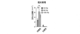
異なる光パルスのセットによる刺激に対する反応は、脳の異なる領域についてfMRIによって測定され得て、および各領域における反応の比較は、脳の第1領域と、1つ以上の他の領域とへの光刺激によって刺激されたニューロン間の機能的結合を示し得る。光刺激が時間パラメータ(例えば周波数またはパルス幅の変化)によって定量的に異なる光パルスセットを含むいくつかの場合では、fMRIによって測定された場合に、直線的でない(例えば第1領域における興奮性ニューロンの活動電位発火振動数の増加は、第2領域における反応の対応する増加を招かない)脳の第2領域における光刺激への反応は、第1領域のニューロンの活動電位発火の時間パターンによっては、第1領域のニューロンと第2領域のニューロンとの間に機能的結合があることを示し得る。光パルスの変動とfMRI反応の変動との間の非直線的関係は、非単調な関係または定性的関係であり得る。いくつかの場合では光パルスの定量的変化は、fMRI血液酸素濃度依存性(BOLD)反応(例えば正または負のBOLD反応は、光パルスの周波数によって測定される)のサインの変化を引き起こし得る。 Responses to stimulation by different sets of light pulses can be measured by fMRI for different regions of the brain, and a comparison of responses in each region can be used to determine the effect of light on a first region of the brain and one or more other regions. A stimulus may indicate functional connections between stimulated neurons. In some cases where light stimulation involves sets of light pulses that differ quantitatively by temporal parameters (e.g. changes in frequency or pulse width), they are non-linear when measured by fMRI (e.g. excitatory neurons in the first region an increase in the frequency of action potential firing in the second region does not lead to a corresponding increase in the response in the second region of the brain. , may indicate that there is a functional connection between the neurons of the first region and the neurons of the second region. The non-linear relationship between light pulse variability and fMRI response variability can be a non-monotonic or qualitative relationship. In some cases, quantitative changes in the light pulse can cause changes in the signature of the fMRI blood oxygenation dependent (BOLD) response (eg, positive or negative BOLD response is measured by the frequency of the light pulse).
例えば第1セットの光パルスおよび異なる時間パターンを持つ第2セットの光パルスを用いて脳の第1領域が照射されると、第1領域のニューロンは、第1セットと第2セットの光パルスによって誘起された活動電位を生成し得る。次いでfMRIは、脳の第2領域における活動を測定するために使用され得る。いくつかの場合では、第2領域における第1セットの光パルスによって誘起された神経活動の第1変化は、fMRIによって測定され得る。加えて第2領域における第2セットの光パルスによって誘起された神経活動の第2変化もまたfMRIによって測定され得る。いくつかの適用例において、神経活動における測定された第1変化と神経活動における測定された第2変化との間に差があり得る。神経活動における測定された第1変化と神経活動における測定された第2変化との間の差に基づいて、脳の第1領域と脳の第2領域とのニューロン間の動的機能的結合が同定され得る。例えば脳の第1領域と脳の第2領域とのニューロン間の動的機能的結合は、神経活動における測定された第1変化と測定された第2変化との間の差を計算することによって同定され得る。神経活動における測定された第1変化と測定された第2変化との間の計算された差が定性的または定量的に異なる場合、これは脳の第1領域のニューロンから脳の第2領域のニューロンへの動的機能的結合が存在するということの指標であり得る。 For example, when a first region of the brain is illuminated with a first set of light pulses and a second set of light pulses with a different temporal pattern, neurons in the first region are exposed to the first and second sets of light pulses. can generate action potentials evoked by fMRI can then be used to measure activity in a second region of the brain. In some cases, a first change in neural activity induced by the first set of light pulses in the second region can be measured by fMRI. Additionally, a second change in neural activity induced by a second set of light pulses in a second region can also be measured by fMRI. In some applications, there may be a difference between the first measured change in neural activity and the second measured change in neural activity. Based on the difference between the first measured change in neural activity and the second measured change in neural activity, dynamic functional connections between neurons in a first region of the brain and a second region of the brain are determined. can be identified. For example, dynamic functional connectivity between neurons in a first region of the brain and a second region of the brain can be determined by calculating the difference between a first measured change in neural activity and a second measured change in neural activity. can be identified. If the calculated difference between the first measured change in neuronal activity and the second measured change in neural activity differs qualitatively or quantitatively, this is the difference between neurons in the first region of the brain and neurons in the second region of the brain. It can be an indication that there are dynamic functional connections to neurons.
いくつかの場合では光刺激は、2セットの光パルスを含み、2セットは光パルスの周波数が異なるなど、異なるパラメータ値を有する特徴がある。2セットの光パルスの周波数が相違する場合、デューティサイクルは同じであったり、相違したりし得る。いくつかの場合では周波数が異なる2セットの光パルスは、パルス幅が同じである。他の事例において、周波数が異なる2セットの光パルスはパルス幅が異なる。 In some cases, the photostimulation is characterized by having different parameter values, such as comprising two sets of light pulses, the two sets having different frequencies of the light pulses. If the frequencies of the two sets of light pulses are different, the duty cycles can be the same or different. The two sets of light pulses, which in some cases differ in frequency, have the same pulse width. In other cases, the two sets of light pulses with different frequencies have different pulse widths.
1セットの光パルスは、任意の好適な周波数を有し得る。いくつかの場合では1セットの光パルスは、光刺激期間全体を通して維持された単一パルスの光を含む。いくつかの場合では1セットの光パルスは、例えば0.5Hz以上、1Hz以上、5Hz以上、10Hz以上、20Hz以上、30Hz以上、40H以上、50Hz以上、または60Hz以上、または70Hz以上、または80Hz以上、または90Hz以上、または100Hz以上など0.1Hz以上の周波数を有し、および例えば10,000Hz以下、1,000Hz以下、500Hz以下、400Hz以下、300Hz以下、200Hz以下、100Hzなど100,000Hz以下の周波数を有する。いくつかの場合では1セットの光パルスは、例えば1~10,000Hz、1~1,000Hz、5~500Hz、または10~100Hzなど0.1~100,000Hzの範囲の周波数を有する。 A set of light pulses may have any suitable frequency. In some cases, a set of light pulses includes a single pulse of light maintained throughout the light stimulation period. In some cases, a set of light pulses is, for example, 0.5 Hz or more, 1 Hz or more, 5 Hz or more, 10 Hz or more, 20 Hz or more, 30 Hz or more, 40 Hz or more, 50 Hz or more, or 60 Hz or more, or 70 Hz or more, or 80 Hz or more. , or has a frequency of 0.1 Hz or higher, such as 90 Hz or higher, or 100 Hz or higher, and 100,000 Hz or lower, such as 10,000 Hz or lower, 1,000 Hz or lower, 500 Hz or lower, 400 Hz or lower, 300 Hz or lower, 200 Hz or lower, 100 Hz have a frequency. In some cases, a set of light pulses has frequencies in the range of 0.1-100,000 Hz, such as 1-10,000 Hz, 1-1,000 Hz, 5-500 Hz, or 10-100 Hz.
いくつかの場合では2セットの光パルスは、異なるパルス幅など、異なるパラメータ値を有する特徴がある。2セットの光パルスのパルス幅が異なる場合、2セットの光パルスのデューティサイクルは同じであったり、相違したりし得る。いくつかの場合ではパルス幅が異なる2セットの光パルスは、周波数が同じである。他の事例において、パルス幅が異なる2セットの光パルスは、周波数が異なる。 In some cases, the two sets of light pulses are characterized by different parameter values, such as different pulse widths. The duty cycles of the two sets of light pulses may be the same or different if the pulse widths of the two sets of light pulses are different. The two sets of light pulses, which in some cases differ in pulse width, have the same frequency. In other cases, the two sets of light pulses with different pulse widths have different frequencies.
光パルスは、任意の好適なパルス幅を有し得る。いくつかの場合ではパルス幅は、例えば0.5ms以上、1ms以上、3ms以上、5ms以上、7.5ms以上、10ms以上、を含む15ms以上、または20ms以上、または25ms以上、または30ms以上、または35ms以上、または40ms以上、または45ms以上、または50ms以上など0.1ms以上であり、および例えば100ms以下、90ms以下、80ms以下、70ms以下、60ms以下、50ms以下、45ms以下、40ms以下、35ms以下、30ms以下、25ms以下、20ms以下など、500ms以下である。いくつかの実施形態において、パルス幅は例えば0.5~100ms、1~80ms、1~60ms、または1~50ms、または1~30msなど0.1~500msの範囲である。 A light pulse may have any suitable pulse width. In some cases, the pulse width is 15 ms or greater, including, for example, 0.5 ms or greater, 1 ms or greater, 3 ms or greater, 5 ms or greater, 7.5 ms or greater, 10 ms or greater, or 20 ms or greater, or 25 ms or greater, or 30 ms or greater, or 0.1 ms or more, such as 35 ms or more, or 40 ms or more, or 45 ms or more, or 50 ms or more, and for example 100 ms or less, 90 ms or less, 80 ms or less, 70 ms or less, 60 ms or less, 50 ms or less, 45 ms or less, 40 ms or less, 35 ms or less , 30 ms or less, 25 ms or less, 20 ms or less, etc., 500 ms or less. In some embodiments, the pulse width ranges from 0.1-500 ms, such as 0.5-100 ms, 1-80 ms, 1-60 ms, or 1-50 ms, or 1-30 ms, for example.
いくつかの場合では2セットの光パルスは、異なるデューティサイクルなど、異なるパラメータ値を有する特徴がある。デューティサイクルは、パルス化シグナルについての「定時」に関する百分率(比)であり、および以下のように計算され得る。
デューティサイクル=(パルス幅)/周波数
In some cases, the two sets of light pulses are characterized by different parameter values, such as different duty cycles. Duty cycle is a percentage (ratio) of "on time" for a pulsed signal, and can be calculated as follows.
Duty Cycle = (Pulse Width)/Frequency
2セットの光パルスのデューティサイクルが異なる場合、2セットの光パルスのパルス幅および/または周波数は同じであったり、相違したりし得る。いくつかの場合では異なるデューティサイクルを有する2セットの光パルスは、パルス幅が異なり、および周波数が異なる。他の事例において、異なるデューティサイクルを有する2セットの光パルスは、パルス幅が異なり、および周波数が同じである。他の事例において、異なるデューティサイクルを有する2セットの光パルスは、パルス幅が同じで、および周波数が異なる。 The pulse widths and/or frequencies of the two sets of light pulses may be the same or different if the duty cycles of the two sets of light pulses are different. The two sets of light pulses, which in some cases have different duty cycles, have different pulse widths and different frequencies. In other cases, the two sets of light pulses with different duty cycles have different pulse widths and the same frequency. In other cases, two sets of light pulses with different duty cycles have the same pulse width and different frequencies.
いくつかの場合では2セットの光パルスは、同じデューティサイクルを有する特徴がある。2セットの光パルスのデューティサイクルが同じ場合、2セットの光パルスのパルス幅および/または周波数は相違し得る。いくつかの場合では同じデューティサイクルを有する2セットの光パルスは、パルス幅が異なり、および周波数も異なる。 In some cases, the two sets of light pulses are characterized by having the same duty cycle. If the duty cycles of the two sets of light pulses are the same, the pulse widths and/or frequencies of the two sets of light pulses may differ. The two sets of light pulses, which in some cases have the same duty cycle, have different pulse widths and different frequencies.
パルスのデューティサイクルは、任意の好適なデューティサイクルであり得る。いくつかの場合ではデューティサイクルは、例えば5%以上、10%以上、15%以上、20%以上、25%以上、または30%以上、または35%以上、または40%以上、または45%以上、または50%以上など1%以上であり、および例えば75%以下、70%以下、65%以下、60%以下、65%以下、50%以下、45%以下、40%以下、または35%以下、または30%以下など、80%以下であり得る。特定の実施形態において、デューティサイクルは例えば5~70%、5~60%、10~50%、または10~40%など、1~80%の範囲である。 The pulse duty cycle can be any suitable duty cycle. In some cases, the duty cycle is, for example, 5% or more, 10% or more, 15% or more, 20% or more, 25% or more, or 30% or more, or 35% or more, or 40% or more, or 45% or more; or 1% or more, such as 50% or more; or may be 80% or less, such as 30% or less. In certain embodiments, the duty cycle ranges from 1-80%, such as 5-70%, 5-60%, 10-50%, or 10-40%.
光パルスを脳の領域に送信する光ファイバーの先端で測定した場合の、光パルスの平均電力値は、任意の好適な電力であり得る。いくつかの場合ではこの電力は例えば0.5mW以上、1mW以上、1.5mW以上、2mW以上、または2.5mW以上、または3mW以上、または3.5mW以上、または4mW以上、または4.5mW以上、または5mW以上など0.1mW以上であり得て、および例えば500mW以下、250mW以下、100mW以下、50mW以下、40mW以下、30mW以下、20mW以下、15mW以下、10mW以下、または5mW以下など1,000mW以下であり得る。いくつかの実施形態において、この電力は例えば0.5~100mW、0.5~50mW、1~20mW、1~10mW、または1~5mWなど、0.1~1,000mWの範囲である。 The average power value of the light pulse, as measured at the tip of the optical fiber that transmits the light pulse to the brain region, can be any suitable power. In some cases, the power is, for example, 0.5 mW or more, 1 mW or more, 1.5 mW or more, 2 mW or more, or 2.5 mW or more, or 3 mW or more, or 3.5 mW or more, or 4 mW or more, or 4.5 mW or more. , or 0.1 mW or more, such as 5 mW or more, and 1,000 mW, such as 500 mW or less, 250 mW or less, 100 mW or less, 50 mW or less, 40 mW or less, 30 mW or less, 20 mW or less, 15 mW or less, 10 mW or less, or 5 mW or less. can be: In some embodiments, this power is in the range of 0.1-1,000 mW, such as 0.5-100 mW, 0.5-50 mW, 1-20 mW, 1-10 mW, or 1-5 mW.
光パルスの波長および強度は変化し得て、および光活性化ポリペプチドの活性化波長、脳の領域の光透過性、脳の照射される所望の容積、などに依存し得る。 The wavelength and intensity of the light pulse can vary and can depend on the activation wavelength of the photoactivatable polypeptide, the optical transparency of the region of the brain, the desired volume of the brain to be illuminated, and the like.
光パルスによって照射された脳領域の容積は、任意の好適な容積であり得る。いくつかの場合では照射された容積は、例えば0.005mm3 以上、0.001mm3 以上、0.005mm3 以上、0.01mm3 以上、0.05mm3 以上、0.1mm3 以上など、0.001mm3 以上であり、および例えば50mm3 以下、20mm3 以下、10mm3以下、5mm3 以下、1mm3 以下、0.1mm3 以下など100mm3 以下である。特定の場合では、照射された容積は、例えば0.005~20mm3 、0.01~10mm3 、0.01~5mm3 、0.05~1mm3 など、0.001~100mm3 の範囲である。 The volume of the brain region illuminated by the light pulse can be any suitable volume. In some cases , the irradiated volume is 0 , e.g. .001 mm 3 or more, and 100 mm 3 or less, such as 50 mm 3 or less, 20 mm 3 or less, 10 mm 3 or less, 5 mm 3 or less, 1 mm 3 or less, 0.1 mm 3 or less. In certain cases, the irradiated volume is in the range of 0.001-100 mm 3 , such as 0.005-20 mm 3 , 0.01-10 mm 3 , 0.01-5 mm 3 , 0.05-1 mm 3 . be.
本方法の別の態様として、前述のように第1領域と第2領域との間の動的機能的結合に介在し得る脳の第3領域においてニューロンを同定するステップを含む。したがって、第3領域のニューロンは、例えば第1領域と第2領域との間の動的機能的結合の調節結節などの結節を表すと言い得て、第3領域のニューロンは第1領域および第2領域両方への機能的結合を有する。いくつかの場合では、動的機能的結合の調節結節を同定するステップは、前述のように電気生理学、神経活動の光誘起変調、EEG、機能イメージング(例えばfMRI)、および行動解析の1つ以上を使用するステップを含み得る。 Another aspect of the method includes identifying neurons in a third region of the brain that may mediate dynamic functional connections between the first and second regions as described above. Thus, the neurons of the third region can be said to represent nodes, such as regulatory nodes of dynamic functional connections between the first and second regions, and the neurons of the third region It has functional connections to both regions. In some cases, identifying regulatory nodes of dynamic functional connectivity involves one or more of electrophysiology, light-induced modulation of neural activity, EEG, functional imaging (e.g., fMRI), and behavioral analysis as described above. may include the step of using
いくつかの実施形態において、第1領域と第2領域との間の動的機能的結合に介在し得る脳の第3領域におけるニューロンは、第1領域と第2領域との間の機能的結合の動的特性に基づいて、第3領域のニューロンの平常活動を中断させる効果を測定することで同定され得る。第3領域のニューロンの平常活動が動的機能的結合にとって必要な場合、第3領域のニューロンは動的機能的結合の調節結節を表し得る。 In some embodiments, neurons in a third region of the brain that can mediate dynamic functional connections between the first and second regions are the functional connections between the first and second regions. can be identified by measuring their effect on disrupting the normal activity of neurons in the third region, based on the dynamic properties of . If normal activity of neurons in the third region is required for dynamic functional connectivity, neurons in the third region may represent regulatory nodes of dynamic functional connectivity.
第3領域のニューロンの平常活動は、任意の好適な方法を使用して中断され得る。いくつかの場合ではこの活動は、例えば第3領域のニューロンにおいて、光活性化ポリペプチドを活性化させるための適切な光刺激を用いた第3領域の照射を介して、正常に活性化されたニューロンに対する光活性化イオンポンプなど、光活性化ポリペプチドを活性化することで中断される。第2領域におけるニューロン反応の変化を、第1領域のニューロンにおいて光活性化ポリペプチドを活性化する第1セットの光パルスと比較することで、第1領域と第2領域との間の動的機能的結合における第3領域のニューロンの役割は、第3領域を照射する第3セットの光パルスの有無で、判定され得る。したがって、第3領域のニューロンの平常活動を中断させる第3領域の照射が、第2領域におけるニューロンの第1セットの光パルスに対する反応を減少、終止、またはさもなければ変更させる場合、第3領域のニューロンは動的機能的結合に介在し得る。いくつかの場合では第2領域におけるニューロンの第1セットの光パルスに対する反応は、例えば30%以上、40%以上、50%以上、60%以上、70%以上、80%以上、90%以上、および100%以内など、20%以上減少され得る。いくつかの場合では第2領域におけるニューロンの第1セットの光パルスに対する反応は、サインの変化を含み得る(例えば、第3領域のニューロンの平常活動による発火頻度の減少が、第3領域のニューロンの平常活動の中断により発火頻度の増加に切り替わる)。 Normal activity of neurons in the third region can be disrupted using any suitable method. In some cases, this activity was successfully activated, e.g., in neurons of the third region, via illumination of the third region with an appropriate light stimulus to activate the photoactivatable polypeptide. Interrupted by activating light-activated polypeptides, such as light-activated ion pumps for neurons. By comparing changes in neuronal responses in the second region to a first set of light pulses that activate photoactivated polypeptides in neurons in the first region, dynamics between the first region and the second region can be determined. The role of neurons in the third region in functional connectivity can be determined in the presence or absence of a third set of light pulses illuminating the third region. Thus, if illumination of a third region that disrupts the normal activity of neurons in the third region reduces, terminates, or otherwise alters the response of neurons in the second region to the first set of light pulses, the third region neurons may mediate dynamic functional connections. In some cases, the response of neurons in the second region to the first set of light pulses is, for example, 30% or more, 40% or more, 50% or more, 60% or more, 70% or more, 80% or more, 90% or more, and may be reduced by 20% or more, such as up to 100%. In some cases, the response of neurons in the second region to the first set of light pulses may include a change in signature (e.g., a decrease in firing frequency due to normal activity of neurons in the third region may cause neurons in the third region to switch to increased firing frequency upon interruption of the normal activity of the brain).
例えば、脳の第3領域における調節結節の有無は、光パルスのセットを用いて脳の第3領域を照射したり、照射しないことで、脳の第1領域(脳の第2領域への動的機能的結合がある)を照射することで判定され得る。第1セットの光パルスを用いた脳の第1領域の照射があると、第1領域のニューロンは第1セットの光パルスによって誘起された活動電位を生成し得る。加えて脳の第3領域は、同時に第3セットの光パルスによって照射され得る。第3領域の照射があると、第3領域のニューロンの活動は、例えば本開示で記述されているように、第3領域におけるニューロンが光活性化ポリペプチドを発現すると、中断され得る。次いで神経活動は、脳の第1領域と動的機能的に結合する脳の第2領域において(例えばfMRIで)測定され得る。例えばfMRIは、脳の第2領域において第1セットの光パルスによって誘起された神経活動における第3変化を、脳の第3領域を照射することなしに、測定するために使用され得る。いくつかの場合では、脳の第3領域を照射せずに、脳の第2領域において第1セットの光パルスによって誘起された神経活動の測定された第3変化は、脳の第1領域と第2領域との間の動的機能的結合のベースライン測定を提供する。加えて第2領域において第1セットの光パルスによって誘起された神経活動における第4変化もまた、脳の第3領域を第3セットの光パルスによって照射している間に測定され得る。いくつかの適用例において、神経活動における測定された第3変化と測定された神経活動における測定された第4変化との間に差があり得る。神経活動における測定された第3変化と神経活動における測定された第4変化との間の差に基づいて、脳の第3領域における調節結節が同定され得る。脳の第3領域における調節結節の存在は、脳の第3領域と脳の第1領域との間の機能的結合および/または脳の第3領域と脳の第2領域との間の機能的結合の指標であり得る。例えば脳の第3領域における調節結節は、神経活動における測定された第3変化と測定された第4変化の間の差を計算することによって同定され得る。神経活動における測定された第3変化と測定された第4変化との間の計算された差が定性的または定量的に異なる場合、これは脳の第3領域において調節結節が存在するということの指標であり得る。例えば、神経活動における測定された第3変化と測定された第4変化との間の計算された差が定性的または定量的に異なる場合、これは、脳の第3領域と脳の第1領域との間に機能的結合があり、および/または脳の第3領域と脳の第2領域との間に機能的結合があることの指標であり得る。
For example, the presence or absence of regulatory nodules in
光遺伝学的刺激は、任意の好適な方法を使用して実施され得る。好適な方法は、例えば米国特許第8,834,546号に記載されており、その特許を参照によってここに組み込む。脳の好適な領域のニューロンの活動は光によって変調されるが、このニューロンは便利な方法を使用して光活性化ポリペプチドを発現するように改変され得る。いくつかの場合では脳領域のニューロンは、遺伝子的に改変されて光活性化ポリペプチドを発現する。いくつかの場合ではニューロンは、例えば光活性化ポリペプチドをエンコードするヌクレオチド配列を有する核酸を含むアデノ随伴ウイルスベクターなどのウイルスベクターを使用して、遺伝子的に改変され得る。ウイルスベクターは、細胞型、時期、誘発因子の存在などに従って光活性化ポリペプチドの発現を制御する任意の好適な制御因子(例えばプロモーター、エンハンサー、組換え部位など)を含み得る。 Optogenetic stimulation can be performed using any suitable method. Suitable methods are described, for example, in US Pat. No. 8,834,546, which is incorporated herein by reference. The activity of neurons in suitable regions of the brain is modulated by light, and the neurons can be modified to express photoactivatable polypeptides using any convenient method. In some cases, neurons in brain regions are genetically modified to express photoactivated polypeptides. In some cases, neurons can be genetically modified using, for example, viral vectors such as adeno-associated viral vectors containing nucleic acids having nucleotide sequences encoding light-activated polypeptides. Viral vectors may contain any suitable regulatory elements (eg, promoters, enhancers, recombination sites, etc.) that control the expression of the photoactivatable polypeptide according to the cell type, timing, presence of inducers, and the like.
好適なニューロン特異的制御配列は、神経特異的エノラーゼ(NSE)プロモーター(例えばEMBL HSENO2,X51956を参照;また例えば米国特許第6,649,811号、米国特許第5,387,742号も参照);芳香族アミノ酸デカルボキシラーゼ(AADC)プロモーター;神経フィラメントプロモーター(例えばGenBank HUMNFL,L04147を参照);シナプシンプロモーター(例えばGenBank HUMSYNIB,M55301を参照);thy-1プロモーター(例えばChen et al.(1987)Cell 51:7-19;およびLlewellyn et al.(2010)Nat.Med.16:1161を参照);セロトニン受容体プロモーター(例えばGenBank S62283を参照);チロシンヒドロキシラーゼプロモーター(TH)(例えばNucl.Acids.Res.15:2363-2384(1987)およびNeuron 6:583-594(1991)を参照);GnRHプロモーター(例えばRadovick et al.,Proc.Natl.Acad.Sci.USA 88:3402-3406(1991)を参照);L7プロモーター(例えばOberdick et al.,Science 248:223-226(1990)を参照);DNMTプロモーター(例えばBartge et al.,Proc.Natl.Acad.Sci.USA 85:3648-3652(1988)を参照);エンケファリンプロモーター(例えばComb et al.,EMBO J. 17:3793-3805(1988)を参照);ミエリン塩基性タンパク質(MBP)プロモーター;CMVエンハンサー/血小板由来成長因子-βプロモーター(例えばLiu et al.(2004)Gene Therapy 11:52-60を参照);運動ニューロン-特異的遺伝子Hb9プロモーター(例えば米国特許第7,632,679号;およびLee et al.(2004)Development 131:3295-3306を参照);およびCa(2+)-カルモジュリン依存性プロテインキナーゼII(CaMKIIα)プロモーターのαサブユニット(例えばMayford et al.(1996)Proc.Natl.Acad.Sci.USA 93:13250を参照)、を含むがそれだけに限らない。他の好適なプロモーターとして、伸張因子(EF)1αおよびドーパミン輸送体(DAT)プロモーターを含む。 A preferred neuron-specific regulatory sequence is the neuron-specific enolase (NSE) promoter (see, eg, EMBL HSENO2, X51956; see also, eg, US Pat. No. 6,649,811, US Pat. No. 5,387,742). the aromatic amino acid decarboxylase (AADC) promoter; the neurofilament promoter (see, eg, GenBank HUMNFL, L04147); the synapsin promoter (see, eg, GenBank HUMSYNIB, M55301); the thy-1 promoter (see, eg, Chen et al. (1987)). Cell 51:7-19; and Llewellyn et al. (2010) Nat. Med. 16:1161); serotonin receptor promoters (see, eg, GenBank S62283); Res. 15:2363-2384 (1987) and Neuron 6:583-594 (1991)); )); the L7 promoter (see, eg, Oberdick et al., Science 248:223-226 (1990)); the DNMT promoter (see, eg, Bartge et al., Proc. Natl. Acad. Sci. USA 85:3648-3652). (1988)); enkephalin promoters (see, eg, Comb et al., EMBO J. 17:3793-3805 (1988)); myelin basic protein (MBP) promoter; (see, eg, Liu et al. (2004) Gene Therapy 11:52-60); motor neuron-specific gene Hb9 promoter (eg, US Pat. No. 7,632,679; and Lee et al. (2004) Development 131). :3295-3306); and the α subunit of the Ca( 2+ )-calmodulin-dependent protein kinase II (CaMKIIα) promoter (eg, Mayford et al. (1996) Proc. Natl. Acad. Sci. USA 93: 13250), including but not limited to. Other suitable promoters include the elongation factor (EF) 1α and dopamine transporter (DAT) promoters.
いくつかの場合では光活性化ポリペプチドの細胞型特異的発現は、例えばCre-Lox組換え、Flp-FRT組換えなど、組換えシステムを使用することで達成され得る。組換えを使用した遺伝子の細胞型特異的発現は、例えばFenno et al.,Nat Methods.2014 Jul;11(7):763;およびGompf et al.,Front Behav Neurosci.2015 Jul 2;9:152に記載されており、これらを参照によってここに組み込む。
In some cases, cell-type specific expression of photoactivatable polypeptides can be achieved using recombinant systems, eg, Cre-Lox recombination, Flp-FRT recombination, and the like. Cell-type specific expression of genes using recombination is described, for example, in Fenno et al. , Nat Methods. 2014 Jul;11(7):763; and Gompf et al. , Front Behav Neurosci. 2015
いくつかの場合では光活性化ペプチドを含むニューロンのある脳の領域は、1つ以上の光ファイバーを使用して照射される。光ファイバーは任意の好適な方法で、例えばレーザー光源または発光ダイオード(LED)光源など、好適な光源から発射された光を脳の領域に導くように構成され得る。光ファイバーは任意の好適な光ファイバーであり得る。いくつかの場合では光ファイバーは、マルチモード光ファイバーである。光ファイバーはコア径を規定するコアを含み得て、光源からの光はコアを通り抜ける。光ファイバーは任意の好適なコア径を有し得る。いくつかの場合では光ファイバーのコア径は、例えば20μm以上、30μm以上、40μm以上、50μm以上、60μm以上、80μm以上など10μm以上であり、および例えば500μm以下、200μm以下、100μm以下、70μm以下など1,000μm以下である。いくつかの実施形態において、光ファイバーのコア径は、例えば20~500μm、30~200μm、40~100μmなど10~1,000μmの範囲である。 In some cases, regions of the brain with neurons containing photoactivatable peptides are illuminated using one or more optical fibers. The optical fiber may be configured in any suitable manner to direct light emitted from a suitable light source, such as a laser light source or a light emitting diode (LED) light source, to a region of the brain. The optical fiber can be any suitable optical fiber. In some cases the optical fiber is a multimode optical fiber. The optical fiber may include a core defining a core diameter through which light from the light source passes. The optical fiber can have any suitable core diameter. In some cases, the core diameter of the optical fiber is 10 μm or greater, such as 20 μm or greater, 30 μm or greater, 40 μm or greater, 50 μm or greater, 60 μm or greater, 80 μm or greater, and 10 μm or greater, such as 500 μm or less, 200 μm or less, 100 μm or less, 70 μm or less. ,000 μm or less. In some embodiments, the core diameter of the optical fiber ranges from 10-1,000 μm, such as 20-500 μm, 30-200 μm, 40-100 μm.
特定の実施形態において、クラッデングは光ファイバーのコアの少なくとも一部分を囲む。例えばクラッデングは、実質的に光ファイバーの外周面全体を囲み得る。いくつかの場合ではクラッデングは、光源から光を受信する光ファイバー先端、および脳の標的領域にあるニューロンに光を発信する光ファイバーの対向する端など、光ファイバーの端には存在しない。クラッデングは、任意の好適なタイプのクラッデングであり得る。いくつかの場合では、クラッデングは光ファイバーのコアより屈折率が低い。クラッデングの好適な材料として、プラスチック、樹脂、および同種のもの、およびそれらの組み合わせを含むがそれだけに限らない。 In certain embodiments, the cladding surrounds at least a portion of the core of the optical fiber. For example, the cladding can surround substantially the entire outer circumference of the optical fiber. In some cases, cladding is absent from the ends of the optical fibers, such as the fiber optic tip that receives the light from the light source and the opposite end of the fiber optic that transmits the light to the neurons in the target region of the brain. The cladding can be any suitable type of cladding. In some cases, the cladding has a lower refractive index than the core of the optical fiber. Suitable materials for the cladding include, but are not limited to, plastics, resins, and the like, and combinations thereof.
いくつかの場合では光ファイバーは、外側コーティングを含む。外側コーティングは、クラッデング表面に配置され得る。コーティングは、実質的に光ファイバーの外周面全体を囲み得る。いくつかの場合ではコーティングは、光源から光を受信する光ファイバー先端、および脳の標的領域にあるニューロンに光を発信する光ファイバーの対向する端など、光ファイバーの端には存在しない。コーティングは生物学的に適合するコーティングであり得る。生物学的に適合するコーティングとして、組織、液体、または光ファイバーが差し込まれた対象に存在する他の物質に有意には反応しないコーティングを含む。いくつかの場合では生物学的に適合するコーティングは、光ファイバーが使用されている周辺環境に対して不活性の(つまり実質的に反応しない)材料から構成される。 In some cases the optical fiber includes an outer coating. An outer coating may be disposed on the cladding surface. The coating may substantially surround the entire outer circumference of the optical fiber. In some cases, the coating is absent from the ends of the optical fibers, such as the fiber optic tip that receives the light from the light source and the opposite end of the fiber optic that transmits the light to the neurons in the target region of the brain. The coating can be a biocompatible coating. Biologically compatible coatings include coatings that do not react significantly with tissue, liquids, or other substances present in the subject into which the optical fiber is inserted. In some cases, the biocompatible coating is composed of materials that are inert (ie, substantially non-reactive) to the surrounding environment in which the optical fiber is used.
脳の標的領域に埋め込まれた光ファイバーの端部は、光ファイバーを通して出力された光刺激を用いて、脳の領域を照射するために好適な任意の好適な構成を有し得る。いくつかの場合では光ファイバーは、光ファイバーの遠位端にまたはその近傍にある取付け装置を含み、光ファイバーの遠位端は対象に差し込まれた端部に対応する。いくつかの場合では取付け装置は光ファイバーに接続するように構成され、および対象の頭部など対象への光ファイバーの取り付けを容易にする。任意の好適な取付け装置が使用され得る。いくつかの場合では取付け装置は、例えば金属の口金、セラミックの口金、またはプラスチックの口金などの口金を含む。口金は、光ファイバーを保持して取り付けるために好適な任意の寸法を有し得る。いくつかの場合では口金は、例えば0.75~2.5mm、または1~2mmなど、0.5~3mmの範囲の直径を有する。 The end of the optical fiber implanted in the target region of the brain may have any suitable configuration suitable for illuminating the region of the brain with light stimulation output through the optical fiber. In some cases, the optical fiber includes an attachment device at or near the distal end of the optical fiber, the distal end of the optical fiber corresponding to the end that is plugged into the subject. In some cases, the attachment device is configured to connect to an optical fiber and facilitate attachment of the optical fiber to a subject, such as the subject's head. Any suitable mounting device may be used. In some cases, the mounting device includes a base, such as a metal base, a ceramic base, or a plastic base. The ferrule may have any dimensions suitable for holding and mounting an optical fiber. In some cases the base has a diameter in the range of 0.5-3 mm, such as 0.75-2.5 mm, or 1-2 mm.
特定の実施形態において、本開示の方法は、任意の好適な電子部品を使用して脳の領域を照射するように使用される様々な光部品を制御および/または調整するように実施され得る。光部品(例えば光源、光ファイバー、レンズ、対物レンズ、ミラー、および同種のもの)は、例えば光パルスを用いて脳の領域を照射する光源を調整するために、制御部によって制御され得る。この制御部は光源の駆動部を含み得て、この駆動部は、光パルスの周波数、パルス幅、デューティサイクル、波長、強度などを含むがそれに限定されない、光パルスに関連する1つ以上のパラメータを制御する。制御部は、光源の部品(例えばコリメータ、シャッター、フィルタホイール、可動ミラー、レンズなど)と通信し得る。 In certain embodiments, the methods of the present disclosure may be implemented using any suitable electronic components to control and/or regulate various optical components used to illuminate regions of the brain. Optical components (eg, light sources, optical fibers, lenses, objectives, mirrors, and the like) can be controlled by the controller, for example, to adjust the light source to illuminate regions of the brain with light pulses. The controller may include a driver of the light source, which drives one or more parameters associated with the light pulse, including but not limited to frequency, pulse width, duty cycle, wavelength, intensity, etc. of the light pulse. to control. The controller may communicate with components of the light source (eg, collimator, shutter, filter wheel, movable mirror, lens, etc.).
演算ユニット(例えばコンピュータ)は、本開示の方法において、1つ以上の制御部を通して光刺激を制御および/または調整するため、およびfMRIによる脳の領域のスキャンからのデータを解析するために使用され得る。演算ユニットは、測定されたfMRI画像を解析する任意の好適な部品を含み得る。したがって演算ユニットは、以下のもののうち1つ以上、プロセッサ、コンピュータ可読媒体などの非一時的コンピュータ可読メモリー、キーボード、マウス、タッチパネルなどの入力装置、モニター、画面、スピーカーなどの出力装置、有線または無線のネットワークインターフェースなどのネットワークインターフェース、および同種のものを含み得る。 A computing unit (e.g., a computer) is used in the methods of the present disclosure to control and/or coordinate light stimulation through one or more controls and to analyze data from fMRI brain region scans. obtain. The computing unit may include any suitable components for analyzing the measured fMRI images. A computing unit may therefore include one or more of the following: a processor, non-transitory computer readable memory such as a computer readable medium, input devices such as keyboards, mice, touch panels, etc., output devices such as monitors, screens, speakers, etc., wired or wireless. and the like.
fMRIは、任意の好適な方法を使用して実施され得る。好適な方法は、例えば米国特許第8,834,546号に記載されており、その特許を参照によってここに組み込む。 fMRI can be performed using any suitable method. Suitable methods are described, for example, in US Pat. No. 8,834,546, which is incorporated herein by reference.
本方法(光遺伝学的に神経活動を刺激するおよび/または測定するため)における脳の関心領域は変化し得て、および任意の好適な領域であり得る。特定の実施形態において、脳領域は、解剖的および/または機能的に規定された脳の領域である。例えばここで記述されているように光パルスによって照射された脳の第1領域と脳の第2領域とは、解剖的に脳の異なる領域であり得る。同様にいくつかの適用例において、脳の第3領域は第1領域と第2領域との間の動的機能的結合を調節するが、その第3領域は脳の第1領域および第2領域とは解剖的に別個であり得る。脳が哺乳類の脳であるいくつかの場合では、脳の関心領域は、視床(中心視床を含む)、感覚皮質(体性感覚皮質を含む)、不確帯(ZI)、腹側被蓋野(VTA)、前頭前皮質(PFC)、側座核(NAc)、扁桃体(BLA)、黒質、腹側淡蒼球、淡蒼球、背側線条体、腹側線条体、視床下核、海馬、歯状回、帯状回、嗅内皮質、嗅覚皮質、一次運動皮質、および小脳、の少なくとも一部分から選択される。いくつかの場合では異なる脳領域(例えば第1脳領域および第2脳領域)は最小でも、例えば2つ以上、3つ以上、4つ以上、5つ以上、7つ以上など1つ以上のシナプス結合によって分離されており、および最小でも例えば12個以下、10個以下、8つ以下、6つ以下など、15個以下のシナプス結合によって分離される。いくつかの実施形態において、異なる脳領域は最小でも例えば1~12シナプス結合、1~10シナプス結合、2~8シナプス結合、3~6シナプス結合など、1~15シナプス結合によって分離される。 The region of interest in the subject methods (for optogenetically stimulating and/or measuring neural activity) can vary and can be any suitable region. In certain embodiments, a brain region is an anatomically and/or functionally defined region of the brain. For example, the first region of the brain and the second region of the brain illuminated by the light pulses as described herein can be anatomically distinct regions of the brain. Similarly, in some applications, a third region of the brain modulates dynamic functional connections between the first and second regions of the brain, wherein the third region is the first and second regions of the brain. can be anatomically distinct from the In some cases where the brain is a mammalian brain, areas of interest in the brain are the thalamus (including the central thalamus), the sensory cortex (including the somatosensory cortex), the zone of incertitude (ZI), the ventral tegmental area. (VTA), prefrontal cortex (PFC), nucleus accumbens (NAc), amygdala (BLA), substantia nigra, ventral pallidus, pallidus, dorsal striatum, ventral striatum, subthalamic nucleus, selected from at least a portion of the hippocampus, dentate gyrus, cingulate gyrus, entorhinal cortex, olfactory cortex, primary motor cortex, and cerebellum. In some cases, different brain regions (e.g., first and second brain regions) have at least one or more synapses, e.g., two or more, three or more, four or more, five or more, seven or more. are separated by connections and are separated by a minimum of 15 synaptic connections or less, such as 12 or less, 10 or less, 8 or less, 6 or less. In some embodiments, different brain regions are separated by at least 1-15 synaptic connections, eg, 1-12 synaptic connections, 1-10 synaptic connections, 2-8 synaptic connections, 3-6 synaptic connections, etc.
脳領域に存在する対象ニューロンは、任意の好適なタイプのニューロンであり得る。いくつかの場合ではこのニューロンは、抑制性ニューロンまたは興奮性ニューロンである。いくつかの場合ではこのニューロンは、感覚ニューロン、介在ニューロン、または運動ニューロンである。いくつかの場合ではこのニューロンは、制限なしでドーパミン作動性ニューロン、コリン作動性ニューロン、GABA作動性神経、グルタミン酸作動性神経、またはペプチド作動性神経である。 A target neuron present in a brain region can be any suitable type of neuron. In some cases this neuron is an inhibitory neuron or an excitatory neuron. In some cases this neuron is a sensory neuron, an interneuron, or a motor neuron. In some cases the neuron is without limitation a dopaminergic neuron, a cholinergic neuron, a GABAergic neuron, a glutamatergic neuron, or a peptidergic neuron.
光活性化ポリペプチド
上記で要約したように本開示の態様は、例えば発現する光活性化ポリペプチドを有するニューロンを含む様々な脳領域を含む。光活性化ポリペプチドは、光活性化イオンチャネルまたは光活性化イオンポンプであり得る。光活性化イオンチャネルポリペプチドは、ポリペプチドに活性化波長の光を照射した場合に、1つ以上のイオンをニューロンの原形質膜を通過可能にすることに適する。光活性化タンパク質はイオンポンプタンパク質を特徴とし得て、このタンパク質は少数のイオンが光の光子によって原形質膜を通過することを容易にする、またはイオンチャネルタンパク質を特徴とし得て、このタンパク質はチャネルが開いている場合にイオン流が自在に原形質膜を貫流することを可能にする。いくつかの実施形態において、光活性化ポリペプチドは、活性化波長の光によって活性化された場合にニューロンを脱分極する。図14は、制限なしで好適な脱分極性光活性化ポリペプチドを示す。いくつかの実施形態において、光活性化ポリペプチドは、活性化波長の光によって活性化された場合にニューロンを過分極する。図15は、制限なしで好適な過分極性光活性化ポリペプチドを示す。
Photoactivated Polypeptides As summarized above, aspects of the present disclosure include various brain regions including, for example, neurons that have photoactivated polypeptides expressed. A photoactivated polypeptide can be a photoactivated ion channel or a photoactivated ion pump. Light-activated ion channel polypeptides are suitable for allowing passage of one or more ions across the plasma membrane of a neuron when the polypeptide is illuminated with an activating wavelength of light. A light-activated protein may be characterized as an ion pump protein, which facilitates the passage of a small number of ions across the plasma membrane by photons of light, or may be characterized as an ion channel protein, which is characterized by Allows free flow of ions through the plasma membrane when the channel is open. In some embodiments, photoactivatable polypeptides depolarize neurons when activated by light at an activation wavelength. Figure 14 shows suitable depolarizing photoactivated polypeptides without limitation. In some embodiments, the photoactivatable polypeptide hyperpolarizes neurons when activated by light at an activation wavelength. Figure 15 shows suitable hyperpolarizable photoactivatable polypeptides without limitation.
いくつかの実施形態において、光活性化ポリペプチドは青色光によって活性化される。いくつかの実施形態において、光活性化ポリペプチドは緑色光によって活性化される。いくつかの実施形態において、光活性化ポリペプチドは黄色光によって活性化される。いくつかの実施形態において、光活性化ポリペプチドは橙色光によって活性化される。いくつかの実施形態において、光活性化ポリペプチドは赤色光によって活性化される。 In some embodiments, photoactivatable polypeptides are activated by blue light. In some embodiments, photoactivatable polypeptides are activated by green light. In some embodiments, the photoactivatable polypeptide is activated by yellow light. In some embodiments, the photoactivatable polypeptide is activated by orange light. In some embodiments, photoactivatable polypeptides are activated by red light.
いくつかの実施形態において、細胞内で発現された光活性化ポリペプチドは、シグナルペプチド、小胞体(ER)輸出シグナル、膜輸送シグナル、および/またはN末端ゴルジ輸出シグナルからなる群から選択された1つ以上のアミノ酸配列モチーフに融合され得る。光活性化タンパク質の哺乳類細胞の原形質膜への輸送を高める1つ以上のアミノ酸配列モチーフは、光活性化ポリペプチドのN末端に、C末端に、またはN末端とC末端との両方に、融合され得る。いくつかの場合では、光活性化ポリペプチドの哺乳類細胞の原形質膜への輸送を高める1つ以上のアミノ酸配列モチーフは、内部的に光活性化ポリペプチド内部で融合される。オプションとして光活性化ポリペプチドと1つ以上のアミノ酸配列モチーフとは、リンカーによって分離され得る。 In some embodiments, the intracellularly expressed photoactivatable polypeptide is selected from the group consisting of a signal peptide, an endoplasmic reticulum (ER) export signal, a membrane trafficking signal, and/or an N-terminal Golgi export signal. It can be fused to one or more amino acid sequence motifs. The one or more amino acid sequence motifs that enhance transport of the photoactivated protein to the plasma membrane of mammalian cells are at the N-terminus, the C-terminus, or at both the N-terminus and the C-terminus of the photoactivated polypeptide. can be fused. In some cases, one or more amino acid sequence motifs that enhance transport of the photoactivated polypeptide to the plasma membrane of mammalian cells are fused internally within the photoactivated polypeptide. Optionally, the photoactivatable polypeptide and one or more amino acid sequence motifs can be separated by a linker.
いくつかの実施形態において、光活性化ポリペプチドは、タンパク質の細胞原形質膜への輸送を高める輸送シグナル(TS)の追加によって改変され得る。いくつかの実施形態において輸送シグナルは、ヒト内向き整流性カリウムチャネルKir2.1のアミノ酸配列に由来し得る。他の実施形態において、輸送シグナルはアミノ酸配列KSRITSEGEYIPLDQIDINV(配列番号56)を含み得る。使用するために好適な輸送配列は、ヒト内向き整流性カリウムチャネルKir2.1(例えばKSRITSEGEYIPLDQIDINV(配列番号56))の輸送配列などのアミノ酸配列と、少なくとも85%、90%、91%、92%、93%、94%、95%、96%、97%、98%、99%、または100%のアミノ酸配列同一性を有するアミノ酸配列を含み得る。 In some embodiments, photoactivated polypeptides can be modified by the addition of trafficking signals (TS) that enhance the transport of proteins to the cell plasma membrane. In some embodiments, the trafficking signal can be derived from the amino acid sequence of the human inward rectifying potassium channel Kir2.1. In other embodiments, the trafficking signal may comprise the amino acid sequence KSRITSEGEYIPLDQIDINV (SEQ ID NO:56). A suitable trafficking sequence for use is an amino acid sequence such as the trafficking sequence of the human inward rectifying potassium channel Kir2.1 (e.g. KSRITSEGEYIPLDQIDINV (SEQ ID NO: 56)) and at least 85%, 90%, 91%, 92% , 93%, 94%, 95%, 96%, 97%, 98%, 99%, or 100% amino acid sequence identity.
輸送配列は、例えば約10アミノ酸~約20アミノ酸、約20アミノ酸~約30アミノ酸、約30アミノ酸~約40アミノ酸、または約40アミノ酸~約50アミノ酸など、約10アミノ酸~約50アミノ酸の長さを持ち得る。 A trafficking sequence has a length of about 10 amino acids to about 50 amino acids, such as about 10 amino acids to about 20 amino acids, about 20 amino acids to about 30 amino acids, about 30 amino acids to about 40 amino acids, or about 40 amino acids to about 50 amino acids. can have
光活性化ポリペプチドと共に使用するために好適なER輸出配列は、例えばVXXSL(ここでXは任意のアミノ酸、配列番号52)(例えばVKESL(配列番号53)、VLGSL(配列番号54)など)、NANSFCYENEVALTSK(配列番号55)、FXYENE(配列番号57)(ここでXは任意のアミノ酸)、例えばFCYENEV(配列番号58)、および同種のものを含む。ER輸送配列は、例えば約5アミノ酸~約10アミノ酸、約10アミノ酸~約15アミノ酸、約15アミノ酸~約20アミノ酸、または約20アミノ酸~約25アミノ酸など、約5アミノ酸~約25アミノ酸の長さを持ち得る。 Suitable ER export sequences for use with photoactivated polypeptides include, for example, VXXSL (where X is any amino acid, SEQ ID NO:52) (e.g., VKESL (SEQ ID NO:53), VLGSL (SEQ ID NO:54), etc.); NANSFCYENEVALTSK (SEQ ID NO: 55), FXYENE (SEQ ID NO: 57) (where X is any amino acid), such as FCYENEV (SEQ ID NO: 58), and congeners. The ER trafficking sequence is from about 5 amino acids to about 25 amino acids in length, such as from about 5 amino acids to about 10 amino acids, from about 10 amino acids to about 15 amino acids, from about 15 amino acids to about 20 amino acids, or from about 20 amino acids to about 25 amino acids. can have
使用するために好適なシグナル配列は、以下の1つなどのアミノ酸配列と、少なくとも85%、90%、91%、92%、93%、94%、95%、96%、97%、98%、99%、または100%のアミノ酸配列同一性を有するアミノ酸配列を含み得る。1)hChR2のシグナルペプチド(例えばMDYGGALSAVGRELLFVTNPVVVNGS(配列番号59))、2)ニューロン性ニコチン性アセチルコリン受容体のβ2サブユニットシグナルペプチド(例えばMAGHSNSMALFSFSLLWLCSGVLGTEF(配列番号60))、3)ニコチン性アセチルコリン受容体シグナル配列(例えばMGLRALMLWLLAAAGLVRESLQG(配列番号64))、および4)ニコチン性アセチルコリン受容体シグナル配列(例えばMRGTPLLLVVSLFSLLQD(配列番号61))。 Signal sequences suitable for use are amino acid sequences such as one of the following and at least 85%, 90%, 91%, 92%, 93%, 94%, 95%, 96%, 97%, 98% , 99%, or 100% amino acid sequence identity. 1) the signal peptide of hChR2 (e.g. MDYGGALSAVGRELLFVTNPVVVVNGS (SEQ ID NO: 59)), 2) the β2 subunit signal peptide of neuronal nicotinic acetylcholine receptors (e.g. MAGHSNSMALFSFSLLWLCSGVLGTEF (SEQ ID NO: 60)), 3) the nicotinic acetylcholine receptor signal sequence. (eg MGLRALMLWLLAAAGLVRESLQG (SEQ ID NO: 64)), and 4) nicotinic acetylcholine receptor signal sequences (eg MRGTPLLLVVSLFSLLQD (SEQ ID NO: 61)).
シグナル配列は、例えば約10アミノ酸~約20アミノ酸、約20アミノ酸~約30アミノ酸、約30アミノ酸~約40アミノ酸、または約40アミノ酸~約50アミノ酸など、約10アミノ酸~約50アミノ酸の長さを持ち得る。 A signal sequence can be from about 10 amino acids to about 50 amino acids in length, such as from about 10 amino acids to about 20 amino acids, from about 20 amino acids to about 30 amino acids, from about 30 amino acids to about 40 amino acids, or from about 40 amino acids to about 50 amino acids. can have
いくつかの実施形態において、タンパク質中のシグナルペプチド配列は、欠失され得る、または相違するタンパク質からのシグナルペプチド配列で置換され得る。 In some embodiments, a signal peptide sequence in a protein can be deleted or replaced with a signal peptide sequence from a different protein.
光活性化ポリペプチドの実施例は、例えば国際特許出願第PCT/US2011/028893号に記載されており、この出願を参照によってここに組み込む。本開示で使用される代表的な光活性化ポリペプチドが、以下に詳細に説明される。 Examples of photoactivatable polypeptides are described, for example, in International Patent Application No. PCT/US2011/028893, which is incorporated herein by reference. Representative photoactivated polypeptides for use in the present disclosure are described in detail below.
脱分極性光活性化ポリペプチド
ChR
いくつかの態様において、脱分極性光活性化ポリペプチドはコナミドリムシに由来するが、細胞に光を照射した場合、ポリペプチドは細胞膜をはさんで陽イオンを輸送可能である。別の実施形態において光活性化ポリペプチドは、配列番号1で示される配列と少なくとも75%、85%、90%、91%、92%、93%、94%、95%、96%、97%、98%、99%、または100%同一のアミノ酸配列を含む。コナミドリムシ由来の光活性化陽イオンチャネルタンパク質を活性化するために使用される光は、約460~約495nmの波長を有し得るか、または約480nmの波長を有し得る。加えて、約100Hzの時間周波数を有する光パルスは、光活性化タンパク質を活性化するために使用され得る。いくつかの実施形態において、約100Hzの時間周波数を有する光パルスを用いてのコナミドリムシ由来の光活性化陽イオンチャネルの活性化は、光活性化陽イオンチャネルを発現するニューロンの脱分極を引き起こし得る。光活性化陽イオンチャネルタンパク質は、天然アミノ酸配列に導入された置換、欠失、および/または挿入を追加的に含み得て、光感度を増減したり、特定の光波長感度を増減したり、および/または細胞の原形質膜の分極状態を調節する光活性化陽イオンチャネルタンパク質の能力を増減したり、し得る。加えて光活性化陽イオンチャネルタンパク質は、1つ以上の保守的アミノ酸置換、および/または1つ以上の非保守的アミノ酸置換を含み得る。天然アミノ酸配列に導入された置換、欠失、および/または挿入を含む光活性化プロトンポンプタンパク質は、細胞膜をはさんで陽イオンを輸送する能力を好適に保持する。
Depolarizing photoactivatable polypeptide ChR
In some embodiments, the depolarizing photoactivatable polypeptide is derived from C. reinhardtii, but the polypeptide is capable of transporting cations across the cell membrane when the cell is illuminated with light. In another embodiment, the photoactivatable polypeptide is at least 75%, 85%, 90%, 91%, 92%, 93%, 94%, 95%, 96%, 97% , contain 98%, 99% or 100% identical amino acid sequences. The light used to activate the photoactivated cation channel protein from Chlamydominus can have a wavelength of about 460 to about 495 nm, or can have a wavelength of about 480 nm. Additionally, light pulses having a temporal frequency of about 100 Hz can be used to activate photoactivated proteins. In some embodiments, activation of a photoactivated cation channel from Ch. reinhardtii with a light pulse having a temporal frequency of about 100 Hz causes depolarization of neurons expressing the photoactivated cation channel. obtain. A light-activated cation channel protein may additionally comprise substitutions, deletions, and/or insertions introduced into the native amino acid sequence to increase or decrease light sensitivity, increase or decrease sensitivity to a particular light wavelength, and/or increase or decrease the ability of light-activated cation channel proteins to modulate the polarized state of the cell's plasma membrane. In addition, the light-activated cation channel protein can contain one or more conservative amino acid substitutions and/or one or more non-conservative amino acid substitutions. Light-activated proton pump proteins containing substitutions, deletions, and/or insertions introduced into the native amino acid sequence preferably retain the ability to transport cations across cell membranes.
いくつかの実施形態において、光活性化陽イオンチャネルは、配列番号1に示すアミノ酸配列のT159C置換を含む。いくつかの実施形態において、光活性化陽イオンチャネルは、配列番号1に示すアミノ酸配列のL132C置換を含む。いくつかの実施形態において、光活性化陽イオンチャネルは、配列番号1に示すアミノ酸配列のE123T置換を含む。いくつかの実施形態において、光活性化陽イオンチャネルは、配列番号1に示すアミノ酸配列のE123A置換を含む。いくつかの実施形態において、光活性化陽イオンチャネルは、配列番号1に示すアミノ酸配列のT159C置換とE123T置換とを含む。いくつかの実施形態において、光活性化陽イオンチャネルは、配列番号1に示すアミノ酸配列のT159C置換とE123A置換とを含む。いくつかの実施形態において、光活性化陽イオンチャネルは、配列番号1に示すアミノ酸配列のT159C置換、L132C置換、およびE123T置換を含む。いくつかの実施形態において、光活性化陽イオンチャネルは、配列番号1に示すアミノ酸配列のT159C置換、L132C置換、およびE123A置換を含む。いくつかの実施形態において、光活性化陽イオンチャネルは、配列番号1に示すアミノ酸配列のL132C置換とE123T置換とを含む。いくつかの実施形態において、光活性化陽イオンチャネルは、配列番号1に示すアミノ酸配列のL132C置換とE123A置換とを含む。 In some embodiments, the light-activated cation channel comprises the T159C substitution of the amino acid sequence set forth in SEQ ID NO:1. In some embodiments, the light-activated cation channel comprises the L132C substitution of the amino acid sequence set forth in SEQ ID NO:1. In some embodiments, the light-activated cation channel comprises an E123T substitution of the amino acid sequence set forth in SEQ ID NO:1. In some embodiments, the light-activated cation channel comprises an E123A substitution of the amino acid sequence set forth in SEQ ID NO:1. In some embodiments, the light-activated cation channel comprises the T159C and E123T substitutions of the amino acid sequence set forth in SEQ ID NO:1. In some embodiments, the light-activated cation channel comprises the T159C and E123A substitutions of the amino acid sequence set forth in SEQ ID NO:1. In some embodiments, the light-activated cation channel comprises T159C, L132C, and E123T substitutions of the amino acid sequence set forth in SEQ ID NO:1. In some embodiments, the light-activated cation channel comprises T159C, L132C, and E123A substitutions of the amino acid sequence set forth in SEQ ID NO:1. In some embodiments, the light-activated cation channel comprises the L132C and E123T substitutions of the amino acid sequence set forth in SEQ ID NO:1. In some embodiments, the light-activated cation channel comprises the L132C and E123A substitutions of the amino acid sequence set forth in SEQ ID NO:1.
いくつかの実施形態において、ChR2タンパク質は、シグナルペプチド、ER輸出シグナル、および膜輸送シグナルからなる群から選択されたニューロンの原形質膜への輸送を高める少なくとも1つの(1、2、3、またはそれ以上の)アミノ酸配列モチーフを含む。いくつかの実施形態において、ChR2タンパク質は、N末端シグナルペプチドとC末端ER輸出シグナルとを含む。いくつかの実施形態において、ChR2タンパク質は、N末端シグナルペプチドとC末端輸送シグナルとを含む。いくつかの実施形態において、ChR2タンパク質は、N末端シグナルペプチドと、C末端ER輸出シグナルと、C末端輸送シグナルとを含む。いくつかの実施形態において、ChR2タンパク質は、C末端ER輸出シグナルとC末端輸送シグナルとを含む。いくつかの実施形態において、C末端ER輸出シグナルとC末端輸送シグナルとはリンカーによって関連付けられる。リンカーは、約5、10、20、30、40、50、75、100、125、150、175、200、225、250、275、300、400、または500の長さのアミノ酸のうち任意のものを含み得る。リンカーは、例えば黄色蛍光タンパク質、赤色蛍光タンパク質、緑色蛍光タンパク質、またはシアン色蛍光タンパク質を含むがそれに限定されない蛍光タンパク質をさらに含み得る。いくつかの実施形態において、ER輸出シグナルは輸送シグナルよりC末端側に位置する。いくつかの実施形態において、輸送シグナルはER輸出シグナルよりC末端側に位置する。 In some embodiments, the ChR2 protein has at least one (1, 2, 3, or more) amino acid sequence motifs. In some embodiments, the ChR2 protein comprises an N-terminal signal peptide and a C-terminal ER export signal. In some embodiments, the ChR2 protein comprises an N-terminal signal peptide and a C-terminal trafficking signal. In some embodiments, the ChR2 protein comprises an N-terminal signal peptide, a C-terminal ER export signal, and a C-terminal trafficking signal. In some embodiments, the ChR2 protein comprises a C-terminal ER export signal and a C-terminal trafficking signal. In some embodiments, the C-terminal ER export signal and the C-terminal trafficking signal are associated by a linker. Linkers are any of about 5, 10, 20, 30, 40, 50, 75, 100, 125, 150, 175, 200, 225, 250, 275, 300, 400, or 500 amino acids in length can include The linker may further comprise a fluorescent protein including, but not limited to, yellow fluorescent protein, red fluorescent protein, green fluorescent protein, or cyan fluorescent protein, for example. In some embodiments, the ER export signal is located C-terminal to the trafficking signal. In some embodiments, the trafficking signal is located C-terminal to the ER export signal.
いくつかの実施形態において輸送シグナルは、ヒト内向き整流性カリウムチャネルKir2.1のアミノ酸配列に由来し得る。他の実施形態において、輸送シグナルはアミノ酸配列KSRITSEGEYIPLDQIDINV(配列番号56)を含み得る。使用するために好適な輸送配列は、ヒト内向き整流性カリウムチャネルKir2.1(例えばKSRITSEGEYIPLDQIDINV(配列番号56))の輸送配列などのアミノ酸配列と、少なくとも85%、90%、91%、92%、93%、94%、95%、96%、97%、98%、99%、または100%のアミノ酸配列同一性を有するアミノ酸配列を含み得る。いくつかの場合ではER輸出シグナルは、例えばVXXSL(ここでXは任意のアミノ酸、配列番号52)(例えばVKESL(配列番号53)、VLGSL(配列番号54)など)、NANSFCYENEVALTSK(配列番号55)、FXYENE(配列番号57)(ここでXは任意のアミノ酸)、例えばFCYENEV(配列番号58)、および同種のものである。 In some embodiments, the trafficking signal can be derived from the amino acid sequence of the human inward rectifying potassium channel Kir2.1. In other embodiments, the trafficking signal may comprise the amino acid sequence KSRITSEGEYIPLDQIDINV (SEQ ID NO:56). A suitable trafficking sequence for use is an amino acid sequence such as the trafficking sequence of the human inward rectifying potassium channel Kir2.1 (e.g. KSRITSEGEYIPLDQIDINV (SEQ ID NO: 56)) and at least 85%, 90%, 91%, 92% , 93%, 94%, 95%, 96%, 97%, 98%, 99%, or 100% amino acid sequence identity. In some cases, the ER export signal is, for example, VXXSL (where X is any amino acid, SEQ ID NO:52) (e.g., VKESL (SEQ ID NO:53), VLGSL (SEQ ID NO:54), etc.), NANSFCYENEVALTSK (SEQ ID NO:55), FXYENE (SEQ ID NO:57) (where X is any amino acid), such as FCYENEV (SEQ ID NO:58), and congeners.
特定の実施形態において、ChR2タンパク質は、配列番号2で示される配列と少なくとも75%、85%、90%、91%、92%、93%、94%、95%、96%、97%、98%、99%、または100%同一のアミノ酸配列を有し得る。 In certain embodiments, the ChR2 protein is at least 75%, 85%, 90%, 91%, 92%, 93%, 94%, 95%, 96%, 97%, 98% identical to the sequence set forth in SEQ ID NO:2. %, 99%, or 100% identical amino acid sequences.
他の実施形態において、光活性化ポリペプチドは、タンパク質の網膜結合ポケットのキー位置で特異的アミノ酸置換を有し得る、階段関数オプシン(SFO)タンパク質または安定化階段関数オプシン(SSFO)タンパク質である。いくつかの実施形態において、SFOタンパク質は配列番号1のアミノ酸残基C128で変異を有し得る。他の実施形態において、SFOタンパク質は配列番号1でC128A変異を有する。他の実施形態において、SFOタンパク質は配列番号1でC128S変異を有する。別の実施形態において、SFOタンパク質は配列番号1でC128T変異を有する。 In other embodiments, the photoactivatable polypeptide is a step function opsin (SFO) protein or stabilized step function opsin (SSFO) protein, which may have specific amino acid substitutions at key positions in the retinal binding pocket of the protein. . In some embodiments, the SFO protein may have a mutation at amino acid residue C128 of SEQ ID NO:1. In other embodiments, the SFO protein has a C128A mutation in SEQ ID NO:1. In other embodiments, the SFO protein has a C128S mutation in SEQ ID NO:1. In another embodiment, the SFO protein has a C128T mutation in SEQ ID NO:1.
いくつかの実施形態において、SSFOタンパク質は配列番号1のアミノ酸残基D156で変異を有し得る。他の実施形態において、SSFOタンパク質は配列番号1のアミノ酸残基C128とD156との両方で変異を有し得る。一つの実施形態において、SSFOタンパク質は配列番号1でC128SとD156Aとの変異を有する。別の実施形態においてSSFOタンパク質は、配列番号1で示される配列と少なくとも75%、85%、90%、91%、92%、93%、94%、95%、96%、97%、98%、99%、または100%同一のアミノ酸配列を含み得て、およびアミノ酸128でアラニン、セリン、またはトレオニンを含み、およびアミノ酸156でアラニンを含む。別の実施形態において、SSFOタンパク質は配列番号1でC128T変異を含み得る。いくつかの実施形態において、SSFOタンパク質は配列番号1でC128TとD156Aとの変異を有する。 In some embodiments, the SSFO protein may have a mutation at amino acid residue D156 of SEQ ID NO:1. In other embodiments, the SSFO protein may have mutations at both amino acid residues C128 and D156 of SEQ ID NO:1. In one embodiment, the SSFO protein has mutations C128S and D156A in SEQ ID NO:1. In another embodiment, the SSFO protein is at least 75%, 85%, 90%, 91%, 92%, 93%, 94%, 95%, 96%, 97%, 98% , 99%, or 100% identical amino acid sequences, and contains an alanine, serine, or threonine at amino acid 128 and an alanine at amino acid 156. In another embodiment, the SSFO protein may contain the C128T mutation in SEQ ID NO:1. In some embodiments, the SSFO protein has mutations C128T and D156A in SEQ ID NO:1.
いくつかの実施形態において、ここで述べたSFOタンパク質またはSSFOタンパク質は、細胞に青色光を照射した場合に、細胞中の脱分極性電流に介在し得る。他の実施形態において、この光は約445nmの波長を有し得る。加えていくつかの実施形態において、SFOとSSFO光電流の長期安定性により、この光は単一パルスの光としてまたは離隔パルスの光として出力し得る。いくつかの実施形態において、単一パルスの光または離隔パルスの光を用いたSFOまたはSSFOタンパク質の活性化は、SFOまたはSSFOタンパク質を発現するニューロンの脱分極を引き起こし得る。いくつかの実施形態において、開示された階段関数オプシンタンパク質と安定化階段関数オプシンタンパク質とのうち各々は、光への反応においてニューロン膜の脱分極において使用するために特異的な特性と特徴とを有し得る。 In some embodiments, the SFO or SSFO proteins described herein can mediate depolarizing currents in cells when the cells are illuminated with blue light. In other embodiments, this light may have a wavelength of approximately 445 nm. Additionally, in some embodiments, due to the long-term stability of the SFO and SSFO photocurrents, this light can be output as a single pulse of light or as spaced pulses of light. In some embodiments, activation of an SFO or SSFO protein with a single pulse of light or spaced pulses of light can cause depolarization of neurons expressing the SFO or SSFO protein. In some embodiments, each of the disclosed step function opsin proteins and stabilized step function opsin proteins possesses specific properties and characteristics for use in depolarizing neuronal membranes in response to light. can have
SFOまたはSSFOタンパク質に関するさらなる開示は、国際特許出願公開第WO2010/056970号で見つかり得て、その開示全体を参照によってここに組み込む。 Further disclosure regarding SFO or SSFO proteins can be found in International Patent Application Publication No. WO2010/056970, the entire disclosure of which is incorporated herein by reference.
いくつかの場合では、ChR2ベースのSFOまたはSSFOは、膜輸送シグナルおよび/またはER輸出シグナルを含む。いくつかの実施形態において輸送シグナルは、ヒト内向き整流性カリウムチャネルKir2.1のアミノ酸配列に由来する。他の実施形態において、輸送シグナルはアミノ酸配列KSRITSEGEYIPLDQIDINV(配列番号56)を含む。使用するために好適な輸送配列は、ヒト内向き整流性カリウムチャネルKir2.1(例えばKSRITSEGEYIPLDQIDINV(配列番号56))の輸送配列などのアミノ酸配列と、少なくとも85%、90%、91%、92%、93%、94%、95%、96%、97%、98%、99%、または100%のアミノ酸配列同一性を持つアミノ酸配列を含む。いくつかの場合ではER輸出シグナルは、例えばVXXSL(ここでXは任意のアミノ酸、配列番号52)(例えばVKESL(配列番号53)、VLGSL(配列番号54)など)、NANSFCYENEVALTSK(配列番号55)、FXYENE(配列番号57)(ここでXは任意のアミノ酸)、例えばFCYENEV(配列番号58)、および同種のものである。 In some cases, a ChR2-based SFO or SSFO comprises a membrane trafficking signal and/or an ER export signal. In some embodiments, the trafficking signal is derived from the amino acid sequence of human inward rectifying potassium channel Kir2.1. In other embodiments, the trafficking signal comprises the amino acid sequence KSRITSEGEYIPLDQIDINV (SEQ ID NO:56). A suitable trafficking sequence for use is an amino acid sequence such as the trafficking sequence of the human inward rectifying potassium channel Kir2.1 (e.g. KSRITSEGEYIPLDQIDINV (SEQ ID NO: 56)) and at least 85%, 90%, 91%, 92% , 93%, 94%, 95%, 96%, 97%, 98%, 99%, or 100% amino acid sequence identity. In some cases, the ER export signal is, for example, VXXSL (where X is any amino acid, SEQ ID NO:52) (e.g., VKESL (SEQ ID NO:53), VLGSL (SEQ ID NO:54), etc.), NANSFCYENEVALTSK (SEQ ID NO:55), FXYENE (SEQ ID NO:57) (where X is any amino acid), such as FCYENEV (SEQ ID NO:58), and congeners.
特定の実施形態において、SSFOタンパク質は、配列番号4で示される配列と少なくとも75%、85%、90%、91%、92%、93%、94%、95%、96%、97%、98%、99%、または100%同一のアミノ酸配列を含む。 In certain embodiments, the SSFO protein comprises at least 75%, 85%, 90%, 91%, 92%, 93%, 94%, 95%, 96%, 97%, 98% of the sequence set forth in SEQ ID NO:4. %, 99%, or 100% identical amino acid sequences.
オオヒゲマワリ光活性化ポリペプチド
いくつかの実施形態において、好適な光活性化ポリペプチドはオオヒゲマワリ(VChR1)由来の陽イオンチャネルであり、例えば約525nm~約550nm、例えば545nmなど、約500nm~約600nmの波長の光を用いた照射によって活性化される。いくつかの実施形態において、光活性化イオンチャネルタンパク質は、配列番号5で示される配列と少なくとも75%、85%、90%、91%、92%、93%、94%、95%、96%、97%、98%、99%、または100%同一のアミノ酸配列とを含む。光活性化イオンチャネルタンパク質は、天然アミノ酸配列に導入された置換、欠失、および/または挿入を追加的に含み得て、光感度を増減したり、特定の光波長感度を増減したり、および/または細胞の原形質膜の分極状態を調節する光活性化イオンチャネルタンパク質の能力を増減したり、し得る。加えて光活性化イオンチャネルタンパク質は、1つ以上の保守的アミノ酸置換、および/または1つ以上の非保守的アミノ酸置換を含み得る。天然アミノ酸配列に導入された置換、欠失、および/または挿入を含む光活性化イオンチャネルタンパク質は、光への反応においてニューロンの原形質膜をはさんでイオンを輸送する能力を好適に保持する。
Bearded Helianthus Photoactivatable Polypeptides In some embodiments, a suitable photoactivated polypeptide is a cation channel from the bearded helianthus (VChR1), such as from about 500 nm to about 600 nm, such as from about 525 nm to about 550 nm, such as 545 nm. It is activated by irradiation with a wavelength of light. In some embodiments, the light-activated ion channel protein is at least 75%, 85%, 90%, 91%, 92%, 93%, 94%, 95%, 96% the sequence set forth in SEQ ID NO:5 , 97%, 98%, 99% or 100% identical amino acid sequences. A light-activated ion channel protein may additionally comprise substitutions, deletions, and/or insertions introduced into the native amino acid sequence to increase or decrease light sensitivity, to increase or decrease sensitivity to a particular light wavelength, and /or may increase or decrease the ability of the light-activated ion channel protein to modulate the polarization state of the cell's plasma membrane. In addition, light-activated ion channel proteins can contain one or more conservative amino acid substitutions and/or one or more non-conservative amino acid substitutions. Light-activated ion channel proteins containing substitutions, deletions, and/or insertions introduced into the native amino acid sequence preferably retain the ability to transport ions across the plasma membrane of neurons in response to light. .
いくつかの場合では、VChR1光活性化陽イオンチャネルタンパク質は、配列番号5で示される配列と少なくとも75%、85%、90%、91%、92%、93%、94%、95%、96%、97%、98%、99%、または100%同一のコアアミノ酸配列と、およびシグナルペプチド、ER輸出シグナル、および膜輸送シグナルからなる群から選択された哺乳類細胞の原形質膜への輸送を高める少なくとも1つの(1、2、3、またはそれ以上の)アミノ酸配列モチーフとを含む。いくつかの実施形態において、光活性化陽子イオンチャネルは、N末端シグナルペプチドとC末端ER輸出シグナルとを含む。いくつかの実施形態において、光活性化イオンチャネルタンパク質は、N末端シグナルペプチドとC末端輸送シグナルとを含む。いくつかの実施形態において、光活性化イオンチャネルタンパク質は、N末端シグナルペプチドと、C末端ER輸出シグナルと、C末端輸送シグナルとを含む。いくつかの実施形態において、光活性化イオンチャネルタンパク質は、C末端ER輸出シグナルとC末端輸送シグナルとを含む。いくつかの実施形態において、C末端ER輸出シグナルとC末端輸送シグナルはリンカーによって関連付けられる。このリンカーは、約5、10、20、30、40、50、75、100、125、150、175、200、225、250、275、300、400、または500の長さのアミノ酸のうち任意のものである。このリンカーは、例えば黄色蛍光タンパク質、赤色蛍光タンパク質、緑色蛍光タンパク質、またはシアン色蛍光タンパク質を含むがそれに限定されない蛍光タンパク質をさらに含み得る。いくつかの実施形態において、ER輸出シグナルは輸送シグナルよりC末端側に位置する。いくつかの実施形態において、輸送シグナルはER輸出シグナルよりC末端側に位置する。 In some cases, the VChR1 light-activated cation channel protein is at least 75%, 85%, 90%, 91%, 92%, 93%, 94%, 95%, 96% the sequence set forth in SEQ ID NO:5 %, 97%, 98%, 99%, or 100% identical core amino acid sequence and transport to the plasma membrane of a mammalian cell selected from the group consisting of a signal peptide, an ER export signal, and a membrane trafficking signal. and at least one (1, 2, 3, or more) amino acid sequence motif that enhances. In some embodiments, a light-activated proton ion channel comprises an N-terminal signal peptide and a C-terminal ER export signal. In some embodiments, the light-activated ion channel protein comprises an N-terminal signal peptide and a C-terminal trafficking signal. In some embodiments, the light-activated ion channel protein comprises an N-terminal signal peptide, a C-terminal ER export signal, and a C-terminal trafficking signal. In some embodiments, the light-activated ion channel protein comprises a C-terminal ER export signal and a C-terminal trafficking signal. In some embodiments, the C-terminal ER export signal and the C-terminal trafficking signal are associated by a linker. The linker can be any of about 5, 10, 20, 30, 40, 50, 75, 100, 125, 150, 175, 200, 225, 250, 275, 300, 400, or 500 amino acids in length. It is a thing. The linker may further comprise a fluorescent protein including, but not limited to, yellow fluorescent protein, red fluorescent protein, green fluorescent protein, or cyan fluorescent protein, for example. In some embodiments, the ER export signal is located C-terminal to the trafficking signal. In some embodiments, the trafficking signal is located C-terminal to the ER export signal.
いくつかの実施形態において輸送シグナルは、ヒト内向き整流性カリウムチャネルKir2.1のアミノ酸配列に由来する。他の実施形態において、輸送シグナルはアミノ酸配列KSRITSEGEYIPLDQIDINV(配列番号56)を含む。使用するために好適な輸送配列は、ヒト内向き整流性カリウムチャネルKir2.1(例えばKSRITSEGEYIPLDQIDINV(配列番号56))の輸送配列などのアミノ酸配列と、少なくとも85%、90%、91%、92%、93%、94%、95%、96%、97%、98%、99%、または100%のアミノ酸配列同一性を有するアミノ酸配列とを含み得る。いくつかの場合ではER輸出シグナルは、例えばVXXSL(ここでXは任意のアミノ酸、配列番号52)(例えばVKESL(配列番号53)、VLGSL(配列番号54)など)、NANSFCYENEVALTSK(配列番号55)、FXYENE(配列番号57)(ここでXは任意のアミノ酸)、例えばFCYENEV(配列番号58)、および同種のものである。 In some embodiments, the trafficking signal is derived from the amino acid sequence of human inward rectifying potassium channel Kir2.1. In other embodiments, the trafficking signal comprises the amino acid sequence KSRITSEGEYIPLDQIDINV (SEQ ID NO:56). A suitable trafficking sequence for use is an amino acid sequence such as the trafficking sequence of the human inward rectifying potassium channel Kir2.1 (e.g. KSRITSEGEYIPLDQIDINV (SEQ ID NO: 56)) and at least 85%, 90%, 91%, 92% , 93%, 94%, 95%, 96%, 97%, 98%, 99%, or 100% amino acid sequence identity. In some cases, the ER export signal is, for example, VXXSL (where X is any amino acid, SEQ ID NO:52) (e.g., VKESL (SEQ ID NO:53), VLGSL (SEQ ID NO:54), etc.), NANSFCYENEVALTSK (SEQ ID NO:55), FXYENE (SEQ ID NO:57) (where X is any amino acid), such as FCYENEV (SEQ ID NO:58), and congeners.
特定の実施形態において、VChR1タンパク質は、配列番号6で示される配列と少なくとも75%、85%、90%、91%、92%、93%、94%、95%、96%、97%、98%、99%、または100%同一のアミノ酸配列を含む。 In certain embodiments, the VChR1 protein is at least 75%, 85%, 90%, 91%, 92%, 93%, 94%, 95%, 96%, 97%, 98% identical to the sequence set forth in SEQ ID NO:6. %, 99%, or 100% identical amino acid sequences.
VChR1に基づく階段関数オプシンと安定化階段関数オプシン
他の実施形態において、光活性化ポリペプチドはVChR1に基づくSFOまたはSSFOである。いくつかの実施形態において、SFOタンパク質は配列番号5のアミノ酸残基C123で変異を有し得る。他の実施形態において、SFOタンパク質は配列番号5中でC123A変異を有する。他の実施形態において、SFOタンパク質は配列番号5中でC123S変異を有する。別の実施形態において、SFOタンパク質は配列番号5中でC123T変異を有する。
VChR1-Based Step Function Opsins and Stabilized Step Function Opsins In other embodiments, the photoactivatable polypeptide is a VChR1-based SFO or SSFO. In some embodiments, the SFO protein may have a mutation at amino acid residue C123 of SEQ ID NO:5. In other embodiments, the SFO protein has a C123A mutation in SEQ ID NO:5. In other embodiments, the SFO protein has a C123S mutation in SEQ ID NO:5. In another embodiment, the SFO protein has a C123T mutation in SEQ ID NO:5.
いくつかの実施形態において、SFOタンパク質は配列番号5のアミノ酸残基D151で変異を有し得る。他の実施形態において、SFOタンパク質は配列番号5のアミノ酸残基C123およびD151の両方で変異を有し得る。一つの実施形態において、SFOタンパク質は配列番号5中でC123SとD151Aの変異を有する。 In some embodiments, the SFO protein may have a mutation at amino acid residue D151 of SEQ ID NO:5. In other embodiments, the SFO protein may have mutations at both amino acid residues C123 and D151 of SEQ ID NO:5. In one embodiment, the SFO protein has the C123S and D151A mutations in SEQ ID NO:5.
いくつかの実施形態において、SFOタンパク質またはSSFOタンパク質は、細胞に青色光を照射した場合に、細胞中の脱分極性電流に介在し得る。いくつかの実施形態において、光は約560nmの波長を有し得る。加えていくつかの実施形態において、SFOとSSFOとの光電流の長期安定性により、光は単一パルスの光としてまたは離隔パルスの光として出力される。いくつかの実施形態において、単一パルスの光または離隔パルスの光を用いたSFOまたはSSFOタンパク質の活性化は、SFOまたはSSFOタンパク質を発現するニューロンの脱分極を引き起こし得る。いくつかの実施形態において、開示された階段関数オプシンタンパク質と安定化階段関数オプシンタンパク質とのうち各々は、光への反応においてニューロン膜の脱分極において使用するため特異的な特性と特徴とを有し得る。 In some embodiments, SFO or SSFO proteins can mediate depolarizing currents in cells when the cells are illuminated with blue light. In some embodiments, the light can have a wavelength of approximately 560 nm. Additionally, in some embodiments, light is output as a single pulse of light or as spaced pulses of light due to the long-term stability of the photocurrents of the SFO and SSFO. In some embodiments, activation of an SFO or SSFO protein with a single pulse of light or spaced pulses of light can cause depolarization of neurons expressing the SFO or SSFO protein. In some embodiments, each of the disclosed step function opsin proteins and stabilized step function opsin proteins has specific properties and characteristics for use in depolarizing neuronal membranes in response to light. can.
いくつかの場合ではVChR1ベースのSFOまたはSSFOは、膜輸送シグナルおよび/またはER輸出シグナルを含む。いくつかの実施形態において輸送シグナルは、ヒト内向き整流性カリウムチャネルKir2.1のアミノ酸配列に由来し得る。使用するために好適な輸送配列は、ヒト内向き整流性カリウムチャネルKir2.1(例えばKSRITSEGEYIPLDQIDINV(配列番号56))の輸送配列などのアミノ酸配列と、少なくとも85%、90%、91%、92%、93%、94%、95%、96%、97%、98%、99%、または100%のアミノ酸配列同一性を有するアミノ酸配列を含み得る。いくつかの場合ではER輸出シグナルは、例えばVXXSL(ここでXは任意のアミノ酸、配列番号52)(例えばVKESL(配列番号53)、VLGSL(配列番号54)など)、NANSFCYENEVALTSK(配列番号55)、FXYENE(配列番号57)(ここでXは任意のアミノ酸)、例えばFCYENEV(配列番号58)、および同種のものである。 In some cases, VChR1-based SFOs or SSFOs include membrane trafficking and/or ER export signals. In some embodiments, the trafficking signal can be derived from the amino acid sequence of the human inward rectifying potassium channel Kir2.1. A suitable trafficking sequence for use is an amino acid sequence such as the trafficking sequence of the human inward rectifying potassium channel Kir2.1 (e.g. KSRITSEGEYIPLDQIDINV (SEQ ID NO: 56)) and at least 85%, 90%, 91%, 92% , 93%, 94%, 95%, 96%, 97%, 98%, 99%, or 100% amino acid sequence identity. In some cases, the ER export signal is, for example, VXXSL (where X is any amino acid, SEQ ID NO:52) (e.g., VKESL (SEQ ID NO:53), VLGSL (SEQ ID NO:54), etc.), NANSFCYENEVALTSK (SEQ ID NO:55), FXYENE (SEQ ID NO:57) (where X is any amino acid), such as FCYENEV (SEQ ID NO:58), and congeners.
C1V1キメラ陽イオンチャネル
他の実施形態において、光活性化陽イオンチャネルタンパク質はオオヒゲマワリのVChR1タンパク質由来のC1V1キメラタンパク質およびコナミドリムシ由来のChR1タンパク質であり、タンパク質は、ChR1の第1および第2の膜貫通ヘリックスによって置換された少なくとも第1および第2の膜貫通ヘリックスを有するVChR1のアミノ酸配列を含み、光に反応し、および細胞に光を照射した場合に、細胞中の脱分極性電流に介在し得る。いくつかの実施形態において、C1V1タンパク質はさらに、キメラ光感受性タンパク質の第2および第3の膜貫通ヘリックス間に位置する細胞内ループドメイン内での置換を含み、細胞内ループドメインの少なくとも一部分は、ChR1の対応する部分によって置換される。別の実施形態において、C1V1キメラタンパク質の細胞内ループドメインの部分は、ChR1のアミノ酸残基A145に伸びるChR1の対応する部分によって置換され得る。他の実施形態において、C1V1キメラタンパク質はさらに、キメラ光感受性タンパク質の第3膜貫通ヘリックス内での置換を含み、第3膜貫通ヘリックスの少なくとも一部分は、ChR1の対応する配列によって置換される。さらに別の実施形態において、C1V1キメラタンパク質の細胞内ループドメインの部分は、ChR1のアミノ酸残基W163に伸びるChR1の対応する部分によって置換され得る。別の実施形態においてC1V1キメラタンパク質は、配列番号7で示される配列と少なくとも75%、85%、90%、91%、92%、93%、94%、95%、96%、97%、98%、99%、または100%同一のアミノ酸配列を含む。
C1V1 Chimeric Cation Channels In other embodiments, the light-activated cation channel protein is a C1V1 chimeric protein derived from the VChR1 protein of Helianthus annuus and a ChR1 protein from Ch. reinhardtii, wherein the proteins comprising an amino acid sequence of VChR1 having at least the first and second transmembrane helices replaced by a spanning helix that responds to light and mediates a depolarizing current in the cell when the cell is illuminated with light; obtain. In some embodiments, the C1V1 protein further comprises a substitution within an intracellular loop domain located between the second and third transmembrane helices of the chimeric photosensitive protein, wherein at least a portion of the intracellular loop domain is Replaced by the corresponding portion of ChR1. In another embodiment, a portion of the intracellular loop domain of the C1V1 chimeric protein can be replaced by the corresponding portion of ChR1 extending to amino acid residue A145 of ChR1. In other embodiments, the C1V1 chimeric protein further comprises a substitution within the third transmembrane helix of the chimeric photosensitive protein, wherein at least a portion of the third transmembrane helix is substituted by the corresponding sequence of ChR1. In yet another embodiment, a portion of the intracellular loop domain of the C1V1 chimeric protein can be replaced by the corresponding portion of ChR1 extending to amino acid residue W163 of ChR1. In another embodiment, the C1V1 chimeric protein is at least 75%, 85%, 90%, 91%, 92%, 93%, 94%, 95%, 96%, 97%, 98% %, 99%, or 100% identical amino acid sequences.
いくつかの実施形態において、C1V1タンパク質は、細胞に緑色光を照射した場合に、細胞中の脱分極性電流に介在する。いくつかの実施形態において、光は約540nm~約560nmの波長を有する。いくつかの実施形態において、光は約542nmの波長を有し得る。いくつかの実施形態において、C1V1キメラタンパク質は、細胞に紫色光を照射した場合に、細胞中の脱分極性電流に介在し得ない。いくつかの実施形態において、キメラタンパク質は、細胞に約405nmの波長の光を用いて照射した場合に、細胞中の脱分極性電流に介在し得ない。加えていくつかの実施形態において、約100Hzの時間周波数を有する光パルスは、C1V1タンパク質を活性化するために使用され得る。 In some embodiments, the C1V1 protein mediates depolarizing currents in cells when the cells are illuminated with green light. In some embodiments, the light has wavelengths from about 540 nm to about 560 nm. In some embodiments, the light can have a wavelength of approximately 542 nm. In some embodiments, the C1V1 chimeric protein is unable to mediate depolarizing currents in cells when the cells are illuminated with violet light. In some embodiments, the chimeric protein is unable to mediate depolarizing currents in cells when the cells are illuminated with light having a wavelength of about 405 nm. Additionally, in some embodiments, light pulses having a temporal frequency of about 100 Hz can be used to activate the C1V1 protein.
いくつかの場合ではC1V1ポリペプチドは、膜輸送シグナルおよび/またはER輸出シグナルを含む。いくつかの実施形態において輸送シグナルは、ヒト内向き整流性カリウムチャネルKir2.1のアミノ酸配列に由来する。他の実施形態において、輸送シグナルはアミノ酸配列KSRITSEGEYIPLDQIDINV(配列番号56)を含む。使用するために好適な輸送配列は、ヒト内向き整流性カリウムチャネルKir2.1(例えばKSRITSEGEYIPLDQIDINV(配列番号56))の輸送配列などのアミノ酸配列と、少なくとも85%、90%、91%、92%、93%、94%、95%、96%、97%、98%、99%、または100%のアミノ酸配列同一性を有するアミノ酸配列とを含み得る。いくつかの場合ではER輸出シグナルは、例えばVXXSL(ここでXは任意のアミノ酸、配列番号52)(例えばVKESL(配列番号53)、VLGSL(配列番号54)など)、NANSFCYENEVALTSK(配列番号55)、FXYENE(配列番号57)(ここでXは任意のアミノ酸)、例えばFCYENEV(配列番号58)、および同種のものである。 In some cases, the C1V1 polypeptide comprises a membrane trafficking signal and/or an ER export signal. In some embodiments, the trafficking signal is derived from the amino acid sequence of human inward rectifying potassium channel Kir2.1. In other embodiments, the trafficking signal comprises the amino acid sequence KSRITSEGEYIPLDQIDINV (SEQ ID NO:56). A suitable trafficking sequence for use is an amino acid sequence such as the trafficking sequence of the human inward rectifying potassium channel Kir2.1 (e.g. KSRITSEGEYIPLDQIDINV (SEQ ID NO: 56)) and at least 85%, 90%, 91%, 92% , 93%, 94%, 95%, 96%, 97%, 98%, 99%, or 100% amino acid sequence identity. In some cases, the ER export signal is, for example, VXXSL (where X is any amino acid, SEQ ID NO:52) (e.g., VKESL (SEQ ID NO:53), VLGSL (SEQ ID NO:54), etc.), NANSFCYENEVALTSK (SEQ ID NO:55), FXYENE (SEQ ID NO:57) (where X is any amino acid), such as FCYENEV (SEQ ID NO:58), and congeners.
特定の実施形態において、C1V1タンパク質は、配列番号8で示される配列と少なくとも75%、85%、90%、91%、92%、93%、94%、95%、96%、97%、98%、99%、または100%同一のアミノ酸配列を含む。 In certain embodiments, the C1V1 protein is at least 75%, 85%, 90%, 91%, 92%, 93%, 94%, 95%, 96%, 97%, 98% the sequence set forth in SEQ ID NO:8 %, 99%, or 100% identical amino acid sequences.
C1V1多様体
いくつかの態様において、好適な光活性化ポリペプチドは置換されたまたは変異されたアミノ酸配列を含み、変異体ポリペプチドは、前駆体C1V1キメラポリペプチドの特徴的な光活性化可能な性質を保持するが、またいくつかの特異的態様において変更された特性を所持し得る。例えばここで記述されている変異体光活性化C1V1キメラタンパク質は、動物細胞内または動物細胞原形質膜上の両方で増加したレベルの発現と、異なる光波長、とりわけ赤色光に曝された場合の反応変化と、および/またはキメラC1V1ポリペプチドが、低脱感作と、高速解除と、他の光活性化陽イオンチャネルを用いた最小限のクロス活性化のための低紫色光活性化と、および/または動物細胞における強い発現の特性と、を有するところのその特性の組み合わせとを示し得る。
C1V1 Variants In some embodiments, preferred photoactivatable polypeptides comprise substituted or mutated amino acid sequences, wherein the variant polypeptide has a characteristic photoactivatable sequence of the precursor C1V1 chimeric polypeptide. It retains the properties, but may also possess altered properties in some specific embodiments. For example, the mutant photoactivatable C1V1 chimeric proteins described herein exhibit increased levels of expression both within animal cells or on animal cell plasma membranes and when exposed to different wavelengths of light, particularly red light. response changes and/or chimeric C1V1 polypeptides with low desensitization, fast release and low violet light activation for minimal cross-activation with other light-activated cation channels; and/or exhibit the property of strong expression in animal cells, and combinations of those properties that have.
したがって好適な光活性化タンパク質は、キメラポリペプチドのVChR1部分の網膜結合ポケット全体にわたってキー位置において特異的アミノ酸置換を有し得るC1V1キメラ光活性化タンパク質を含む。いくつかの実施形態において、C1V1タンパク質は配列番号7のアミノ酸残基E122でアミノ酸置換を含む。いくつかの実施形態において、C1V1タンパク質は配列番号7のアミノ酸残基E162で置換を含む。他の実施形態において、C1V1タンパク質は配列番号7のアミノ酸残基E162およびE122の両方で置換を含む。 Suitable photoactivatable proteins therefore include C1V1 chimeric photoactivatable proteins that may have specific amino acid substitutions at key positions throughout the retinal binding pocket of the VChR1 portion of the chimeric polypeptide. In some embodiments, the C1V1 protein comprises an amino acid substitution at amino acid residue E122 of SEQ ID NO:7. In some embodiments, the C1V1 protein comprises a substitution at amino acid residue E162 of SEQ ID NO:7. In other embodiments, the C1V1 protein comprises substitutions at both amino acid residues E162 and E122 of SEQ ID NO:7.
いくつかの実施形態において、C1V1-E122変異体キメラタンパク質は、細胞に光を照射した場合に、細胞中の脱分極性電流に介在し得る。いくつかの実施形態において、光は緑色光である。他の実施形態において、光は約540nm~約560nmの波長を有する。いくつかの実施形態において、光は約546nmの波長を有する。他の実施形態において、C1V1-E122変異体キメラタンパク質は、細胞に赤色光を照射した場合に、細胞中の脱分極性電流に介在する。いくつかの実施形態において、赤色光は約630nmの波長を有する。いくつかの実施形態において、C1V1-E122変異体キメラタンパク質は、細胞に紫色光を照射した場合に、細胞中の脱分極性電流に介在しない。いくつかの実施形態において、キメラタンパク質は、細胞に約405nmの波長の光を照射した場合に、細胞中の脱分極性電流に介在しない。加えていくつかの実施形態において、約100Hzの時間周波数を有する光パルスは、C1V1-E122変異体キメラタンパク質を活性化するために使用され得る。いくつかの実施形態において、周波数100Hzを有する光パルスを用いたC1V1-E122変異体キメラタンパク質の活性化は、C1V1-E122変異体キメラタンパク質を発現するニューロンの脱分極を引き起こし得る。 In some embodiments, the C1V1-E122 mutant chimeric protein can mediate depolarizing currents in cells when the cells are illuminated with light. In some embodiments the light is green light. In other embodiments, the light has a wavelength of about 540nm to about 560nm. In some embodiments, the light has a wavelength of approximately 546 nm. In other embodiments, the C1V1-E122 mutant chimeric protein mediates a depolarizing current in cells when the cells are illuminated with red light. In some embodiments, red light has a wavelength of approximately 630 nm. In some embodiments, the C1V1-E122 mutant chimeric protein does not mediate depolarizing currents in cells when the cells are illuminated with violet light. In some embodiments, the chimeric protein does not mediate a depolarizing current in the cell when the cell is illuminated with light having a wavelength of about 405 nm. Additionally, in some embodiments, light pulses having a temporal frequency of about 100 Hz can be used to activate the C1V1-E122 mutant chimeric protein. In some embodiments, activation of the C1V1-E122 mutant chimeric protein with light pulses having a frequency of 100 Hz can cause depolarization of neurons expressing the C1V1-E122 mutant chimeric protein.
他の態様において、C1V1-E162変異体キメラタンパク質は、細胞に光を照射した場合に、細胞中の脱分極性電流に介在し得る。いくつかの実施形態において、光は緑色光であり得る。他の実施形態において、光は約535nm~約540nmの波長を有し得る。いくつかの実施形態において、光は約542nmの波長を有し得る。他の実施形態において、光は約530nmの波長を有し得る。いくつかの実施形態において、C1V1-E162変異体キメラタンパク質は、細胞に紫色光を照射した場合に、細胞中の脱分極性電流に介在しない。いくつかの実施形態において、キメラタンパク質は、細胞に約405nmの波長の光を照射した場合に、細胞中の脱分極性電流に介在しない。加えていくつかの実施形態において、約100Hzの時間周波数を有する光パルスは、C1V1-E162変異体キメラタンパク質を活性化するために使用され得る。いくつかの実施形態において、周波数100Hzを有する光パルスを用いたC1V1-E162変異体キメラタンパク質の活性化は、C1V1-E162変異体キメラタンパク質を発現するニューロンの脱分極誘発シナプス除去を引き起こし得る。 In other embodiments, the C1V1-E162 mutant chimeric protein can mediate depolarizing currents in cells when the cells are illuminated with light. In some embodiments, the light can be green light. In other embodiments, the light can have wavelengths from about 535 nm to about 540 nm. In some embodiments, the light can have a wavelength of approximately 542 nm. In other embodiments, the light may have a wavelength of approximately 530 nm. In some embodiments, the C1V1-E162 mutant chimeric protein does not mediate depolarizing currents in cells when the cells are illuminated with violet light. In some embodiments, the chimeric protein does not mediate a depolarizing current in the cell when the cell is illuminated with light having a wavelength of about 405 nm. Additionally, in some embodiments, light pulses having a temporal frequency of about 100 Hz can be used to activate the C1V1-E162 mutant chimeric protein. In some embodiments, activation of the C1V1-E162 mutant chimeric protein with light pulses having a frequency of 100 Hz can cause depolarization-induced synaptic ablation of neurons expressing the C1V1-E162 mutant chimeric protein.
他の態様において、C1V1-E122/E162変異体キメラタンパク質は、細胞に光を照射した場合に、細胞中の脱分極性電流に介在し得る。いくつかの実施形態において、光は緑色光であり得る。他の実施形態において、光は約540nm~約560nmの波長を有し得る。いくつかの実施形態において、光は約546nmの波長を有し得る。いくつかの実施形態において、C1V1-E122/162変異体キメラタンパク質は、細胞に紫色光を照射した場合に、細胞中の脱分極性電流に介在しない。いくつかの実施形態において、キメラタンパク質は、細胞に約405nmの波長の光を照射した場合に、細胞中の脱分極性電流に介在しない。いくつかの実施形態において、C1V1-E122/162変異体キメラタンパク質は、E122/162で変異を欠くC1V1変異体キメラタンパク質に比較して、または他の光活性化陽イオンチャネルタンパク質に比較して、紫色光に曝された場合は低い活性化を示し得る。加えていくつかの実施形態において、約100Hzの時間周波数を有する光パルスは、C1V1-E122/E162変異体キメラタンパク質を活性化するために使用され得る。いくつかの実施形態において、周波数100Hzを有する光パルスを用いたC1V1-E122/E1262変異体キメラタンパク質の活性化は、C1V1-E122/E162変異体キメラタンパク質を発現するニューロンの脱分極誘発シナプス除去を引き起こし得る。 In other embodiments, the C1V1-E122/E162 mutant chimeric protein can mediate depolarizing currents in cells when the cells are illuminated with light. In some embodiments, the light can be green light. In other embodiments, the light can have wavelengths from about 540 nm to about 560 nm. In some embodiments, the light can have a wavelength of approximately 546 nm. In some embodiments, the C1V1-E122/162 mutant chimeric protein does not mediate depolarizing currents in cells when the cells are illuminated with violet light. In some embodiments, the chimeric protein does not mediate a depolarizing current in the cell when the cell is illuminated with light having a wavelength of about 405 nm. In some embodiments, the C1V1-E122/162 mutant chimeric protein compares to a C1V1 mutant chimeric protein lacking a mutation at E122/162 or compared to other light-activated cation channel proteins It may show less activation when exposed to violet light. Additionally, in some embodiments, light pulses having a temporal frequency of about 100 Hz can be used to activate the C1V1-E122/E162 mutant chimeric protein. In some embodiments, activation of the C1V1-E122/E1262 mutant chimeric protein with a light pulse having a frequency of 100 Hz results in depolarization-induced synaptic ablation of neurons expressing the C1V1-E122/E162 mutant chimeric protein. can cause.
いくつかの場合ではC1V1多様体ポリペプチドは、膜輸送シグナルおよび/またはER輸出シグナルを含む。いくつかの実施形態において輸送シグナルは、ヒト内向き整流性カリウムチャネルKir2.1のアミノ酸配列に由来し得る。他の実施形態において、輸送シグナルはアミノ酸配列KSRITSEGEYIPLDQIDINV(配列番号56)を含む。使用するために好適な輸送配列は、ヒト内向き整流性カリウムチャネルKir2.1(例えばKSRITSEGEYIPLDQIDINV(配列番号56))の輸送配列などのアミノ酸配列と、少なくとも85%、90%、91%、92%、93%、94%、95%、96%、97%、98%、99%、または100%のアミノ酸配列同一性を有するアミノ酸配列を含み得る。いくつかの場合ではER輸出シグナルは、例えばVXXSL(ここでXは任意のアミノ酸、配列番号52)(例えばVKESL(配列番号53)、VLGSL(配列番号54)など)、NANSFCYENEVALTSK(配列番号55)、FXYENE(配列番号57)(ここでXは任意のアミノ酸)、例えばFCYENEV(配列番号58)、および同種のものである。 In some cases, the C1V1 variant polypeptide comprises a membrane trafficking signal and/or an ER export signal. In some embodiments, the trafficking signal can be derived from the amino acid sequence of the human inward rectifying potassium channel Kir2.1. In other embodiments, the trafficking signal comprises the amino acid sequence KSRITSEGEYIPLDQIDINV (SEQ ID NO:56). A suitable trafficking sequence for use is an amino acid sequence such as the trafficking sequence of the human inward rectifying potassium channel Kir2.1 (e.g. KSRITSEGEYIPLDQIDINV (SEQ ID NO: 56)) and at least 85%, 90%, 91%, 92% , 93%, 94%, 95%, 96%, 97%, 98%, 99%, or 100% amino acid sequence identity. In some cases, the ER export signal is, for example, VXXSL (where X is any amino acid, SEQ ID NO:52) (e.g., VKESL (SEQ ID NO:53), VLGSL (SEQ ID NO:54), etc.), NANSFCYENEVALTSK (SEQ ID NO:55), FXYENE (SEQ ID NO:57) (where X is any amino acid), such as FCYENEV (SEQ ID NO:58), and congeners.
C1C2キメラ陽イオンチャネル
他の実施形態において、光活性化陽イオンチャネルタンパク質は、コナミドリムシからのChR1とChR2タンパク質由来のC1C2キメラタンパク質であり、このタンパク質は光に反応し、および細胞に光を照射した場合に、細胞中の脱分極性電流に介在し得る。別の実施形態において光活性化ポリペプチドは、配列番号9で示される配列と少なくとも75%、85%、90%、91%、92%、93%、94%、95%、96%、97%、98%、99%、または100%同一のアミノ酸配列を含む。光活性化陽イオンチャネルタンパク質は、天然アミノ酸配列に導入された置換、欠失、および/または挿入を追加的に含み得て、光感度を増減したり、特定の光波長感度を増減したり、および/または細胞の原形質膜の分極状態を調節する光活性化陽イオンチャネルタンパク質の能力を増減したり、し得る。加えて光活性化陽イオンチャネルタンパク質は、1つ以上の保守的アミノ酸置換、および/または1つ以上の非保守的アミノ酸置換を含む。天然アミノ酸配列に導入された置換、欠失、および/または挿入を含む光活性化プロトンポンプタンパク質は、細胞膜をはさんで陽イオンを輸送する能力を好適に保持する。
C1C2 Chimeric Cation Channel In another embodiment, the light-activated cation channel protein is a C1C2 chimeric protein derived from ChR1 and ChR2 proteins from Chlamydominus, which reacts to light and illuminates cells. may mediate depolarizing currents in cells when In another embodiment, the photoactivatable polypeptide is at least 75%, 85%, 90%, 91%, 92%, 93%, 94%, 95%, 96%, 97% , contain 98%, 99% or 100% identical amino acid sequences. A light-activated cation channel protein may additionally comprise substitutions, deletions, and/or insertions introduced into the native amino acid sequence to increase or decrease light sensitivity, increase or decrease sensitivity to a particular light wavelength, and/or increase or decrease the ability of light-activated cation channel proteins to modulate the polarized state of the cell's plasma membrane. Additionally, the light-activated cation channel protein comprises one or more conservative amino acid substitutions and/or one or more non-conservative amino acid substitutions. Light-activated proton pump proteins containing substitutions, deletions, and/or insertions introduced into the native amino acid sequence preferably retain the ability to transport cations across cell membranes.
いくつかの実施形態において、C1C2タンパク質は、シグナルペプチド、ER輸出シグナル、および膜輸送シグナルからなる群から選択されたニューロンの原形質膜への輸送を高める少なくとも1つの(1、2、3、またはそれ以上の)アミノ酸配列モチーフを含む。いくつかの実施形態において、C1C2タンパク質は、N末端シグナルペプチドとC末端ER輸出シグナルとを含む。いくつかの実施形態において、C1C2タンパク質は、N末端シグナルペプチドとC末端輸送シグナルとを含む。いくつかの実施形態において、C1C2タンパク質は、N末端シグナルペプチドと、C末端ER輸出シグナルと、C末端輸送シグナルとを含む。いくつかの実施形態において、C1C2タンパク質は、C末端ER輸出シグナルとC末端輸送シグナルとを含む。いくつかの実施形態において、C末端ER輸出シグナルとC末端輸送シグナルとはリンカーによって関連付けられる。このリンカーは、約5、10、20、30、40、50、75、100、125、150、175、200、225、250、275、300、400、または500の長さのアミノ酸のうち任意のものである。このリンカーは、例えば黄色蛍光タンパク質、赤色蛍光タンパク質、緑色蛍光タンパク質、またはシアン色蛍光タンパク質を含むがそれに限定されない蛍光タンパク質をさらに含み得る。いくつかの実施形態において、ER輸出シグナルは輸送シグナルよりC末端側に位置する。いくつかの実施形態において、輸送シグナルはER輸出シグナルよりC末端側に位置する。 In some embodiments, the C1C2 protein has at least one (1, 2, 3, or more) amino acid sequence motifs. In some embodiments, the C1C2 protein comprises an N-terminal signal peptide and a C-terminal ER export signal. In some embodiments, the C1C2 protein includes an N-terminal signal peptide and a C-terminal trafficking signal. In some embodiments, the C1C2 protein comprises an N-terminal signal peptide, a C-terminal ER export signal and a C-terminal export signal. In some embodiments, the C1C2 protein comprises a C-terminal ER export signal and a C-terminal trafficking signal. In some embodiments, the C-terminal ER export signal and the C-terminal trafficking signal are associated by a linker. The linker can be any of about 5, 10, 20, 30, 40, 50, 75, 100, 125, 150, 175, 200, 225, 250, 275, 300, 400, or 500 amino acids in length. It is a thing. The linker may further comprise a fluorescent protein including, but not limited to, yellow fluorescent protein, red fluorescent protein, green fluorescent protein, or cyan fluorescent protein, for example. In some embodiments, the ER export signal is located C-terminal to the trafficking signal. In some embodiments, the trafficking signal is located C-terminal to the ER export signal.
いくつかの実施形態において輸送シグナルは、ヒト内向き整流性カリウムチャネルKir2.1のアミノ酸配列に由来し得る。他の実施形態において、輸送シグナルはアミノ酸配列KSRITSEGEYIPLDQIDINV(配列番号56)を含み得る。使用するために好適な輸送配列は、ヒト内向き整流性カリウムチャネルKir2.1(例えばKSRITSEGEYIPLDQIDINV(配列番号56))の輸送配列などのアミノ酸配列と、少なくとも85%、90%、91%、92%、93%、94%、95%、96%、97%、98%、99%、または100%のアミノ酸配列同一性を有するアミノ酸配列を含み得る。いくつかの場合ではER輸出シグナルは、例えばVXXSL(ここでXは任意のアミノ酸、配列番号52)(例えばVKESL(配列番号53)、VLGSL(配列番号54)など)、NANSFCYENEVALTSK(配列番号55)、FXYENE(配列番号57)(ここでXは任意のアミノ酸)、例えばFCYENEV(配列番号58)、および同種のものである。 In some embodiments, the trafficking signal can be derived from the amino acid sequence of the human inward rectifying potassium channel Kir2.1. In other embodiments, the trafficking signal may comprise the amino acid sequence KSRITSEGEYIPLDQIDINV (SEQ ID NO:56). A suitable trafficking sequence for use is an amino acid sequence such as the trafficking sequence of the human inward rectifying potassium channel Kir2.1 (e.g. KSRITSEGEYIPLDQIDINV (SEQ ID NO: 56)) and at least 85%, 90%, 91%, 92% , 93%, 94%, 95%, 96%, 97%, 98%, 99%, or 100% amino acid sequence identity. In some cases, the ER export signal is, for example, VXXSL (where X is any amino acid, SEQ ID NO:52) (e.g., VKESL (SEQ ID NO:53), VLGSL (SEQ ID NO:54), etc.), NANSFCYENEVALTSK (SEQ ID NO:55), FXYENE (SEQ ID NO:57) (where X is any amino acid), such as FCYENEV (SEQ ID NO:58), and congeners.
特定の実施形態において、C1C2タンパク質は、配列番号10で示される配列と少なくとも75%、85%、90%、91%、92%、93%、94%、95%、96%、97%、98%、99%、または100%同一のアミノ酸配列を含む。 In certain embodiments, the C1C2 protein is at least 75%, 85%, 90%, 91%, 92%, 93%, 94%, 95%, 96%, 97%, 98% identical to the sequence set forth in SEQ ID NO: 10. %, 99%, or 100% identical amino acid sequences.
ReaChR
いくつかの態様において、脱分極性光活性化ポリペプチドは、コナミドリムシ由来の脱分極性光活性化ポリペプチドの赤色シフト多様体であり、そのような光活性化ポリペプチドは本開示では「ReaChRポリペプチド」、または「ReaChRタンパク質」、または「ReaChR」と呼ばれる。別の実施形態において光活性化ポリペプチドは、配列番号11で示される配列と少なくとも75%、85%、90%、91%、92%、93%、94%、95%、96%、97%、98%、99%、または100%同一のアミノ酸配列を含む。ReaChRポリペプチドを活性化するために使用される光は、約590~約630nmの波長を有し得るか、または約610nmの波長を有し得る。ReaChRタンパク質は、天然アミノ酸配列に導入された置換、欠失、および/または挿入を追加的に含み得て、光感度を増減したり、特定の光波長感度を増減したり、および/または細胞の原形質膜の分極状態を調節する光活性化陽イオンチャネルタンパク質の能力を増減したり、し得る。加えてReaChRタンパク質は、1つ以上の保守的アミノ酸置換、および/または1つ以上の非保守的アミノ酸置換を含み得る。天然アミノ酸配列に導入された置換、欠失、および/または挿入を含むReaChRは、細胞膜をはさんで陽イオンを輸送する能力を好適に保持する。
ReaChR
In some embodiments, the depolarizing photoactivatable polypeptide is a red-shifted variant of a depolarizing photoactivatable polypeptide from Chlamydomonas reinhardtii, such photoactivatable polypeptide being referred to in this disclosure as "ReaChR polypeptide", or "ReaChR protein", or "ReaChR". In another embodiment, the photoactivatable polypeptide is at least 75%, 85%, 90%, 91%, 92%, 93%, 94%, 95%, 96%, 97% , contain 98%, 99% or 100% identical amino acid sequences. Light used to activate ReaChR polypeptides can have a wavelength of about 590 to about 630 nm, or can have a wavelength of about 610 nm. ReaChR proteins may additionally contain substitutions, deletions, and/or insertions introduced into the native amino acid sequence to increase or decrease light sensitivity, to increase or decrease sensitivity to specific light wavelengths, and/or to increase or decrease cellular sensitivity. The ability of light-activated cation channel proteins to modulate the polarized state of the plasma membrane may be increased or decreased. In addition, ReaChR proteins can contain one or more conservative amino acid substitutions and/or one or more non-conservative amino acid substitutions. ReaChRs containing substitutions, deletions and/or insertions introduced into the native amino acid sequence preferably retain the ability to transport cations across cell membranes.
いくつかの実施形態において、ReaChRタンパク質は、シグナルペプチド、ER輸出シグナル、および膜輸送シグナルからなる群から選択されたニューロンの原形質膜への輸送を高める少なくとも1つの(1、2、3、またはそれ以上の)アミノ酸配列モチーフを含む。いくつかの実施形態において、ReaChRタンパク質は、N末端シグナルペプチドとC末端ER輸出シグナルとを含む。いくつかの実施形態において、ReaChRタンパク質は、N末端シグナルペプチドとC末端輸送シグナルとを含む。いくつかの実施形態において、ReaChRタンパク質は、N末端シグナルペプチドと、C末端ER輸出シグナルと、C末端輸送シグナルとを含む。いくつかの実施形態において、ReaChRタンパク質は、C末端ER輸出シグナルとC末端輸送シグナルとを含む。いくつかの実施形態において、C末端ER輸出シグナルとC末端輸送シグナルとはリンカーによって関連付けられる。このリンカーは、約5、10、20、30、40、50、75、100、125、150、175、200、225、250、275、300、400、または500の長さのアミノ酸のうち任意のものである。このリンカーは、例えば黄色蛍光タンパク質、赤色蛍光タンパク質、緑色蛍光タンパク質、またはシアン色蛍光タンパク質を含むがそれに限定されない蛍光タンパク質をさらに含み得る。いくつかの実施形態において、ER輸出シグナルは輸送シグナルよりC末端側に位置する。いくつかの実施形態において、輸送シグナルはER輸出シグナルよりC末端側に位置する。 In some embodiments, the ReaChR protein has at least one (1, 2, 3, or more) amino acid sequence motifs. In some embodiments, the ReaChR protein comprises an N-terminal signal peptide and a C-terminal ER export signal. In some embodiments, the ReaChR protein comprises an N-terminal signal peptide and a C-terminal trafficking signal. In some embodiments, the ReaChR protein comprises an N-terminal signal peptide, a C-terminal ER export signal, and a C-terminal trafficking signal. In some embodiments, the ReaChR protein comprises a C-terminal ER export signal and a C-terminal trafficking signal. In some embodiments, the C-terminal ER export signal and the C-terminal trafficking signal are associated by a linker. The linker can be any of about 5, 10, 20, 30, 40, 50, 75, 100, 125, 150, 175, 200, 225, 250, 275, 300, 400, or 500 amino acids in length. It is. The linker may further comprise a fluorescent protein including, but not limited to, yellow fluorescent protein, red fluorescent protein, green fluorescent protein, or cyan fluorescent protein, for example. In some embodiments, the ER export signal is located C-terminal to the trafficking signal. In some embodiments, the trafficking signal is located C-terminal to the ER export signal.
いくつかの実施形態において輸送シグナルは、ヒト内向き整流性カリウムチャネルKir2.1のアミノ酸配列に由来し得る。他の実施形態において、輸送シグナルはアミノ酸配列KSRITSEGEYIPLDQIDINV(配列番号56)を含み得る。使用するために好適な輸送配列は、ヒト内向き整流性カリウムチャネルKir2.1(例えばKSRITSEGEYIPLDQIDINV(配列番号56))の輸送配列などのアミノ酸配列と、少なくとも85%、90%、91%、92%、93%、94%、95%、96%、97%、98%、99%、または100%のアミノ酸配列同一性を有するアミノ酸配列を含み得る。いくつかの場合ではER輸出シグナルは、例えばVXXSL(ここでXは任意のアミノ酸、配列番号52)(例えばVKESL(配列番号53)、VLGSL(配列番号54)など)、NANSFCYENEVALTSK(配列番号55)、FXYENE(配列番号57)(ここでXは任意のアミノ酸)、例えばFCYENEV(配列番号58)、および同種のものである。 In some embodiments, the trafficking signal can be derived from the amino acid sequence of the human inward rectifying potassium channel Kir2.1. In other embodiments, the trafficking signal may comprise the amino acid sequence KSRITSEGEYIPLDQIDINV (SEQ ID NO:56). A suitable trafficking sequence for use is an amino acid sequence such as the trafficking sequence of the human inward rectifying potassium channel Kir2.1 (e.g. KSRITSEGEYIPLDQIDINV (SEQ ID NO: 56)) and at least 85%, 90%, 91%, 92% , 93%, 94%, 95%, 96%, 97%, 98%, 99%, or 100% amino acid sequence identity. In some cases, the ER export signal is, for example, VXXSL (where X is any amino acid, SEQ ID NO:52) (e.g., VKESL (SEQ ID NO:53), VLGSL (SEQ ID NO:54), etc.), NANSFCYENEVALTSK (SEQ ID NO:55), FXYENE (SEQ ID NO:57) (where X is any amino acid), such as FCYENEV (SEQ ID NO:58), and congeners.
特定の実施形態において、ReaChRタンパク質は、配列番号12で示される配列と少なくとも75%、85%、90%、91%、92%、93%、94%、95%、96%、97%、98%、99%、または100%同一のアミノ酸配列を含む。 In certain embodiments, the ReaChR protein has at least 75%, 85%, 90%, 91%, 92%, 93%, 94%, 95%, 96%, 97%, 98% %, 99%, or 100% identical amino acid sequences.
SdChR
いくつかの態様において、脱分極性光活性化ポリペプチドはシェルフェリアデュビア由来のSdChRポリペプチドであるが、細胞に光を照射した場合、SdChRポリペプチドは細胞膜をはさんで陽イオンを輸送可能である。いくつかの場合ではSdChRポリペプチドは、配列番号13で示される配列と少なくとも75%、85%、90%、91%、92%、93%、94%、95%、96%、97%、98%、99%、または100%同一のアミノ酸配列を含む。SdChRポリペプチドを活性化するために使用される光は、約440~約490nmの波長を有し得るか、または約460nmの波長を有し得る。SdChRタンパク質は、天然アミノ酸配列に導入された置換、欠失、および/または挿入を追加的に含み得て、光感度を増減したり、特定の光波長感度を増減したり、および/または細胞の原形質膜の分極状態を調節するSdChRタンパク質の能力を増減したり、し得る。いくつかの適用例においてSdChRタンパク質は、1つ以上の保守的アミノ酸置換、および/または1つ以上の非保守的アミノ酸置換を含む。天然アミノ酸配列に導入された置換、欠失、および/または挿入を含むSdChRタンパク質は、細胞膜をはさんで陽イオンを輸送する能力を好適に保持する。
SdChR
In some embodiments, the depolarizing photoactivatable polypeptide is an SdChR polypeptide from Scherferia dubia, wherein the SdChR polypeptide is capable of transporting cations across the cell membrane when the cell is irradiated with light. be. In some cases, the SdChR polypeptide is at least 75%, 85%, 90%, 91%, 92%, 93%, 94%, 95%, 96%, 97%, 98% of the sequence set forth in SEQ ID NO: 13 %, 99%, or 100% identical amino acid sequences. The light used to activate the SdChR polypeptide can have a wavelength of about 440 to about 490 nm, or can have a wavelength of about 460 nm. The SdChR protein may additionally contain substitutions, deletions, and/or insertions introduced into the native amino acid sequence to increase or decrease light sensitivity, increase or decrease sensitivity to specific light wavelengths, and/or increase or decrease cellular sensitivity. The ability of SdChR proteins to modulate the polarized state of the plasma membrane may be increased, decreased, or reduced. In some applications, the SdChR protein contains one or more conservative amino acid substitutions and/or one or more non-conservative amino acid substitutions. SdChR proteins containing substitutions, deletions and/or insertions introduced into the native amino acid sequence preferably retain the ability to transport cations across cell membranes.
いくつかの実施形態において、SdChRタンパク質は、シグナルペプチド、ER輸出シグナル、および膜輸送シグナルからなる群から選択されたニューロンの原形質膜への輸送を高める少なくとも1つの(1、2、3、またはそれ以上の)アミノ酸配列モチーフを含む。いくつかの実施形態において、SdChRタンパク質は、N末端シグナルペプチドとC末端ER輸出シグナルとを含む。いくつかの実施形態において、SdChRタンパク質は、N末端シグナルペプチドとC末端輸送シグナルとを含む。いくつかの実施形態において、SdChRタンパク質は、N末端シグナルペプチドと、C末端ER輸出シグナルと、C末端輸送シグナルとを含む。いくつかの実施形態において、SdChRタンパク質は、C末端ER輸出シグナルとC末端輸送シグナルとを含む。いくつかの実施形態において、C末端ER輸出シグナルとC末端輸送シグナルとはリンカーによって関連付けられる。このリンカーは、約5、10、20、30、40、50、75、100、125、150、175、200、225、250、275、300、400、または500の長さのアミノ酸のうち任意のものである。このリンカーは、例えば黄色蛍光タンパク質、赤色蛍光タンパク質、緑色蛍光タンパク質、またはシアン色蛍光タンパク質を含むがそれに限定されない蛍光タンパク質をさらに含み得る。いくつかの実施形態において、ER輸出シグナルは輸送シグナルよりC末端側に位置する。いくつかの実施形態において、輸送シグナルはER輸出シグナルよりC末端側に位置する。 In some embodiments, the SdChR protein has at least one (1, 2, 3, or more) amino acid sequence motifs. In some embodiments, the SdChR protein comprises an N-terminal signal peptide and a C-terminal ER export signal. In some embodiments, the SdChR protein comprises an N-terminal signal peptide and a C-terminal trafficking signal. In some embodiments, the SdChR protein comprises an N-terminal signal peptide, a C-terminal ER export signal and a C-terminal trafficking signal. In some embodiments, the SdChR protein comprises a C-terminal ER export signal and a C-terminal trafficking signal. In some embodiments, the C-terminal ER export signal and the C-terminal trafficking signal are associated by a linker. The linker can be any of about 5, 10, 20, 30, 40, 50, 75, 100, 125, 150, 175, 200, 225, 250, 275, 300, 400, or 500 amino acids in length. It is a thing. The linker may further comprise a fluorescent protein including, but not limited to, yellow fluorescent protein, red fluorescent protein, green fluorescent protein, or cyan fluorescent protein, for example. In some embodiments, the ER export signal is located C-terminal to the trafficking signal. In some embodiments, the trafficking signal is located C-terminal to the ER export signal.
いくつかの実施形態において輸送シグナルは、ヒト内向き整流性カリウムチャネルKir2.1のアミノ酸配列に由来し得る。他の実施形態において、輸送シグナルはアミノ酸配列KSRITSEGEYIPLDQIDINV(配列番号56)を含む。使用するために好適な輸送配列は、ヒト内向き整流性カリウムチャネルKir2.1(例えばKSRITSEGEYIPLDQIDINV(配列番号56))の輸送配列などのアミノ酸配列と、少なくとも85%、90%、91%、92%、93%、94%、95%、96%、97%、98%、99%、または100%のアミノ酸配列同一性を持つアミノ酸配列を含む。いくつかの場合ではER輸出シグナルは、例えばVXXSL(ここでXは任意のアミノ酸、配列番号52)(例えばVKESL(配列番号53)、VLGSL(配列番号54)など)、NANSFCYENEVALTSK(配列番号55)、FXYENE(配列番号57)(ここでXは任意のアミノ酸)、例えばFCYENEV(配列番号58)、および同種のものである。 In some embodiments, the trafficking signal can be derived from the amino acid sequence of the human inward rectifying potassium channel Kir2.1. In other embodiments, the trafficking signal comprises the amino acid sequence KSRITSEGEYIPLDQIDINV (SEQ ID NO:56). A suitable trafficking sequence for use is an amino acid sequence such as the trafficking sequence of the human inward rectifying potassium channel Kir2.1 (e.g. KSRITSEGEYIPLDQIDINV (SEQ ID NO: 56)) and at least 85%, 90%, 91%, 92% , 93%, 94%, 95%, 96%, 97%, 98%, 99%, or 100% amino acid sequence identity. In some cases, the ER export signal is, for example, VXXSL (where X is any amino acid, SEQ ID NO:52) (e.g., VKESL (SEQ ID NO:53), VLGSL (SEQ ID NO:54), etc.), NANSFCYENEVALTSK (SEQ ID NO:55), FXYENE (SEQ ID NO:57) (where X is any amino acid), such as FCYENEV (SEQ ID NO:58), and congeners.
特定の実施形態において、SdChRタンパク質は、配列番号14で示される配列と少なくとも75%、85%、90%、91%、92%、93%、94%、95%、96%、97%、98%、99%、または100%同一のアミノ酸配列を含む。 In certain embodiments, the SdChR protein is at least 75%, 85%, 90%, 91%, 92%, 93%, 94%, 95%, 96%, 97%, 98% identical to the sequence set forth in SEQ ID NO: 14. %, 99%, or 100% identical amino acid sequences.
CnChR1
いくつかの態様において、脱分極性光活性化ポリペプチドは例えばCnChR1など、クラミドモナス・ノクチガマに由来し得るが、細胞に光を照射した場合、CnChR1ポリペプチドは細胞膜をはさんで陽イオンを輸送可能である。いくつかの場合ではCnChR1ポリペプチドは、配列番号15で示される配列と少なくとも75%、85%、90%、91%、92%、93%、94%、95%、96%、97%、98%、99%、または100%同一のアミノ酸配列を含む。CnChR1ポリペプチドを活性化するために使用される光は、約560~約630nmの波長を有し得るか、または約600nmの波長を有し得る。CnChR1タンパク質は、天然アミノ酸配列に導入された置換、欠失、および/または挿入を追加的に含み得て、光感度を増減したり、特定の光波長感度を増減したり、および/または細胞の原形質膜の分極状態を調節するCnChR1タンパク質の能力を増減したり、し得る。いくつかの場合ではCnChR1タンパク質は、1つ以上の保守的アミノ酸置換、および/または1つ以上の非保守的アミノ酸置換を含む。天然アミノ酸配列に導入された置換、欠失、および/または挿入を含むCnChR1タンパク質は、細胞膜をはさんで陽イオンを輸送する能力を好適に保持する。
CnChR1
In some embodiments, the depolarizing photoactivatable polypeptide can be derived from Chlamydomonas noctigama, such as CnChR1, wherein the CnChR1 polypeptide is capable of transporting cations across the cell membrane when the cell is illuminated with light. is. In some cases, the CnChR1 polypeptide is at least 75%, 85%, 90%, 91%, 92%, 93%, 94%, 95%, 96%, 97%, 98% %, 99%, or 100% identical amino acid sequences. The light used to activate the CnChR1 polypeptide can have a wavelength of about 560 to about 630 nm, or can have a wavelength of about 600 nm. The CnChR1 protein may additionally comprise substitutions, deletions, and/or insertions introduced into the native amino acid sequence to increase or decrease light sensitivity, increase or decrease sensitivity to particular light wavelengths, and/or increase or decrease cellular sensitivity. The ability of the CnChR1 protein to modulate the polarized state of the plasma membrane may be increased, decreased, or reduced. In some cases, the CnChR1 protein contains one or more conservative amino acid substitutions and/or one or more non-conservative amino acid substitutions. CnChR1 proteins containing substitutions, deletions, and/or insertions introduced into the native amino acid sequence preferably retain the ability to transport cations across cell membranes.
いくつかの実施形態において、CnChR1タンパク質は、シグナルペプチド、ER輸出シグナル、および膜輸送シグナルからなる群から選択されたニューロンの原形質膜への輸送を高める少なくとも1つの(1、2、3、またはそれ以上の)アミノ酸配列モチーフを含む。いくつかの実施形態において、CnChR1タンパク質は、N末端シグナルペプチドとC末端ER輸出シグナルとを含む。いくつかの実施形態において、CnChR1タンパク質は、N末端シグナルペプチドとC末端輸送シグナルとを含む。いくつかの実施形態において、CnChR1タンパク質は、N末端シグナルペプチドと、C末端ER輸出シグナルと、C末端輸送シグナルとを含む。いくつかの実施形態において、CnChR1タンパク質は、C末端ER輸出シグナルとC末端輸送シグナルとを含む。いくつかの実施形態において、C末端ER輸出シグナルとC末端輸送シグナルとはリンカーによって関連付けられる。このリンカーは、約5、10、20、30、40、50、75、100、125、150、175、200、225、250、275、300、400、または500の長さのアミノ酸のうち任意のものである。このリンカーは、例えば黄色蛍光タンパク質、赤色蛍光タンパク質、緑色蛍光タンパク質、またはシアン色蛍光タンパク質を含むがそれに限定されない蛍光タンパク質をさらに含み得る。いくつかの実施形態において、ER輸出シグナルは輸送シグナルよりC末端側に位置する。いくつかの実施形態において、輸送シグナルはER輸出シグナルよりC末端側に位置する。 In some embodiments, the CnChR1 protein has at least one (1, 2, 3, or more) amino acid sequence motifs. In some embodiments, the CnChR1 protein comprises an N-terminal signal peptide and a C-terminal ER export signal. In some embodiments, the CnChR1 protein comprises an N-terminal signal peptide and a C-terminal trafficking signal. In some embodiments, the CnChR1 protein comprises an N-terminal signal peptide, a C-terminal ER export signal and a C-terminal export signal. In some embodiments, the CnChR1 protein comprises a C-terminal ER export signal and a C-terminal trafficking signal. In some embodiments, the C-terminal ER export signal and the C-terminal trafficking signal are associated by a linker. The linker can be any of about 5, 10, 20, 30, 40, 50, 75, 100, 125, 150, 175, 200, 225, 250, 275, 300, 400, or 500 amino acids in length. It is. The linker may further comprise a fluorescent protein including, but not limited to, yellow fluorescent protein, red fluorescent protein, green fluorescent protein, or cyan fluorescent protein, for example. In some embodiments, the ER export signal is located C-terminal to the trafficking signal. In some embodiments, the trafficking signal is located C-terminal to the ER export signal.
いくつかの実施形態において輸送シグナルは、ヒト内向き整流性カリウムチャネルKir2.1のアミノ酸配列に由来する。他の実施形態において、輸送シグナルはアミノ酸配列KSRITSEGEYIPLDQIDINV(配列番号56)を含む。使用するために好適な輸送配列は、ヒト内向き整流性カリウムチャネルKir2.1(例えばKSRITSEGEYIPLDQIDINV(配列番号56))の輸送配列などのアミノ酸配列と、少なくとも85%、90%、91%、92%、93%、94%、95%、96%、97%、98%、99%、または100%のアミノ酸配列同一性を有するアミノ酸配列を含み得る。いくつかの場合ではER輸出シグナルは、例えばVXXSL(ここでXは任意のアミノ酸、配列番号52)(例えばVKESL(配列番号53)、VLGSL(配列番号54)など)、NANSFCYENEVALTSK(配列番号55)、FXYENE(配列番号57)(ここでXは任意のアミノ酸)、例えばFCYENEV(配列番号58)、および同種のものである。 In some embodiments, the trafficking signal is derived from the amino acid sequence of human inward rectifying potassium channel Kir2.1. In other embodiments, the trafficking signal comprises the amino acid sequence KSRITSEGEYIPLDQIDINV (SEQ ID NO:56). A suitable trafficking sequence for use is an amino acid sequence such as the trafficking sequence of the human inward rectifying potassium channel Kir2.1 (e.g. KSRITSEGEYIPLDQIDINV (SEQ ID NO: 56)) and at least 85%, 90%, 91%, 92% , 93%, 94%, 95%, 96%, 97%, 98%, 99%, or 100% amino acid sequence identity. In some cases, the ER export signal is, for example, VXXSL (where X is any amino acid, SEQ ID NO:52) (e.g., VKESL (SEQ ID NO:53), VLGSL (SEQ ID NO:54), etc.), NANSFCYENEVALTSK (SEQ ID NO:55), FXYENE (SEQ ID NO:57) (where X is any amino acid), such as FCYENEV (SEQ ID NO:58), and congeners.
特定の実施形態において、CnChR1タンパク質は、配列番号16で示される配列と少なくとも75%、85%、90%、91%、92%、93%、94%、95%、96%、97%、98%、99%、または100%同一のアミノ酸配列を含む。 In certain embodiments, the CnChR1 protein is at least 75%, 85%, 90%, 91%, 92%, 93%, 94%, 95%, 96%, 97%, 98% identical to the sequence set forth in SEQ ID NO: 16. %, 99%, or 100% identical amino acid sequences.
CsChrimson
他の実施形態において、光活性化陽イオンチャネルタンパク質はクロロモナス・スブジビザのCsChRタンパク質由来のCsChrimsonキメラタンパク質とクラミドモナス・ノクチガマ由来のCnChR1タンパク質とであり、このタンパク質のN末端は、CnChR1のアミノ酸配列の残基79~350が後に続くCsChRの残基1~73のアミノ酸配列を含み、光に反応し、および細胞に光を照射した場合に、細胞中の脱分極性電流に介在し得る。別の実施形態においてCsChrimsonポリペプチドは、配列番号17で示される配列と少なくとも75%、85%、90%、91%、92%、93%、94%、95%、96%、97%、98%、99%、または100%同一のアミノ酸配列を含む。CsChrimsonタンパク質は、天然アミノ酸配列に導入された置換、欠失、および/または挿入を追加的に含み得て、光感度を増減したり、特定の光波長感度を増減したり、および/または細胞の原形質膜の分極状態を調節するCsChrimsonタンパク質の能力を増減したり、し得る。加えてCsChrimsonタンパク質は、1つ以上の保守的アミノ酸置換、および/または1つ以上の非保守的アミノ酸置換を含み得る。天然アミノ酸配列に導入された置換、欠失、および/または挿入を含むCsChrimsonタンパク質は、細胞膜をはさんで陽イオンを輸送する能力を好適に保持する。
Cs Crimson
In another embodiment, the light-activated cation channel protein is a CsChrimson chimeric protein from the CsChR protein of Chloromonas subdivida and a CnChR1 protein from Chlamydomonas noctigama, wherein the N-terminus of the protein is the rest of the amino acid sequence of CnChR1. Groups 79-350 comprise the amino acid sequence of residues 1-73 of CsChR followed by and are capable of responding to light and mediating depolarizing currents in cells when the cells are exposed to light. In another embodiment, the CsChrimson polypeptide is at least 75%, 85%, 90%, 91%, 92%, 93%, 94%, 95%, 96%, 97%, 98% of the sequence set forth in SEQ ID NO: 17 %, 99%, or 100% identical amino acid sequences. CsChrimson proteins may additionally contain substitutions, deletions, and/or insertions introduced into the native amino acid sequence to increase or decrease light sensitivity, to increase or decrease sensitivity to particular light wavelengths, and/or to increase or decrease cellular sensitivity. The ability of the CsChrimson protein to modulate the polarized state of the plasma membrane may be increased, decreased, or reduced. In addition, CsChrimson proteins may contain one or more conservative amino acid substitutions and/or one or more non-conservative amino acid substitutions. CsChrimson proteins containing substitutions, deletions and/or insertions introduced into the native amino acid sequence preferably retain the ability to transport cations across cell membranes.
いくつかの実施形態において、CsChrimsonタンパク質は、シグナルペプチド、ER輸出シグナル、および膜輸送シグナルからなる群から選択されたニューロンの原形質膜への輸送を高める少なくとも1つの(1、2、3、またはそれ以上の)アミノ酸配列モチーフを含む。いくつかの実施形態において、CsChrimsonタンパク質は、N末端シグナルペプチドとC末端ER輸出シグナルとを含む。いくつかの実施形態において、CsChrimsonタンパク質は、N末端シグナルペプチドとC末端輸送シグナルとを含む。いくつかの実施形態において、CsChrimsonタンパク質は、N末端シグナルペプチドと、C末端ER輸出シグナルと、C末端輸送シグナルとを含む。いくつかの実施形態において、CsChrimsonタンパク質は、C末端ER輸出シグナルとC末端輸送シグナルとを含む。いくつかの実施形態において、C末端ER輸出シグナルとC末端輸送シグナルとはリンカーによって関連付けられる。このリンカーは、約5、10、20、30、40、50、75、100、125、150、175、200、225、250、275、300、400、または500の長さのアミノ酸のうち任意のものである。このリンカーは、例えば黄色蛍光タンパク質、赤色蛍光タンパク質、緑色蛍光タンパク質、またはシアン色蛍光タンパク質を含むがそれに限定されない蛍光タンパク質をさらに含み得る。いくつかの実施形態において、ER輸出シグナルは輸送シグナルよりC末端側に位置する。いくつかの実施形態において、輸送シグナルはER輸出シグナルよりC末端側に位置する。 In some embodiments, the CsChrimson protein has at least one (1, 2, 3, or more) amino acid sequence motifs. In some embodiments, the CsChrimson protein comprises an N-terminal signal peptide and a C-terminal ER export signal. In some embodiments, the CsChrimson protein comprises an N-terminal signal peptide and a C-terminal trafficking signal. In some embodiments, the CsChrimson protein comprises an N-terminal signal peptide, a C-terminal ER export signal and a C-terminal trafficking signal. In some embodiments, the CsChrimson protein comprises a C-terminal ER export signal and a C-terminal trafficking signal. In some embodiments, the C-terminal ER export signal and the C-terminal trafficking signal are associated by a linker. The linker can be any of about 5, 10, 20, 30, 40, 50, 75, 100, 125, 150, 175, 200, 225, 250, 275, 300, 400, or 500 amino acids in length. It is a thing. The linker may further comprise a fluorescent protein including, but not limited to, yellow fluorescent protein, red fluorescent protein, green fluorescent protein, or cyan fluorescent protein, for example. In some embodiments, the ER export signal is located C-terminal to the trafficking signal. In some embodiments, the trafficking signal is located C-terminal to the ER export signal.
いくつかの実施形態において輸送シグナルは、ヒト内向き整流性カリウムチャネルKir2.1のアミノ酸配列に由来する。他の実施形態において、輸送シグナルはアミノ酸配列KSRITSEGEYIPLDQIDINV(配列番号56)を含む。使用するために好適な輸送配列は、ヒト内向き整流性カリウムチャネルKir2.1(例えばKSRITSEGEYIPLDQIDINV(配列番号56))の輸送配列などのアミノ酸配列と、少なくとも85%、90%、91%、92%、93%、94%、95%、96%、97%、98%、99%、または100%のアミノ酸配列同一性を有するアミノ酸配列を含み得る。いくつかの場合ではER輸出シグナルは、例えばVXXSL(ここでXは任意のアミノ酸、配列番号52)(例えばVKESL(配列番号53)、VLGSL(配列番号54)など)、NANSFCYENEVALTSK(配列番号55)、FXYENE(配列番号57)(ここでXは任意のアミノ酸)、例えばFCYENEV(配列番号58)、および同種のものである。 In some embodiments, the trafficking signal is derived from the amino acid sequence of human inward rectifying potassium channel Kir2.1. In other embodiments, the trafficking signal comprises the amino acid sequence KSRITSEGEYIPLDQIDINV (SEQ ID NO:56). A suitable trafficking sequence for use is an amino acid sequence such as the trafficking sequence of the human inward rectifying potassium channel Kir2.1 (e.g. KSRITSEGEYIPLDQIDINV (SEQ ID NO: 56)) and at least 85%, 90%, 91%, 92% , 93%, 94%, 95%, 96%, 97%, 98%, 99%, or 100% amino acid sequence identity. In some cases, the ER export signal is, for example, VXXSL (where X is any amino acid, SEQ ID NO:52) (e.g., VKESL (SEQ ID NO:53), VLGSL (SEQ ID NO:54), etc.), NANSFCYENEVALTSK (SEQ ID NO:55), FXYENE (SEQ ID NO:57) (where X is any amino acid), such as FCYENEV (SEQ ID NO:58), and congeners.
特定の実施形態において、CsChrimsonタンパク質は、配列番号18で示される配列と少なくとも75%、85%、90%、91%、92%、93%、94%、95%、96%、97%、98%、99%、または100%同一のアミノ酸配列を含む。 In certain embodiments, the CsChrimson protein has at least 75%, 85%, 90%, 91%, 92%, 93%, 94%, 95%, 96%, 97%, 98% %, 99%, or 100% identical amino acid sequences.
ShChR1
いくつかの態様において、脱分極性光活性化ポリペプチドは、例えばShChR1などスチゲオクロニウム・ヘルベチクムに由来し得るが、細胞に光を照射した場合、ShChR1ポリペプチドは細胞膜をはさんで陽イオンを輸送可能である。いくつかの場合ではShChR1ポリペプチドは、配列番号19で示される配列と少なくとも75%、85%、90%、91%、92%、93%、94%、95%、96%、97%、98%、99%、または100%同一のアミノ酸配列を含む。スチゲオクロニウム・ヘルベチクム由来のShChR1タンパク質を活性化するために使用される光は、約480~約510nmの波長を有し得るか、または約500nmの波長を有し得る。ShChR1タンパク質は、天然アミノ酸配列に導入された置換、欠失、および/または挿入を追加的に含み得て、光感度を増減したり、特定の光波長感度を増減したり、および/または細胞の原形質膜の分極状態を調節するShChR1タンパク質の能力を増減したり、し得る。加えてShChR1タンパク質は、1つ以上の保守的アミノ酸置換、および/または1つ以上の非保守的アミノ酸置換を含み得る。天然アミノ酸配列に導入された置換、欠失、および/または挿入を含むShChR1タンパク質は、細胞膜をはさんで陽イオンを輸送する能力を好適に保持する。
ShChR1
In some embodiments, the depolarizing photoactivatable polypeptide can be derived from Stygeocronium helveticum, such as ShChR1, wherein when the cell is irradiated with light, the ShChR1 polypeptide is cationic across the cell membrane. can be transported. In some cases, the ShChR1 polypeptide is at least 75%, 85%, 90%, 91%, 92%, 93%, 94%, 95%, 96%, 97%, 98% of the sequence set forth in SEQ ID NO: 19 %, 99%, or 100% identical amino acid sequences. The light used to activate the ShChR1 protein from Stygeocuronium helveticum can have a wavelength of about 480 to about 510 nm, or can have a wavelength of about 500 nm. ShChR1 proteins may additionally contain substitutions, deletions, and/or insertions introduced into the native amino acid sequence to increase or decrease light sensitivity, to increase or decrease sensitivity to particular light wavelengths, and/or to increase or decrease cellular sensitivity. The ability of the ShChR1 protein to modulate the polarized state of the plasma membrane may be increased, decreased, or reduced. In addition, ShChR1 proteins may contain one or more conservative amino acid substitutions and/or one or more non-conservative amino acid substitutions. ShChR1 proteins containing substitutions, deletions, and/or insertions introduced into the native amino acid sequence preferably retain the ability to transport cations across cell membranes.
いくつかの実施形態において、ShChR1タンパク質は、シグナルペプチド、ER輸出シグナル、および膜輸送シグナルからなる群から選択されたニューロンの原形質膜への輸送を高める少なくとも1つの(1、2、3、またはそれ以上の)アミノ酸配列モチーフを含む。いくつかの実施形態において、ShChR1タンパク質は、N末端シグナルペプチドとC末端ER輸出シグナルとを含む。いくつかの実施形態において、ShChR1タンパク質は、N末端シグナルペプチドとC末端輸送シグナルとを含む。いくつかの実施形態において、ShChR1タンパク質は、N末端シグナルペプチドと、C末端ER輸出シグナルと、C末端輸送シグナルとを含む。いくつかの実施形態において、ShChR1タンパク質は、C末端ER輸出シグナルとC末端輸送シグナルとを含む。いくつかの実施形態において、C末端ER輸出シグナルとC末端輸送シグナルとはリンカーによって関連付けられる。このリンカーは、約5、10、20、30、40、50、75、100、125、150、175、200、225、250、275、300、400、または500の長さのアミノ酸のうち任意のものである。このリンカーは、例えば黄色蛍光タンパク質、赤色蛍光タンパク質、緑色蛍光タンパク質、またはシアン色蛍光タンパク質を含むがそれに限定されない蛍光タンパク質をさらに含み得る。いくつかの実施形態において、ER輸出シグナルは輸送シグナルよりC末端側に位置する。いくつかの実施形態において、輸送シグナルはER輸出シグナルよりC末端側に位置する。 In some embodiments, the ShChR1 protein has at least one (1, 2, 3, or more) amino acid sequence motifs. In some embodiments, the ShChR1 protein comprises an N-terminal signal peptide and a C-terminal ER export signal. In some embodiments, the ShChR1 protein comprises an N-terminal signal peptide and a C-terminal trafficking signal. In some embodiments, the ShChR1 protein comprises an N-terminal signal peptide, a C-terminal ER export signal and a C-terminal trafficking signal. In some embodiments, the ShChR1 protein comprises a C-terminal ER export signal and a C-terminal trafficking signal. In some embodiments, the C-terminal ER export signal and the C-terminal trafficking signal are associated by a linker. The linker can be any of about 5, 10, 20, 30, 40, 50, 75, 100, 125, 150, 175, 200, 225, 250, 275, 300, 400, or 500 amino acids in length. It is. The linker may further comprise a fluorescent protein including, but not limited to, yellow fluorescent protein, red fluorescent protein, green fluorescent protein, or cyan fluorescent protein, for example. In some embodiments, the ER export signal is located C-terminal to the trafficking signal. In some embodiments, the trafficking signal is located C-terminal to the ER export signal.
いくつかの実施形態において輸送シグナルは、ヒト内向き整流性カリウムチャネルKir2.1のアミノ酸配列に由来し得る。他の実施形態において、輸送シグナルはアミノ酸配列KSRITSEGEYIPLDQIDINV(配列番号56)を含む。使用するために好適な輸送配列は、ヒト内向き整流性カリウムチャネルKir2.1(例えばKSRITSEGEYIPLDQIDINV(配列番号56))の輸送配列などのアミノ酸配列と、少なくとも85%、90%、91%、92%、93%、94%、95%、96%、97%、98%、99%、または100%のアミノ酸配列同一性を有するアミノ酸配列を含み得る。いくつかの場合ではER輸出シグナルは、例えばVXXSL(ここでXは任意のアミノ酸、配列番号52)(例えばVKESL(配列番号53)、VLGSL(配列番号54)など)、NANSFCYENEVALTSK(配列番号55)、FXYENE(配列番号57)(ここでXは任意のアミノ酸)、例えばFCYENEV(配列番号58)、および同種のものである。 In some embodiments, the trafficking signal can be derived from the amino acid sequence of the human inward rectifying potassium channel Kir2.1. In other embodiments, the trafficking signal comprises the amino acid sequence KSRITSEGEYIPLDQIDINV (SEQ ID NO:56). A suitable trafficking sequence for use is an amino acid sequence such as the trafficking sequence of the human inward rectifying potassium channel Kir2.1 (e.g. KSRITSEGEYIPLDQIDINV (SEQ ID NO: 56)) and at least 85%, 90%, 91%, 92% , 93%, 94%, 95%, 96%, 97%, 98%, 99%, or 100% amino acid sequence identity. In some cases, the ER export signal is, for example, VXXSL (where X is any amino acid, SEQ ID NO:52) (e.g., VKESL (SEQ ID NO:53), VLGSL (SEQ ID NO:54), etc.), NANSFCYENEVALTSK (SEQ ID NO:55), FXYENE (SEQ ID NO:57) (where X is any amino acid), such as FCYENEV (SEQ ID NO:58), and congeners.
特定の実施形態において、ShChR1タンパク質は、配列番号20で示される配列と少なくとも75%、85%、90%、91%、92%、93%、94%、95%、96%、97%、98%、99%、または100%同一のアミノ酸配列を含む。 In certain embodiments, the ShChR1 protein is at least 75%, 85%, 90%, 91%, 92%, 93%, 94%, 95%, 96%, 97%, 98% identical to the sequence set forth in SEQ ID NO:20. %, 99%, or 100% identical amino acid sequences.
他の好適な脱分極性光活性化ポリペプチドは、例えばKlapoetke et al.Nat Methods 2014 11:338に記載されている。 Other suitable depolarizing photoactivatable polypeptides are described, for example, in Klapoetke et al. Nat Methods 2014 11:338.
光活性化ポリペプチド過分極化
Arch
いくつかの実施形態において、好適な光活性化ポリペプチドはアーキロドプシン(Arch)プロトンポンプ(例えばハロルブルム・ソドメンス由来のプロトンポンプ)であり、このプロトンポンプは細胞に光を照射した場合に、細胞の原形質膜をはさんで1つ以上の陽子を輸送できる。光は約530~約595nmの波長を有し得るか、または約560nmの波長を有し得る。いくつかの実施形態において、Archタンパク質は、配列番号21で示される配列と少なくとも75%、85%、90%、91%、92%、93%、94%、95%、96%、97%、98%、99%、または100%同一のアミノ酸配列を含む。Archタンパク質は、天然アミノ酸配列に導入された置換、欠失、および/または挿入を追加的に有して、光感度を増減したり、特定の光波長感度を増減したり、および/またはニューロンの原形質膜をはさんでイオンを輸送するArchタンパク質の能力を増減したり、し得る。加えてArchタンパク質は、1つ以上の保守的アミノ酸置換、および/または1つ以上の非保守的アミノ酸置換を含み得る。天然アミノ酸配列に導入された置換、欠失、および/または挿入を含むArchタンパク質は、光への反応においてニューロンの原形質膜をはさんでイオンを輸送する能力を好適に保持する。
Photoactivated Polypeptide Hyperpolarized Arch
In some embodiments, a suitable photoactivatable polypeptide is an archidopsin (Arch) proton pump (e.g., the proton pump from Harlorblum sodomens), which, when the cell is irradiated with light, activates the It can transport one or more protons across the plasma membrane. The light can have a wavelength of about 530 to about 595 nm, or can have a wavelength of about 560 nm. In some embodiments, the Arch protein is at least 75%, 85%, 90%, 91%, 92%, 93%, 94%, 95%, 96%, 97%, Contain amino acid sequences that are 98%, 99%, or 100% identical. Arch proteins may additionally have substitutions, deletions, and/or insertions introduced into the native amino acid sequence to increase or decrease light sensitivity, to increase or decrease sensitivity to specific light wavelengths, and/or to increase or decrease neuronal sensitivity. The ability of the Arch protein to transport ions across the plasma membrane may be increased, decreased, or reduced. In addition, Arch proteins may contain one or more conservative amino acid substitutions and/or one or more non-conservative amino acid substitutions. Arch proteins containing substitutions, deletions and/or insertions introduced into the native amino acid sequence preferably retain the ability to transport ions across the neuronal plasma membrane in response to light.
いくつかの実施形態において、Archタンパク質は、シグナルペプチド、ER輸出シグナル、および膜輸送シグナルから選択された、ニューロンの原形質膜への輸送を高める、少なくとも1つの(1、2、3、またはそれ以上の)アミノ酸配列モチーフを含む。いくつかの実施形態において、Archタンパク質は、N末端シグナルペプチドとC末端ER輸出シグナルとを含む。いくつかの実施形態において、Archタンパク質は、N末端シグナルペプチドとC末端輸送シグナルとを含む。いくつかの実施形態において、Archタンパク質は、N末端シグナルペプチドと、C末端ER輸出シグナルと、C末端輸送シグナルとを含む。いくつかの実施形態において、Archタンパク質は、C末端ER輸出シグナルとC末端輸送シグナルとを含む。いくつかの実施形態において、C末端ER輸出シグナルとC末端輸送シグナルとはリンカーによって関連付けられる。このリンカーは、約5、10、20、30、40、50、75、100、125、150、175、200、225、250、275、300、400、または500の長さのアミノ酸のうち任意のものである。このリンカーは、例えば黄色蛍光タンパク質、赤色蛍光タンパク質、緑色蛍光タンパク質、またはシアン色蛍光タンパク質を含むがそれに限定されない蛍光タンパク質をさらに含み得る。いくつかの実施形態において、ER輸出シグナルは輸送シグナルよりC末端側に位置する。いくつかの実施形態において、輸送シグナルはER輸出シグナルよりC末端側に位置する。 In some embodiments, the Arch protein comprises at least one (1, 2, 3, or more) that enhances transport to the neuronal plasma membrane selected from a signal peptide, an ER export signal, and a membrane trafficking signal. (above) amino acid sequence motifs. In some embodiments, the Arch protein comprises an N-terminal signal peptide and a C-terminal ER export signal. In some embodiments, the Arch protein includes an N-terminal signal peptide and a C-terminal trafficking signal. In some embodiments, the Arch protein comprises an N-terminal signal peptide, a C-terminal ER export signal, and a C-terminal export signal. In some embodiments, the Arch protein comprises a C-terminal ER export signal and a C-terminal trafficking signal. In some embodiments, the C-terminal ER export signal and the C-terminal trafficking signal are associated by a linker. The linker can be any of about 5, 10, 20, 30, 40, 50, 75, 100, 125, 150, 175, 200, 225, 250, 275, 300, 400, or 500 amino acids in length. It is. The linker may further comprise a fluorescent protein including, but not limited to, yellow fluorescent protein, red fluorescent protein, green fluorescent protein, or cyan fluorescent protein, for example. In some embodiments, the ER export signal is located C-terminal to the trafficking signal. In some embodiments, the trafficking signal is located C-terminal to the ER export signal.
いくつかの実施形態において輸送シグナルは、ヒト内向き整流性カリウムチャネルKir2.1のアミノ酸配列に由来する。他の実施形態において、輸送シグナルはアミノ酸配列KSRITSEGEYIPLDQIDINV(配列番号56)を含み得る。使用するために好適な輸送配列は、ヒト内向き整流性カリウムチャネルKir2.1(例えばKSRITSEGEYIPLDQIDINV(配列番号56))の輸送配列などのアミノ酸配列と、少なくとも85%、90%、91%、92%、93%、94%、95%、96%、97%、98%、99%、または100%のアミノ酸配列同一性を有するアミノ酸配列を含み得る。いくつかの場合ではER輸出シグナルは、例えばVXXSL(ここでXは任意のアミノ酸、配列番号52)(例えばVKESL(配列番号53)、VLGSL(配列番号54)など)、NANSFCYENEVALTSK(配列番号55)、FXYENE(配列番号57)(ここでXは任意のアミノ酸)、例えばFCYENEV(配列番号58)、および同種のものである。 In some embodiments, the trafficking signal is derived from the amino acid sequence of human inward rectifying potassium channel Kir2.1. In other embodiments, the trafficking signal may comprise the amino acid sequence KSRITSEGEYIPLDQIDINV (SEQ ID NO:56). A suitable trafficking sequence for use is an amino acid sequence such as the trafficking sequence of the human inward rectifying potassium channel Kir2.1 (e.g. KSRITSEGEYIPLDQIDINV (SEQ ID NO: 56)) and at least 85%, 90%, 91%, 92% , 93%, 94%, 95%, 96%, 97%, 98%, 99%, or 100% amino acid sequence identity. In some cases, the ER export signal is, for example, VXXSL (where X is any amino acid, SEQ ID NO:52) (e.g., VKESL (SEQ ID NO:53), VLGSL (SEQ ID NO:54), etc.), NANSFCYENEVALTSK (SEQ ID NO:55), FXYENE (SEQ ID NO:57) (where X is any amino acid), such as FCYENEV (SEQ ID NO:58), and congeners.
特定の実施形態において、Archタンパク質は、配列番号22で示される配列と少なくとも75%、85%、90%、91%、92%、93%、94%、95%、96%、97%、98%、99%、または100%同一のアミノ酸配列を含む。 In certain embodiments, the Arch protein has at least 75%, 85%, 90%, 91%, 92%, 93%, 94%, 95%, 96%, 97%, 98% %, 99%, or 100% identical amino acid sequences.
ArchT
いくつかの実施形態において、好適な光活性化タンパク質はアーキロドプシン(ArchT)プロトンポンプ(例えばハロルブルムsp.TP009由来のプロトンポンプ)であり、このプロトンポンプは細胞に光を照射した場合に、細胞の原形質膜をはさんで1つ以上の陽子を輸送できる。光は約530~約595nmの波長を有し得るか、または約560nmの波長を有し得る。いくつかの実施形態において、ArchTタンパク質は、配列番号23(ArchT)で示される配列と少なくとも75%、85%、90%、91%、92%、93%、94%、95%、96%、97%、98%、99%、または100%同一のアミノ酸配列を含む。ArchTタンパク質は、天然アミノ酸配列に導入された置換、欠失、および/または挿入を追加的に含み得て、光感度を増減したり、特定の光波長感度を増減したり、および/またはニューロンの原形質膜をはさんでイオンを輸送するArchTタンパク質の能力を増減したり、し得る。加えてArchTタンパク質は、1つ以上の保守的アミノ酸置換、および/または1つ以上の非保守的アミノ酸置換を含み得る。天然アミノ酸配列に導入された置換、欠失、および/または挿入を含むArchTタンパク質は、光への反応においてニューロンの原形質膜をはさんでイオンを輸送する能力を好適に保持する。
ArchT
In some embodiments, a suitable light-activated protein is the archidopsin (ArchT) proton pump (eg, the proton pump from Hallorblum sp. TP009), which, when the cell is irradiated with light, activates the It can transport one or more protons across the plasma membrane. The light can have a wavelength of about 530 to about 595 nm, or can have a wavelength of about 560 nm. In some embodiments, the ArchT protein is at least 75%, 85%, 90%, 91%, 92%, 93%, 94%, 95%, 96%, Contains 97%, 98%, 99% or 100% identical amino acid sequences. The ArchT protein may additionally contain substitutions, deletions, and/or insertions introduced into the native amino acid sequence to increase or decrease light sensitivity, increase or decrease sensitivity to specific light wavelengths, and/or increase or decrease neuronal sensitivity. It may increase, decrease, or decrease the ability of the ArchT protein to transport ions across the plasma membrane. Additionally, the ArchT protein may contain one or more conservative amino acid substitutions and/or one or more non-conservative amino acid substitutions. ArchT proteins containing substitutions, deletions, and/or insertions introduced into the native amino acid sequence preferably retain the ability to transport ions across the plasma membrane of neurons in response to light.
いくつかの場合ではArchTポリペプチドは、膜輸送シグナルおよび/またはER輸出シグナルを含む。いくつかの実施形態において輸送シグナルは、ヒト内向き整流性カリウムチャネルKir2.1のアミノ酸配列に由来し得る。他の実施形態において、輸送シグナルはアミノ酸配列KSRITSEGEYIPLDQIDINV(配列番号56)を含む。使用するために好適な輸送配列は、ヒト内向き整流性カリウムチャネルKir2.1(例えばKSRITSEGEYIPLDQIDINV(配列番号56))の輸送配列などのアミノ酸配列と、少なくとも85%、90%、91%、92%、93%、94%、95%、96%、97%、98%、99%、または100%のアミノ酸配列同一性を有するアミノ酸配列を含み得る。いくつかの場合ではER輸出シグナルは、例えばVXXSL(ここでXは任意のアミノ酸、配列番号52)(例えばVKESL(配列番号53)、VLGSL(配列番号54)など)、NANSFCYENEVALTSK(配列番号55)、FXYENE(配列番号57)(ここでXは任意のアミノ酸)、例えばFCYENEV(配列番号58)、および同種のものである。 In some cases, an ArchT polypeptide includes a membrane trafficking signal and/or an ER export signal. In some embodiments, the trafficking signal can be derived from the amino acid sequence of the human inward rectifying potassium channel Kir2.1. In other embodiments, the trafficking signal comprises the amino acid sequence KSRITSEGEYIPLDQIDINV (SEQ ID NO:56). A suitable trafficking sequence for use is an amino acid sequence such as the trafficking sequence of the human inward rectifying potassium channel Kir2.1 (e.g. KSRITSEGEYIPLDQIDINV (SEQ ID NO: 56)) and at least 85%, 90%, 91%, 92% , 93%, 94%, 95%, 96%, 97%, 98%, 99%, or 100% amino acid sequence identity. In some cases, the ER export signal is, for example, VXXSL (where X is any amino acid, SEQ ID NO:52) (e.g., VKESL (SEQ ID NO:53), VLGSL (SEQ ID NO:54), etc.), NANSFCYENEVALTSK (SEQ ID NO:55), FXYENE (SEQ ID NO:57) (where X is any amino acid), such as FCYENEV (SEQ ID NO:58), and congeners.
特定の実施形態において、ArchTタンパク質は、配列番号24で示される配列と少なくとも75%、85%、90%、91%、92%、93%、94%、95%、96%、97%、98%、99%、または100%同一のアミノ酸配列を含む。 In certain embodiments, the ArchT protein is at least 75%, 85%, 90%, 91%, 92%, 93%, 94%, 95%, 96%, 97%, 98% %, 99%, or 100% identical amino acid sequences.
GtR3
いくつかの実施形態において、光活性化ポリペプチドは、青色光に反応し、およびグィラルディア・セータ由来のプロトンポンプタンパク質であり、プロトンポンプタンパク質は、細胞に青色光を照射した場合に、細胞中の過分極性電流に介在でき、そのようなタンパク質は本開示では「GtR3タンパク質」または「GtR3ポリペプチド」と呼ばれる。光は約450~約495nmの波長を有し得るか、または約490nmの波長を有し得る。いくつかの実施形態において、GtR3タンパク質は、配列番号25(GtR3)で示される配列と少なくとも85%、90%、91%、92%、93%、94%、95%、96%、97%、98%、99%、または100%同一のアミノ酸配列を含む。GtR3タンパク質は、天然アミノ酸配列に導入された置換、欠失、および/または挿入を追加的に含み得て、光感度を増減したり、特定の光波長感度を増減したり、および/または細胞の原形質膜の分極状態を調節するGtR3タンパク質の能力を増減したり、し得る。加えてGtR3タンパク質は、1つ以上の保守的アミノ酸置換、および/または1つ以上の非保守的アミノ酸置換を含み得る。天然アミノ酸配列に導入された置換、欠失、および/または挿入を含むGtR3タンパク質は、光への反応においてニューロンの原形質膜を過分極する能力を好適に保持する。
GtR3
In some embodiments, the photoactivatable polypeptide responds to blue light and is a proton pump protein from Ghirardia theta, wherein the proton pump protein responds to Such proteins are capable of mediating hyperpolarizing currents and are referred to in this disclosure as "GtR3 proteins" or "GtR3 polypeptides." The light can have a wavelength of about 450 to about 495 nm, or can have a wavelength of about 490 nm. In some embodiments, the GtR3 protein is at least 85%, 90%, 91%, 92%, 93%, 94%, 95%, 96%, 97%, Contain amino acid sequences that are 98%, 99%, or 100% identical. The GtR3 protein may additionally contain substitutions, deletions, and/or insertions introduced into the native amino acid sequence to increase or decrease light sensitivity, increase or decrease sensitivity to particular light wavelengths, and/or increase or decrease cellular sensitivity. It may increase, decrease, or increase the ability of the GtR3 protein to modulate the polarized state of the plasma membrane. Additionally, the GtR3 protein may contain one or more conservative amino acid substitutions and/or one or more non-conservative amino acid substitutions. GtR3 proteins containing substitutions, deletions and/or insertions introduced into the native amino acid sequence preferably retain the ability to hyperpolarize the neuronal plasma membrane in response to light.
いくつかの場合ではGtR3タンパク質は、配列番号25で示される配列と少なくとも75%、85%、90%、91%、92%、93%、94%、95%、96%、97%、98%、99%、または100%同一のコアアミノ酸配列と、およびシグナルペプチド、ER輸出シグナル、および膜輸送シグナルからなる群から選択された哺乳類細胞の原形質膜への輸送を高める少なくとも1つの(1、2、3、またはそれ以上の)アミノ酸配列モチーフと、を含む。いくつかの実施形態において、GtR3タンパク質は、N末端シグナルペプチドとC末端ER輸出シグナルとを含む。いくつかの実施形態において、GtR3タンパク質は、N末端シグナルペプチドとC末端輸送シグナルとを含む。いくつかの実施形態において、光活性化プロトンポンプタンパク質は、N末端シグナルペプチドと、C末端ER輸出シグナルと、C末端輸送シグナルとを含む。いくつかの実施形態において、GtR3タンパク質は、C末端ER輸出シグナルとC末端輸送シグナルとを含む。いくつかの実施形態において、シグナルペプチドはアミノ酸配列MDYGGALSAVGRELLFVTNPVVVNGS(配列番号59)を含む。いくつかの実施形態において、先頭の19アミノ酸はMDYGGALSAVGRELLFVTNPVVVNGS(配列番号59)によって置換される。いくつかの実施形態において、C末端ER輸出シグナルとC末端輸送シグナルとはリンカーによって関連付けられる。このリンカーは、約5、10、20、30、40、50、75、100、125、150、175、200、225、250、275、300、400、または500の長さのアミノ酸のうち任意のものである。GtR3タンパク質は、例えば黄色蛍光タンパク質、赤色蛍光タンパク質、緑色蛍光タンパク質、またはシアン色蛍光タンパク質を含むがそれに限定されない蛍光タンパク質をさらに含み得る。いくつかの実施形態において、ER輸出シグナルは輸送シグナルよりC末端側に位置する。いくつかの実施形態において、輸送シグナルはER輸出シグナルよりC末端側に位置する。 In some cases, the GtR3 protein is at least 75%, 85%, 90%, 91%, 92%, 93%, 94%, 95%, 96%, 97%, 98% , 99%, or 100% identical core amino acid sequence, and at least one (1, 2, 3, or more) amino acid sequence motifs. In some embodiments, the GtR3 protein comprises an N-terminal signal peptide and a C-terminal ER export signal. In some embodiments, the GtR3 protein comprises an N-terminal signal peptide and a C-terminal trafficking signal. In some embodiments, the light-activated proton pump protein comprises an N-terminal signal peptide, a C-terminal ER export signal, and a C-terminal trafficking signal. In some embodiments, the GtR3 protein comprises a C-terminal ER export signal and a C-terminal trafficking signal. In some embodiments, the signal peptide comprises the amino acid sequence MDYGGALSAVGRELLFVTNPVVVNGS (SEQ ID NO:59). In some embodiments, the first 19 amino acids are replaced by MDYGGALSAVGRELLFVTNPVVVNGS (SEQ ID NO:59). In some embodiments, the C-terminal ER export signal and the C-terminal trafficking signal are associated by a linker. The linker can be any of about 5, 10, 20, 30, 40, 50, 75, 100, 125, 150, 175, 200, 225, 250, 275, 300, 400, or 500 amino acids in length. It is. The GtR3 protein may further comprise fluorescent proteins including, but not limited to, yellow fluorescent protein, red fluorescent protein, green fluorescent protein, or cyan fluorescent protein, for example. In some embodiments, the ER export signal is located C-terminal to the trafficking signal. In some embodiments, the trafficking signal is located C-terminal to the ER export signal.
いくつかの実施形態において輸送シグナルは、ヒト内向き整流性カリウムチャネルKir2.1のアミノ酸配列に由来する。他の実施形態において、輸送シグナルはアミノ酸配列KSRITSEGEYIPLDQIDINV(配列番号56)を含む。使用するために好適な輸送配列は、ヒト内向き整流性カリウムチャネルKir2.1(例えばKSRITSEGEYIPLDQIDINV(配列番号56))の輸送配列などのアミノ酸配列と、少なくとも85%、90%、91%、92%、93%、94%、95%、96%、97%、98%、99%、または100%のアミノ酸配列同一性を有するアミノ酸配列を含み得る。いくつかの場合ではER輸出シグナルは、例えばVXXSL(ここでXは任意のアミノ酸、配列番号52)(例えばVKESL(配列番号53)、VLGSL(配列番号54)など)、NANSFCYENEVALTSK(配列番号55)、FXYENE(配列番号57)(ここでXは任意のアミノ酸)、例えばFCYENEV(配列番号58)、および同種のものである。 In some embodiments, the trafficking signal is derived from the amino acid sequence of human inward rectifying potassium channel Kir2.1. In other embodiments, the trafficking signal comprises the amino acid sequence KSRITSEGEYIPLDQIDINV (SEQ ID NO:56). A suitable trafficking sequence for use is an amino acid sequence such as the trafficking sequence of the human inward rectifying potassium channel Kir2.1 (e.g. KSRITSEGEYIPLDQIDINV (SEQ ID NO: 56)) and at least 85%, 90%, 91%, 92% , 93%, 94%, 95%, 96%, 97%, 98%, 99%, or 100% amino acid sequence identity. In some cases, the ER export signal is, for example, VXXSL (where X is any amino acid, SEQ ID NO:52) (e.g., VKESL (SEQ ID NO:53), VLGSL (SEQ ID NO:54), etc.), NANSFCYENEVALTSK (SEQ ID NO:55), FXYENE (SEQ ID NO:57) (where X is any amino acid), such as FCYENEV (SEQ ID NO:58), and congeners.
特定の実施形態において、GtR3タンパク質は、配列番号26で示される配列と少なくとも75%、85%、90%、91%、92%、93%、94%、95%、96%、97%、98%、99%、または100%同一のアミノ酸配列を含む。 In certain embodiments, the GtR3 protein is at least 75%, 85%, 90%, 91%, 92%, 93%, 94%, 95%, 96%, 97%, 98% identical to the sequence set forth in SEQ ID NO:26. %, 99%, or 100% identical amino acid sequences.
Oxy
いくつかの実施形態において、光活性化タンパク質はオキシリス・マリーナ(Oxy)プロトンポンプであり、このプロトンポンプは細胞に光を照射した場合に、細胞の原形質膜をはさんで1つ以上の陽子を輸送できる。光は約500~約560nmの波長を有し得るか、または約530nmの波長を有し得る。いくつかの実施形態において、Oxyタンパク質は、配列番号27で示される配列と少なくとも75%、85%、90%、91%、92%、93%、94%、95%、96%、97%、98%、99%、または100%同一のアミノ酸配列を含む。Oxyタンパク質は、天然アミノ酸配列に導入された置換、欠失、および/または挿入を追加的に含み得て、光感度を増減したり、特定の光波長感度を増減したり、および/またはニューロンの原形質膜をはさんでイオンを輸送するOxyタンパク質の能力を増減したり、し得る。加えてOxyタンパク質は、1つ以上の保守的アミノ酸置換、および/または1つ以上の非保守的アミノ酸置換を含み得る。天然アミノ酸配列に導入された置換、欠失、および/または挿入を含むOxyタンパク質は、光への反応においてニューロンの原形質膜をはさんでイオンを輸送する能力を好適に保持する。
Oxy
In some embodiments, the photoactivatable protein is the Oxylis marina (Oxy) proton pump, which pumps one or more protons across the cell's plasma membrane when the cell is irradiated with light. can be transported. The light can have a wavelength of about 500 to about 560 nm, or can have a wavelength of about 530 nm. In some embodiments, the Oxy protein is at least 75%, 85%, 90%, 91%, 92%, 93%, 94%, 95%, 96%, 97%, Contain amino acid sequences that are 98%, 99%, or 100% identical. The Oxy protein may additionally contain substitutions, deletions, and/or insertions introduced into the native amino acid sequence to increase or decrease light sensitivity, increase or decrease sensitivity to specific light wavelengths, and/or reduce neuronal sensitivity. The ability of the Oxy protein to transport ions across the plasma membrane may be increased or decreased. Additionally, the Oxy protein may contain one or more conservative amino acid substitutions and/or one or more non-conservative amino acid substitutions. Oxy proteins containing substitutions, deletions and/or insertions introduced into the native amino acid sequence preferably retain the ability to transport ions across the plasma membrane of neurons in response to light.
いくつかの実施形態において、Oxyタンパク質は、シグナルペプチド、ER輸出シグナル、および膜輸送シグナルからなる群から選択されたニューロンの原形質膜への輸送を高める少なくとも1つの(1、2、3、またはそれ以上の)アミノ酸配列モチーフを含む。いくつかの実施形態において、Oxyタンパク質は、N末端シグナルペプチドとC末端ER輸出シグナルとを含む。いくつかの実施形態において、Oxyタンパク質は、N末端シグナルペプチドとC末端輸送シグナルとを含む。いくつかの実施形態において、Oxyタンパク質は、N末端シグナルペプチドと、C末端ER輸出シグナルと、C末端輸送シグナルとを含む。いくつかの実施形態において、Oxyタンパク質は、C末端ER輸出シグナルとC末端輸送シグナルとを含む。いくつかの実施形態において、C末端ER輸出シグナルとC末端輸送シグナルとはリンカーによって関連付けられる。このリンカーは、約5、10、20、30、40、50、75、100、125、150、175、200、225、250、275、300、400、または500の長さのアミノ酸のうち任意のものである。Oxyタンパク質は、例えば黄色蛍光タンパク質、赤色蛍光タンパク質、緑色蛍光タンパク質、またはシアン色蛍光タンパク質を含むがそれに限定されない蛍光タンパク質をさらに含み得る。いくつかの実施形態において、ER輸出シグナルは輸送シグナルよりC末端側に位置する。いくつかの実施形態において、輸送シグナルはER輸出シグナルよりC末端側に位置する。 In some embodiments, the Oxy protein has at least one (1, 2, 3, or more) amino acid sequence motifs. In some embodiments, the Oxy protein comprises an N-terminal signal peptide and a C-terminal ER export signal. In some embodiments, the Oxy protein comprises an N-terminal signal peptide and a C-terminal trafficking signal. In some embodiments, the Oxy protein comprises an N-terminal signal peptide, a C-terminal ER export signal and a C-terminal trafficking signal. In some embodiments, the Oxy protein comprises a C-terminal ER export signal and a C-terminal trafficking signal. In some embodiments, the C-terminal ER export signal and the C-terminal trafficking signal are associated by a linker. The linker can be any of about 5, 10, 20, 30, 40, 50, 75, 100, 125, 150, 175, 200, 225, 250, 275, 300, 400, or 500 amino acids in length. It is. The Oxy protein may further comprise fluorescent proteins including, but not limited to, yellow fluorescent protein, red fluorescent protein, green fluorescent protein, or cyan fluorescent protein, for example. In some embodiments, the ER export signal is located C-terminal to the trafficking signal. In some embodiments, the trafficking signal is located C-terminal to the ER export signal.
いくつかの場合ではOxyポリペプチドは、膜輸送シグナルおよび/またはER輸出シグナルを含む。いくつかの実施形態において輸送シグナルは、ヒト内向き整流性カリウムチャネルKir2.1のアミノ酸配列に由来し得る。他の実施形態において、輸送シグナルはアミノ酸配列KSRITSEGEYIPLDQIDINV(配列番号56)を含む。使用するために好適な輸送配列は、ヒト内向き整流性カリウムチャネルKir2.1(例えばKSRITSEGEYIPLDQIDINV(配列番号56))の輸送配列などのアミノ酸配列と、少なくとも85%、90%、91%、92%、93%、94%、95%、96%、97%、98%、99%、または100%のアミノ酸配列同一性を有するアミノ酸配列を含み得る。いくつかの場合ではER輸出シグナルは、例えばVXXSL(ここでXは任意のアミノ酸、配列番号52)(例えばVKESL(配列番号53)、VLGSL(配列番号54)など)、NANSFCYENEVALTSK(配列番号55)、FXYENE(配列番号57)(ここでXは任意のアミノ酸)、例えばFCYENEV(配列番号58)、および同種のものである。 In some cases, the Oxy polypeptide comprises a membrane trafficking signal and/or an ER export signal. In some embodiments, the trafficking signal can be derived from the amino acid sequence of the human inward rectifying potassium channel Kir2.1. In other embodiments, the trafficking signal comprises the amino acid sequence KSRITSEGEYIPLDQIDINV (SEQ ID NO:56). A suitable trafficking sequence for use is an amino acid sequence such as the trafficking sequence of the human inward rectifying potassium channel Kir2.1 (e.g. KSRITSEGEYIPLDQIDINV (SEQ ID NO: 56)) and at least 85%, 90%, 91%, 92% , 93%, 94%, 95%, 96%, 97%, 98%, 99%, or 100% amino acid sequence identity. In some cases, the ER export signal is, for example, VXXSL (where X is any amino acid, SEQ ID NO:52) (e.g., VKESL (SEQ ID NO:53), VLGSL (SEQ ID NO:54), etc.), NANSFCYENEVALTSK (SEQ ID NO:55), FXYENE (SEQ ID NO:57) (where X is any amino acid), such as FCYENEV (SEQ ID NO:58), and congeners.
特定の実施形態において、Oxyタンパク質は、配列番号28で示される配列と少なくとも75%、85%、90%、91%、92%、93%、94%、95%、96%、97%、98%、99%、または100%同一のアミノ酸配列を含む。 In certain embodiments, the Oxy protein is at least 75%, 85%, 90%, 91%, 92%, 93%, 94%, 95%, 96%, 97%, 98% identical to the sequence set forth in SEQ ID NO:28. %, 99%, or 100% identical amino acid sequences.
Mac
いくつかの実施形態において、光活性化プロトンポンプタンパク質(ここでは「Macタンパク質」と呼ばれる)は光に反応し、およびレプトスファエリア・マヌランス由来であり、細胞に520nm~560nmの光を照射した場合に、Macプロトンポンプタンパク質は細胞膜をはさんでプロトンをポンピングし得る。光は約520nm~約560nmの波長を有し得る。いくつかの場合ではMacタンパク質は、配列番号29または配列番号30(Mac、Mac3.0)で示される配列と少なくとも75%、85%、90%、91%、92%、93%、94%、95%、96%、97%、98%、99%、または100%同一のアミノ酸配列を含む。Macタンパク質は、天然アミノ酸配列に導入された置換、欠失、および/または挿入を追加的に含み得て、光感度を増減したり、特定の光波長感度を増減したり、および/または細胞の原形質膜の分極状態を調節するMacタンパク質の能力を増減したり、し得る。加えてMacタンパク質は、1つ以上の保守的アミノ酸置換、および/または1つ以上の非保守的アミノ酸置換を含み得る。天然アミノ酸配列に導入された置換、欠失、および/または挿入を含むMacタンパク質は、光への反応においてニューロンの原形質膜をはさんで陽子をポンピングする能力を好適に保持する。
Macs
In some embodiments, the photoactivatable proton pump protein (referred to herein as a "Mac protein") responds to light and is from Leptosphaeria manulans, and when the cell is irradiated with light at 520 nm to 560 nm In addition, the Mac proton pump protein can pump protons across cell membranes. The light can have a wavelength of about 520nm to about 560nm. In some cases, the Mac protein is at least 75%, 85%, 90%, 91%, 92%, 93%, 94%, Contains 95%, 96%, 97%, 98%, 99% or 100% identical amino acid sequences. The Mac protein may additionally contain substitutions, deletions, and/or insertions introduced into the native amino acid sequence to increase or decrease light sensitivity, increase or decrease sensitivity to particular light wavelengths, and/or increase or decrease cellular sensitivity. The Mac protein's ability to modulate the polarized state of the plasma membrane may be increased, decreased, or reduced. Additionally, the Mac protein may contain one or more conservative amino acid substitutions and/or one or more non-conservative amino acid substitutions. Mac proteins containing substitutions, deletions and/or insertions introduced into the native amino acid sequence preferably retain the ability to pump protons across the plasma membrane of neurons in response to light.
他の態様において、Macタンパク質は、配列番号29で示される配列と少なくとも75%、85%、90%、91%、92%、93%、94%、95%、96%、97%、98%、99%、または100%同一のコアアミノ酸配列と、およびシグナルペプチド、ER輸出シグナル、および膜輸送シグナルからなる群から選択された哺乳類細胞の原形質膜への輸送を高める少なくとも1つの(1、2、3、またはそれ以上の)アミノ酸配列モチーフと、を含む。いくつかの実施形態において、Macタンパク質は、N末端シグナルペプチドとC末端ER輸出シグナルとを含む。いくつかの実施形態において、Macタンパク質は、N末端シグナルペプチドとC末端輸送シグナルとを含む。いくつかの実施形態において、Macタンパク質は、N末端シグナルペプチドと、C末端ER輸出シグナルと、C末端輸送シグナルとを含む。いくつかの実施形態において、Macタンパク質は、C末端ER輸出シグナルとC末端輸送シグナルとを含む。いくつかの実施形態において、C末端ER輸出シグナルとC末端輸送シグナルとはリンカーによって関連付けられる。このリンカーは、約5、10、20、30、40、50、75、100、125、150、175、200、225、250、275、300、400、または500の長さのアミノ酸のうち任意のものを含み得る。Macタンパク質は、例えば黄色蛍光タンパク質、赤色蛍光タンパク質、緑色蛍光タンパク質、またはシアン色蛍光タンパク質を含むがそれに限定されない蛍光タンパク質をさらに含み得る。いくつかの実施形態において、ER輸出シグナルは輸送シグナルよりC末端側に位置する。いくつかの実施形態において、輸送シグナルはER輸出シグナルよりC末端側に位置する。 In other embodiments, the Mac protein is at least 75%, 85%, 90%, 91%, 92%, 93%, 94%, 95%, 96%, 97%, 98% , 99%, or 100% identical core amino acid sequence, and at least one (1, 2, 3, or more) amino acid sequence motifs. In some embodiments, the Mac protein comprises an N-terminal signal peptide and a C-terminal ER export signal. In some embodiments, the Mac protein comprises an N-terminal signal peptide and a C-terminal trafficking signal. In some embodiments, the Mac protein comprises an N-terminal signal peptide, a C-terminal ER export signal and a C-terminal export signal. In some embodiments, the Mac protein comprises a C-terminal ER export signal and a C-terminal trafficking signal. In some embodiments, the C-terminal ER export signal and the C-terminal trafficking signal are associated by a linker. The linker can be any of about 5, 10, 20, 30, 40, 50, 75, 100, 125, 150, 175, 200, 225, 250, 275, 300, 400, or 500 amino acids in length. can contain things. Mac proteins may further comprise fluorescent proteins including, but not limited to, yellow fluorescent protein, red fluorescent protein, green fluorescent protein, or cyan fluorescent protein, for example. In some embodiments, the ER export signal is located C-terminal to the trafficking signal. In some embodiments, the trafficking signal is located C-terminal to the ER export signal.
いくつかの場合ではMacポリペプチドは、膜輸送シグナルおよび/またはER輸出シグナルを含む。いくつかの実施形態において輸送シグナルは、ヒト内向き整流性カリウムチャネルKir2.1のアミノ酸配列に由来し得る。他の実施形態において、輸送シグナルはアミノ酸配列KSRITSEGEYIPLDQIDINV(配列番号56)を含む。使用するために好適な輸送配列は、ヒト内向き整流性カリウムチャネルKir2.1(例えばKSRITSEGEYIPLDQIDINV(配列番号56))の輸送配列などのアミノ酸配列と、少なくとも85%、90%、91%、92%、93%、94%、95%、96%、97%、98%、99%、または100%のアミノ酸配列同一性を有するアミノ酸配列を含み得る。いくつかの場合ではER輸出シグナルは、例えばVXXSL(ここでXは任意のアミノ酸、配列番号52)(例えばVKESL(配列番号53)、VLGSL(配列番号54)など)、NANSFCYENEVALTSK(配列番号55)、FXYENE(配列番号57)(ここでXは任意のアミノ酸)、例えばFCYENEV(配列番号58)、および同種のものである。 In some cases, the Mac polypeptide includes a membrane trafficking signal and/or an ER export signal. In some embodiments, the trafficking signal can be derived from the amino acid sequence of the human inward rectifying potassium channel Kir2.1. In other embodiments, the trafficking signal comprises the amino acid sequence KSRITSEGEYIPLDQIDINV (SEQ ID NO:56). A suitable trafficking sequence for use is an amino acid sequence such as the trafficking sequence of the human inward rectifying potassium channel Kir2.1 (e.g. KSRITSEGEYIPLDQIDINV (SEQ ID NO: 56)) and at least 85%, 90%, 91%, 92% , 93%, 94%, 95%, 96%, 97%, 98%, 99%, or 100% amino acid sequence identity. In some cases, the ER export signal is, for example, VXXSL (where X is any amino acid, SEQ ID NO:52) (e.g., VKESL (SEQ ID NO:53), VLGSL (SEQ ID NO:54), etc.), NANSFCYENEVALTSK (SEQ ID NO:55), FXYENE (SEQ ID NO:57) (where X is any amino acid), such as FCYENEV (SEQ ID NO:58), and congeners.
光活性化プロトンポンプタンパク質に関するさらなる開示は、国際特許出願第PCT/US2011/028893号で見つかり得て、その開示全体を参照によってここに組み込む。 Further disclosure regarding light-activated proton pump proteins can be found in International Patent Application No. PCT/US2011/028893, the entire disclosure of which is incorporated herein by reference.
NpHR
いくつかの場合では、好適な光活性化塩素ポンプタンパク質はナトロモナス・ファラオニスに由来し、そのようなタンパク質は本開示では「NpHRタンパク質」または「NpHRポリペプチド」と呼ばれる。いくつかの実施形態において、NpHRタンパク質はアンバー色光ならびに赤色光に反応し得て、NpHRタンパク質にアンバー色光または赤色光を照射した場合に、ニューロン中の過分極性電流に介在し得る。NpHRタンパク質を活性化し得る光の波長は、580nm~630nmの間であり得る。いくつかの実施形態において、光は約589nmの波長であり得る、または光は約630nmより大きい(例えば約740nm未満の)波長を有し得る。別の実施形態において、光は約630nmの波長を有し得る。いくつかの実施形態において、NpHRタンパク質は、連続光パルスに曝された場合、少なくとも約90分の間に神経膜を過分極し得る。いくつかの実施形態において、NpHRタンパク質は、配列番号31で示される配列と少なくとも約75%、85%、90%、91%、92%、93%、94%、95%、96%、97%、98%、99%、または100%同一のアミノ酸配列を含む。加えて、NpHRタンパク質は、天然アミノ酸配列に導入された置換、欠失、および/または挿入を追加的に含み得て、光感度を増減したり、特定の光波長感度を増減したり、および/または細胞の原形質膜の分極状態を調節するMacタンパク質の能力を増減したり、し得る。いくつかの実施形態において、NpHRタンパク質は、1つ以上の保守的アミノ酸置換を含む。いくつかの実施形態において、NpHRタンパク質は、1つ以上の非保守的アミノ酸置換を含む。天然アミノ酸配列に導入された置換、欠失、および/または挿入を含むNpHRタンパク質は、光への反応においてニューロンの原形質膜を過分極する能力を好適に保持する。
NpHR
In some cases, suitable light-activated chlorine pump proteins are derived from Natromonas pharaonis, and such proteins are referred to in this disclosure as "NpHR proteins" or "NpHR polypeptides." In some embodiments, the NpHR protein can respond to amber light as well as red light and can mediate hyperpolarizing currents in neurons when the NpHR protein is illuminated with amber or red light. The wavelength of light that can activate NpHR protein can be between 580 nm and 630 nm. In some embodiments, the light can have a wavelength of about 589 nm, or the light can have a wavelength greater than about 630 nm (eg, less than about 740 nm). In another embodiment, the light can have a wavelength of approximately 630 nm. In some embodiments, the NpHR protein is capable of hyperpolarizing neural membranes for at least about 90 minutes when exposed to continuous light pulses. In some embodiments, the NpHR protein is at least about 75%, 85%, 90%, 91%, 92%, 93%, 94%, 95%, 96%, 97% the sequence set forth in SEQ ID NO:31 , contain 98%, 99% or 100% identical amino acid sequences. In addition, NpHR proteins may additionally contain substitutions, deletions, and/or insertions introduced into the native amino acid sequence to increase or decrease light sensitivity, increase or decrease sensitivity to particular light wavelengths, and/or or increase or decrease the ability of the Mac protein to modulate the polarized state of the cell's plasma membrane. In some embodiments, the NpHR protein contains one or more conservative amino acid substitutions. In some embodiments, the NpHR protein comprises one or more non-conservative amino acid substitutions. NpHR proteins containing substitutions, deletions and/or insertions introduced into the native amino acid sequence preferably retain the ability to hyperpolarize the neuronal plasma membrane in response to light.
いくつかの場合ではNpHRタンパク質は、配列番号31で示される配列と少なくとも約75%、85%、90%、91%、92%、93%、94%、95%、96%、97%、98%、99%、または100%同一のコアアミノ酸配列と、小胞体(ER)輸出シグナルとを含む。このER輸出シグナルは、コアアミノ酸配列のC末端に融合され得る、または、コアアミノ酸配列のN末端に融合され得る。いくつかの実施形態において、ER輸出シグナルは、リンカーによってコアアミノ酸配列に関連付けられる。このリンカーは、約5、10、20、30、40、50、75、100、125、150、175、200、225、250、275、300、400、または500の長さのアミノ酸のうち任意のものである。このリンカーは、例えば黄色蛍光タンパク質、赤色蛍光タンパク質、緑色蛍光タンパク質、またはシアン色蛍光タンパク質を含むがそれに限定されない蛍光タンパク質をさらに含み得る。いくつかの実施形態において、ER輸出シグナルはアミノ酸配列FXYENE(配列番号57)を含むが、ここでXは任意のアミノ酸であり得る。別の実施形態において、ER輸出シグナルはアミノ酸配列VXXSLを含むが、ここでXは任意のアミノ酸であり得る。いくつかの実施形態において、ER輸出シグナルはアミノ酸配列FCYENEV(配列番号58)を含む。 In some cases, the NpHR protein is at least about 75%, 85%, 90%, 91%, 92%, 93%, 94%, 95%, 96%, 97%, 98% %, 99% or 100% identical core amino acid sequence and endoplasmic reticulum (ER) export signal. This ER export signal can be fused to the C-terminus of the core amino acid sequence or can be fused to the N-terminus of the core amino acid sequence. In some embodiments, the ER export signal is linked to the core amino acid sequence by a linker. The linker can be any of about 5, 10, 20, 30, 40, 50, 75, 100, 125, 150, 175, 200, 225, 250, 275, 300, 400, or 500 amino acids in length. It is a thing. The linker may further comprise a fluorescent protein including, but not limited to, yellow fluorescent protein, red fluorescent protein, green fluorescent protein, or cyan fluorescent protein, for example. In some embodiments, the ER export signal comprises the amino acid sequence FXYENE (SEQ ID NO:57), where X can be any amino acid. In another embodiment, the ER export signal comprises the amino acid sequence VXXSL, where X can be any amino acid. In some embodiments, the ER export signal comprises the amino acid sequence FCYENEV (SEQ ID NO:58).
使用に好適な小胞体(ER)輸出配列は、例えばVXXSL(ここでXは任意のアミノ酸、配列番号52)(例えばVKESL(配列番号53)、VLGSL(配列番号54)など)、NANSFCYENEVALTSK(配列番号55)、FXYENE(配列番号57)(ここでXは任意のアミノ酸)、例えばFCYENEV(配列番号58)、および同種のものを含む。ER輸出配列は、例えば約5アミノ酸~約10アミノ酸、約10アミノ酸~約15アミノ酸、約15アミノ酸~約20アミノ酸、または約20アミノ酸~約25アミノ酸など、約5アミノ酸~約25アミノ酸の長さを持ち得る。 Endoplasmic reticulum (ER) export sequences suitable for use include, for example, VXXSL (where X is any amino acid, SEQ ID NO: 52) (e.g., VKESL (SEQ ID NO: 53), VLGSL (SEQ ID NO: 54), etc.), NANSFCYENEVALTSK (SEQ ID NO: 55), FXYENE (SEQ ID NO:57) (where X is any amino acid), such as FCYENEV (SEQ ID NO:58), and the like. The ER export sequence is from about 5 amino acids to about 25 amino acids in length, such as from about 5 amino acids to about 10 amino acids, from about 10 amino acids to about 15 amino acids, from about 15 amino acids to about 20 amino acids, or from about 20 amino acids to about 25 amino acids. can have
他の態様においてNpHRタンパク質は、配列番号31で示される配列と少なくとも75%、85%、90%、91%、92%、93%、94%、95%、96%、97%、98%、99%、または100%同一のコアアミノ酸配列と、(例えばNpHRタンパク質の原形質膜への輸送を高め得る)輸送シグナルとを含む。輸送シグナルは、コアアミノ酸配列のC末端に融合され得る、または、コアアミノ酸配列のN末端に融合され得る。いくつかの実施形態において、輸送シグナルはリンカーによってコアアミノ酸配列に関連付けられ得て、この配列は約5、10、20、30、40、50、75、100、125、150、175、200、225、250、275、300、400、または500の長さのアミノ酸のうち任意のものであり得る。NpHRタンパク質は、例えば黄色蛍光タンパク質、赤色蛍光タンパク質、緑色蛍光タンパク質、またはシアン色蛍光タンパク質を含むがそれに限定されない蛍光タンパク質をさらに含み得る。いくつかの実施形態において輸送シグナルは、ヒト内向き整流性カリウムチャネルKir2.1のアミノ酸配列に由来し得る。他の実施形態において、輸送シグナルはアミノ酸配列KSRITSEGEYIPLDQIDINV(配列番号56)を含み得る。 In other embodiments, the NpHR protein is at least 75%, 85%, 90%, 91%, 92%, 93%, 94%, 95%, 96%, 97%, 98%, It contains a 99% or 100% identical core amino acid sequence and a trafficking signal (eg, which can enhance trafficking of the NpHR protein to the plasma membrane). The trafficking signal can be fused to the C-terminus of the core amino acid sequence or can be fused to the N-terminus of the core amino acid sequence. In some embodiments, the trafficking signal can be associated with a core amino acid sequence by a linker, which sequence is about , 250, 275, 300, 400, or 500 amino acids in length. The NpHR protein may further comprise fluorescent proteins including, but not limited to, yellow fluorescent protein, red fluorescent protein, green fluorescent protein, or cyan fluorescent protein, for example. In some embodiments, the trafficking signal can be derived from the amino acid sequence of the human inward rectifying potassium channel Kir2.1. In other embodiments, the trafficking signal may comprise the amino acid sequence KSRITSEGEYIPLDQIDINV (SEQ ID NO:56).
いくつかの態様において、NpHRタンパク質は、配列番号31で示される配列と少なくとも75%、85%、90%、91%、92%、93%、94%、95%、96%、97%、98%、99%、または100%同一のコアアミノ酸配列と、シグナルペプチド、ER輸出シグナル、および膜輸送シグナルからなる群から選択された哺乳類細胞の原形質膜への輸送を高める少なくとも1つの(1、2、3、またはそれ以上の)アミノ酸配列モチーフと、を含む。いくつかの実施形態において、NpHRタンパク質は、N末端シグナルペプチドと、C末端ER輸出シグナルと、C末端輸送シグナルとを含む。いくつかの実施形態において、C末端ER輸出シグナルとC末端輸送シグナルとはリンカーによって関連付けられる。このリンカーは、約5、10、20、30、40、50、75、100、125、150、175、200、225、250、275、300、400、または500の長さのアミノ酸のうち任意のものである。NpHRタンパク質はまた、例えば黄色蛍光タンパク質、赤色蛍光タンパク質、緑色蛍光タンパク質、またはシアン色蛍光タンパク質を含むがそれに限定されない蛍光タンパク質をさらに含み得る。いくつかの実施形態において、ER輸出シグナルは輸送シグナルよりC末端側に位置し得る。他の実施形態において、輸送シグナルはER輸出シグナルよりC末端側に位置する。いくつかの実施形態において、シグナルペプチドはアミノ酸配列MTETLPPVTESAVALQAE(配列番号62)を含む。別の実施形態においてNpHRタンパク質は、少なくとも95%配列番号31に等しいアミノ酸配列を含む。 In some embodiments, the NpHR protein is at least 75%, 85%, 90%, 91%, 92%, 93%, 94%, 95%, 96%, 97%, 98% of the sequence set forth in SEQ ID NO:31 %, 99% or 100% identical core amino acid sequence and at least one (1, 2, 3, or more) amino acid sequence motifs. In some embodiments, the NpHR protein comprises an N-terminal signal peptide, a C-terminal ER export signal, and a C-terminal trafficking signal. In some embodiments, the C-terminal ER export signal and the C-terminal trafficking signal are associated by a linker. The linker can be any of about 5, 10, 20, 30, 40, 50, 75, 100, 125, 150, 175, 200, 225, 250, 275, 300, 400, or 500 amino acids in length. It is. The NpHR protein may also further comprise fluorescent proteins including, but not limited to, yellow fluorescent protein, red fluorescent protein, green fluorescent protein, or cyan fluorescent protein, for example. In some embodiments, the ER export signal may be located C-terminal to the trafficking signal. In other embodiments, the trafficking signal is located C-terminal to the ER export signal. In some embodiments, the signal peptide comprises the amino acid sequence MTETLPPVTESAVALQAE (SEQ ID NO:62). In another embodiment, the NpHR protein comprises an amino acid sequence that is at least 95% identical to SEQ ID NO:31.
さらに他の態様においてNpHRタンパク質は、配列番号31で示される配列と少なくとも75%、85%、90%、91%、92%、93%、94%、95%、96%、97%、98%、99%、または100%同一のコアアミノ酸配列を含み、配列番号31のN末端シグナルペプチドは欠失または置換される。いくつかの実施形態において、他のシグナルペプチド(他のオプシンからのシグナルペプチドなどの)が使用され得る。光活性化タンパク質はさらに、ER輸出シグナルおよび/または膜輸送シグナルを含み得る。 In still other embodiments, the NpHR protein is at least 75%, 85%, 90%, 91%, 92%, 93%, 94%, 95%, 96%, 97%, 98% , 99%, or 100% identical core amino acid sequence, with the N-terminal signal peptide of SEQ ID NO: 31 deleted or substituted. In some embodiments, other signal peptides (such as signal peptides from other opsins) may be used. Photoactivated proteins may further include ER export and/or membrane trafficking signals.
いくつかの実施形態において、光活性化タンパク質はNpHRタンパク質であり、このタンパク質は配列番号31で示される配列と少なくとも75%、少なくとも85%、少なくとも90%、少なくとも95%、少なくとも96%、少なくとも97%、少なくとも98%、少なくとも99%、または100%同一のアミノ酸配列を含む。いくつかの実施形態において、NpHRタンパク質はさらに、小胞体(ER)輸出シグナル、および/または膜輸送シグナルを含む。例えばNpHRタンパク質は、配列番号31で示される配列に少なくとも95%同一のアミノ酸配列および小胞体(ER)輸出シグナルを含む。いくつかの実施形態において、配列番号31で示される配列に少なくとも95%同一のアミノ酸配列は、リンカーを通してER輸出シグナルに関連付けられる。いくつかの実施形態において、ER輸出シグナルはアミノ酸配列FXYENE(配列番号57)を含むが、ここでXは任意のアミノ酸であり得る。別の実施形態において、ER輸出シグナルはアミノ酸配列VXXSLを含むが、ここでXは任意のアミノ酸であり得る。いくつかの実施形態において、ER輸出シグナルはアミノ酸配列FCYENEV(配列番号58)を含む。いくつかの実施形態においてNpHRタンパク質は、配列番号31で示される配列に少なくとも95%同一のアミノ酸配列、ER輸出シグナル、および膜輸送シグナルを含む。他の実施形態においてNpHRタンパク質は、N末端からC末端に向かって、配列番号31で示される配列に少なくとも95%同一のアミノ酸配列、ER輸出シグナル、および膜輸送シグナルを含む。他の実施形態においてNpHRタンパク質は、N末端からC末端に向かって、配列番号31で示される配列に少なくとも95%同一のアミノ酸配列、膜輸送シグナル、およびER輸出シグナルを含む。いくつかの実施形態において膜輸送シグナルは、ヒト内向き整流性カリウムチャネルKir2.1のアミノ酸配列に由来する。いくつかの実施形態において輸送シグナルは、アミノ酸配列KSRITSEGEYIPLDQIDINV(配列番号56)を含む。いくつかの実施形態において、膜輸送シグナルは、配列番号31で示される配列に少なくとも95%同一のアミノ酸配列とリンカーによって関連付けられる。いくつかの実施形態において、膜輸送シグナルはリンカーを通してER輸出シグナル関連付けられる。このリンカーは、5、10、20、30、40、50、75、100、125、150、175、200、225、250、275、300、400、または500の長さのアミノ酸のうち任意のものである。このリンカーは、例えば黄色蛍光タンパク質、赤色蛍光タンパク質、緑色蛍光タンパク質、またはシアン色蛍光タンパク質を含むがそれに限定されない蛍光タンパク質をさらに含み得る。いくつかの実施形態において、光活性化タンパク質はさらにN末端シグナルペプチドを含む。 In some embodiments, the photoactivatable protein is the NpHR protein, which is at least 75%, at least 85%, at least 90%, at least 95%, at least 96%, at least 97% the sequence shown in SEQ ID NO:31. %, at least 98%, at least 99%, or 100% identical amino acid sequences. In some embodiments, the NpHR protein further comprises an endoplasmic reticulum (ER) export signal, and/or a membrane trafficking signal. For example, the NpHR protein comprises an amino acid sequence that is at least 95% identical to the sequence shown in SEQ ID NO:31 and an endoplasmic reticulum (ER) export signal. In some embodiments, an amino acid sequence that is at least 95% identical to the sequence set forth in SEQ ID NO:31 is associated with the ER export signal through a linker. In some embodiments, the ER export signal comprises the amino acid sequence FXYENE (SEQ ID NO:57), where X can be any amino acid. In another embodiment, the ER export signal comprises the amino acid sequence VXXSL, where X can be any amino acid. In some embodiments, the ER export signal comprises the amino acid sequence FCYENEV (SEQ ID NO:58). In some embodiments, the NpHR protein comprises an amino acid sequence at least 95% identical to the sequence set forth in SEQ ID NO:31, an ER export signal, and a membrane trafficking signal. In other embodiments, the NpHR protein comprises an amino acid sequence that is at least 95% identical from N-terminus to C-terminus to the sequence set forth in SEQ ID NO:31, an ER export signal, and a membrane trafficking signal. In other embodiments, the NpHR protein comprises an amino acid sequence that is at least 95% identical from N-terminus to C-terminus to the sequence set forth in SEQ ID NO:31, a membrane trafficking signal, and an ER export signal. In some embodiments, the membrane trafficking signal is derived from the amino acid sequence of human inward rectifying potassium channel Kir2.1. In some embodiments, the trafficking signal comprises the amino acid sequence KSRITSEGEYIPLDQIDINV (SEQ ID NO:56). In some embodiments, the membrane trafficking signal is associated by a linker with an amino acid sequence that is at least 95% identical to the sequence set forth in SEQ ID NO:31. In some embodiments, the membrane trafficking signal is linked to the ER export signal through a linker. The linker is any of 5, 10, 20, 30, 40, 50, 75, 100, 125, 150, 175, 200, 225, 250, 275, 300, 400, or 500 amino acids in length is. The linker may further comprise a fluorescent protein including, but not limited to, yellow fluorescent protein, red fluorescent protein, green fluorescent protein, or cyan fluorescent protein, for example. In some embodiments, the photoactivatable protein further comprises an N-terminal signal peptide.
光活性化塩素ポンプタンパク質に関するさらなる開示は米国特許出願公開第2009/0093403号と第2010/0145418号、ならびに国際特許出願第PCT/US2011/028893号に見出し得るが、それぞれの開示全体を参照によってここに組み込む。 Additional disclosure regarding light-activated chlorine pump proteins can be found in US Patent Application Publication Nos. 2009/0093403 and 2010/0145418, and International Patent Application No. PCT/US2011/028893, the entire disclosure of each of which is incorporated herein by reference. incorporate into
ドナリエラ・サリナ光活性化ポリペプチド
いくつかの実施形態において、好適な光活性化イオンチャネルタンパク質は、例えばドナリエラ・サリナ由来のDsChRタンパク質であり、イオンチャネルタンパク質は、細胞に光を照射した場合に、細胞中の過分極性電流に介在し得る。光は約470nm~約510nmの波長を有し得るか、または約490nmの波長を有し得る。いくつかの実施形態において、DsChRタンパク質は、配列番号34で示される配列と少なくとも75%、85%、90%、91%、92%、93%、94%、95%、96%、97%、98%、99%、または100%同一のアミノ酸配列を含む。DsChRタンパク質は、天然アミノ酸配列に導入された置換、欠失、および/または挿入を追加的に含み得て、光感度を増減したり、特定の光波長感度を増減したり、および/または細胞の原形質膜の分極状態を調節するDsChRタンパク質の能力を増減したり、し得る。加えてDsChRタンパク質は、1つ以上の保守的アミノ酸置換、および/または1つ以上の非保守的アミノ酸置換を含み得る。天然アミノ酸配列に導入された置換、欠失、および/または挿入を含むDsChRタンパク質は、光への反応においてニューロンの原形質膜をはさんでイオンを輸送する能力を好適に保持する。
Donaliella salina photoactivatable polypeptides In some embodiments, a suitable photoactivatable ion channel protein is, for example, the DsChR protein from Dunaliella salina, which ion channel protein reacts when a cell is irradiated with light. May mediate hyperpolarizing currents in cells. The light may have a wavelength of approximately 470 nm to approximately 510 nm, or may have a wavelength of approximately 490 nm. In some embodiments, the DsChR protein is at least 75%, 85%, 90%, 91%, 92%, 93%, 94%, 95%, 96%, 97%, Contain amino acid sequences that are 98%, 99%, or 100% identical. The DsChR protein may additionally contain substitutions, deletions, and/or insertions introduced into the native amino acid sequence to increase or decrease light sensitivity, increase or decrease sensitivity to particular light wavelengths, and/or increase or decrease cellular sensitivity. The ability of DsChR proteins to modulate the polarized state of the plasma membrane may be increased, decreased, or reduced. In addition, DsChR proteins may contain one or more conservative amino acid substitutions and/or one or more non-conservative amino acid substitutions. DsChR proteins containing substitutions, deletions and/or insertions introduced into the native amino acid sequence preferably retain the ability to transport ions across the plasma membrane of neurons in response to light.
いくつかの場合ではDsChRタンパク質は、配列番号34で示される配列と少なくとも75%、85%、90%、91%、92%、93%、94%、95%、96%、97%、98%、99%、または100%同一のコアアミノ酸配列と、シグナルペプチド、ER輸出シグナル、および膜輸送シグナルからなる群から選択された哺乳類細胞の原形質膜への輸送を高める少なくとも1つの(1、2、3、またはそれ以上の)アミノ酸配列モチーフと、を含む。いくつかの実施形態において、DsChRタンパク質は、N末端シグナルペプチドとC末端ER輸出シグナルとを含む。いくつかの実施形態において、DsChRタンパク質は、N末端シグナルペプチドとC末端輸送シグナルとを含む。いくつかの実施形態において、DsChRタンパク質は、N末端シグナルペプチドと、C末端ER輸出シグナルと、C末端輸送シグナルとを含む。いくつかの実施形態において、DsChRタンパク質は、C末端ER輸出シグナルとC末端輸送シグナルとを含む。いくつかの実施形態において、C末端ER輸出シグナルとC末端輸送シグナルとはリンカーによって関連付けられる。このリンカーは、約5、10、20、30、40、50、75、100、125、150、175、200、225、250、275、300、400、または500の長さのアミノ酸のうち任意のものである。DsChRタンパク質は、例えば黄色蛍光タンパク質、赤色蛍光タンパク質、緑色蛍光タンパク質、またはシアン色蛍光タンパク質を含むがそれに限定されない蛍光タンパク質をさらに含み得る。いくつかの実施形態において、ER輸出シグナルは輸送シグナルよりC末端側に位置する。いくつかの実施形態において、輸送シグナルはER輸出シグナルよりC末端側に位置する。 In some cases, the DsChR protein is at least 75%, 85%, 90%, 91%, 92%, 93%, 94%, 95%, 96%, 97%, 98% , 99% or 100% identical core amino acid sequence and at least one (1, 2 , 3 or more) amino acid sequence motifs. In some embodiments, the DsChR protein comprises an N-terminal signal peptide and a C-terminal ER export signal. In some embodiments, the DsChR protein comprises an N-terminal signal peptide and a C-terminal trafficking signal. In some embodiments, the DsChR protein comprises an N-terminal signal peptide, a C-terminal ER export signal and a C-terminal trafficking signal. In some embodiments, the DsChR protein comprises a C-terminal ER export signal and a C-terminal trafficking signal. In some embodiments, the C-terminal ER export signal and the C-terminal trafficking signal are associated by a linker. The linker can be any of about 5, 10, 20, 30, 40, 50, 75, 100, 125, 150, 175, 200, 225, 250, 275, 300, 400, or 500 amino acids in length. It is. DsChR proteins may further comprise fluorescent proteins including, but not limited to, yellow fluorescent protein, red fluorescent protein, green fluorescent protein, or cyan fluorescent protein, for example. In some embodiments, the ER export signal is located C-terminal to the trafficking signal. In some embodiments, the trafficking signal is located C-terminal to the ER export signal.
いくつかの場合ではDsChRポリペプチドは、膜輸送シグナルおよび/またはER輸出シグナルを含む。いくつかの実施形態において輸送シグナルは、ヒト内向き整流性カリウムチャネルKir2.1のアミノ酸配列に由来する。他の実施形態において、輸送シグナルはアミノ酸配列KSRITSEGEYIPLDQIDINV(配列番号56)を含む。使用するために好適な輸送配列は、ヒト内向き整流性カリウムチャネルKir2.1(例えばKSRITSEGEYIPLDQIDINV(配列番号56))の輸送配列などのアミノ酸配列と、少なくとも85%、90%、91%、92%、93%、94%、95%、96%、97%、98%、99%、または100%のアミノ酸配列同一性を有するアミノ酸配列を含み得る。いくつかの場合ではER輸出シグナルは、例えばVXXSL(ここでXは任意のアミノ酸、配列番号52)(例えばVKESL(配列番号53)、VLGSL(配列番号54)など)、NANSFCYENEVALTSK(配列番号55)、FXYENE(配列番号57)(ここでXは任意のアミノ酸)、例えばFCYENEV(配列番号58)、および同種のものである。 In some cases, the DsChR polypeptide comprises a membrane trafficking signal and/or an ER export signal. In some embodiments, the trafficking signal is derived from the amino acid sequence of human inward rectifying potassium channel Kir2.1. In other embodiments, the trafficking signal comprises the amino acid sequence KSRITSEGEYIPLDQIDINV (SEQ ID NO:56). A suitable trafficking sequence for use is an amino acid sequence such as the trafficking sequence of the human inward rectifying potassium channel Kir2.1 (e.g. KSRITSEGEYIPLDQIDINV (SEQ ID NO: 56)) and at least 85%, 90%, 91%, 92% , 93%, 94%, 95%, 96%, 97%, 98%, 99%, or 100% amino acid sequence identity. In some cases, the ER export signal is, for example, VXXSL (where X is any amino acid, SEQ ID NO:52) (e.g., VKESL (SEQ ID NO:53), VLGSL (SEQ ID NO:54), etc.), NANSFCYENEVALTSK (SEQ ID NO:55), FXYENE (SEQ ID NO:57) (where X is any amino acid), such as FCYENEV (SEQ ID NO:58), and congeners.
特定の実施形態において、DsChRタンパク質は、配列番号35で示される配列と少なくとも75%、85%、90%、91%、92%、93%、94%、95%、96%、97%、98%、99%、または100%同一のアミノ酸配列を含む。 In certain embodiments, the DsChR protein is at least 75%, 85%, 90%, 91%, 92%, 93%, 94%, 95%, 96%, 97%, 98% identical to the sequence set forth in SEQ ID NO:35. %, 99%, or 100% identical amino acid sequences.
C1C2ベースのアニオンチャネルポリペプチド
いくつかの実施形態において、光活性化アニオンチャネルポリペプチドはC1C2タンパク質である。いくつかの実施形態においてC1C2ポリペプチドは、配列番号36に示すアミノ酸配列と少なくとも58%、少なくとも60%、少なくとも65%、少なくとも70%、少なくとも75%、少なくとも80%、少なくとも85%、少なくとも90%、少なくとも95%、少なくとも98%、少なくとも99%、または100%のアミノ酸配列同一性を有するアミノ酸配列を含む。いくつかの実施形態において、C1C2タンパク質のアミノ酸配列は、T98S、E129S、E140S、E162S、V156K、H173R、T285N、V281K、および/またはN297Qの1つ以上の変異をアミノ酸配列に導入することで改変される。いくつかの実施形態においてC1C2タンパク質は、前記リストのアミノ酸置換9つすべてを含むタンパク質C1C2のアミノ酸配列を含み、その結果C1C2ポリペプチドのアミノ酸配列は配列番号36に示す配列となる。
C1C2-Based Anion Channel Polypeptides In some embodiments, the light-activated anion channel polypeptide is a C1C2 protein. In some embodiments, the C1C2 polypeptide is at least 58%, at least 60%, at least 65%, at least 70%, at least 75%, at least 80%, at least 85%, at least 90% the amino acid sequence set forth in SEQ ID NO:36 , includes amino acid sequences having at least 95%, at least 98%, at least 99%, or 100% amino acid sequence identity. In some embodiments, the amino acid sequence of the C1C2 protein is modified by introducing one or more of the following mutations into the amino acid sequence: T98S, E129S, E140S, E162S, V156K, H173R, T285N, V281K, and/or N297Q. be. In some embodiments, the C1C2 protein comprises the amino acid sequence of protein C1C2 containing all nine of the amino acid substitutions listed above, such that the amino acid sequence of the C1C2 polypeptide is the sequence shown in SEQ ID NO:36.
いくつかの実施形態においてC1C2ポリペプチドは、配列番号36に示すアミノ酸配列と少なくとも58%、少なくとも60%、少なくとも65%、少なくとも70%、少なくとも75%、少なくとも80%、少なくとも85%、少なくとも90%、少なくとも95%、少なくとも98%、少なくとも99%、または100%のアミノ酸配列同一性を有するアミノ酸配列を含み、また、C1C2(配列番号36)のアミノ酸配列に比較してT98S、E129S、E140S、E162S、V156K、H173R、T285N、V281Kおよび/またはN297Qから選択された1、2、3、4、5、6、7、8、または9のアミノ酸置換を含む。いくつかの実施形態においてC1C2ポリペプチドは、配列番号36に示すアミノ酸配列と少なくとも58%、少なくとも60%、少なくとも65%、少なくとも70%、少なくとも75%、少なくとも80%、少なくとも85%、少なくとも90%、少なくとも95%、少なくとも98%、少なくとも99%、または100%のアミノ酸配列同一性を有するアミノ酸配列を含み、また、C1C2のアミノ酸配列に比較してT98S、E129S、E140S、E162S、およびT285Nの置換を含む。いくつかの実施形態においてC1C2ポリペプチドは、配列番号36に示すアミノ酸配列と少なくとも58%、少なくとも60%、少なくとも65%、少なくとも70%、少なくとも75%、少なくとも80%、少なくとも85%、少なくとも90%、少なくとも95%、少なくとも98%、少なくとも99%、または100%のアミノ酸配列同一性を有するアミノ酸配列を含み、また、C1C2のアミノ酸配列に比較してV156K、H173R、V281K、およびN297Qの置換を含む。 In some embodiments, the C1C2 polypeptide is at least 58%, at least 60%, at least 65%, at least 70%, at least 75%, at least 80%, at least 85%, at least 90% the amino acid sequence set forth in SEQ ID NO:36 , at least 95%, at least 98%, at least 99%, or 100% amino acid sequence identity, and T98S, E129S, E140S, E162S as compared to the amino acid sequence of C1C2 (SEQ ID NO: 36) , V156K, H173R, T285N, V281K and/or N297Q. In some embodiments, the C1C2 polypeptide is at least 58%, at least 60%, at least 65%, at least 70%, at least 75%, at least 80%, at least 85%, at least 90% the amino acid sequence set forth in SEQ ID NO:36 , at least 95%, at least 98%, at least 99%, or 100% amino acid sequence identity, and the substitutions T98S, E129S, E140S, E162S, and T285N as compared to the amino acid sequence of C1C2 including. In some embodiments, the C1C2 polypeptide is at least 58%, at least 60%, at least 65%, at least 70%, at least 75%, at least 80%, at least 85%, at least 90% the amino acid sequence set forth in SEQ ID NO:36 , at least 95%, at least 98%, at least 99%, or 100% amino acid sequence identity, and includes substitutions V156K, H173R, V281K, and N297Q compared to the amino acid sequence of C1C2 .
いくつかの実施形態においてC1C2ポリペプチドは、配列番号36に示すアミノ酸配列と少なくとも58%、少なくとも60%、少なくとも65%、少なくとも70%、少なくとも75%、少なくとも80%、少なくとも85%、少なくとも90%、少なくとも95%、少なくとも98%、少なくとも99%、または100%のアミノ酸配列同一性を有するアミノ酸配列を含み、また、S98、S129、S140、S162、K156、R173、N285、K281、およびQ297のうちの1つ、2つ、3つ、4つ、5つ、6つ、7つ、8つ、または9つを含む。ここでアミノ酸付番は配列番号36に示す通りである。いくつかの実施形態においてC1C2ポリペプチドは、配列番号36に示すアミノ酸配列と少なくとも58%、少なくとも60%、少なくとも65%、少なくとも70%、少なくとも75%、少なくとも80%、少なくとも85%、少なくとも90%、少なくとも95%、少なくとも98%、少なくとも99%、または100%のアミノ酸配列同一性を有するアミノ酸配列を含み、また、S98、S129、S140、S162、K156、R173、N285、K281、およびQ297を含む。ここでアミノ酸付番は配列番号36に示す通りである。これらの実施形態のいずれか1つにおいて、C1C2ポリペプチドは膜輸送シグナルを含み得る(例えばKSRITSEGEYIPLDQIDINV(配列番号56))。これらの実施形態のいずれか1つにおいて、C1C2ポリペプチドはER輸出シグナルを含み得る(例えばFCYENEV(配列番号58))。これらの実施形態のいずれか1つにおいて、C1C2ポリペプチドは膜輸送シグナル(例えばKSRITSEGEYIPLDQIDINV(配列番号56))およびER輸出シグナル(例えばFCYENEV(配列番号58))の両方を含む。したがって特定の実施形態において、C1C2タンパク質は、配列番号36で示される配列と少なくとも75%、85%、90%、91%、92%、93%、94%、95%、96%、97%、98%、99%、または100%同一のアミノ酸配列を含む。
In some embodiments, the C1C2 polypeptide is at least 58%, at least 60%, at least 65%, at least 70%, at least 75%, at least 80%, at least 85%, at least 90% the amino acid sequence set forth in SEQ ID NO:36 , at least 95%, at least 98%, at least 99%, or 100% amino acid sequence identity, and among S98, S129, S140, S162, K156, R173, N285, K281, and
いくつかの実施形態においてC1C2ポリペプチドは、タンパク質C1C2(配列番号36)のアミノ酸配列ベースであり、このアミノ酸配列は、C1C2の先頭50のN末端アミノ酸をタンパク質ChR2(MDYGGALSAVG)(配列番号63)からのアミノ酸1~11で置換することで改変される。いくつかの実施形態において好適な光活性化アニオンチャネルポリペプチドは「ibC1C2」と呼ばれ、配列番号40に示すアミノ酸配列と少なくとも58%、少なくとも60%、少なくとも65%、少なくとも70%、少なくとも75%、少なくとも80%、少なくとも85%、少なくとも90%、少なくとも95%、少なくとも98%、少なくとも99%、または100%のアミノ酸配列同一性を有するアミノ酸配列を含み、また、S59、S90、S101、S123、K117、R134、N246、K242、およびQ258のうちの1つ、2つ、3つ、4つ、5つ、6つ、7つ、8つ、または9つを含む。ここでアミノ酸付番は配列番号40に示す通りである。いくつかの実施形態において好適な光活性化アニオンチャネルポリペプチドは、配列番号40に示すアミノ酸配列と少なくとも58%、少なくとも60%、少なくとも65%、少なくとも70%、少なくとも75%、少なくとも80%、少なくとも85%、少なくとも90%、少なくとも95%、少なくとも98%、少なくとも99%、または100%のアミノ酸配列同一性を有するアミノ酸配列を含み、また、S59、S90、S101、S123、K117、R134、N246、K242、およびQ258を含む。ここでアミノ酸付番は配列番号40に示す通りである。いくつかの実施形態において、好適な光活性化アニオンチャネルポリペプチドは、配列番号40に示すアミノ酸配列を含む。これらの実施形態のいずれか1つにおいて、好適なアニオンチャネルポリペプチドは膜輸送シグナル(例えばKSRITSEGEYIPLDQIDINV(配列番号56))を含む。これらの実施形態のいずれか1つにおいて、好適なアニオンチャネルポリペプチドはER輸出シグナル(例えばFCYENEV(配列番号58))を含む。これらの実施形態のいずれか1つにおいて、好適なアニオンチャネルポリペプチドは、膜輸送シグナル(例えばKSRITSEGEYIPLDQIDINV(配列番号56))およびER輸出シグナル(例えばFCYENEV(配列番号58))の両方を含む。したがって特定の実施形態において、ibC1C2タンパク質は、配列番号40で示される配列と少なくとも75%、85%、90%、91%、92%、93%、94%、95%、96%、97%、98%、99%、または100%同一のアミノ酸配列を含む。 In some embodiments, the C1C2 polypeptide is based on the amino acid sequence of protein C1C2 (SEQ ID NO: 36), which amino acid sequence extends the first 50 N-terminal amino acids of C1C2 from protein ChR2 (MDYGGALSAVG) (SEQ ID NO: 63). is modified by substituting amino acids 1-11 of In some embodiments, a preferred light-activated anion channel polypeptide is referred to as "ibC1C2" and is at least 58%, at least 60%, at least 65%, at least 70%, at least 75% the amino acid sequence set forth in SEQ ID NO:40. , at least 80%, at least 85%, at least 90%, at least 95%, at least 98%, at least 99%, or 100% amino acid sequence identity; 1, 2, 3, 4, 5, 6, 7, 8, or 9 of K117, R134, N246, K242, and Q258. Here, amino acid numbering is as shown in SEQ ID NO:40. In some embodiments, a suitable light-activated anion channel polypeptide is at least 58%, at least 60%, at least 65%, at least 70%, at least 75%, at least 80%, at least comprising amino acid sequences having 85%, at least 90%, at least 95%, at least 98%, at least 99%, or 100% amino acid sequence identity; K242, and Q258. Here, amino acid numbering is as shown in SEQ ID NO:40. In some embodiments, a suitable light-activated anion channel polypeptide comprises the amino acid sequence set forth in SEQ ID NO:40. In any one of these embodiments, a suitable anion channel polypeptide comprises a membrane trafficking signal (eg, KSRITSEGEYIPLDQIDINV (SEQ ID NO:56)). In any one of these embodiments, a suitable anion channel polypeptide comprises an ER export signal (eg FCYENEV (SEQ ID NO:58)). In any one of these embodiments, a suitable anion channel polypeptide comprises both a membrane trafficking signal (eg KSRITSEGEYIPLDQIDINV (SEQ ID NO:56)) and an ER export signal (eg FCYENEV (SEQ ID NO:58)). Thus, in certain embodiments, the ibC1C2 protein is at least 75%, 85%, 90%, 91%, 92%, 93%, 94%, 95%, 96%, 97%, Contain amino acid sequences that are 98%, 99%, or 100% identical.
いくつかの実施形態において、好適な光活性化アニオンチャネルポリペプチドはタンパク質C1C2(配列番号36)のアミノ酸配列ベースであり、167位のシステインアミノ酸残基はトレオニン残基によって置換されている。いくつかの実施形態において、例えばSwiChRCTなど好適な光活性化アニオンチャネルポリペプチドは、配列番号38に示すアミノ酸配列と少なくとも58%、少なくとも60%、少なくとも65%、少なくとも70%、少なくとも75%、少なくとも80%、少なくとも85%、少なくとも90%、少なくとも95%、少なくとも98%、少なくとも99%、または100%のアミノ酸配列同一性を有するアミノ酸配列を含み、また、S98、S129、S140、S162、K156、R173、N285、K281、およびQ297のうちの1つ、2つ、3つ、4つ、5つ、6つ、7つ、8つ、または9つを含み、さらにT167を含む。いくつかの実施形態において好適な光活性化アニオンチャネルポリペプチドは、配列番号38に示すアミノ酸配列と少なくとも58%、少なくとも60%、少なくとも65%、少なくとも70%、少なくとも75%、少なくとも80%、少なくとも85%、少なくとも90%、少なくとも95%、少なくとも98%、少なくとも99%、または100%のアミノ酸配列同一性を有するアミノ酸配列を含み、また、S98、S129、S140、S162、K156、R173、N285、K281、およびQ297を含み、さらに、T167を含む。ここでアミノ酸付番は配列番号38に示す通りである。いくつかの実施形態において、光活性化アニオンチャネルポリペプチドは、配列番号38に示すアミノ酸配列を含む。いくつかの実施形態において、光活性化ポリペプチドは、光電流の長期安定性を示す。いくつかの実施形態において、先頭50のアミノ酸はMDYGGALSAVG(配列番号63)によって置換される。これらの実施形態のいずれか1つにおいて、好適なアニオンチャネルポリペプチドは膜輸送シグナル(例えばKSRITSEGEYIPLDQIDINV(配列番号56))を含む。これらの実施形態のいずれか1つにおいて、好適なアニオンチャネルポリペプチドはER輸出シグナル(例えばFCYENEV(配列番号58))を含む。これらの実施形態のいずれか1つにおいて、好適なアニオンチャネルポリペプチドは、膜輸送シグナル(例えばKSRITSEGEYIPLDQIDINV(配列番号56))およびER輸出シグナル(例えばFCYENEV(配列番号58))の両方を含む。 In some embodiments, a suitable light-activated anion channel polypeptide is based on the amino acid sequence of protein C1C2 (SEQ ID NO:36), wherein the cysteine amino acid residue at position 167 is replaced by a threonine residue. In some embodiments, a suitable light-activated anion channel polypeptide, e.g., SwiChRCT, is at least 58%, at least 60%, at least 65%, at least 70%, at least 75%, at least comprising amino acid sequences having 80%, at least 85%, at least 90%, at least 95%, at least 98%, at least 99%, or 100% amino acid sequence identity and also S98, S129, S140, S162, K156, 1, 2, 3, 4, 5, 6, 7, 8, or 9 of R173, N285, K281, and Q297, and T167. In some embodiments, a suitable light-activated anion channel polypeptide is at least 58%, at least 60%, at least 65%, at least 70%, at least 75%, at least 80%, at least comprising amino acid sequences having 85%, at least 90%, at least 95%, at least 98%, at least 99%, or 100% amino acid sequence identity; Including K281, and Q297, and also including T167. Here, amino acid numbering is as shown in SEQ ID NO:38. In some embodiments, the light-activated anion channel polypeptide comprises the amino acid sequence set forth in SEQ ID NO:38. In some embodiments, photoactivatable polypeptides exhibit long-term photocurrent stability. In some embodiments, the first 50 amino acids are replaced with MDYGGALSAVG (SEQ ID NO:63). In any one of these embodiments, a suitable anion channel polypeptide comprises a membrane trafficking signal (eg, KSRITSEGEYIPLDQIDINV (SEQ ID NO:56)). In any one of these embodiments, a suitable anion channel polypeptide comprises an ER export signal (eg FCYENEV (SEQ ID NO:58)). In any one of these embodiments, a suitable anion channel polypeptide comprises both a membrane trafficking signal (eg KSRITSEGEYIPLDQIDINV (SEQ ID NO:56)) and an ER export signal (eg FCYENEV (SEQ ID NO:58)).
いくつかの実施形態において、好適な光活性化アニオンチャネルポリペプチドはタンパク質C1C2のアミノ酸配列ベースであり、167位のシステインアミノ酸残基はアラニン残基によって置換されている。いくつかの実施形態において、好適な光活性化アニオンチャネルポリペプチド、SwiChRCAは、配列番号38に示すアミノ酸配列と少なくとも58%、少なくとも60%、少なくとも65%、少なくとも70%、少なくとも75%、少なくとも80%、少なくとも85%、少なくとも90%、少なくとも95%、少なくとも98%、少なくとも99%、または100%のアミノ酸配列同一性を有するアミノ酸配列を含み、また、S98、S129、S140、S162、K156、R173、N285、K281、およびQ297のうちの1つ、2つ、3つ、4つ、5つ、6つ、7つ、8つ、または9つを含み、さらに、A167を含む。ここでアミノ酸付番は配列番号38に示す通りである。いくつかの実施形態において好適な光活性化アニオンチャネルポリペプチドは、配列番号38に示すアミノ酸配列と少なくとも58%、少なくとも60%、少なくとも65%、少なくとも70%、少なくとも75%、少なくとも80%、少なくとも85%、少なくとも90%、少なくとも95%、少なくとも98%、少なくとも99%、または100%のアミノ酸配列同一性を有するアミノ酸配列を含み、また、S98、S129、S140、S162、K156、R173、N285、K281、およびQ297を含み、さらにA167を含む。ここでアミノ酸付番は配列番号38に示す通りである。いくつかの実施形態において、先頭50のアミノ酸はMDYGGALSAVG(配列番号63)によって置換される。これらの実施形態のいずれか1つにおいて、好適なアニオンチャネルポリペプチドは膜輸送シグナル(例えばKSRITSEGEYIPLDQIDINV(配列番号56))を含む。これらの実施形態のいずれか1つにおいて、標記アニオンチャネルポリペプチドはER輸出シグナル(例えばFCYENEV(配列番号58))を含む。これらの実施形態のいずれか1つにおいて、標記アニオンチャネルポリペプチドは、膜輸送シグナル(例えばKSRITSEGEYIPLDQIDINV(配列番号56))およびER輸出シグナル(例えばFCYENEV(配列番号58))の両方を含む。 In some embodiments, a suitable light-activated anion channel polypeptide is based on the amino acid sequence of protein C1C2, wherein the cysteine amino acid residue at position 167 is replaced by an alanine residue. In some embodiments, a suitable light-activated anion channel polypeptide, SwiChRCA, is at least 58%, at least 60%, at least 65%, at least 70%, at least 75%, at least 80% with the amino acid sequence set forth in SEQ ID NO:38. %, at least 85%, at least 90%, at least 95%, at least 98%, at least 99%, or 100% amino acid sequence identity and also S98, S129, S140, S162, K156, R173 , N285, K281, and Q297, and A167. Here, amino acid numbering is as shown in SEQ ID NO:38. In some embodiments, a suitable light-activated anion channel polypeptide is at least 58%, at least 60%, at least 65%, at least 70%, at least 75%, at least 80%, at least comprising amino acid sequences having 85%, at least 90%, at least 95%, at least 98%, at least 99%, or 100% amino acid sequence identity; K281, and Q297, and also A167. Here, amino acid numbering is as shown in SEQ ID NO:38. In some embodiments, the first 50 amino acids are replaced with MDYGGALSAVG (SEQ ID NO:63). In any one of these embodiments, a suitable anion channel polypeptide comprises a membrane trafficking signal (eg, KSRITSEGEYIPLDQIDINV (SEQ ID NO:56)). In any one of these embodiments, the subject anion channel polypeptide comprises an ER export signal (eg, FCYENEV (SEQ ID NO:58)). In any one of these embodiments, the subject anion channel polypeptide comprises both a membrane trafficking signal (eg KSRITSEGEYIPLDQIDINV (SEQ ID NO:56)) and an ER export signal (eg FCYENEV (SEQ ID NO:58)).
いくつかの実施形態において、好適な光活性化アニオンチャネルポリペプチドはタンパク質C1C2のアミノ酸配列ベースであり、167位のシステインアミノ酸残基はセリン残基によって置換されている。いくつかの実施形態において、好適な光活性化アニオンチャネルポリペプチド、SwiChRCSは、配列番号38に示すアミノ酸配列と少なくとも58%、少なくとも60%、少なくとも65%、少なくとも70%、少なくとも75%、少なくとも80%、少なくとも85%、少なくとも90%、少なくとも95%、少なくとも98%、少なくとも99%、または100%のアミノ酸配列同一性を有するアミノ酸配列を含み、また、S98、S129、S140、S162、K156、R173、N285、K281、およびQ297のうちの1つ、2つ、3つ、4つ、5つ、6つ、7つ、8つ、または9つを含み、さらに、A167を含む。ここでアミノ酸付番は配列番号38に示す通りである。いくつかの実施形態において好適な光活性化アニオンチャネルポリペプチドは、配列番号38に示すアミノ酸配列と少なくとも58%、少なくとも60%、少なくとも65%、少なくとも70%、少なくとも75%、少なくとも80%、少なくとも85%、少なくとも90%、少なくとも95%、少なくとも98%、少なくとも99%、または100%のアミノ酸配列同一性を有するアミノ酸配列を含み、また、S98、S129、S140、S162、K156、R173、N285、K281、およびQ297を含み、さらに、S167を含む。ここでアミノ酸付番は配列番号38に示す通りである。いくつかの実施形態において、先頭50のアミノ酸はMDYGGALSAVG(配列番号63)によって置換される。これらの実施形態のいずれか1つにおいて、好適なアニオンチャネルポリペプチドは膜輸送シグナル(例えばKSRITSEGEYIPLDQIDINV(配列番号56))を含む。これらの実施形態のいずれか1つにおいて、標記アニオンチャネルポリペプチドはER輸出シグナル(例えばFCYENEV(配列番号58))を含む。これらの実施形態のいずれか1つにおいて、標記アニオンチャネルポリペプチドは、膜輸送シグナル(例えばKSRITSEGEYIPLDQIDINV(配列番号56))およびER輸出シグナル(例えばFCYENEV(配列番号58))の両方を含む。 In some embodiments, a suitable light-activated anion channel polypeptide is based on the amino acid sequence of protein C1C2, wherein the cysteine amino acid residue at position 167 is replaced by a serine residue. In some embodiments, a suitable light-activated anion channel polypeptide, SwiChRCS, is at least 58%, at least 60%, at least 65%, at least 70%, at least 75%, at least 80% with the amino acid sequence set forth in SEQ ID NO:38. %, at least 85%, at least 90%, at least 95%, at least 98%, at least 99%, or 100% amino acid sequence identity and also S98, S129, S140, S162, K156, R173 , N285, K281, and Q297, and A167. Here, amino acid numbering is as shown in SEQ ID NO:38. In some embodiments, a suitable light-activated anion channel polypeptide is at least 58%, at least 60%, at least 65%, at least 70%, at least 75%, at least 80%, at least comprising amino acid sequences having 85%, at least 90%, at least 95%, at least 98%, at least 99%, or 100% amino acid sequence identity; Including K281, and Q297, and also including S167. Here, amino acid numbering is as shown in SEQ ID NO:38. In some embodiments, the first 50 amino acids are replaced with MDYGGALSAVG (SEQ ID NO:63). In any one of these embodiments, a suitable anion channel polypeptide comprises a membrane trafficking signal (eg, KSRITSEGEYIPLDQIDINV (SEQ ID NO:56)). In any one of these embodiments, the subject anion channel polypeptide comprises an ER export signal (eg, FCYENEV (SEQ ID NO:58)). In any one of these embodiments, the subject anion channel polypeptide comprises both a membrane trafficking signal (eg KSRITSEGEYIPLDQIDINV (SEQ ID NO:56)) and an ER export signal (eg FCYENEV (SEQ ID NO:58)).
特定の実施形態において、SwiChRタンパク質は、配列番号39で示される配列と少なくとも75%、85%、90%、91%、92%、93%、94%、95%、96%、97%、98%、99%、または100%同一のアミノ酸配列を含む。 In certain embodiments, the SwiChR protein is at least 75%, 85%, 90%, 91%, 92%, 93%, 94%, 95%, 96%, 97%, 98% %, 99%, or 100% identical amino acid sequences.
いくつかの実施形態において、好適な光活性化アニオンチャネルポリペプチド、SwiChRは、配列番号38に示すアミノ酸配列と少なくとも58%、少なくとも60%、少なくとも65%、少なくとも70%、少なくとも75%、少なくとも80%、少なくとも85%、少なくとも90%、少なくとも95%、少なくとも98%、少なくとも99%、または100%のアミノ酸配列同一性を有するアミノ酸配列を含み、また、S98、S129、S140、S162、K156、R173、N285、K281、およびQ297のうちの1つ、2つ、3つ、4つ、5つ、6つ、7つ、8つ、または9つを含み、また、N195またはA195を含み、さらに、A167を含む。ここでアミノ酸付番は配列番号38に示す通りである。いくつかの実施形態において好適な光活性化アニオンチャネルポリペプチドは、配列番号38に示すアミノ酸配列と少なくとも58%、少なくとも60%、少なくとも65%、少なくとも70%、少なくとも75%、少なくとも80%、少なくとも85%、少なくとも90%、少なくとも95%、少なくとも98%、少なくとも99%、または100%のアミノ酸配列同一性を有するアミノ酸配列を含み、また、S98、S129、S140、S162、K156、R173、N285、K281、およびQ297を含み、また、A167を含み、さらに、N195またはA195を含む。ここでアミノ酸付番は配列番号38に示す通りである。いくつかの実施形態において、先頭50のアミノ酸はMDYGGALSAVG(配列番号63)によって置換される。これらの実施形態のいずれか1つにおいて、標記アニオンチャネルポリペプチドは膜輸送シグナル(例えばKSRITSEGEYIPLDQIDINV(配列番号56))を含む。これらの実施形態のいずれか1つにおいて、標記アニオンチャネルポリペプチドはER輸出シグナル(例えばFCYENEV(配列番号58))を含む。これらの実施形態のいずれか1つにおいて、標記アニオンチャネルポリペプチドは、膜輸送シグナル(例えばKSRITSEGEYIPLDQIDINV(配列番号56))およびER輸出シグナル(例えばFCYENEV(配列番号58))の両方を含む。 In some embodiments, a suitable light-activated anion channel polypeptide, SwiChR, is at least 58%, at least 60%, at least 65%, at least 70%, at least 75%, at least 80% the amino acid sequence set forth in SEQ ID NO:38. %, at least 85%, at least 90%, at least 95%, at least 98%, at least 99%, or 100% amino acid sequence identity and also S98, S129, S140, S162, K156, R173 , N285, K281, and Q297, 1, 2, 3, 4, 5, 6, 7, 8, or 9 of Q297; Including A167. Here, amino acid numbering is as shown in SEQ ID NO:38. In some embodiments, a suitable light-activated anion channel polypeptide is at least 58%, at least 60%, at least 65%, at least 70%, at least 75%, at least 80%, at least comprising amino acid sequences having 85%, at least 90%, at least 95%, at least 98%, at least 99%, or 100% amino acid sequence identity; Including K281, and Q297, also including A167, and further including N195 or A195. Here, amino acid numbering is as shown in SEQ ID NO:38. In some embodiments, the first 50 amino acids are replaced with MDYGGALSAVG (SEQ ID NO:63). In any one of these embodiments, the subject anion channel polypeptide comprises a membrane trafficking signal (eg, KSRITSEGEYIPLDQIDINV (SEQ ID NO:56)). In any one of these embodiments, the subject anion channel polypeptide comprises an ER export signal (eg, FCYENEV (SEQ ID NO:58)). In any one of these embodiments, the subject anion channel polypeptide comprises both a membrane trafficking signal (eg KSRITSEGEYIPLDQIDINV (SEQ ID NO:56)) and an ER export signal (eg FCYENEV (SEQ ID NO:58)).
いくつかの実施形態において、好適な光活性化アニオンチャネルポリペプチドは、前述改変の1つ以上を有するタンパク質C1C2のアミノ酸配列ベースであり、元195位のアスパラギン酸アミノ酸残基はアラニン残基によって置換されている。特定の実施形態において、タンパク質の先頭50のN末端アミノ酸はタンパク質ChR2からのアミノ酸1~11により置換されており、156位のアスパラギン酸アミノ酸残基(配列番号36に示すC1C2アミノ酸配列の元195位に対応)はアラニン残基により置換される。 In some embodiments, a suitable light-activated anion channel polypeptide is based on the amino acid sequence of protein C1C2 having one or more of the aforementioned modifications, wherein the original aspartate amino acid residue at position 195 is replaced by an alanine residue. It is In a specific embodiment, the first 50 N-terminal amino acids of the protein are replaced by amino acids 1-11 from protein ChR2 and the aspartic acid amino acid residue at position 156 (originally position 195 of the C1C2 amino acid sequence shown in SEQ ID NO:36). ) is replaced by an alanine residue.
いくつかの実施形態において、好適な過分極性光活性化ポリペプチドは、前述改変の1つ以上を有するタンパク質C1C2のアミノ酸配列ベースであり、元195位のアスパラギン酸アミノ酸残基はアスパラギン残基によって置換されている。特定の実施形態において、タンパク質の先頭50のN末端アミノ酸はタンパク質ChR2からのアミノ酸1~11により置換されており、156位のアスパラギン酸アミノ酸残基(配列番号36に示すC1C2アミノ酸配列の元195位に対応)はアスパラギン残基により置換される。 In some embodiments, a suitable hyperpolarizable photoactivatable polypeptide is based on the amino acid sequence of protein C1C2 with one or more of the aforementioned modifications, wherein the original aspartic acid residue at position 195 is replaced by an asparagine residue. It is In a specific embodiment, the first 50 N-terminal amino acids of the protein are replaced by amino acids 1-11 from protein ChR2 and the aspartic acid amino acid residue at position 156 (originally position 195 of the C1C2 amino acid sequence shown in SEQ ID NO:36). ) is replaced by an asparagine residue.
いくつかの実施形態において好適な過分極性光活性化ポリペプチドは、配列番号40に示すアミノ酸配列と少なくとも58%、少なくとも60%、少なくとも65%、少なくとも70%、少なくとも75%、少なくとも80%、少なくとも85%、少なくとも90%、少なくとも95%、少なくとも98%、少なくとも99%、または100%のアミノ酸配列同一性を有するアミノ酸配列を含み、また、S59、S90、S101、S123、K117、R134、N246、K242、およびQ258のうちの1つ、2つ、3つ、4つ、5つ、6つ、7つ、8つ、または9つを含み、さらに、A128、T128またはS128を含む。ここでアミノ酸付番は配列番号40に示す通りである。いくつかの実施形態において好適な過分極性光活性化ポリペプチドは、配列番号40に示すアミノ酸配列と少なくとも58%、少なくとも60%、少なくとも65%、少なくとも70%、少なくとも75%、少なくとも80%、少なくとも85%、少なくとも90%、少なくとも95%、少なくとも98%、少なくとも99%、または100%のアミノ酸配列同一性を有するアミノ酸配列を含み、また、S59、S90、S101、S123、K117、R134、N246、K242、およびQ258を含み、さらにA128、T128またはS128を含む。ここでアミノ酸付番は配列番号40に示す通りである。これらの実施形態のいずれか1つにおいて、標記アニオンチャネルポリペプチドは膜輸送シグナル(例えばKSRITSEGEYIPLDQIDINV(配列番号56))を含む。これらの実施形態のいずれか1つにおいて、好適なアニオンチャネルポリペプチドはER輸出シグナル(例えばFCYENEV(配列番号58))を含む。これらの実施形態のいずれか1つにおいて、好適なアニオンチャネルポリペプチドは、膜輸送シグナル(例えばKSRITSEGEYIPLDQIDINV(配列番号56))およびER輸出シグナル(例えばFCYENEV(配列番号58))の両方を含む。 In some embodiments, a suitable hyperpolarizable photoactivatable polypeptide is at least 58%, at least 60%, at least 65%, at least 70%, at least 75%, at least 80%, at least comprising amino acid sequences having 85%, at least 90%, at least 95%, at least 98%, at least 99%, or 100% amino acid sequence identity; K242, and 1, 2, 3, 4, 5, 6, 7, 8, or 9 of Q258, and A128, T128 or S128. Here, amino acid numbering is as shown in SEQ ID NO:40. In some embodiments, a suitable hyperpolarizable photoactivatable polypeptide is at least 58%, at least 60%, at least 65%, at least 70%, at least 75%, at least 80%, at least comprising amino acid sequences having 85%, at least 90%, at least 95%, at least 98%, at least 99%, or 100% amino acid sequence identity; K242, and Q258, and also A128, T128 or S128. Here, amino acid numbering is as shown in SEQ ID NO:40. In any one of these embodiments, the subject anion channel polypeptide comprises a membrane trafficking signal (eg, KSRITSEGEYIPLDQIDINV (SEQ ID NO:56)). In any one of these embodiments, a suitable anion channel polypeptide comprises an ER export signal (eg FCYENEV (SEQ ID NO:58)). In any one of these embodiments, a suitable anion channel polypeptide comprises both a membrane trafficking signal (eg KSRITSEGEYIPLDQIDINV (SEQ ID NO:56)) and an ER export signal (eg FCYENEV (SEQ ID NO:58)).
ChR2ベースのアニオンチャネルタンパク質
いくつかの実施形態において、好適な過分極性光活性化ポリペプチドは、タンパク質ChR2のアミノ酸配列ベースである。ChR2のアミノ酸配列は、配列番号42に示す。いくつかの実施形態において、ChR2タンパク質のアミノ酸配列は、A59S、E90S、E101S、E123S、Q117K、H134R、V242K、T246Nおよび/またはN258Qの1つ以上の変異をアミノ酸配列に導入することで改変される。いくつかの実施形態において好適な過分極性光活性化ポリペプチドは、前記リストのアミノ酸置換9つすべてを含むタンパク質ChR2のアミノ酸配列を含み、その結果ポリペプチドのアミノ酸配列は配列番号42に示される(iChR2)。
ChR2-Based Anion Channel Proteins In some embodiments, suitable hyperpolarizable photoactivatable polypeptides are based on the amino acid sequence of protein ChR2. The amino acid sequence of ChR2 is shown in SEQ ID NO:42. In some embodiments, the amino acid sequence of the ChR2 protein is modified by introducing one or more of the following mutations into the amino acid sequence: A59S, E90S, E101S, E123S, Q117K, H134R, V242K, T246N and/or N258Q . A suitable hyperpolarizable photoactivatable polypeptide in some embodiments comprises an amino acid sequence of protein ChR2 containing all nine amino acid substitutions of the above list, such that the amino acid sequence of the polypeptide is shown in SEQ ID NO: 42 ( iChR2).
いくつかの実施形態において好適な光活性化アニオンチャネルポリペプチドiChR2は、配列番号42に示すアミノ酸配列と少なくとも58%、少なくとも60%、少なくとも65%、少なくとも70%、少なくとも75%、少なくとも80%、少なくとも85%、少なくとも90%、少なくとも95%、少なくとも98%、少なくとも99%、または100%のアミノ酸配列同一性を有するアミノ酸配列を含み、また、ChR2(配列番号1)のアミノ酸配列に比較してA59S、E90S、E101S、E123S、Q117K、H134R、V242K、T246Nおよび/またはN258Qから選択された1つ、2つ、3つ、4つ、5つ、6つ、7つ、8つ、または9つのアミノ酸置換を含む。 In some embodiments, a suitable light-activated anion channel polypeptide iChR2 is at least 58%, at least 60%, at least 65%, at least 70%, at least 75%, at least 80%, comprising an amino acid sequence having at least 85%, at least 90%, at least 95%, at least 98%, at least 99%, or 100% amino acid sequence identity and compared to the amino acid sequence of ChR2 (SEQ ID NO: 1) 1, 2, 3, 4, 5, 6, 7, 8 or 9 selected from A59S, E90S, E101S, E123S, Q117K, H134R, V242K, T246N and/or N258Q Including amino acid substitutions.
いくつかの実施形態において好適な光活性化ポリペプチド(「iChR2」)は、配列番号42に示すアミノ酸配列と少なくとも58%、少なくとも60%、少なくとも65%、少なくとも70%、少なくとも75%、少なくとも80%、少なくとも85%、少なくとも90%、少なくとも95%、少なくとも98%、少なくとも99%、または100%のアミノ酸配列同一性を有するアミノ酸配列を含み、また、S59、S90、S101、S123、K117、R134、K242、N246、およびQ258のうちの1つ、2つ、3つ、4つ、5つ、6つ、7つ、8つ、または9つを含む。ここでアミノ酸付番は配列番号42に示す通りである。いくつかの実施形態においてiChR2ポリペプチドは、配列番号42に示すアミノ酸配列と少なくとも58%、少なくとも60%、少なくとも65%、少なくとも70%、少なくとも75%、少なくとも80%、少なくとも85%、少なくとも90%、少なくとも95%、少なくとも98%、少なくとも99%、または100%のアミノ酸配列同一性を有するアミノ酸配列を含み、また、S59、S90、S101、S123、K117、R134、K242、N246、Q258、およびN156またはA156、およびT128、A128またはS128のうちの1つ、2つ、3つ、4つ、5つ、6つ、7つ、8つ、9つ、10個、または11個を含む。ここでアミノ酸付番は配列番号42に示す通りである。いくつかの実施形態においてiChR2ポリペプチドは、配列番号42に示すアミノ酸配列と少なくとも58%、少なくとも60%、少なくとも65%、少なくとも70%、少なくとも75%、少なくとも80%、少なくとも85%、少なくとも90%、少なくとも95%、少なくとも98%、少なくとも99%、または100%のアミノ酸配列同一性を有するアミノ酸配列を含み、また、S59、S90、S101、S123、K117、R134、K242、N246、およびQ258を含む。ここでアミノ酸付番は配列番号42に示す通りである。これらの実施形態のいずれか1つにおいて、iChR2ポリペプチドは膜輸送シグナルを含み得る(例えばKSRITSEGEYIPLDQIDINV(配列番号56))。これらの実施形態のいずれか1つにおいて、iChR2ポリペプチドはER輸出シグナルを含み得る(例えばFCYENEV(配列番号58))。これらの実施形態のいずれか1つにおいて、iChR2ポリペプチドは膜輸送シグナル(例えばKSRITSEGEYIPLDQIDINV(配列番号56))およびER輸出シグナル(例えばFCYENEV(配列番号58))の両方を含み得る。したがって特定の実施形態において、iChR2タンパク質は、配列番号43で示される配列と少なくとも75%、85%、90%、91%、92%、93%、94%、95%、96%、97%、98%、99%、または100%同一のアミノ酸配列を含む。 In some embodiments, a suitable photoactivatable polypeptide (“iChR2”) is at least 58%, at least 60%, at least 65%, at least 70%, at least 75%, at least 80% the amino acid sequence set forth in SEQ ID NO:42. %, at least 85%, at least 90%, at least 95%, at least 98%, at least 99%, or 100% amino acid sequence identity and also S59, S90, S101, S123, K117, R134 , K242, N246, and Q258. Here, amino acid numbering is as shown in SEQ ID NO:42. In some embodiments, the iChR2 polypeptide is at least 58%, at least 60%, at least 65%, at least 70%, at least 75%, at least 80%, at least 85%, at least 90% the amino acid sequence set forth in SEQ ID NO:42 , at least 95%, at least 98%, at least 99%, or 100% amino acid sequence identity, and S59, S90, S101, S123, K117, R134, K242, N246, Q258, and N156 or A156 and 1, 2, 3, 4, 5, 6, 7, 8, 9, 10, or 11 of T128, A128 or S128. Here, amino acid numbering is as shown in SEQ ID NO:42. In some embodiments, the iChR2 polypeptide is at least 58%, at least 60%, at least 65%, at least 70%, at least 75%, at least 80%, at least 85%, at least 90% the amino acid sequence set forth in SEQ ID NO:42 , at least 95%, at least 98%, at least 99%, or 100% amino acid sequence identity and also includes S59, S90, S101, S123, K117, R134, K242, N246, and Q258 . Here, amino acid numbering is as shown in SEQ ID NO:42. In any one of these embodiments, the iChR2 polypeptide can include a membrane trafficking signal (eg, KSRITSEGEYIPLDQIDINV (SEQ ID NO:56)). In any one of these embodiments, the iChR2 polypeptide can include an ER export signal (eg, FCYENEV (SEQ ID NO:58)). In any one of these embodiments, the iChR2 polypeptide may include both a membrane trafficking signal (eg KSRITSEGEYIPLDQIDINV (SEQ ID NO:56)) and an ER export signal (eg FCYENEV (SEQ ID NO:58)). Thus, in certain embodiments, the iChR2 protein is at least 75%, 85%, 90%, 91%, 92%, 93%, 94%, 95%, 96%, 97%, Contain amino acid sequences that are 98%, 99%, or 100% identical.
C1V1ベースのアニオンチャネルポリペプチド
いくつかの実施形態において、好適な過分極性光活性化ポリペプチドは、タンパク質C1V1のアミノ酸配列ベースである。C1V1のアミノ酸配列は、配列番号44に示す。いくつかの実施形態において、C1V1タンパク質のアミノ酸配列は、T98S、E129S、E140S、E162S、V156K、H173R、A285N、P281K、および/またはN297Qの1つ以上の変異をアミノ酸配列に導入することで改変される。いくつかの実施形態において過分極性光活性化ポリペプチドは、前記リストのアミノ酸置換9つすべてを含むタンパク質C1V1のアミノ酸配列を含み、その結果ポリペプチドのアミノ酸配列は配列番号44に示される。
C1V1-Based Anion Channel Polypeptides In some embodiments, suitable hyperpolarizable photoactivatable polypeptides are based on the amino acid sequence of protein C1V1. The amino acid sequence of C1V1 is shown in SEQ ID NO:44. In some embodiments, the amino acid sequence of the C1V1 protein is modified by introducing one or more of the following mutations into the amino acid sequence: T98S, E129S, E140S, E162S, V156K, H173R, A285N, P281K, and/or N297Q. be. In some embodiments, the hyperpolarizable photoactivatable polypeptide comprises an amino acid sequence of protein C1V1 containing all nine amino acid substitutions of the above list, such that the amino acid sequence of the polypeptide is shown in SEQ ID NO:44.
いくつかの実施形態において好適な光活性化アニオンチャネルポリペプチド、iC1V1は、配列番号44に示すアミノ酸配列と少なくとも58%、少なくとも60%、少なくとも65%、少なくとも70%、少なくとも75%、少なくとも80%、少なくとも85%、少なくとも90%、少なくとも95%、少なくとも98%、少なくとも99%、または100%のアミノ酸配列同一性を有するアミノ酸配列を含み、また、C1V1(配列番号7)のアミノ酸配列に比較してT98S、E129S、E140S、E162S、V156K、H173R、A285N、P281Kおよび/またはN297Qから選択された1つ、2つ、3つ、4つ、5つ、6つ、7つ、8つ、または9つのアミノ酸置換を含む。 In some embodiments, a preferred light-activated anion channel polypeptide, iC1V1, is at least 58%, at least 60%, at least 65%, at least 70%, at least 75%, at least 80% the amino acid sequence set forth in SEQ ID NO:44. , at least 85%, at least 90%, at least 95%, at least 98%, at least 99%, or 100% amino acid sequence identity, and compared to the amino acid sequence of C1V1 (SEQ ID NO: 7) 1, 2, 3, 4, 5, 6, 7, 8 or 9 selected from T98S, E129S, E140S, E162S, V156K, H173R, A285N, P281K and/or N297Q contains one amino acid substitution.
いくつかの実施形態において好適な光活性化アニオンチャネルポリペプチド、iC1V1は、配列番号44に示すアミノ酸配列と少なくとも58%、少なくとも60%、少なくとも65%、少なくとも70%、少なくとも75%、少なくとも80%、少なくとも85%、少なくとも90%、少なくとも95%、少なくとも98%、少なくとも99%、または100%のアミノ酸配列同一性を有するアミノ酸配列を含み、また、S98、S129、S140、S162、K156、R173、N285、K281、およびQ297のうちの1つ、2つ、3つ、4つ、5つ、6つ、7つ、8つ、または9つを含む。ここでアミノ酸付番は配列番号44に示す通りである。いくつかの実施形態において、好適な光活性化アニオンチャネルポリペプチド(「iC1V1」と呼ぶ)は、配列番号44に示すアミノ酸配列と少なくとも58%、少なくとも60%、少なくとも65%、少なくとも70%、少なくとも75%、少なくとも80%、少なくとも85%、少なくとも90%、少なくとも95%、少なくとも98%、少なくとも99%、または100%のアミノ酸配列同一性を有するアミノ酸配列を含み、また、S98、S129、S140、S162、K156、R173、N285、K281、およびQ297のうちの1つ、2つ、3つ、4つ、5つ、6つ、7つ、8つ、または9つを含み、さらに、N195を含む。ここでアミノ酸付番は配列番号44に示す通りである。いくつかの実施形態において好適な光活性化アニオンチャネルポリペプチドは、配列番号44に示すアミノ酸配列と少なくとも58%、少なくとも60%、少なくとも65%、少なくとも70%、少なくとも75%、少なくとも80%、少なくとも85%、少なくとも90%、少なくとも95%、少なくとも98%、少なくとも99%、または100%のアミノ酸配列同一性を有するアミノ酸配列を含み、また、S98、S129、S140、S162、K156、R173、N285、K281、およびQ297を含む。ここでアミノ酸付番は配列番号44に示す通りである。これらの実施形態のいずれか1つにおいて、好適なアニオンチャネルポリペプチドは膜輸送シグナル(例えばKSRITSEGEYIPLDQIDINV(配列番号56))を含む。これらの実施形態のいずれか1つにおいて、標記アニオンチャネルポリペプチドはER輸出シグナル(例えばFCYENEV(配列番号58))を含む。これらの実施形態のいずれか1つにおいて、好適なアニオンチャネルポリペプチドは、膜輸送シグナル(例えばKSRITSEGEYIPLDQIDINV(配列番号56))およびER輸出シグナル(例えばFCYENEV(配列番号58))の両方を含む。したがって特定の実施形態において、iC1V1タンパク質は、配列番号45で示される配列と少なくとも75%、85%、90%、91%、92%、93%、94%、95%、96%、97%、98%、99%、または100%同一のアミノ酸配列を含む。 In some embodiments, a preferred light-activated anion channel polypeptide, iC1V1, is at least 58%, at least 60%, at least 65%, at least 70%, at least 75%, at least 80% the amino acid sequence set forth in SEQ ID NO:44. , at least 85%, at least 90%, at least 95%, at least 98%, at least 99%, or 100% amino acid sequence identity; 1, 2, 3, 4, 5, 6, 7, 8, or 9 of N285, K281, and Q297. Here, amino acid numbering is as shown in SEQ ID NO:44. In some embodiments, a suitable light-activated anion channel polypeptide (referred to as "iC1V1") is at least 58%, at least 60%, at least 65%, at least 70%, at least comprising amino acid sequences having 75%, at least 80%, at least 85%, at least 90%, at least 95%, at least 98%, at least 99%, or 100% amino acid sequence identity, and also S98, S129, S140, 1, 2, 3, 4, 5, 6, 7, 8, or 9 of S162, K156, R173, N285, K281, and Q297, and N195 . Here, amino acid numbering is as shown in SEQ ID NO:44. In some embodiments, a suitable light-activated anion channel polypeptide is at least 58%, at least 60%, at least 65%, at least 70%, at least 75%, at least 80%, at least comprising amino acid sequences having 85%, at least 90%, at least 95%, at least 98%, at least 99%, or 100% amino acid sequence identity; Including K281, and Q297. Here, amino acid numbering is as shown in SEQ ID NO:44. In any one of these embodiments, a suitable anion channel polypeptide comprises a membrane trafficking signal (eg, KSRITSEGEYIPLDQIDINV (SEQ ID NO:56)). In any one of these embodiments, the subject anion channel polypeptide comprises an ER export signal (eg, FCYENEV (SEQ ID NO:58)). In any one of these embodiments, a suitable anion channel polypeptide comprises both a membrane trafficking signal (eg KSRITSEGEYIPLDQIDINV (SEQ ID NO:56)) and an ER export signal (eg FCYENEV (SEQ ID NO:58)). Thus, in certain embodiments, the iC1V1 protein is at least 75%, 85%, 90%, 91%, 92%, 93%, 94%, 95%, 96%, 97%, Contain amino acid sequences that are 98%, 99%, or 100% identical.
いくつかの実施形態において好適な過分極性光活性化ポリペプチドは、タンパク質C1V1(配列番号7)のアミノ酸配列ベースであり、このアミノ酸配列は、C1V1の先頭50のN末端アミノ酸をタンパク質ChR2(MDYGGALSAVG)(配列番号63)からのアミノ酸1~11で置換することで改変されている。いくつかの実施形態において好適な過分極性光活性化ポリペプチド、ibC1V1は、配列番号46に示すアミノ酸配列と少なくとも58%、少なくとも60%、少なくとも65%、少なくとも70%、少なくとも75%、少なくとも80%、少なくとも85%、少なくとも90%、少なくとも95%、少なくとも98%、少なくとも99%、または100%のアミノ酸配列同一性を有するアミノ酸配列を含み、また、S59、S90、S101、S123、K117、R134、N246、K242、およびQ258のうちの1つ、2つ、3つ、4つ、5つ、6つ、7つ、8つ、または9つを含む。ここでアミノ酸付番は配列番号46に示す通りである。いくつかの実施形態において好適な過分極性光活性化ポリペプチド(「ibC1V1」と呼ぶ)は、配列番号46に示すアミノ酸配列と少なくとも58%、少なくとも60%、少なくとも65%、少なくとも70%、少なくとも75%、少なくとも80%、少なくとも85%、少なくとも90%、少なくとも95%、少なくとも98%、少なくとも99%、または100%のアミノ酸配列同一性を有するアミノ酸配列を含み、また、S59、S90、S101、S123、K117、R134、N246、K242、およびQ258のうちの1つ、2つ、3つ、4つ、5つ、6つ、7つ、8つ、または9つを含み、さらに、N156を含む。ここでアミノ酸付番は配列番号46に示す通りである。いくつかの実施形態において好適な過分極性光活性化ポリペプチドは、配列番号46に示すアミノ酸配列と少なくとも58%、少なくとも60%、少なくとも65%、少なくとも70%、少なくとも75%、少なくとも80%、少なくとも85%、少なくとも90%、少なくとも95%、少なくとも98%、少なくとも99%、または100%のアミノ酸配列同一性を有するアミノ酸配列を含み、また、S59、S90、S101、S123、K117、R134、N246、K242、およびQ258を含む。ここでアミノ酸付番は配列番号46に示す通りである。いくつかの実施形態において、好適な光活性化アニオンチャネルポリペプチドは、配列番号46に示すアミノ酸配列を含む。これらの実施形態のいずれか1つにおいて、好適なアニオンチャネルポリペプチドは膜輸送シグナル(例えばKSRITSEGEYIPLDQIDINV(配列番号56))を含む。これらの実施形態のいずれか1つにおいて、好適なアニオンチャネルポリペプチドはER輸出シグナル(例えばFCYENEV(配列番号58))を含む。これらの実施形態のいずれか1つにおいて、標記アニオンチャネルポリペプチドは、膜輸送シグナル(例えばKSRITSEGEYIPLDQIDINV(配列番号56))およびER輸出シグナル(例えばFCYENEV(配列番号58))の両方を含む。したがって特定の実施形態において、ibC1V1タンパク質は、配列番号47で示される配列と少なくとも75%、85%、90%、91%、92%、93%、94%、95%、96%、97%、98%、99%、または100%同一のアミノ酸配列を含む。 A preferred hyperpolarizable photoactivatable polypeptide in some embodiments is based on the amino acid sequence of protein C1V1 (SEQ ID NO: 7), which merges the first 50 N-terminal amino acids of C1V1 with protein ChR2 (MDYGGALSAVG). It has been modified by substituting amino acids 1-11 from (SEQ ID NO: 63). In some embodiments, a preferred hyperpolarizable photoactivatable polypeptide, ibC1V1, is at least 58%, at least 60%, at least 65%, at least 70%, at least 75%, at least 80% the amino acid sequence shown in SEQ ID NO:46. , at least 85%, at least 90%, at least 95%, at least 98%, at least 99%, or 100% amino acid sequence identity; 1, 2, 3, 4, 5, 6, 7, 8, or 9 of N246, K242, and Q258. Here, amino acid numbering is as shown in SEQ ID NO:46. In some embodiments, a suitable hyperpolarizable photoactivatable polypeptide (referred to as “ibC1V1”) has an amino acid sequence set forth in SEQ ID NO: 46 that is at least 58%, at least 60%, at least 65%, at least 70%, at least 75% %, at least 80%, at least 85%, at least 90%, at least 95%, at least 98%, at least 99%, or 100% amino acid sequence identity, and S59, S90, S101, S123 , K117, R134, N246, K242, and Q258, and N156. Here, amino acid numbering is as shown in SEQ ID NO:46. In some embodiments, a suitable hyperpolarizable photoactivatable polypeptide is at least 58%, at least 60%, at least 65%, at least 70%, at least 75%, at least 80%, at least comprising amino acid sequences having 85%, at least 90%, at least 95%, at least 98%, at least 99%, or 100% amino acid sequence identity; Including K242, and Q258. Here, amino acid numbering is as shown in SEQ ID NO:46. In some embodiments, a suitable light-activated anion channel polypeptide comprises the amino acid sequence set forth in SEQ ID NO:46. In any one of these embodiments, a suitable anion channel polypeptide comprises a membrane trafficking signal (eg, KSRITSEGEYIPLDQIDINV (SEQ ID NO:56)). In any one of these embodiments, a suitable anion channel polypeptide comprises an ER export signal (eg FCYENEV (SEQ ID NO:58)). In any one of these embodiments, the subject anion channel polypeptide comprises both a membrane trafficking signal (eg KSRITSEGEYIPLDQIDINV (SEQ ID NO:56)) and an ER export signal (eg FCYENEV (SEQ ID NO:58)). Thus, in certain embodiments, the ibC1V1 protein is at least 75%, 85%, 90%, 91%, 92%, 93%, 94%, 95%, 96%, 97%, Contain amino acid sequences that are 98%, 99%, or 100% identical.
いくつかの実施形態において、好適な過分極性光活性化ポリペプチドはタンパク質C1V1(配列番号7)のアミノ酸配列ベースであり、167位のシステインアミノ酸残基はトレオニン残基によって置換されている。いくつかの実施形態において好適な過分極性光活性化ポリペプチドは、配列番号7に示すアミノ酸配列と少なくとも58%、少なくとも60%、少なくとも65%、少なくとも70%、少なくとも75%、少なくとも80%、少なくとも85%、少なくとも90%、少なくとも95%、少なくとも98%、少なくとも99%、または100%のアミノ酸配列同一性を有するアミノ酸配列を含み、また、S98、S129、S140、S162、K156、R173、N285、K281、およびQ297のうちの1つ、2つ、3つ、4つ、5つ、6つ、7つ、8つ、または9つを含み、さらに、T167を含む。いくつかの実施形態において好適な過分極性光活性化ポリペプチドは、配列番号44に示すアミノ酸配列と少なくとも58%、少なくとも60%、少なくとも65%、少なくとも70%、少なくとも75%、少なくとも80%、少なくとも85%、少なくとも90%、少なくとも95%、少なくとも98%、少なくとも99%、または100%のアミノ酸配列同一性を有するアミノ酸配列を含み、また、S98、S129、S140、S162、K156、R173、N285、K281、およびQ297を含み、さらに、T167、S167またはA167を含む。ここでアミノ酸付番は配列番号44に示す通りである。いくつかの実施形態において好適な過分極性光活性化ポリペプチドは、配列番号46に示すアミノ酸配列と少なくとも58%、少なくとも60%、少なくとも65%、少なくとも70%、少なくとも75%、少なくとも80%、少なくとも85%、少なくとも90%、少なくとも95%、少なくとも98%、少なくとも99%、または100%のアミノ酸配列同一性を有するアミノ酸配列を含み、また、S98、S129、S140、S162、K156、R173、N285、K281、およびQ297を含み、また、T167、S167またはA167を含み、さらに、A195またはN195を含む。ここでアミノ酸付番は配列番号46に示す通りである。いくつかの実施形態において、先頭50のアミノ酸はMDYGGALSAVG(配列番号63)によって置換される。これらの実施形態のいずれか1つにおいて、好適な過分極性光活性化ポリペプチドは膜輸送シグナル(例えばKSRITSEGEYIPLDQIDINV(配列番号56))を含む。これらの実施形態のいずれか1つにおいて、好適な過分極性光活性化ポリペプチドはER輸出シグナル(例えばFCYENEV(配列番号58))を含む。これらの実施形態のいずれか1つにおいて、好適な過分極性光活性化ポリペプチドは、膜輸送シグナル(例えばKSRITSEGEYIPLDQIDINV(配列番号56))およびER輸出シグナル(例えばFCYENEV(配列番号58))の両方を含む。 In some embodiments, a suitable hyperpolarizable photoactivatable polypeptide is based on the amino acid sequence of protein C1V1 (SEQ ID NO:7), wherein the cysteine amino acid residue at position 167 is replaced by a threonine residue. In some embodiments, a suitable hyperpolarizable photoactivatable polypeptide is at least 58%, at least 60%, at least 65%, at least 70%, at least 75%, at least 80%, at least comprising amino acid sequences having 85%, at least 90%, at least 95%, at least 98%, at least 99%, or 100% amino acid sequence identity; 1, 2, 3, 4, 5, 6, 7, 8, or 9 of K281 and Q297, and T167. In some embodiments, a suitable hyperpolarizable photoactivatable polypeptide is at least 58%, at least 60%, at least 65%, at least 70%, at least 75%, at least 80%, at least comprising amino acid sequences having 85%, at least 90%, at least 95%, at least 98%, at least 99%, or 100% amino acid sequence identity; K281, and Q297, and also T167, S167 or A167. Here, amino acid numbering is as shown in SEQ ID NO:44. In some embodiments, a suitable hyperpolarizable photoactivatable polypeptide is at least 58%, at least 60%, at least 65%, at least 70%, at least 75%, at least 80%, at least comprising amino acid sequences having 85%, at least 90%, at least 95%, at least 98%, at least 99%, or 100% amino acid sequence identity; K281, and Q297, and also T167, S167 or A167, and A195 or N195. Here, amino acid numbering is as shown in SEQ ID NO:46. In some embodiments, the first 50 amino acids are replaced with MDYGGALSAVG (SEQ ID NO:63). In any one of these embodiments, a suitable hyperpolarizable photoactivatable polypeptide comprises a membrane trafficking signal (eg, KSRITSEGEYIPLDQIDINV (SEQ ID NO:56)). In any one of these embodiments, a suitable hyperpolarizable photoactivatable polypeptide comprises an ER export signal (eg FCYENEV (SEQ ID NO:58)). In any one of these embodiments, a suitable hyperpolarizable photoactivatable polypeptide has both a membrane trafficking signal (e.g. KSRITSEGEYIPLDQIDINV (SEQ ID NO:56)) and an ER export signal (e.g. FCYENEV (SEQ ID NO:58)). include.
いくつかの実施形態において、好適な過分極性光活性化ポリペプチドは、前述した改変の1つ以上を有するタンパク質C1V1のアミノ酸配列ベースであり、元195位のアスパラギン酸アミノ酸残基はアラニン残基によって置換されている。特定の実施形態において、タンパク質の先頭50のN末端アミノ酸はタンパク質ChR2からのアミノ酸1~11により置換されており、156位のアスパラギン酸アミノ酸残基(配列番号7に示すC1V1アミノ酸配列の元195位に対応)はアラニン残基により置換される。 In some embodiments, a suitable hyperpolarizable photoactivatable polypeptide is based on the amino acid sequence of protein C1V1 having one or more of the modifications described above, wherein the original aspartic acid residue at position 195 is replaced by an alanine residue. has been replaced. In a specific embodiment, the first 50 N-terminal amino acids of the protein are replaced by amino acids 1-11 from the protein ChR2 and the aspartic acid amino acid residue at position 156 (originally at position 195 of the C1V1 amino acid sequence shown in SEQ ID NO:7). ) is replaced by an alanine residue.
いくつかの実施形態において、好適な過分極性光活性化ポリペプチドは、前述した改変の1つ以上を有するタンパク質C1V1のアミノ酸配列ベースであり、元195位のアスパラギン酸アミノ酸残基はアスパラギン残基によって置換されている。特定の実施形態において、タンパク質の先頭50のN末端アミノ酸はタンパク質ChR2からのアミノ酸1~11により置換されており、156位のアスパラギン酸アミノ酸残基(配列番号7に示すC1V1アミノ酸配列の元195位に対応)はアスパラギン残基により置換される。 In some embodiments, a suitable hyperpolarizable photoactivatable polypeptide is based on the amino acid sequence of protein C1V1 having one or more of the modifications described above, wherein the original aspartic acid residue at position 195 is replaced by an asparagine residue. has been replaced. In a specific embodiment, the first 50 N-terminal amino acids of the protein are replaced by amino acids 1-11 from the protein ChR2 and the aspartic acid amino acid residue at position 156 (originally at position 195 of the C1V1 amino acid sequence shown in SEQ ID NO:7). ) is replaced by an asparagine residue.
いくつかの実施形態において好適な過分極性光活性化ポリペプチド、ibC1V1は、配列番号46に示すアミノ酸配列と少なくとも58%、少なくとも60%、少なくとも65%、少なくとも70%、少なくとも75%、少なくとも80%、少なくとも85%、少なくとも90%、少なくとも95%、少なくとも98%、少なくとも99%、または100%のアミノ酸配列同一性を有するアミノ酸配列を含み、また、S59、S90、S101、S123、K117、R134、N246、K242、およびQ258のうちの1つ、2つ、3つ、4つ、5つ、6つ、7つ、8つ、または9つを含み、さらに、T128、A128またはS128を含む。ここでアミノ酸付番は配列番号46に示す通りである。いくつかの実施形態において好適な過分極性光活性化ポリペプチドは、配列番号46に示すアミノ酸配列と少なくとも58%、少なくとも60%、少なくとも65%、少なくとも70%、少なくとも75%、少なくとも80%、少なくとも85%、少なくとも90%、少なくとも95%、少なくとも98%、少なくとも99%、または100%のアミノ酸配列同一性を有するアミノ酸配列を含み、また、S59、S90、S101、S123、K117、R134、N246、K242、およびQ258を含み、さらに、T128、A128またはS128を含む。ここでアミノ酸付番は配列番号46に示す通りである。これらの実施形態のいずれか1つにおいて、好適なアニオンチャネルポリペプチドは膜輸送シグナル(例えばKSRITSEGEYIPLDQIDINV(配列番号56))を含む。これらの実施形態のいずれか1つにおいて、好適なアニオンチャネルポリペプチドはER輸出シグナル(例えばFCYENEV(配列番号58))を含む。これらの実施形態のいずれか1つにおいて、好適なアニオンチャネルポリペプチドは、膜輸送シグナル(例えばKSRITSEGEYIPLDQIDINV(配列番号56))およびER輸出シグナル(例えばFCYENEV(配列番号58))の両方を含む。 In some embodiments, a preferred hyperpolarizable photoactivatable polypeptide, ibC1V1, is at least 58%, at least 60%, at least 65%, at least 70%, at least 75%, at least 80% the amino acid sequence shown in SEQ ID NO:46. , at least 85%, at least 90%, at least 95%, at least 98%, at least 99%, or 100% amino acid sequence identity; 1, 2, 3, 4, 5, 6, 7, 8, or 9 of N246, K242, and Q258, and T128, A128, or S128. Here, amino acid numbering is as shown in SEQ ID NO:46. In some embodiments, a suitable hyperpolarizable photoactivatable polypeptide is at least 58%, at least 60%, at least 65%, at least 70%, at least 75%, at least 80%, at least comprising amino acid sequences having 85%, at least 90%, at least 95%, at least 98%, at least 99%, or 100% amino acid sequence identity; K242, and Q258, and also T128, A128 or S128. Here, amino acid numbering is as shown in SEQ ID NO:46. In any one of these embodiments, a suitable anion channel polypeptide comprises a membrane trafficking signal (eg, KSRITSEGEYIPLDQIDINV (SEQ ID NO:56)). In any one of these embodiments, a suitable anion channel polypeptide comprises an ER export signal (eg FCYENEV (SEQ ID NO:58)). In any one of these embodiments, a suitable anion channel polypeptide comprises both a membrane trafficking signal (eg KSRITSEGEYIPLDQIDINV (SEQ ID NO:56)) and an ER export signal (eg FCYENEV (SEQ ID NO:58)).
いくつかの実施形態において好適な過分極性光活性化ポリペプチドは、配列番号46に示すアミノ酸配列と少なくとも58%、少なくとも60%、少なくとも65%、少なくとも70%、少なくとも75%、少なくとも80%、少なくとも85%、少なくとも90%、少なくとも95%、少なくとも98%、少なくとも99%、または100%のアミノ酸配列同一性を有するアミノ酸配列を含み、また、S59、S90、S101、S123、K117、R134、N246、K242、およびQ258のうちの1つ、2つ、3つ、4つ、5つ、6つ、7つ、8つ、または9つを含み、また、T128、A128またはS128を含み、さらに、A156またはN156を含む。ここでアミノ酸付番は配列番号46に示す通りである。いくつかの実施形態において好適な過分極性光活性化ポリペプチドは、配列番号46に示すアミノ酸配列と少なくとも58%、少なくとも60%、少なくとも65%、少なくとも70%、少なくとも75%、少なくとも80%、少なくとも85%、少なくとも90%、少なくとも95%、少なくとも98%、少なくとも99%、または100%のアミノ酸配列同一性を有するアミノ酸配列を含み、また、S59、S90、S101、S123、K117、R134、N246、K242、およびQ258を含み、また、T128、A128またはS128を含み、さらに、A156またはN156を含む。ここでアミノ酸付番は配列番号46に示す通りである。これらの実施形態のいずれか1つにおいて、好適な過分極性光活性化ポリペプチドは膜輸送シグナル(例えばKSRITSEGEYIPLDQIDINV(配列番号56))を含む。これらの実施形態のいずれか1つにおいて、好適な過分極性光活性化ポリペプチドはER輸出シグナル(例えばFCYENEV(配列番号58))を含む。これらの実施形態のいずれか1つにおいて、標記アニオンチャネルポリペプチドは、膜輸送シグナル(例えばKSRITSEGEYIPLDQIDINV(配列番号56))およびER輸出シグナル(例えばFCYENEV(配列番号58))の両方を含む。 In some embodiments, a suitable hyperpolarizable photoactivatable polypeptide is at least 58%, at least 60%, at least 65%, at least 70%, at least 75%, at least 80%, at least comprising amino acid sequences having 85%, at least 90%, at least 95%, at least 98%, at least 99%, or 100% amino acid sequence identity; K242, and 1, 2, 3, 4, 5, 6, 7, 8, or 9 of Q258, also including T128, A128, or S128, and A156 or including N156. Here, amino acid numbering is as shown in SEQ ID NO:46. In some embodiments, a suitable hyperpolarizable photoactivatable polypeptide is at least 58%, at least 60%, at least 65%, at least 70%, at least 75%, at least 80%, at least comprising amino acid sequences having 85%, at least 90%, at least 95%, at least 98%, at least 99%, or 100% amino acid sequence identity; K242, and Q258, and also T128, A128 or S128, and A156 or N156. Here, amino acid numbering is as shown in SEQ ID NO:46. In any one of these embodiments, a suitable hyperpolarizable photoactivatable polypeptide comprises a membrane trafficking signal (eg, KSRITSEGEYIPLDQIDINV (SEQ ID NO:56)). In any one of these embodiments, a suitable hyperpolarizable photoactivatable polypeptide comprises an ER export signal (eg FCYENEV (SEQ ID NO:58)). In any one of these embodiments, the subject anion channel polypeptide comprises both a membrane trafficking signal (eg KSRITSEGEYIPLDQIDINV (SEQ ID NO:56)) and an ER export signal (eg FCYENEV (SEQ ID NO:58)).
ReaChRベースのアニオンチャネルポリペプチド
いくつかの実施形態において、標記過分極性光活性化ポリペプチドは、タンパク質ReaChRのアミノ酸配列ベースである。ReaChRのアミノ酸配列は、配列番号11に示す。いくつかの実施形態において、ReaChRタンパク質のアミノ酸配列は、T99S、E130S、E141S、E163S、V157K、H174R、A286N、P282Kおよび/またはN298Qの1つ以上の変異をアミノ酸配列に導入することで改変される。いくつかの実施形態において過分極性光活性化ポリペプチドは、前記リストのアミノ酸置換9つすべてを有するタンパク質ReaChRのアミノ酸配列を含み、その結果ポリペプチドのアミノ酸配列は配列番号48に示される。
ReaChR-Based Anion Channel Polypeptides In some embodiments, a subject hyperpolarizable photoactivatable polypeptide is based on the amino acid sequence of the protein ReaChR. The amino acid sequence of ReaChR is shown in SEQ ID NO:11. In some embodiments, the amino acid sequence of the ReaChR protein is modified by introducing one or more of the following mutations into the amino acid sequence: T99S, E130S, E141S, E163S, V157K, H174R, A286N, P282K and/or N298Q . In some embodiments, the hyperpolarizable photoactivatable polypeptide comprises an amino acid sequence of protein ReaChR having all nine amino acid substitutions of the above list, such that the amino acid sequence of the polypeptide is shown in SEQ ID NO:48.
いくつかの実施形態において標記光活性化アニオンチャネルポリペプチドは、配列番号48に示すアミノ酸配列と少なくとも58%、少なくとも60%、少なくとも65%、少なくとも70%、少なくとも75%、少なくとも80%、少なくとも85%、少なくとも90%、少なくとも95%、少なくとも98%、少なくとも99%、または100%のアミノ酸配列同一性を有するアミノ酸配列を含み、また、ReaChR(配列番号11)のアミノ酸配列に比較してT99S、E130S、E141S、E163S、V157K、H174R、A286N、P282K、および/またはN298Qから選択された1つ、2つ、3つ、4つ、5つ、6つ、7つ、8つ、または9つのアミノ酸置換を含む。 In some embodiments, the subject light-activated anion channel polypeptide is at least 58%, at least 60%, at least 65%, at least 70%, at least 75%, at least 80%, at least 85% the amino acid sequence set forth in SEQ ID NO:48. %, at least 90%, at least 95%, at least 98%, at least 99%, or 100% amino acid sequence identity; 1, 2, 3, 4, 5, 6, 7, 8, or 9 amino acids selected from E130S, E141S, E163S, V157K, H174R, A286N, P282K, and/or N298Q Including substitutions.
いくつかの実施形態において標記光活性化アニオンチャネルポリペプチド、iReaChRは、配列番号48に示すアミノ酸配列と少なくとも58%、少なくとも60%、少なくとも65%、少なくとも70%、少なくとも75%、少なくとも80%、少なくとも85%、少なくとも90%、少なくとも95%、少なくとも98%、少なくとも99%、または100%のアミノ酸配列同一性を有するアミノ酸配列を含み、また、S99、S130、S141、S163、K157、R174、N286、K281、およびQ298のうちの1つ、2つ、3つ、4つ、5つ、6つ、7つ、8つ、または9つを含む。ここでアミノ酸付番は配列番号48に示す通りである。いくつかの実施形態において標記光活性化アニオンチャネルポリペプチドは、配列番号48に示すアミノ酸配列と少なくとも58%、少なくとも60%、少なくとも65%、少なくとも70%、少なくとも75%、少なくとも80%、少なくとも85%、少なくとも90%、少なくとも95%、少なくとも98%、少なくとも99%、または100%のアミノ酸配列同一性を有するアミノ酸配列を含み、また、S99、S130、S141、S163、K157、R174、N286、K281、およびQ298を含む。ここでアミノ酸付番は配列番号48に示す通りである。これらの実施形態のいずれか1つにおいて、標記アニオンチャネルポリペプチドは膜輸送シグナル(例えばKSRITSEGEYIPLDQIDINV(配列番号56))を含む。これらの実施形態のいずれか1つにおいて、標記アニオンチャネルポリペプチドはER輸出シグナル(例えばFCYENEV(配列番号58))を含む。これらの実施形態のいずれか1つにおいて、標記アニオンチャネルポリペプチドは、膜輸送シグナル(例えばKSRITSEGEYIPLDQIDINV(配列番号56))およびER輸出シグナル(例えばFCYENEV(配列番号58))の両方を含む。したがって特定の実施形態において、iReaChRタンパク質は、配列番号49で示される配列と少なくとも75%、85%、90%、91%、92%、93%、94%、95%、96%、97%、98%、99%、または100%同一のアミノ酸配列を含む。 In some embodiments, the subject light-activated anion channel polypeptide, iReaChR, is at least 58%, at least 60%, at least 65%, at least 70%, at least 75%, at least 80%, comprising amino acid sequences having at least 85%, at least 90%, at least 95%, at least 98%, at least 99%, or 100% amino acid sequence identity and also S99, S130, S141, S163, K157, R174, N286 , K281, and Q298. Here, amino acid numbering is as shown in SEQ ID NO:48. In some embodiments, the subject light-activated anion channel polypeptide is at least 58%, at least 60%, at least 65%, at least 70%, at least 75%, at least 80%, at least 85% the amino acid sequence set forth in SEQ ID NO:48. %, at least 90%, at least 95%, at least 98%, at least 99%, or 100% amino acid sequence identity and also S99, S130, S141, S163, K157, R174, N286, K281 , and Q298. Here, amino acid numbering is as shown in SEQ ID NO:48. In any one of these embodiments, the subject anion channel polypeptide comprises a membrane trafficking signal (eg, KSRITSEGEYIPLDQIDINV (SEQ ID NO:56)). In any one of these embodiments, the subject anion channel polypeptide comprises an ER export signal (eg, FCYENEV (SEQ ID NO:58)). In any one of these embodiments, the subject anion channel polypeptide comprises both a membrane trafficking signal (eg KSRITSEGEYIPLDQIDINV (SEQ ID NO:56)) and an ER export signal (eg FCYENEV (SEQ ID NO:58)). Thus, in certain embodiments, the iReaChR protein is at least 75%, 85%, 90%, 91%, 92%, 93%, 94%, 95%, 96%, 97%, Contain amino acid sequences that are 98%, 99%, or 100% identical.
いくつかの実施形態において標記光活性化アニオンチャネルポリペプチド、iReaChRは、配列番号48に示すアミノ酸配列と少なくとも58%、少なくとも60%、少なくとも65%、少なくとも70%、少なくとも75%、少なくとも80%、少なくとも85%、少なくとも90%、少なくとも95%、少なくとも98%、少なくとも99%、または100%のアミノ酸配列同一性を有するアミノ酸配列を含み、また、S99、S130、S141、S163、K157、R174、N286、K281、およびQ298のうちの1つ、2つ、3つ、4つ、5つ、6つ、7つ、8つ、または9つを含み、さらに、N196を含む。ここでアミノ酸付番は配列番号48に示す通りである。いくつかの実施形態において標記光活性化アニオンチャネルポリペプチドは、配列番号48に示すアミノ酸配列と少なくとも58%、少なくとも60%、少なくとも65%、少なくとも70%、少なくとも75%、少なくとも80%、少なくとも85%、少なくとも90%、少なくとも95%、少なくとも98%、少なくとも99%、または100%のアミノ酸配列同一性を有するアミノ酸配列を含み、また、S99、S130、S141、S163、K157、R174、N286、K281、およびQ298を含み、さらに、N196を含む。ここでアミノ酸付番は配列番号48に示す通りである。これらの実施形態のいずれか1つにおいて、標記アニオンチャネルポリペプチドは膜輸送シグナル(例えばKSRITSEGEYIPLDQIDINV(配列番号56))を含む。これらの実施形態のいずれか1つにおいて、標記アニオンチャネルポリペプチドはER輸出シグナル(例えばFCYENEV(配列番号58))を含む。これらの実施形態のいずれか1つにおいて、標記アニオンチャネルポリペプチドは、膜輸送シグナル(例えばKSRITSEGEYIPLDQIDINV(配列番号56))およびER輸出シグナル(例えばFCYENEV(配列番号58))の両方を含む。 In some embodiments, the subject light-activated anion channel polypeptide, iReaChR, is at least 58%, at least 60%, at least 65%, at least 70%, at least 75%, at least 80%, comprising amino acid sequences having at least 85%, at least 90%, at least 95%, at least 98%, at least 99%, or 100% amino acid sequence identity and also S99, S130, S141, S163, K157, R174, N286 , K281, and Q298, and N196. Here, amino acid numbering is as shown in SEQ ID NO:48. In some embodiments, the subject light-activated anion channel polypeptide is at least 58%, at least 60%, at least 65%, at least 70%, at least 75%, at least 80%, at least 85% the amino acid sequence set forth in SEQ ID NO:48. %, at least 90%, at least 95%, at least 98%, at least 99%, or 100% amino acid sequence identity and also S99, S130, S141, S163, K157, R174, N286, K281 , and Q298, and further includes N196. Here, amino acid numbering is as shown in SEQ ID NO:48. In any one of these embodiments, the subject anion channel polypeptide comprises a membrane trafficking signal (eg, KSRITSEGEYIPLDQIDINV (SEQ ID NO:56)). In any one of these embodiments, the subject anion channel polypeptide comprises an ER export signal (eg, FCYENEV (SEQ ID NO:58)). In any one of these embodiments, the subject anion channel polypeptide comprises both a membrane trafficking signal (eg KSRITSEGEYIPLDQIDINV (SEQ ID NO:56)) and an ER export signal (eg FCYENEV (SEQ ID NO:58)).
いくつかの実施形態において標記過分極性光活性化ポリペプチドは、タンパク質ReaChR(配列番号11)のアミノ酸配列ベースであり、アミノ酸配列は、ReaChRの先頭51のN末端アミノ酸をタンパク質ChR2(MDYGGALSAVG)(配列番号63)からのアミノ酸1~11で置換することで改変されている。いくつかの実施形態において標記過分極性光活性化ポリペプチド、ibReaChRは、配列番号50に示すアミノ酸配列と少なくとも58%、少なくとも60%、少なくとも65%、少なくとも70%、少なくとも75%、少なくとも80%、少なくとも85%、少なくとも90%、少なくとも95%、少なくとも98%、少なくとも99%、または100%のアミノ酸配列同一性を有するアミノ酸配列を含み、また、S59、S90、S101、S123、K117、R134、N246、K242、およびQ258のうちの1つ、2つ、3つ、4つ、5つ、6つ、7つ、8つ、または9つを含む。ここでアミノ酸付番は配列番号50に示す通りである。いくつかの実施形態において標記過分極性光活性化ポリペプチドは、配列番号50に示すアミノ酸配列と少なくとも58%、少なくとも60%、少なくとも65%、少なくとも70%、少なくとも75%、少なくとも80%、少なくとも85%、少なくとも90%、少なくとも95%、少なくとも98%、少なくとも99%、または100%のアミノ酸配列同一性を有するアミノ酸配列を含み、また、S59、S90、S101、S123、K117、R134、N246、K242、およびQ258を含む。ここでアミノ酸付番は配列番号50に示す通りである。いくつかの実施形態において、標記光活性化アニオンチャネルポリペプチドは、配列番号50に示すアミノ酸配列を含む。これらの実施形態のいずれか1つにおいて、標記アニオンチャネルポリペプチドは膜輸送シグナル(例えばKSRITSEGEYIPLDQIDINV(配列番号56))を含む。これらの実施形態のいずれか1つにおいて、標記アニオンチャネルポリペプチドはER輸出シグナル(例えばFCYENEV(配列番号58))を含む。これらの実施形態のいずれか1つにおいて、標記アニオンチャネルポリペプチドは、膜輸送シグナル(例えばKSRITSEGEYIPLDQIDINV(配列番号56))およびER輸出シグナル(例えばFCYENEV(配列番号58))の両方を含む。したがって特定の実施形態において、ibReaChRタンパク質は、配列番号51で示される配列と少なくとも75%、85%、90%、91%、92%、93%、94%、95%、96%、97%、98%、99%、または100%同一のアミノ酸配列を含む。 In some embodiments, the subject hyperpolarizable photoactivatable polypeptide is based on the amino acid sequence of protein ReaChR (SEQ ID NO: 11), wherein the amino acid sequence is the first 51 N-terminal amino acids of ReaChR to protein ChR2 (MDYGGALSAVG) (sequence 63) by substituting with amino acids 1-11. In some embodiments, the subject hyperpolarizable photoactivatable polypeptide, ibReaChR, is at least 58%, at least 60%, at least 65%, at least 70%, at least 75%, at least 80%, comprising amino acid sequences having at least 85%, at least 90%, at least 95%, at least 98%, at least 99%, or 100% amino acid sequence identity and also S59, S90, S101, S123, K117, R134, N246 , K242, and Q258. Here, amino acid numbering is as shown in SEQ ID NO:50. In some embodiments, the subject hyperpolarizable photoactivatable polypeptide is at least 58%, at least 60%, at least 65%, at least 70%, at least 75%, at least 80%, at least 85% the amino acid sequence set forth in SEQ ID NO:50. %, at least 90%, at least 95%, at least 98%, at least 99%, or 100% amino acid sequence identity and also S59, S90, S101, S123, K117, R134, N246, K242 , and Q258. Here, amino acid numbering is as shown in SEQ ID NO:50. In some embodiments, a subject light-activated anion channel polypeptide comprises the amino acid sequence set forth in SEQ ID NO:50. In any one of these embodiments, the subject anion channel polypeptide comprises a membrane trafficking signal (eg, KSRITSEGEYIPLDQIDINV (SEQ ID NO:56)). In any one of these embodiments, the subject anion channel polypeptide comprises an ER export signal (eg, FCYENEV (SEQ ID NO:58)). In any one of these embodiments, the subject anion channel polypeptide comprises both a membrane trafficking signal (eg KSRITSEGEYIPLDQIDINV (SEQ ID NO:56)) and an ER export signal (eg FCYENEV (SEQ ID NO:58)). Thus, in certain embodiments, the ibReaChR protein is at least 75%, 85%, 90%, 91%, 92%, 93%, 94%, 95%, 96%, 97%, Contain amino acid sequences that are 98%, 99%, or 100% identical.
いくつかの実施形態において標記過分極性光活性化ポリペプチドは、タンパク質ReaChR(配列番号11)のアミノ酸配列ベースであり、アミノ酸配列は、ReaChRの先頭51のN末端アミノ酸をタンパク質ChR2(MDYGGALSAVG)(配列番号63)からのアミノ酸1~11で置換することで改変されている。いくつかの実施形態において標記過分極性光活性化ポリペプチドは、配列番号11に示すアミノ酸配列と少なくとも58%、少なくとも60%、少なくとも65%、少なくとも70%、少なくとも75%、少なくとも80%、少なくとも85%、少なくとも90%、少なくとも95%、少なくとも98%、少なくとも99%、または100%のアミノ酸配列同一性を有するアミノ酸配列を含み、また、S59、S90、S101、S123、K117、R134、N246、K242、およびQ258のうちの1つ、2つ、3つ、4つ、5つ、6つ、7つ、8つ、または9つを含み、さらに、N156を含む。ここでアミノ酸付番は配列番号11に示す通りである。いくつかの実施形態において標記過分極性光活性化ポリペプチドは、配列番号11に示すアミノ酸配列と少なくとも58%、少なくとも60%、少なくとも65%、少なくとも70%、少なくとも75%、少なくとも80%、少なくとも85%、少なくとも90%、少なくとも95%、少なくとも98%、少なくとも99%、または100%のアミノ酸配列同一性を有するアミノ酸配列を含み、また、S59、S90、S101、S123、K117、R134、N246、K242、およびQ258を含み、さらに、N156を含む。ここでアミノ酸付番は配列番号11に示す通りである。これらの実施形態のいずれか1つにおいて、標記アニオンチャネルポリペプチドは膜輸送シグナル(例えばKSRITSEGEYIPLDQIDINV(配列番号56))を含む。これらの実施形態のいずれか1つにおいて、標記アニオンチャネルポリペプチドはER輸出シグナル(例えばFCYENEV(配列番号58))を含む。これらの実施形態のいずれか1つにおいて、標記アニオンチャネルポリペプチドは、膜輸送シグナル(例えばKSRITSEGEYIPLDQIDINV(配列番号56))およびER輸出シグナル(例えばFCYENEV(配列番号58))の両方を含む。 In some embodiments, the subject hyperpolarizable photoactivatable polypeptide is based on the amino acid sequence of protein ReaChR (SEQ ID NO: 11), wherein the amino acid sequence is the first 51 N-terminal amino acids of ReaChR to protein ChR2 (MDYGGALSAVG) (sequence 63) by substituting with amino acids 1-11. In some embodiments, the subject hyperpolarizable photoactivatable polypeptide is at least 58%, at least 60%, at least 65%, at least 70%, at least 75%, at least 80%, at least 85% the amino acid sequence set forth in SEQ ID NO: 11 %, at least 90%, at least 95%, at least 98%, at least 99%, or 100% amino acid sequence identity and also S59, S90, S101, S123, K117, R134, N246, K242 , and 1, 2, 3, 4, 5, 6, 7, 8, or 9 of Q258, and N156. Here, the amino acid numbering is as shown in SEQ ID NO:11. In some embodiments, the subject hyperpolarizable photoactivatable polypeptide is at least 58%, at least 60%, at least 65%, at least 70%, at least 75%, at least 80%, at least 85% the amino acid sequence set forth in SEQ ID NO: 11 %, at least 90%, at least 95%, at least 98%, at least 99%, or 100% amino acid sequence identity and also S59, S90, S101, S123, K117, R134, N246, K242 , and Q258, and further includes N156. Here, the amino acid numbering is as shown in SEQ ID NO:11. In any one of these embodiments, the subject anion channel polypeptide comprises a membrane trafficking signal (eg, KSRITSEGEYIPLDQIDINV (SEQ ID NO:56)). In any one of these embodiments, the subject anion channel polypeptide comprises an ER export signal (eg, FCYENEV (SEQ ID NO:58)). In any one of these embodiments, the subject anion channel polypeptide comprises both a membrane trafficking signal (eg KSRITSEGEYIPLDQIDINV (SEQ ID NO:56)) and an ER export signal (eg FCYENEV (SEQ ID NO:58)).
いくつかの実施形態において、標記過分極性光活性化ポリペプチドはタンパク質ReaChR(配列番号11)のアミノ酸配列ベースであり、168位のシステインアミノ酸残基はトレオニン残基によって置換されている。いくつかの実施形態において標記過分極性光活性化ポリペプチドは、配列番号11に示すアミノ酸配列と少なくとも58%、少なくとも60%、少なくとも65%、少なくとも70%、少なくとも75%、少なくとも80%、少なくとも85%、少なくとも90%、少なくとも95%、少なくとも98%、少なくとも99%、または100%のアミノ酸配列同一性を有するアミノ酸配列を含み、また、S99、S130、S141、S163、K157、R174、N286、K281、およびQ298のうちの1つ、2つ、3つ、4つ、5つ、6つ、7つ、8つ、または9つを含み、さらに、T168、S168またはA168を含む。いくつかの実施形態において標記過分極性光活性化ポリペプチドは、配列番号11に示すアミノ酸配列と少なくとも58%、少なくとも60%、少なくとも65%、少なくとも70%、少なくとも75%、少なくとも80%、少なくとも85%、少なくとも90%、少なくとも95%、少なくとも98%、少なくとも99%、または100%のアミノ酸配列同一性を有するアミノ酸配列を含み、また、S99、S130、S141、S163、K157、R174、N286、K281、およびQ298を含み、さらに、T168、S168またはA168を含む。ここでアミノ酸付番は配列番号11に示す通りである。いくつかの実施形態において、先頭51のアミノ酸はMDYGGALSAVG(配列番号63)によって置換される。これらの実施形態のいずれか1つにおいて、標記アニオンチャネルポリペプチドは膜輸送シグナル(例えばKSRITSEGEYIPLDQIDINV(配列番号56))を含む。これらの実施形態のいずれか1つにおいて、標記アニオンチャネルポリペプチドはER輸出シグナル(例えばFCYENEV(配列番号58))を含む。これらの実施形態のいずれか1つにおいて、標記アニオンチャネルポリペプチドは、膜輸送シグナル(例えばKSRITSEGEYIPLDQIDINV(配列番号56))およびER輸出シグナル(例えばFCYENEV(配列番号58))の両方を含む。 In some embodiments, a subject hyperpolarizable photoactivatable polypeptide is based on the amino acid sequence of protein ReaChR (SEQ ID NO: 11), wherein the cysteine amino acid residue at position 168 is replaced by a threonine residue. In some embodiments, the subject hyperpolarizable photoactivatable polypeptide is at least 58%, at least 60%, at least 65%, at least 70%, at least 75%, at least 80%, at least 85% the amino acid sequence set forth in SEQ ID NO: 11 %, at least 90%, at least 95%, at least 98%, at least 99%, or 100% amino acid sequence identity and also S99, S130, S141, S163, K157, R174, N286, K281 , and 1, 2, 3, 4, 5, 6, 7, 8, or 9 of Q298, and T168, S168 or A168. In some embodiments, the subject hyperpolarizable photoactivatable polypeptide is at least 58%, at least 60%, at least 65%, at least 70%, at least 75%, at least 80%, at least 85% the amino acid sequence set forth in SEQ ID NO: 11 %, at least 90%, at least 95%, at least 98%, at least 99%, or 100% amino acid sequence identity and also S99, S130, S141, S163, K157, R174, N286, K281 , and Q298, and further including T168, S168 or A168. Here, the amino acid numbering is as shown in SEQ ID NO:11. In some embodiments, the first 51 amino acids are replaced with MDYGGALSAVG (SEQ ID NO:63). In any one of these embodiments, the subject anion channel polypeptide comprises a membrane trafficking signal (eg, KSRITSEGEYIPLDQIDINV (SEQ ID NO:56)). In any one of these embodiments, the subject anion channel polypeptide comprises an ER export signal (eg, FCYENEV (SEQ ID NO:58)). In any one of these embodiments, the subject anion channel polypeptide comprises both a membrane trafficking signal (eg KSRITSEGEYIPLDQIDINV (SEQ ID NO:56)) and an ER export signal (eg FCYENEV (SEQ ID NO:58)).
いくつかの実施形態において標記過分極性光活性化ポリペプチド、iReaChRは、配列番号48に示すアミノ酸配列と少なくとも58%、少なくとも60%、少なくとも65%、少なくとも70%、少なくとも75%、少なくとも80%、少なくとも85%、少なくとも90%、少なくとも95%、少なくとも98%、少なくとも99%、または100%のアミノ酸配列同一性を有するアミノ酸配列を含み、また、S99、S130、S141、S163、K157、R174、N286、K281、およびQ298のうちの1つ、2つ、3つ、4つ、5つ、6つ、7つ、8つ、または9つを含み、また、A196またはN196を含み、さらに、T168、S168またはA168を含み、ここでアミノ酸付番は配列番号48に示す通りである。いくつかの実施形態において標記過分極性光活性化ポリペプチドは、配列番号48に示すアミノ酸配列と少なくとも58%、少なくとも60%、少なくとも65%、少なくとも70%、少なくとも75%、少なくとも80%、少なくとも85%、少なくとも90%、少なくとも95%、少なくとも98%、少なくとも99%、または100%のアミノ酸配列同一性を有するアミノ酸配列を含み、また、S99、S130、S141、S163、K157、R174、N286、K281、およびQ298を含み、また、A196またはN196を含み、さらにT168、S168またはA168を含む。ここでアミノ酸付番は配列番号48に示す通りである。いくつかの実施形態において、先頭51のアミノ酸はMDYGGALSAVG(配列番号63)によって置換される。これらの実施形態のいずれか1つにおいて、標記アニオンチャネルポリペプチドは膜輸送シグナル(例えばKSRITSEGEYIPLDQIDINV(配列番号56))を含む。これらの実施形態のいずれか1つにおいて、標記アニオンチャネルポリペプチドはER輸出シグナル(例えばFCYENEV(配列番号58))を含む。これらの実施形態のいずれか1つにおいて、標記アニオンチャネルポリペプチドは、膜輸送シグナル(例えばKSRITSEGEYIPLDQIDINV(配列番号56))およびER輸出シグナル(例えばFCYENEV(配列番号58))の両方を含む。 In some embodiments, the subject hyperpolarizable photoactivatable polypeptide, iReaChR, is at least 58%, at least 60%, at least 65%, at least 70%, at least 75%, at least 80%, comprising amino acid sequences having at least 85%, at least 90%, at least 95%, at least 98%, at least 99%, or 100% amino acid sequence identity and also S99, S130, S141, S163, K157, R174, N286 , K281, and 1, 2, 3, 4, 5, 6, 7, 8, or 9 of Q298, and also A196 or N196, and T168, S168 or A168, where the amino acid numbering is as shown in SEQ ID NO:48. In some embodiments, the subject hyperpolarizable photoactivatable polypeptide is at least 58%, at least 60%, at least 65%, at least 70%, at least 75%, at least 80%, at least 85% the amino acid sequence set forth in SEQ ID NO:48. %, at least 90%, at least 95%, at least 98%, at least 99%, or 100% amino acid sequence identity and also S99, S130, S141, S163, K157, R174, N286, K281 , and Q298, also including A196 or N196, and further including T168, S168 or A168. Here, amino acid numbering is as shown in SEQ ID NO:48. In some embodiments, the first 51 amino acids are replaced with MDYGGALSAVG (SEQ ID NO:63). In any one of these embodiments, the subject anion channel polypeptide comprises a membrane trafficking signal (eg, KSRITSEGEYIPLDQIDINV (SEQ ID NO:56)). In any one of these embodiments, the subject anion channel polypeptide comprises an ER export signal (eg, FCYENEV (SEQ ID NO:58)). In any one of these embodiments, the subject anion channel polypeptide comprises both a membrane trafficking signal (eg KSRITSEGEYIPLDQIDINV (SEQ ID NO:56)) and an ER export signal (eg FCYENEV (SEQ ID NO:58)).
いくつかの実施形態において、標記過分極性光活性化ポリペプチドは、前述した改変の1つ以上を有するタンパク質ReaChRのアミノ酸配列ベースであり、元196位のアスパラギン酸アミノ酸残基はアラニン残基によって置換されている。特定の実施形態において、タンパク質の先頭51のN末端アミノ酸はタンパク質ChR2からのアミノ酸1~11により置換されており、156位のアスパラギン酸アミノ酸残基(配列番号11に示すReaChRアミノ酸配列の元196位に対応)はアラニン残基により置換される。 In some embodiments, a subject hyperpolarizable photoactivatable polypeptide is based on the amino acid sequence of protein ReaChR having one or more of the aforementioned modifications, wherein the original aspartic acid residue at position 196 is replaced by an alanine residue. It is In a particular embodiment, the first 51 N-terminal amino acids of the protein are replaced by amino acids 1-11 from protein ChR2 and the aspartic acid amino acid residue at position 156 (originally at position 196 of the ReaChR amino acid sequence shown in SEQ ID NO: 11). ) is replaced by an alanine residue.
いくつかの実施形態において、標記過分極性光活性化ポリペプチドは、前述した改変の1つ以上を有するタンパク質ReaChRのアミノ酸配列ベースであり、元196位のアスパラギン酸アミノ酸残基はアスパラギン残基によって置換されている。特定の実施形態において、タンパク質の先頭51のN末端アミノ酸はタンパク質ChR2からのアミノ酸1~11により置換されており、156位のアスパラギン酸アミノ酸残基(配列番号11に示すReaChRアミノ酸配列の元196位に対応)はアスパラギン残基により置換される。 In some embodiments, a subject hyperpolarizable photoactivatable polypeptide is based on the amino acid sequence of protein ReaChR having one or more of the modifications described above, wherein the original aspartic acid residue at position 196 is replaced by an asparagine residue. It is In a particular embodiment, the first 51 N-terminal amino acids of the protein are replaced by amino acids 1-11 from protein ChR2 and the aspartic acid amino acid residue at position 156 (originally at position 196 of the ReaChR amino acid sequence shown in SEQ ID NO: 11). ) is replaced by an asparagine residue.
いくつかの実施形態において標記過分極性光活性化ポリペプチド、ibReaChRは、配列番号50に示すアミノ酸配列と少なくとも58%、少なくとも60%、少なくとも65%、少なくとも70%、少なくとも75%、少なくとも80%、少なくとも85%、少なくとも90%、少なくとも95%、少なくとも98%、少なくとも99%、または100%のアミノ酸配列同一性を有するアミノ酸配列を含み、また、S59、S90、S101、S123、K117、R134、N246、K242、およびQ258のうちの1つ、2つ、3つ、4つ、5つ、6つ、7つ、8つ、または9つを含み、さらに、T128、S128またはA128を含む。ここでアミノ酸付番は配列番号50に示す通りである。いくつかの実施形態において標記過分極性光活性化ポリペプチドは、配列番号50に示すアミノ酸配列と少なくとも58%、少なくとも60%、少なくとも65%、少なくとも70%、少なくとも75%、少なくとも80%、少なくとも85%、少なくとも90%、少なくとも95%、少なくとも98%、少なくとも99%、または100%のアミノ酸配列同一性を有するアミノ酸配列を含み、また、S59、S90、S101、S123、K117、R134、N246、K242、およびQ258を含み、さらにT128を含む。ここでアミノ酸付番は配列番号50に示す通りである。これらの実施形態のいずれか1つにおいて、標記アニオンチャネルポリペプチドは膜輸送シグナル(例えばKSRITSEGEYIPLDQIDINV(配列番号56))を含む。これらの実施形態のいずれか1つにおいて、標記アニオンチャネルポリペプチドはER輸出シグナル(例えばFCYENEV(配列番号58))を含む。これらの実施形態のいずれか1つにおいて、標記アニオンチャネルポリペプチドは、膜輸送シグナル(例えばKSRITSEGEYIPLDQIDINV(配列番号56))およびER輸出シグナル(例えばFCYENEV(配列番号58))の両方を含む。 In some embodiments, the subject hyperpolarizable photoactivatable polypeptide, ibReaChR, is at least 58%, at least 60%, at least 65%, at least 70%, at least 75%, at least 80%, comprising amino acid sequences having at least 85%, at least 90%, at least 95%, at least 98%, at least 99%, or 100% amino acid sequence identity and also S59, S90, S101, S123, K117, R134, N246 , K242, and Q258, and T128, S128 or A128. Here, amino acid numbering is as shown in SEQ ID NO:50. In some embodiments, the subject hyperpolarizable photoactivatable polypeptide is at least 58%, at least 60%, at least 65%, at least 70%, at least 75%, at least 80%, at least 85% the amino acid sequence set forth in SEQ ID NO:50. %, at least 90%, at least 95%, at least 98%, at least 99%, or 100% amino acid sequence identity and also S59, S90, S101, S123, K117, R134, N246, K242 , and Q258, further including T128. Here, amino acid numbering is as shown in SEQ ID NO:50. In any one of these embodiments, the subject anion channel polypeptide comprises a membrane trafficking signal (eg, KSRITSEGEYIPLDQIDINV (SEQ ID NO:56)). In any one of these embodiments, the subject anion channel polypeptide comprises an ER export signal (eg, FCYENEV (SEQ ID NO:58)). In any one of these embodiments, the subject anion channel polypeptide comprises both a membrane trafficking signal (eg KSRITSEGEYIPLDQIDINV (SEQ ID NO:56)) and an ER export signal (eg FCYENEV (SEQ ID NO:58)).
いくつかの実施形態において標記過分極性光活性化ポリペプチド、ibReaChRは、配列番号50に示すアミノ酸配列と少なくとも58%、少なくとも60%、少なくとも65%、少なくとも70%、少なくとも75%、少なくとも80%、少なくとも85%、少なくとも90%、少なくとも95%、少なくとも98%、少なくとも99%、または100%のアミノ酸配列同一性を有するアミノ酸配列を含み、また、S59、S90、S101、S123、K117、R134、N246、K242、およびQ258のうちの1つ、2つ、3つ、4つ、5つ、6つ、7つ、8つ、または9つを含み、また、T128、S128またはA128を含み、さらに、A156またはN156を含む。ここでアミノ酸付番は配列番号50に示す通りである。いくつかの実施形態において標記過分極性光活性化ポリペプチドは、配列番号50に示すアミノ酸配列と少なくとも58%、少なくとも60%、少なくとも65%、少なくとも70%、少なくとも75%、少なくとも80%、少なくとも85%、少なくとも90%、少なくとも95%、少なくとも98%、少なくとも99%、または100%のアミノ酸配列同一性を有するアミノ酸配列を含み、また、S59、S90、S101、S123、K117、R134、N246、K242、およびQ258を含み、また、T128、S128またはA128を含み、さらに、A156またはN156を含む。ここでアミノ酸付番は配列番号50に示す通りである。これらの実施形態のいずれか1つにおいて、標記アニオンチャネルポリペプチドは膜輸送シグナル(例えばKSRITSEGEYIPLDQIDINV(配列番号56))を含む。これらの実施形態のいずれか1つにおいて、標記アニオンチャネルポリペプチドはER輸出シグナル(例えばFCYENEV(配列番号58))を含む。これらの実施形態のいずれか1つにおいて、標記アニオンチャネルポリペプチドは、膜輸送シグナル(例えばKSRITSEGEYIPLDQIDINV(配列番号56))およびER輸出シグナル(例えばFCYENEV(配列番号58))の両方を含む。 In some embodiments, the subject hyperpolarizable photoactivatable polypeptide, ibReaChR, is at least 58%, at least 60%, at least 65%, at least 70%, at least 75%, at least 80%, comprising amino acid sequences having at least 85%, at least 90%, at least 95%, at least 98%, at least 99%, or 100% amino acid sequence identity and also S59, S90, S101, S123, K117, R134, N246 , K242, and 1, 2, 3, 4, 5, 6, 7, 8, or 9 of Q258; Including A156 or N156. Here, amino acid numbering is as shown in SEQ ID NO:50. In some embodiments, the subject hyperpolarizable photoactivatable polypeptide is at least 58%, at least 60%, at least 65%, at least 70%, at least 75%, at least 80%, at least 85% the amino acid sequence set forth in SEQ ID NO:50. %, at least 90%, at least 95%, at least 98%, at least 99%, or 100% amino acid sequence identity and also S59, S90, S101, S123, K117, R134, N246, K242 , and Q258, also including T128, S128 or A128, and further including A156 or N156. Here, amino acid numbering is as shown in SEQ ID NO:50. In any one of these embodiments, the subject anion channel polypeptide comprises a membrane trafficking signal (eg, KSRITSEGEYIPLDQIDINV (SEQ ID NO:56)). In any one of these embodiments, the subject anion channel polypeptide comprises an ER export signal (eg, FCYENEV (SEQ ID NO:58)). In any one of these embodiments, the subject anion channel polypeptide comprises both a membrane trafficking signal (eg KSRITSEGEYIPLDQIDINV (SEQ ID NO:56)) and an ER export signal (eg FCYENEV (SEQ ID NO:58)).
効用
本開示の方法にはさまざまな利用法がある。前述のように本開示の方法は、機能的脳回路を解剖および解析するために使用される。いくつかの場合、本方法は機能回路において、解剖的および/または機能的に規定されたニューロンの新しい役割を同定する方法を提供し得る。いくつかの場合、脳の領域間の解剖的または構造的結合は結合の機能的役割についての洞察を与えるために十分ではなく、および本開示の方法が機能的結合を明らかにし得る。
Utility The methods of the present disclosure have a variety of uses. As noted above, the methods of the present disclosure are used to dissect and analyze functional brain circuits. In some cases, the methods may provide a way to identify new roles for anatomically and/or functionally defined neurons in functional circuits. In some cases, anatomical or structural connections between brain regions are not sufficient to provide insight into the functional role of the connections, and the methods of the present disclosure may reveal functional connections.
いくつかの場合、本方法は相関データのみ使用可能な脳の領域間の機能的結合について、細胞レベルで因果関係を確立する方法を提供し得る。したがっていくつかの場合、本方法は脳回路における相関性の観察の基底にある因果関係の有無を実証し得る。 In some cases, the method may provide a way to establish causality at the cellular level for functional connections between brain regions for which only correlation data are available. Thus, in some cases, the method may demonstrate the presence or absence of causal relationships underlying observations of correlations in brain circuits.
特定の実施形態において、本方法は、光活性化ポリペプチドの選択的発現と脳領域の選択的照射との組み合わせを介して、異なる時間周波数で選択的に特定のニューロン集団を活性化する方法を提供するが、ここで各周波数で活性化されるニューロン数は実質的に同一である。したがって、機能的結合にある脳の第2領域での反応にあたって第1領域を活性化する光パルスの周波数増加の影響は、主として周波数の変更によると考えられ、例えば周波数依存的方法により多くのニューロンが漸増するなど他の要因によるものではない。 In certain embodiments, the method provides a method of selectively activating specific neuronal populations at different temporal frequencies through a combination of selective expression of a photoactivatable polypeptide and selective irradiation of brain regions. Provided, here the number of neurons activated at each frequency is substantially the same. Thus, the effect of increasing the frequency of light pulses activating the first region on responses in the second region of the brain in functional connectivity is thought to be primarily due to frequency changes, e.g., increasing the number of neurons in a frequency-dependent manner is not due to other factors such as a gradual increase in
本方法はまた、例えば中心視床、島葉、帯状、視床下核(STN)、淡蒼球内節(GPI)、不確帯(ZI)など脳領域の脳深部刺激(DBS)の影響を探査するために使用され、痛み、うつ病、依存症、アルツハイマー病、注意欠陥障害、自閉症、無オルガスム症、脳性小児まひ、双極性うつ病、単極性うつ病、てんかん、全般性不安障害、急性頭部外傷、快楽主義、肥満、強迫障害(OCD)、急性とう痛、慢性とう痛、パーキンソン病、遷延性植物状態、恐怖症、外傷後ストレス障害、脳卒中後のリハビリテーション/リジェネシス、頭部後外傷、社交不安障害、トゥーレット症候群、出血性卒中、虚血性脳卒中などの様々な神経疾患の治療に使用され得る。本方法はいくつかの場合、世界の脳ダイナミクスならびに細胞レベル機能回路において、定義済みニューロン集団の光パルス周波数またはパルス幅など、刺激の単一パラメータの影響を探査する方法を提供し得る。 The method also explores the effects of deep brain stimulation (DBS) on brain regions such as the central thalamus, insular lobe, cingulate, subthalamic nucleus (STN), globus pallidus inner segment (GPI), zona indefinite (ZI). Used for pain, depression, addiction, Alzheimer's disease, attention deficit disorder, autism, anorgasmia, cerebral palsy, bipolar depression, unipolar depression, epilepsy, generalized anxiety disorder, Acute head injury, hedonism, obesity, obsessive-compulsive disorder (OCD), acute pain, chronic pain, Parkinson's disease, persistent vegetative state, phobia, post-traumatic stress disorder, post-stroke rehabilitation/regeneration, head It can be used in the treatment of various neurological disorders such as post trauma, social anxiety disorder, Tourette's syndrome, hemorrhagic stroke, ischemic stroke. The method may in some cases provide a way to explore the effects of single parameters of stimulation, such as light pulse frequency or pulse width, on defined neuronal populations on global brain dynamics as well as on cellular level functional circuits.
実施例
以下の実施例は当業者に本発明を使用する方法を完全に開示し、および説明するために提示されており、および発明者が発明者の発明として見なすものの範囲を制限することを意図したものではなく、また以下の実験が実施されたすべてのまたは唯一の実験であると示すことを意図したものでもない。使用された数(例えば数量、温度、など)に関して正確性を確保するための努力が費やされたが、いくつかの実験エラーおよび偏差は考慮すべきである。別途指定がない限り、部品は部品数×重量、分子量は重量平均分子量、温度は摂氏温度、および圧力は大気圧またはその近傍である。例えば、bp(塩基対)、kb(キロベース)、pl(ピコリットル)、sまたはsec(秒)、min(分)、hまたはhr(時)、aa(アミノ酸)、kb(キロベース)、bp(塩基対)、nt(ヌクレオチド)、i.m.(分子内の)、i.p.(腹腔内の)、s.c.(皮下の)、および同種のものなどの標準略語が使用され得る。
EXAMPLES The following examples are presented to fully disclose and illustrate the manner in which the invention may be used by those skilled in the art, and are intended to limit the scope of what the inventors regard as their invention. nor is it intended to represent that the experiments below are all or the only experiments performed. Efforts have been made to ensure accuracy with respect to numbers used (eg quantities, temperature, etc.) but some experimental errors and deviations should be accounted for. Unless indicated otherwise, parts is number of parts x weight, molecular weight is weight average molecular weight, temperature is degrees Celsius, and pressure is at or near atmospheric. For example, bp (base pairs), kb (kilobases), pl (picoliter), s or sec (seconds), min (minutes), h or hr (hours), aa (amino acids), kb (kilobases), bp (base pairs), nt (nucleotides), i. m. (intramolecular), i. p. (intraperitoneal), s. c. Standard abbreviations such as (subcutaneous), and the like may be used.
実施例1:材料および方法
動物。スプラーグドーリーラット、メス(>11週齢、250~350g)を、全視床注入の被検体として使用した。動物は個別ケージに入れ、各12時間の明暗周期下におき、食料および水分は自由に与えた。動物の飼育と実験上の操作とは、米国立衛生研究所、カリフォルニ大学ロサンゼルス校(UCLA)、動物実験委員会(IACUC)、およびスタンフォード大学IACUCのガイドラインに厳密に従った。
Example 1: Materials and Methods Animals. Female Sprague-Dawley rats (>11 weeks old, 250-350 g) were used as subjects for whole thalamic injections. Animals were housed in individual cages under a 12 hour light/dark cycle and given food and water ad libitum. Animal care and experimental manipulations strictly followed the guidelines of the National Institutes of Health, University of California, Los Angeles (UCLA), Institutional Animal Care and Use Committee (IACUC), and Stanford University IACUC.
ウィルス注入およびファイバー固定。スタンフォード大学のDeisseroth研究室から、pAAV5-CaMKIIa-hChR2(H134R)-EYFP-WPREプラスミドを得た。ノースカロライナ大学チャペルヒル校のベクトルコアで、濃縮ウィルスが産生された。イソフルラン(導入に5%、維持に2~3%、Sigma-Aldrich、St.Louis、Missouri、USA)を使ってラットに麻酔をかけて、定位フレームに固定した。滅菌手術の標準手順に従った。痛みを最小限に抑えるためブプレノルフィンが投与された。目には人工涙液をさした。頭部を剃髪し、正中切開に続いて70%エタノールおよびbetadineでむき出しの頭皮を消毒した。歯科用ドリルを用いて標的座標上で小さく頭骨切開を実施した。マイクロシリンジポンプ制御装置を使い、34ゲージ針(World Precision Instruments Inc.,Sarasota,FL)を通して150nl/minの速度で、中心視床または対照実験用の他の皮質下標的における所望の座標に2マイクロリットルのウィルスを注入した。I)CLおよびPC中心視床細胞核(-3.2mmAP、+1.5mmML、-5.6mmDV、n=47イメージング用動物)、II)後内側腹側核(-2.5mmAP、+2.6mmML、-6.0mmDV)、III)視床前核(-3.1mmAP、+1.8mmML、-5.3mmDV)、IV)視床後核(-4.6mmAP、+1.8mmML、-5.0mmDV)、V)中間海馬(-5.8mmAP、+5.2mmML、-3.4mmDV、n=8動物)。注入はすべて右半球で実施された。ゆっくり抜き出す前に、注射針はさらに10分間その場所に置いておかれた。カスタマイズされたガイドカニューレ(Plastics One製)または光ファイバーカニューレ(Doric Lenses Inc.製)を頭がい骨に取り付けて、metabond(Parkell)を使用して固定した。切開は縫合され、また、動物は麻酔から回復するまで加熱パッドの上に置かれた。手術後の48時間毎日2回、不快症状を最小限に抑えるためブプレノルフィンを皮下に注入した。47の中心視床動物の元のコホートを、さらに標的座標から0.85mm未満の埋め込み場所でスクリーニングした後に、18の群に絞り込んだ(T2磁気共鳴画像法(MRI)スキャンを用いて推定、図1B)。視床活性化の欠落により2つの動物を除外し、解析には16の動物が残った。 Virus injection and fiber fixation. The pAAV5-CaMKIIa-hChR2(H134R)-EYFP-WPRE plasmid was obtained from the Deisseroth lab at Stanford University. Concentrated virus was produced at the Vector Core at the University of North Carolina at Chapel Hill. Rats were anesthetized with isoflurane (5% induction, 2-3% maintenance, Sigma-Aldrich, St. Louis, Missouri, USA) and held in a stereotaxic frame. Standard procedures for sterile surgery were followed. Buprenorphine was administered to minimize pain. I put artificial tears in my eyes. The head was shaved and the bare scalp was disinfected with 70% ethanol and betadine following a midline incision. A small craniotomy was performed on the target coordinate using a dental drill. Using a microsyringe pump controller, 2 microliters were dispensed through a 34 gauge needle (World Precision Instruments Inc., Sarasota, Fla.) at a rate of 150 nl/min to the desired coordinates in the central thalamus or other subcortical targets for control experiments. of virus was injected. I) CL and PC centrothalamic nuclei (−3.2 mm AP, +1.5 mm ML, −5.6 mm DV, n=47 animals for imaging), II) posteromedial ventral nuclei (−2.5 mm AP, +2.6 mm ML, −6 0 mm DV), III) anterior thalamic nucleus (−3.1 mm AP, +1.8 mm ML, −5.3 mm DV), IV) posterior nucleus of the thalamus (−4.6 mm AP, +1.8 mm ML, −5.0 mm DV), V) midhippocampus (-5.8 mm AP, +5.2 mm ML, -3.4 mm DV, n=8 animals). All injections were performed in the right hemisphere. The needle was left in place for an additional 10 minutes before slowly withdrawing. A customized guide cannula (Plastics One) or fiber optic cannula (Doric Lenses Inc.) was attached to the skull and secured using metabond (Parkell). The incision was sutured and the animal was placed on a heating pad until recovery from anesthesia. Buprenorphine was injected subcutaneously twice daily for 48 hours after surgery to minimize discomfort. The original cohort of 47 centrothalamic animals was narrowed down to 18 groups after further screening with implantation sites less than 0.85 mm from the target coordinates (estimated using T2 magnetic resonance imaging (MRI) scans, Fig. 1B ). Two animals were excluded due to lack of thalamic activation, leaving 16 animals for analysis.
ラットの第2コホートにおいて、前述のように中心視床へのChR2注入の完了後、ノースカロライナ大学チャペルヒル校ベクトルコアで産生された濃縮AAV5-hSyn-eNpHR3.0-mCherryウィルスが右不確帯に注入された(-3.96mmAP、+2.8mmML、+7.4mmDV、n=4動物)。両方の注入は同じ手術で実施された。0.5マイクロリットルのeNpHRウィルスが34ゲージ針を通して100nl/minの速度で注入された。注入に続いて、ゆっくり抜き出す前に、注射針は約10分間その場所に置いておかれた。回復についての詳細は、前述と同じである。 In a second cohort of rats, concentrated AAV5-hSyn-eNpHR3.0-mCherry virus produced at the University of North Carolina at Chapel Hill Vector Core was injected into the right zona incerta after completion of ChR2 injection into the central thalamus as previously described. (-3.96 mm AP, +2.8 mm ML, +7.4 mm DV, n=4 animals). Both injections were performed in the same surgery. 0.5 microliters of eNpHR virus was injected through a 34 gauge needle at a rate of 100 nl/min. Following injection, the needle was left in place for approximately 10 minutes before slowly withdrawing. The details of recovery are the same as before.
光遺伝学機能的MRI(ofMRI)のデータ取得。UCLAで、7T Bruker Biospec小動物MRIシステムを使用して機能的MRI(fMRI)スキャニングが実施された。最初に、5%イソフルランを使って動物を麻酔にかけて、耳および歯を固定するカスタムメイドのMRI適合クレードルに固定する前に挿管された。外径39mm、内径25mmの特注設計の送受信単ループ表面コイルを頭がい骨上の関心領域の上中央に置いて、信号対雑音比を最大化した。コア径62.5μmの光ファイバーを473nmのレーザー源(Laserglow Technologies、Toronto、Canada)に接続して、埋込式光ファイバーカニューレに連結した。単一のofMRIスキャンは、6つの20s光パルス列(10、40、または100Hz、順序はランダム)、1分間に1回出力し、それが6分続くブロックデザインで構成された。各セッションは、5~6回の連続的なスキャンにより採取を行った。実験全体で、光ファイバー出力電力は2.5mWに校正された。全周波数にわたって30%のデューティサイクルを使用して全光出力量を維持し、結果として10、40、および100Hzのそれぞれについて30、7.5、および3msの一意のパルス幅になった。プローブ場所を確認済みの第2コホート動物を使用した一連の対照実験(n=3)において、パルス幅は一定の3msを保ちながらデューティサイクルは変化した(図10)。 Data acquisition of optogenetic functional MRI (of MRI). Functional MRI (fMRI) scanning was performed at UCLA using a 7T Bruker Biospec small animal MRI system. Animals were first anesthetized using 5% isoflurane and intubated before being secured in custom-made MRI compatible cradles that secure the ears and teeth. A custom-designed transmit-receive single-loop surface coil with an outer diameter of 39 mm and an inner diameter of 25 mm was centered over the region of interest on the skull to maximize the signal-to-noise ratio. A 62.5 μm core diameter optical fiber was connected to a 473 nm laser source (Laserglow Technologies, Toronto, Canada) and coupled to an implantable fiber optic cannula. A single ofMRI scan consisted of a block design of six 20 s light pulse trains (10, 40, or 100 Hz, random order), delivered once per minute, lasting 6 minutes. Each session was collected with 5-6 consecutive scans. The optical fiber output power was calibrated to 2.5 mW throughout the experiment. A 30% duty cycle was used across all frequencies to maintain the amount of total light output, resulting in unique pulse widths of 30, 7.5, and 3 ms for 10, 40, and 100 Hz, respectively. In a series of control experiments (n=3) using a second cohort of animals with confirmed probe location, the pulse width was kept constant at 3 ms while the duty cycle was varied (FIG. 10).
fMRIスキャンニングの際は、動物を磁石のアイソセンターに配置し、さらに送風機と校正済み気化器、およびO2 (35%)、N2 O(63.5%)、イソフルラン(1.3~1.5%)の混合物を使用した軽麻酔下で人為的に換気を行った(45~60ストローク/分)。安定した(血液酸素濃度依存性)BOLDシグナルを確保するため、呼気のCO2 を3~4%に維持し、加熱空気流を使用して体温を36.5~37.5℃に保った。fMRIスキャンニングに先立ってファストスピンエコーシーケンスを用いてT2重み付き高解像度解剖画像を得て、脳の損傷を確認し、光ファイバーの場所を検証した(面内分解能:137μm、視界(FOV):35×35mm2 、スライス厚:0.5mm、コロナルスライス:32)。グラジェントエコー(GRE)BOLD法を使用して、光刺激時のfMRI画像を得た。fMRI画像の取得は、35×35mm2 の面内視野(FOV)および0.5×0.5×0.5mm3 の空間解像度を得るように設計されており、スライディングウィンドウ再構成を用いて繰り返し時間(TR)毎に画像を更新した。2次元マルチスライスグラジェントエコーシーケンスは4インターリーブスパイラルリードアウト(96,97)、30°フリップ角、750msTR、および12msエコー時間を使用し、その結果23個のコロナルスライス(128×128マトリックスサイズ)を得た。スパイラルk空間サンプルは、2次元グリッド再構成法を通して再構成された。最後に前述の画像処理装置(GPU)ベースのシステムを使用して、リアルタイム動き補正を実施した。スパイラルアーチファクトおよび大きな動きを示す脳境界での活性化について注意深く目視検査を行うことで同定される有意の動きのスキャンは、解析から除外された。この除外条件は、データ収集の前に設定された。 During fMRI scanning, the animal was positioned at the isocenter of the magnet, and a blower and calibrated vaporizer and O 2 (35%), N 2 O (63.5%), isoflurane (1.3-1 Ventilation was artificially performed (45-60 strokes/min) under light anesthesia using a mixture of .5%). To ensure a stable (blood oxygen concentration dependent) BOLD signal, exhaled CO 2 was maintained at 3-4% and body temperature was maintained at 36.5-37.5° C. using a heated air stream. T2-weighted high-resolution anatomical images were obtained using fast spin-echo sequences prior to fMRI scanning to confirm brain damage and verify fiber optic location (in-plane resolution: 137 μm, field of view (FOV): 35 ×35 mm 2 , slice thickness: 0.5 mm, coronal slice: 32). Gradient echo (GRE) BOLD method was used to obtain fMRI images upon photostimulation. Acquisition of fMRI images was designed to obtain an in - plane field of view (FOV) of 35 × 35 mm and a spatial resolution of 0.5 × 0.5 × 0.5 mm and repeated using sliding window reconstruction. Images were updated every time (TR). The two-dimensional multislice gradient echo sequence used 4 interleaved spiral readouts (96, 97), 30° flip angle, 750 ms TR, and 12 ms echo time, resulting in 23 coronal slices (128 × 128 matrix size). Obtained. Spiral k-space samples were reconstructed through a two-dimensional grid reconstruction method. Finally, real-time motion correction was performed using the aforementioned GPU-based system. Significant motion scans identified by careful visual inspection for activation at the brain boundary exhibiting spiral artifacts and large motion were excluded from the analysis. This exclusion criterion was set prior to data collection.
fMRIデータ解析。すべてのfMRIデータ処理は、Matlab(登録商標)ソフトウェア環境(MathWorks,Inc.,Natick、MA)とmrVista(Stanford Vision and Imaging Science and Technology Laboratory、Stanford、CA;web.stanford.edu/group/vista/cgi-bin/wiki/index.php/MrVista)を使用して実施された。同じ刺激パラダイムとスキャンニングセッションの連続的スキャンに属する動き補正した画像について、最初にまとめて平均化した。次いで平均の4次元(4D)画像を、6自由度剛体変換を使用して共通座標フレームに位置合わせした。同じ動物に同じ周波数(通常は1、最大でも4)で複数のスキャンセッションを実施した場合、各セッションの結果得られた画像は、任意の被検体間解析を行う前に最初にまとめて平均化し、全動物被検体からの画像に均しくウェイトを付けながら、最高の信号対雑音比(SNR)を実現した。 fMRI data analysis. All fMRI data processing was performed using the Matlab® software environment (MathWorks, Inc., Natick, Mass.) and mrVista (Stanford Vision and Imaging Science and Technology Laboratory, Stanford, Calif.; web.stanford.edu/group/vista cgi-bin/wiki/index.php/MrVista). Motion-corrected images belonging to successive scans of the same stimulation paradigm and scanning session were first averaged together. The average four-dimensional (4D) image was then registered to a common coordinate frame using a 6-DOF rigid body transformation. When multiple scan sessions are performed on the same animal at the same frequency (usually 1, maximum 4), the images resulting from each session are first averaged together prior to any intersubject analysis. , achieved the highest signal-to-noise ratio (SNR) while equally weighting images from all animal subjects.
各ボクセルの時系列は、この動物個体の画像において、刺激に先立って収集された30秒のベースライン期間と比較したBOLDシグナルの変調率として計算された。また、1分のウィンドウサイズでトレンド除去するBoxcar関数を実施して、スキャナードリフトの可能性を補正した。次に、各ボクセルの時系列について、全周波数要素の二乗和で除算した反復刺激ブロック(つまり1/60Hz)の周波数でのフーリエ変換の大きさとして、コヒーレンス値を計算した。0.35より大きいコヒーレンス値のボクセルは、刺激に有意に同調したと見なされた。ガウス雑音および470以内の自由度(統計的パラメトリックマッピング(SPM)ソフトウェア環境を使用して計算)を仮定すると、このしきい値のボンフェローニ補正P値は10-9未満であると推定し得る。活性化容積(図2)は有意ボクセル数として定義されたが、この数値はボクセルあたりの容積で乗算された定義済み関心領域内で正の反応を示した。正の反応は、間隔[0,π/2]∪[3π/2,2π]のフェーズとして同定された。フェーズは、正弦波としてモデル化された場合の反応の時間的シフトを表し、および反復刺激ブロックの周波数(つまり1/60Hz)での各ボクセルのフーリエ変換の引数として計算された。
The time series for each voxel was calculated as the percent modulation of the BOLD signal compared to the 30 second baseline period collected prior to stimulation in this individual animal image. A Boxcar function was also performed to detrend with a window size of 1 minute to correct for possible scanner drift. For each voxel time series, the coherence value was then calculated as the magnitude of the Fourier transform at the frequency of the repeated stimulus block (ie, 1/60 Hz) divided by the sum of squares of all frequency components. Voxels with coherence values greater than 0.35 were considered significantly entrained to the stimulus. Assuming Gaussian noise and within 470 degrees of freedom (calculated using the Statistical Parametric Mapping (SPM) software environment), the Bonferroni-corrected P-value for this threshold can be estimated to be less than 10 −9 . Activation volume (Fig. 2) was defined as the number of significant voxels, which showed a positive response within the defined region of interest multiplied by the volume per voxel. Positive reactions were identified as phases in the interval [0,π/2]∪[3π/2,2π]. Phase represents the temporal shift of the response when modeled as a sinusoid and was calculated as the argument of the Fourier transform of each voxel at the frequency of the repetitive stimulus block (
血液動態学的反応関数(HRF)は、ボクセルの6サイクルの60s平均反応、6分の時系列で計算された。図に表示される時系列とHRFとは、動物にまたがっての対応ROIにおいて0.35より大きいコヒーレンス値を持つボクセルの平均時系列または平均HRFを平均することで生成された。いくつかの場合では第1データポイントの値は、刺激発現からの相対的変調率を定義するために、各ボクセルのHRFから減算された。 The hemodynamic response function (HRF) was calculated with a 60 s average response of 6 cycles of voxels, a 6 min time series. The time series and HRF displayed in the figure were generated by averaging the mean time series or mean HRF of voxels with coherence values greater than 0.35 in corresponding ROIs across animals. In some cases the value of the first data point was subtracted from each voxel's HRF to define the relative modulation rate from the stimulus expression.
平均活性化マップ(図2)を生成するため、同じ刺激場所と周波数の実験から得た4DのfMRI画像は、まとめて動物全体にまたがって正規化され、平均化された。次いで平均化された画像を、上記フーリエドメイン解析に従って処理した。コヒーレンス値は、しきい値0.35を上回るコヒーレンスを持つ全ボクセルにオーバレイされた。正および負の反応それぞれについて、Matlab(登録商標)の「hot」と「winter」関数を使用して生成された温冷カラーマップを使用して、体性感覚皮質に対する負のBOLDの定位を図示した。これらの活性化マップは、デジタル標準ラット脳アトラスを使って対応するT2重み付き解剖画像にオーバレイされた。同じアトラスはセグメントROIにも使用された。代表的動物の図10において、同じ解析パイプラインを活性化マップに使用した。 4D fMRI images from experiments with the same stimulation location and frequency were collectively normalized and averaged across animals to generate an average activation map (Fig. 2). The averaged images were then processed according to the Fourier domain analysis described above. Coherence values were overlaid on all voxels with coherence above a threshold of 0.35. Illustrate the localization of negative BOLD to the somatosensory cortex using hot and cold color maps generated using Matlab's 'hot' and 'winter' functions for positive and negative responses, respectively. did. These activation maps were overlaid on corresponding T2-weighted anatomical images using a digital standard rat brain atlas. The same atlas was also used for segmented ROI. The same analysis pipeline was used for activation maps in FIG. 10 of a representative animal.
脳波図(EEG)の電極埋込み。動物亜集団におけるofMRI実験の完了と共に、EEG電極が埋込まれた。外科的準備と回復についての詳細は、ウィルス注入で使用されたものと同一であった。2cmの絶縁電線(30ゲージ、R30Y0100、WireWrappingWire、O.K.Industries)に取り付けられたステンレス製ネジ(0~80、直径1.5mm、Plastics One)をEEG電極として使用し、および歯科用セメントを使用して頭がい骨に固定した。記録用電極は、ブレグマの約2mm前、および2mm中心線の右側に取り付けられた。基準電極は、ブレグマの約5mm前に、および3mm中心線の左側に取り付けられた。 Electrode implantation for electroencephalogram (EEG). EEG electrodes were implanted upon completion of the ofMRI experiment in the animal subpopulation. Details of surgical preparation and recovery were identical to those used for virus injection. Stainless steel screws (0-80, 1.5 mm diameter, Plastics One) attached to 2 cm insulated wire (30 gauge, R30Y0100, WireWrapping Wire, OK Industries) were used as EEG electrodes and dental cement was applied. It was used to fixate to the skull. Recording electrodes were placed approximately 2 mm anterior to bregma and 2 mm to the right of the midline. A reference electrode was placed approximately 5 mm anterior to bregma and 3 mm to the left of the centerline.
ビデオEEGの取得と解析。ビデオEEG記録の前に、光ファイバーの連結とEEGの結線とのため、5%以下のイソフルランで約5分間、動物を麻酔にかけた。次いで動物は光および音が制御された実験室に移されたが、動物はそこで自由に動くことが許された。動物が麻酔から回復してその後15分間睡眠した後に(動きの欠如とリアルタイムのEEG出力読み取りで示す)、行動実験が開始された。それぞれの実験について、動物は5分間の睡眠の間ビデオに記録され、その後20秒光刺激を与え(10、40、または100Hz、473nmレーザー、2.5mWレーザー電力、デューティサイクル30%)、および次いで5分間、刺激後の期間を与えた。この実験を通して、MP150データ取得ユニットおよびEEG100Cアンプ(BiopacSystemsInc.,SantaBarbara、CA)、EL254S Ag-AgCl電極とGel102導電性EEGペーストを使用して、1kHzでEEGデータを取得した。デジタルカメラを使用して実験をビデオに記録した。すべての行動実験は、動物の光サイクルの間に実施された。 Video EEG acquisition and analysis. Prior to video-EEG recording, animals were anesthetized with <5% isoflurane for approximately 5 minutes for fiber optic coupling and EEG wiring. The animals were then transferred to a light and sound controlled laboratory where they were allowed to move freely. Behavioral experiments were initiated after the animals recovered from anesthesia and then slept for 15 minutes (indicated by lack of movement and real-time EEG output readings). For each experiment, animals were video recorded during 5 minutes of sleep, followed by 20 seconds of light stimulation (10, 40, or 100 Hz, 473 nm laser, 2.5 mW laser power, 30% duty cycle), and then A post-stimulus period of 5 minutes was allowed. Throughout this experiment, EEG data were acquired at 1 kHz using an MP150 data acquisition unit and an EEG100C amplifier (BiopacSystems Inc., SantaBarbara, CA), EL254S Ag-AgCl electrodes and Gel102 conductive EEG paste. Experiments were video recorded using a digital camera. All behavioral experiments were performed during the animal's light cycle.
EEG記録はBiopac Acqknowledge(登録商標)ソフトウェアを使用して、経験豊富な実験内容を知らない脳波測定者によって、正常、低電位速波脳波、棘波、棘徐波、または進展する電子写真発作の最適カテゴリーの1つに分類された。ビデオクリップと各EEG記録でペアを作り、ビデオクリップを睡眠(つまり変化なし)、覚醒(動物は機敏で探索している)、欠神発作(動物は刺激が持続するあいだ動かず凍ったように見えるが、刺激が終わると睡眠状態に戻る)、またはけいれん発作のカテゴリーのうちの1つに分類して、さらに動物の脳状態を診断した。観察された行動反応は、すべてこれらのカテゴリーの1つに分類され得た。図13の帯域電力は、Matlab(登録商標)のbandpower関数を使用して数値化され、および0Hzからサンプリングレートの半分(500Hz)までのシグナルの全電力によって正規化された。 EEG recordings were made of normal, low-voltage fast wave EEG, spikes, spikes and slow waves, or developing electrographic seizures by an experienced, laboratory-blind electroencephalograph using Biopac Acqknowledge® software. fell into one of the optimal categories. Each EEG recording was paired with a video clip, and the video clips were categorized as sleep (i.e., no change), awake (animal alert and exploring), and absence seizure (animal frozen motionless for the duration of the stimulus). (visible, but reverts to sleep when stimulation ceases), or classified into one of the seizure categories to further diagnose the animal's brain condition. All observed behavioral responses could be classified into one of these categories. The band power in FIG. 13 was quantified using the Matlab® bandpower function and normalized by the total power of the signal from 0 Hz to half the sampling rate (500 Hz).
生体内電気生理学。ofMRIとEEGとの行動実験の完了時に、動物の亜集団において生体内電気生理学実験を実施した。導入のため5%のイソフルランで動物に麻酔をかけて、任意の頭骨切開が完了するまで2~3%のイソフルランで麻酔を維持した。記録セッションの間、イソフルランを0.8~1.2%に保つとともに、人工涙液で目に点眼した。図4および図5の記録は、fMRI実験と同一の換気条件下で実施されたものである。定位フレームの中に動物を固定した後、関心領域の上で歯科用ドリルを使用して小さく頭骨切開を実施した。刺激について、中心視床に埋込まれたカニューレを、光ファイバーを介して2.5mWの出力電力レベルの473nmレーザー源(Laserglow Technologies)に接続した。不確帯に埋込まれたカニューレは、2.5~3.0mWに校正された593nmレーザー源(Laserglow Technologies)に接続された。鋭い16チャネル微小電極アレイは、定位装置(NeuroNexus Technologies、A1x16標準モデルリニア電極アレイ、ローカル皮質記録用、V1-16-Poly2多極アレイ、ZI記録用、0.35±0.5MΩインピーダンス)を使用して記録部位を標的に取り付けられた。ステンレス製基準ネジを小脳上に配置した。連続的電場電位と単一ユニットのスパイキング事象を、Plexon omniplexシステムとplexcontrolソフトウェア(Plexon Inc.,TX、USA)とを使用して記録した。ChR2のみ活性化された場合、20秒間刺激なしのまま記録を実施し、その後10、40、または100Hz、30%デューティサイクルで反復刺激サイクル(20sオン、40sオフ)を実施した。ChR2およびeNpHRが一緒に活性化された場合、同じ刺激パラダイムが続くが、ただしChR2興奮発現の5s前に、30sの連続的593nm光出力の期間を開始した。eNpHRのみ活性化された場合(図5D)、20sまたは30sの連続的な593nm光出力の期間、および繰り返しサイクルの間に40sまたは30sの光出力なしの期間をそれぞれ適用した。単一ユニット反応について、Plexon Multichannel Acquisition Processorを使用してニューロンシグナル(150Hz~8kHz)を増幅およびバンドパスフィルターした。シグナルは40kHzでデジタル化して処理され、リアルタイムで活動電位を抽出した。電場電位を分離するため、ローパスフィルター(200Hz遮断周波数、4極Besselフィルター)を使用し、シグナルは1kHzにダウンサンプリングした。図4の不確帯記録の間、MP150データ取得ユニットとEEG100Cアンプ(Biopac)とを使用して、1kHzで同時EEGデータを収集した。 In vivo electrophysiology. Upon completion of the ofMRI and EEG behavioral experiments, in vivo electrophysiology experiments were performed in a subpopulation of animals. Animals were anesthetized with 5% isoflurane for induction and maintained with 2-3% isoflurane until any craniotomy was completed. Artificial tears were instilled into the eyes while isoflurane was maintained at 0.8-1.2% during the recording session. The recordings in Figures 4 and 5 were performed under the same ventilation conditions as the fMRI experiments. After immobilizing the animal in a stereotaxic frame, a small craniotomy was performed using a dental drill over the region of interest. For stimulation, the cannula implanted in the central thalamus was connected via an optical fiber to a 473 nm laser source (Laserglow Technologies) with an output power level of 2.5 mW. The cannula implanted in the zone of uncertainty was connected to a 593 nm laser source (Laserglow Technologies) calibrated to 2.5-3.0 mW. A sharp 16-channel microelectrode array uses a stereotaxic instrument (NeuroNexus Technologies, A1x16 standard model linear electrode array, for local cortical recording, V1-16-Poly2 multipolar array, for ZI recording, 0.35±0.5 MΩ impedance). The recording site was then attached to the target. A stainless steel reference screw was placed over the cerebellum. Continuous field potentials and single unit spiking events were recorded using the Plexon omniplex system and plexcontrol software (Plexon Inc., TX, USA). When only ChR2 was activated, recordings were performed without stimulation for 20 seconds, followed by repeated stimulation cycles (20 s on, 40 s off) at 10, 40, or 100 Hz, 30% duty cycle. When ChR2 and eNpHR were activated together, the same stimulation paradigm followed, but 5 s before ChR2 excitatory onset, a period of 30 s of continuous 593 nm light output was initiated. When only eNpHR was activated (Fig. 5D), periods of continuous 593 nm light output of 20 s or 30 s and periods of no light output of 40 s or 30 s between repeated cycles were applied, respectively. For single unit reactions, neuronal signals (150 Hz-8 kHz) were amplified and bandpass filtered using a Plexon Multichannel Acquisition Processor. Signals were digitized and processed at 40 kHz to extract action potentials in real time. A low-pass filter (200 Hz cut-off frequency, 4-pole Bessel filter) was used to isolate the field potentials and the signal was downsampled to 1 kHz. Simultaneous EEG data were collected at 1 kHz using an MP150 data acquisition unit and an EEG100C amplifier (Biopac) during the zone of uncertainty recording of FIG.
不確帯電気生理学解析。図4の解析について、電場電位記録は2Hzの遮断周波数でハイパスフィルターして呼吸アーチファクトを除去した。次いで刺激間に発生するスピンドル状発振(SLO)は、事後カスタムアルゴリズムを使用して各試験ベースで数値化された。手短かにいうと、記録値の大きさが絶対平均を上回って少なくとも6標準偏差に到達した場合、SLOが同定された。先行する500msの期間に記録がこの値を越えず、および次の500msを越える期間に少なくともサンプルの2%がこの値を上回った場合、SLOがカウントされた。この定量化の方法は、視覚的に識別可能な大振幅発振を正確に捕捉した(図4D参照)。 Zone of indecision electrophysiology analysis. For the analysis of Figure 4, field potential recordings were high-pass filtered with a cut-off frequency of 2 Hz to remove respiratory artifacts. Spindle-like oscillations (SLO) occurring between stimuli were then quantified on each trial basis using a post hoc custom algorithm. Briefly, an SLO was identified if the magnitude of the recorded value reached at least 6 standard deviations above the absolute mean. An SLO was counted if no recording exceeded this value during the preceding 500 ms period and if at least 2% of the samples exceeded this value during the following 500 ms period. This method of quantification accurately captured visually discernible large-amplitude oscillations (see FIG. 4D).
蛍光イメージングと免疫組織化学。生体内ofMRI、行動実験、および電気生理学的実験が完了すると、組立ボックス内でイソフルランを用いてラットを深く麻酔にかけて、および0.1Mリン酸緩衝食塩水(PBS)とPBS中の4%氷冷パラアルデヒド(PFA)とを経心的に潅流させた。脳が抽出されて、および4℃で夜通し4%PEA中で固定された。脳は4℃でPEA中、10%、20%、および30%スクロース中で平衡化された。冠状断面(50μm)が凍結ミクロトーム(HM 430 Sliding Microtome,Thermo Scientific Inc.)で準備された。連続断面(500μm間隔)は、蛍光顕微鏡(Leica EL6000)に取り付けられ、蛍光顕微鏡を用いて検査された。定量的免疫組織化学(図7)について、浮動断面は、5%正常ロバ血清、および0.4%のTriton X-100を使って60分間処理された。次いで断面は4℃で48時間、マウスモノクローナルCaMKIIα(CaMKIIα、1:500、05-532、Millipore(登録商標)、Billerica、MA)に対する一次抗体にさらされた。PBSを使って洗浄した後、断面はAlexa Fluor(登録商標) 647-conjugated AffiniPure donkey anti-mouse IgG(1:250、Jackson Laboratories、West Grove、PA)を使い、2時間室内温度で培養された。次いでスライスは洗浄されて取り付けられた(DAPI-Fluoromount G、SouthernBiotech、Birmingham、AL)。レーザー共焦点顕微鏡を用いて免疫蛍光法を評価した(Leica CTR 6500)。 Fluorescent imaging and immunohistochemistry. Upon completion of in vivo of MRI, behavioral, and electrophysiological experiments, rats were deeply anesthetized with isoflurane in an assembly box and placed in 0.1 M phosphate-buffered saline (PBS) and 4% ice-cold water in PBS. Perfused transcardially with paraldehyde (PFA). Brains were extracted and fixed in 4% PEA overnight at 4°C. Brains were equilibrated in 10%, 20%, and 30% sucrose in PEA at 4°C. Coronal sections (50 μm) were prepared with a freezing microtome (HM 430 Sliding Microtome, Thermo Scientific Inc.). Serial sections (500 μm intervals) were mounted on a fluorescence microscope (Leica EL6000) and examined using a fluorescence microscope. For quantitative immunohistochemistry (FIG. 7), floating sections were treated with 5% normal donkey serum and 0.4% Triton X-100 for 60 minutes. Sections were then exposed to a primary antibody against mouse monoclonal CaMKIIα (CaMKIIα, 1:500, 05-532, Millipore®, Billerica, MA) for 48 hours at 4°C. After washing with PBS, sections were incubated with Alexa Fluor® 647-conjugated AffiniPure donkey anti-mouse IgG (1:250, Jackson Laboratories, West Grove, Pa.) for 2 hours at room temperature. Slices were then washed and mounted (DAPI-Fluoromount G, SouthernBiotech, Birmingham, Ala.). Immunofluorescence was evaluated using a laser confocal microscope (Leica CTR 6500).
高解像度、全脳蛍光イメージング(図1A、図4H、および図8)について、凍結脳は、ステンレス製Tissue-Tek(登録商標)ベースモールドとNeg-50埋込み媒体(Richard-Allan Scientific (Thermo)、n=2動物)を使用して埋め込まれた(103)。凍結後、Neg-50埋込み脳は、スライドに直接取り付けされた断面すべてについて、Microm(登録商標) HM550低温保持装置の上で、テープ転写法を使用して切断された。代替の20μmにカットされた断面は分離されて2つの異なる脳ごとの系列を形成した。断面材料の1スライド系列は、チオニンベースのプロトコルを使用し、DPX封入剤を用いて封入して、ニッスル細胞体染色法向けに処理された。代替系列は脱水され、蛍光イメージング用にDPXを用いて直接に封入された。0.46μm/ピクセルでHamamatsu NanoZoomer(登録商標) 2.0-HTシステムを使用し、トライパスフィルターキューブを使用した12ビット深度で蛍光スキャンを用いて、ホールスライドデジタルイメージングを実施した。無損失jp2(JPEG2000)へのデータ変換に続いて、剛性2次元(2D)画像変換を使用して、個々の脳断面を位置合わせして登録された。 For high-resolution, whole-brain fluorescence imaging (FIGS. 1A, 4H, and 8), frozen brains were placed in stainless steel Tissue-Tek® base molds and Neg-50 embedding media (Richard-Allan Scientific (Thermo), n=2 animals) were implanted using (103). After freezing, the Neg-50 embedded brains were cut using the tape transfer method on a Microm® HM550 cryostat with all sections mounted directly on slides. Alternate 20 μm cut sections were separated to form two different per-brain series. One slide series of section material was processed for Nissl cell body staining using a thionine-based protocol, mounted with DPX mounting medium. Alternate series were dehydrated and directly mounted with DPX for fluorescence imaging. Whole-slide digital imaging was performed using a Hamamatsu NanoZoomer® 2.0-HT system at 0.46 μm/pixel with fluorescence scans at 12-bit depth using a trip-pass filter cube. Following data conversion to lossless jp2 (JPEG2000), rigid two-dimensional (2D) image transformations were used to register and register individual brain sections.
統計情報。Matlab(登録商標)において、すべての統計的検定が実施された。解析全体にわたってノンパラメトリック検定が使用された。視床と不確帯とでの生体内電気生理学的測定について、片側ウィルコクソン符号順位検定を使用して、予備刺激期間と刺激期間との間の発火頻度の変化の評価を行った。図3Gの感覚皮質での測定について、検定の両側バージョンを使用して発火頻度増減の評価を行った。表1の結果について、平均前置刺激発火頻度(20sビン)を、片側ウィルコクソン符号順位検定を使用して、複数比較について未補正の、20sの刺激期間を通して4回の5sビンの平均発火頻度と比較した。片側ウィルコクソン順位和検定を使用して、eNpHR活性化が中心視床刺激に連結された場合に、SLO発生の差異(図4E)、ならびに皮質発火または不確発火の変化について評価を行った(図5Fおよび図5H)。電気生理学的結果について、繰り返し試験間で非依存性を想定した。この試験について他のすべての想定は満たされた。図2E~Gの容積測定の比較について、片側ウィルコクソン符号順位検定を使用して、10Hzと40Hzとの間および10Hzと100Hzとの間で、BOLD活性化の容積増加を同定した(複数比較について補正を実施)。概して分散は比較対象群にわたって類似していることに注意。α=0.05カットオフレベルで、有意性が判定された。サンプルサイズを推定するために、統計的方法は使用されなかった。変化を周波数と比較するために使用されたすべての統計的検定(図2E~2G、および図9B)は、各周波数で使用した動物の数を等しくしてペアで実施された。 Statistics. All statistical tests were performed in Matlab®. Nonparametric tests were used throughout the analysis. For in vivo electrophysiological measurements in the thalamus and zona incerta, one-tailed Wilcoxon signed-rank test was used to assess changes in firing frequency between prestimulation and stimulation periods. For measurements in the sensory cortex of Figure 3G, a two-tailed version of the test was used to assess increases and decreases in firing frequency. For the results in Table 1, the mean prestimulus firing frequency (20 s bins) was compared with the mean firing frequency of four 5 s bins across the 20 s stimulation period, uncorrected for multiple comparisons, using a one-sided Wilcoxon signed-rank test. compared. A one-sided Wilcoxon rank sum test was used to assess differences in SLO development (Fig. 4E), as well as changes in cortical or indeterminate firing when eNpHR activation was coupled to central thalamic stimulation (Fig. 5F). and FIG. 5H). For electrophysiological results, independence was assumed between replicates. All other assumptions for this trial were met. For the volumetric comparisons in Figures 2E-G, a one-tailed Wilcoxon signed-rank test was used to identify volumetric increases in BOLD activation between 10 and 40 Hz and between 10 and 100 Hz (corrected for multiple comparisons). implementation). Note that the variances are generally similar across the comparison groups. Significance was determined at the α=0.05 cutoff level. No statistical methods were used to estimate the sample size. All statistical tests used to compare changes with frequency (FIGS. 2E-2G and FIG. 9B) were performed in pairs with equal numbers of animals used at each frequency.
実施例2:中心視床リレーニューロンの高周波刺激は、生体内で広範な前脳活性化を推進
中心視床の特異的役割を調査するため、光遺伝学的技術を適用して空間的、時間的に高精度な方法で中継細胞を制御した。チャネルロドプシン2(ChR2)と蛍光レポータータンパク質EYFPとを搭載したアデノ随伴ウィルスを用いて、CaMKIIaプロモーターの制御下で、中心視床の右のCLおよびPCの髄板内核に定位注入を実施した。このプロモーターは、主として興奮性ニューロン中で発現するが、視床中のその興奮性ニューロンの大多数は中継細胞である。バルク注入領域内の同定された細胞のうち35%がEYFPポジティブで、EYFPポジティブ細胞のうち97%は共発現CaMKIIaであり、興奮性ニューロンの刺激に対して高い感度を示した(n=2ラット、831細胞、図7)。ChR2-EYFP発現はこれらの2つの細胞核(図1A)を越えて広がったが、髄板内核の標的刺激は、高解像度のT2重み付き構造MR画像によって確認されたように、(a)埋め込まれた光ファイバーの定位固定(図1B、1C)、および(b)空間的に限定された照射によって達成された(図1A、1B)。47匹のラットは当初、定位標的としての中心視床を使用して注入およびカニューレされた(-3.2mm AP、+1.5mm ML、-5.5mm DV)。ただし、髄板内核は比較的小さく、および正確に標的とすることが困難である。それゆえこの動物のサブセットのみ、T2重み付きMRIスキャンを使用して、光ファイバーチップの場所の経験的に観察された分布に基づいて使用された(図1B、標的座標から<0.85mmの距離)。正確に位置指定された埋め込み場所を持つ18のラットのうち、2つのラットはfMRI活動の一般的な非存在-刺激部位の中にあって最も注目すべき-を示して除外され、さらなる解析のために16匹の動物が残った。
Example 2: High-frequency stimulation of centrothalamic relay neurons drives widespread forebrain activation in vivo. A highly precise method was used to control the relay cells. An adeno-associated virus carrying channelrhodopsin 2 (ChR2) and the fluorescent reporter protein EYFP was used to perform stereotaxic injections into the intramedullary nuclei of the right CL and PC of the centrothalamus under the control of the CaMKIIa promoter. This promoter is expressed primarily in excitatory neurons, the majority of which in the thalamus are relay cells. Thirty-five percent of the identified cells within the bulk-injected area were EYFP-positive, and 97% of the EYFP-positive cells co-expressed CaMKIIa, demonstrating high sensitivity to stimulation of excitatory neurons (n=2 rats). , 831 cells, Fig. 7). Although ChR2-EYFP expression extended beyond these two cell nuclei (Fig. 1A), targeted stimulation of the intramedullary nucleus was (a) embedded, as confirmed by high-resolution T2-weighted structural MR images. (Figs. 1B, 1C) and (b) spatially confined illumination (Figs. 1A, 1B). 47 rats were initially injected and cannulated using the central thalamus as the stereotaxic target (-3.2 mm AP, +1.5 mm ML, -5.5 mm DV). However, the intramedullary nucleus is relatively small and difficult to target accurately. Therefore, only this subset of animals was used based on the empirically observed distribution of fiber optic tip locations using T2-weighted MRI scans (Fig. 1B, <0.85 mm distance from target coordinates). . Of the 18 rats with precisely localized implantation sites, 2 rats exhibited a general absence of fMRI activity--most notably among the stimulation sites--and were excluded for further analysis. 16 animals remained.
図1.中心視床の標的刺激は、正のBOLD変化を誘発し、刺激部位でのニューロン発火を増加させる。(図1A)代表的な広視野蛍光画像は、拡大して示した興奮した組織の推定コーンでオーバレイされたように、中心視床全体にわたってロバストなChR2-EYFP発現を示す。(図1B)高解像度構造MRIスキャンを使用して推定された、47匹のラットの初期コホート中の経験的に観察された光ファイバー埋め込み場所。この動物のうち18匹は、中心視床に正確に位置指定された埋め込み場所を持つ(標的部位から<0.85mm、破線の円と十字形とで示す)。2匹の動物は、視床活性化の欠落に基づいて除外され、それ以上の解析にはn=16のラットが残された。黒点は、解析に使用された16匹の動物の埋め込み座標を示す。灰色の点は、31匹の却下された動物の埋め込み座標を示す。(図1C)矢印でマークされた埋め込み場所の推定に使用された、代表的なT2重み付き解剖学的MRIスキャン。(図1D)ofMRI実験の際に取得された23個のコロナルスライスの概略図。スライス番号は、図2の活性化マップに対応する。(図1E)同側視床ROI(図2D参照)内で有意に変調されたボクセルの平均時系列は、青色のバーで示した10、40、100Hzでの繰り返しの20s刺激期間の際にロバストな正のBOLD反応を示す。値は平均値±動物全体のs.e.m.である(各周波数に対してそれぞれ、n=16、10、および16)。(図1F)中心視床光刺激の際の局所生体内オプトロード記録の図。差し込み図は記録されたニューロンの棘波波形を示す。(図1G)この3つの試験された周波数の各々での光刺激の際に、中心視床内で発火頻度の増加を示す記録されたニューロンの代表的ペリイベント時間ヒストグラム。図7も参照。 Figure 1. Targeted stimulation of the central thalamus induces positive BOLD changes and increases neuronal firing at the stimulation site. (FIG. 1A) A representative wide-field fluorescence image shows robust ChR2-EYFP expression throughout the central thalamus as overlaid with a putative cone of aroused tissue shown enlarged. (Fig. 1B) Empirically observed optic fiber implantation locations in an initial cohort of 47 rats estimated using high-resolution structural MRI scans. Eighteen of these animals had implantation sites precisely localized to the central thalamus (<0.85 mm from the target site, indicated by dashed circles and crosses). Two animals were excluded based on lack of thalamic activation, leaving n=16 rats for further analysis. Black dots indicate the embedded coordinates of the 16 animals used for analysis. Gray dots indicate the embedded coordinates of 31 rejected animals. (Fig. 1C) Representative T2-weighted anatomical MRI scans used for estimation of implantation sites marked by arrows. (Fig. 1D) Schematic representation of 23 coronal slices acquired during the ofMRI experiment. The slice numbers correspond to the activation map of FIG. (Fig. 1E) Mean time series of significantly modulated voxels within the ipsilateral thalamic ROI (see Fig. 2D) are robust during repeated 20 s stimulation periods at 10, 40, 100 Hz indicated by blue bars. A positive BOLD response is indicated. Values are mean±s.d. of whole animals. e. m. (n=16, 10, and 16 for each frequency, respectively). (FIG. 1F) Illustration of local in vivo optrode recordings during central thalamic photostimulation. The inset shows the recorded neuronal spike waveform. (FIG. 1G) Representative peri-event time histograms of recorded neurons showing an increase in firing frequency within the central thalamus upon light stimulation at each of the three frequencies tested. See also FIG.
図7.CaMKIIaポジティブ細胞を標的としたChR2の特異性。免疫組織化学では、ChR2-EYFPが中心視床中のCaMKIIaポジティブニューロンを特異的に標的としていることが確認されている。バルク注入領域内の同定された細胞のうち35%がEYFPポジティブで、EYFPポジティブ細胞のうち97%は共発現CaMKIIaである(n=2ラット、831細胞)。縮尺バー、10μm。 Figure 7. Specificity of ChR2 targeting CaMKIIa positive cells. Immunohistochemistry confirms that ChR2-EYFP specifically targets CaMKIIa-positive neurons in the central thalamus. 35% of the identified cells within the bulk injection area are EYFP positive and 97% of the EYFP positive cells co-express CaMKIIa (n=2 rats, 831 cells). Scale bar, 10 μm.
髄板内核に限定された小容積の直接興奮した組織を実現するために、直径62.5μmの光ファイバーを使用した。ChR2の活性化には1mW/mm2 の強度が必要であると仮定すると、本実験において(2.5mW)光ファイバーのチップから出た比出力は、ChR2+ニューロンが興奮する1.08mmの侵入深さおよび0.08mm3 の容積に対応する。図1Aは、標的細胞核に対応するこの侵入深さと活性化コーン(発散半角11.7°)とを図示し、刺激は上手に中心視床に限定されていることを示す。この2つのファクター(MR検証の定位ファイバー固定と小容積の興奮した組織)は、ここに報告した影響が主に中心視床内の興奮性リレーニューロンの刺激に由来することを示す。 A 62.5 μm diameter optical fiber was used to achieve a small volume of directly excited tissue confined to the intramedullary nucleus. Assuming that activation of ChR2 requires an intensity of 1 mW/mm 2 , the specific power output from the tip of the fiber optic (2.5 mW) in this experiment is 1.08 mm penetration depth at which ChR2+ neurons are excited. and corresponds to a volume of 0.08 mm 3 . FIG. 1A illustrates this penetration depth and activation cone (divergence half-angle 11.7°) corresponding to the target cell nucleus, indicating that stimulation is well confined to the centrothalamus. These two factors (stereotactic fiber fixation for MR validation and a small volume of excited tissue) indicate that the effects reported here derive primarily from stimulation of excitatory relay neurons within the central thalamus.
中心視床中でトランスフェクトしたニューロンの解剖学的結合を探索するため、ChR2-EYFP発現の生体外蛍光顕微鏡画像を収集した。ウィルス性トランスフェクションの拡散のため(図1A)、報告された蛍光は隣接する視床核からの投射も反映することが可能である。にもかかわらず公知の中心視床の投射系に一致して、前頭皮質および線条体を含む前脳の全体にわたってEYFP発現軸索が観察された(図8)。特に内側前頭前皮質、外側前頭前皮質、帯状皮質、運動皮質、および感覚皮質はすべて強い投射を受けた。このインプットは表面層に非常に収束性があり、中間層にも中程度ではあるが弱い投射が存在した。さらに投射は、両側の皮質および線条体に関するウィルス注入に対して有意に同側半球に限定されていた。この解剖学的結合は中心視床がどのように脳状態に影響を及ぼし得るかという理解に強力な基礎を提供するが、この回路のダイナミックな性質-例えば中心視床の異なる周波数での刺激がどのように別個の行動反応に導き得るか-をほとんど説明しない。それゆえ、この大量前脳投射の機能的有意性を解剖して、中心視床刺激によって誘発された大規模な空間的、時間的ダイナミックスを可視化するため、光刺激を同時生体内全脳機能イメージングと組み合わせた。 To explore the anatomical connectivity of transfected neurons in the central thalamus, ex vivo fluorescence microscopy images of ChR2-EYFP expression were collected. Due to the spread of viral transfection (FIG. 1A), it is possible that the reported fluorescence also reflects projections from adjacent thalamic nuclei. Nevertheless, consistent with the known central thalamic projection system, EYFP-expressing axons were observed throughout the forebrain, including the frontal cortex and striatum (Fig. 8). In particular, the medial prefrontal cortex, lateral prefrontal cortex, cingulate cortex, motor cortex, and sensory cortex all received strong projections. This input was highly convergent in the surface layer and had moderate but weak projection in the intermediate layer. Moreover, projections were significantly restricted to the ipsilateral hemisphere for virus injections on both sides of the cortex and striatum. While this anatomical connection provides a strong foundation for understanding how the central thalamus can influence brain states, the dynamic nature of this circuit—for example, how stimulation of the central thalamus at different frequencies can lead to distinct behavioral responses—too little. Therefore, to dissect the functional significance of this massive forebrain projection to visualize the large-scale spatial and temporal dynamics evoked by central thalamic stimulation, simultaneous in vivo whole-brain functional imaging with optical stimulation is proposed. combined with
図8.遠隔標的のChR2-EYFPの代表的蛍光画像は、右中心視床においてトランスフェクトされたリレーニューロンから前脳への大量投射を図示する。下2列は、定量ofMRI解析に使用された皮質領域および線条体領域の拡大画像を示す。上2列は、この拡大画像が由来する全脳スライスを示す。EYFP発現軸索投射は主として、同側半球と皮質中の表面層とに局在する。 Figure 8. Representative fluorescence images of distant targets ChR2-EYFP illustrate mass projections from transfected relay neurons in the right central thalamus to the forebrain. The bottom two rows show magnified images of cortical and striatal regions used for quantitative of MRI analysis. The top two rows show the whole brain slice from which this magnified image is derived. EYFP-expressing axonal projections are primarily localized to the ipsilateral hemisphere and superficial layers in the cortex.
光遺伝学fMRI実験の際に、面内分解能0.5×0.5mm2 、厚さ0.5mmの23個のコロナルスライスを、スパイラルk空間軌道とスライディングウィンドウ再構成アルゴリズムを使用してフレームレート750msで得て、全脳をカバーする高時空間解像度を実現した(ブレグマ+5.2~-5.3mm、図1D)。新しい逆ガウスニュートン法も使用して、可能な運動アーチファクトを補正し、光遺伝学的に誘発された反応を検出するロバスト性を最適化した。それぞれの実験について、20sの期間の刺激が10、40、または100Hzで6分間の間、毎分出力された。この形式の連続的定常状態の刺激は、臨床DBSで使用されたアプローチを模倣し、光遺伝学的刺激によってロバストなfMRI反応を誘発することが示された。実際に3つすべての周波数での刺激は、結果として光出力と大きく同期した刺激部位で、光活性化で増加したロバストな正の血液-酸素-レベル-依存(BOLD)シグナルとなり、次第に刺激の終りに続くベースラインに復帰した(図1E)。このBOLDシグナルがベースのニューロン発火パターンを反映したことを確認するため、中心視床でのオプトロードを使用して刺激を用いて単一ユニットの同時記録を実施した(図1F)。fMRIシグナルに一致して、10、40、100Hzでの刺激はすべて、ローカルニューロン発火頻度のロバストな増加となった(図1G、n=5ニューロン、P<0.05、20s予備刺激と刺激期間との間のウィルコクソン符号順位検定、各ニューロンについて12試験を実施)。 During the optogenetic fMRI experiment, 23 coronal slices with an in-plane resolution of 0.5 × 0.5 mm 2 and a thickness of 0.5 mm were scanned at frame rate using spiral k-space trajectories and a sliding window reconstruction algorithm. Acquired at 750 ms and achieved high spatiotemporal resolution covering the whole brain (Bregma +5.2 to −5.3 mm, FIG. 1D). A new inverse Gauss-Newton method was also used to correct for possible motion artifacts and optimize robustness in detecting optogenetically evoked responses. For each experiment, stimuli of 20 s duration were delivered every minute for 6 minutes at 10, 40, or 100 Hz. This form of continuous steady-state stimulation mimics the approach used in clinical DBS and was shown to induce robust fMRI responses by optogenetic stimulation. Indeed, stimulation at all three frequencies resulted in a robust positive blood-oxygen-level-dependent (BOLD) signal that increased upon photoactivation, at stimulation sites that were largely synchronous with light output and progressively less stimulating. There was a return to baseline following the end (Fig. 1E). To confirm that this BOLD signal reflected the basal neuronal firing pattern, single-unit simultaneous recordings were performed with stimulation using an optrode in the central thalamus (Fig. 1F). Consistent with fMRI signals, stimulation at 10, 40 and 100 Hz all resulted in robust increases in local neuronal firing frequency (Fig. 1G, n = 5 neurons, P < 0.05, 20 s prestimulation and stimulation period Wilcoxon signed-rank test between , 12 tests performed for each neuron).
刺激の周波数は、同側性および対側性のBOLD活性化の範囲を、視床中の局所および下流、シナプスによって連結された脳領域、と決定することに影響を与えたが、本開示では、ブロック刺激パラダイムに有意に同期した正のBOLDシグナルとして規定された(実施例1参照)。一般に、10Hzの刺激に比較して40Hzと100Hzとの刺激によって、より多くの容積の脳組織が活性化され、前頭皮質野および線条体が高周波数で強く活性化された(図2A~2C)。漸増パターンにおけるこの空間的差異を定量化するため、正で統計的に有意で、選択ROIにおいて刺激によって誘発されたBOLDシグナルの全容積を計算した(図2D)。低(10Hz)および高(40Hzと100Hz)刺激周波数間の活性化容積のこの差異は、視床、線条体、および内側前頭前皮質、外側前頭前皮質、帯状皮質、運動皮質、および感覚皮質で有意であった(図2E~G)。線条体活動は主として背側肝に局在し、腹側領域において無視できる活動が発生した(図2B、C)。さらに、BOLD活性化は概して同側半球に限定されていた、ただ10Hzの刺激に比較して100Hzの刺激の際には、対側線条体、外側前頭前皮質、運動皮質、および感覚皮質における活性化容積はすべて有意に大きかった(図2F~H)。 Although the frequency of stimulation influenced determining the extent of ipsilateral and contralateral BOLD activation, local and downstream in the thalamus, brain regions connected by synapses, in the present disclosure, Defined as a positive BOLD signal significantly synchronized to the blocking stimulation paradigm (see Example 1). In general, greater volumes of brain tissue were activated by 40 Hz and 100 Hz stimulation compared to 10 Hz stimulation, and the frontal cortex and striatum were strongly activated at high frequencies (Figs. 2A-2C). ). To quantify this spatial difference in recruitment patterns, we calculated the total volume of BOLD signals that were positive, statistically significant, and evoked by stimulation in selected ROIs (Fig. 2D). This difference in activation volume between low (10 Hz) and high (40 Hz and 100 Hz) stimulation frequencies was found in the thalamus, striatum, and medial prefrontal cortex, lateral prefrontal cortex, cingulate cortex, motor cortex, and sensory cortex. was significant (Fig. 2E-G). Striatal activity was localized primarily to the dorsal liver, with negligible activity occurring in the ventral region (Fig. 2B,C). Furthermore, BOLD activation was largely restricted to the ipsilateral hemisphere, with activity in the contralateral striatum, lateral prefrontal cortex, motor cortex, and sensory cortex upon 100 Hz stimulation compared to just 10 Hz stimulation. All of the nucleation volumes were significantly larger (FIGS. 2F-H).
図2.誘発されたfMRIシグナルの空間的特徴付け。(図2A~2C)興奮性中心視床リレーニューロンを10、40、100Hzで刺激した際の脳全体の活動のコヒーレンス平均値マップ。暖色は正のBOLD反応を示し、他方寒色は負のBOLD反応を示す(方法を参照)。(図2D)空間的ofMRI活性化パターンの定量解析に使用された関心領域(ROI)。(図2E)同側視床における活動容積量(コヒーレンス>0.35の正のシグナル)は、40Hzと100Hzとの刺激の際の方が10Hzの刺激の際より有意に大きい。視床漸増は、比較的対側性側に限定される。(図2F)同側線条体の活性化は、40Hzと100Hzとの刺激の際の方が10Hzの刺激の際より有意に大きい。対側線条体の活性化は全周波数にわたって限定されるが、それでも10Hzから100Hzに向かって増加する。(図2G)内側前頭前皮質と外側前頭前皮質とは40Hzおよび/または100Hzの際の刺激の方が10Hzの刺激の際より有意に大きい活性化容積を示す。対側半球の活動は試験した全周波数にわたって限定されているが、それでも10Hzから100Hzに向かって増加する。(図2H)帯状皮質、運動皮質、および体性感覚皮質の活性化はそれぞれ、40Hzと100Hzとの際の方が10Hzの刺激の際より大きい。対側運動皮質および感覚皮質もまた、40Hzおよび/または100Hzの刺激の際により大きい範囲で活性化される。図2A~図2Cの縮尺バーは2mmを表す。図2E~図2Hの星印は、10Hz対40Hzと10Hz対100Hzとについて刺激の有意な差異を示す。*P<0.05、**P<0.005、***P<0.001、片側ウィルコクソン符号順位検定、複数比較のために補正。エラーバーは平均値±動物全体のs.e.m.を表す。それぞれ10、40、100Hzについてn=16、10、および16の動物である。i-(同側性)、c-(対側性)、Cg(帯状皮質)、MC(運動皮質)、MPFC(内側前頭前皮質)、LPFC(外側前頭前皮質)、SC(感覚皮質)、Str(線条体)、Th(視床)の略称を使用。また図8、図9A~図9B、および図10A~図10Cを参照。 Figure 2. Spatial characterization of evoked fMRI signals. (FIGS. 2A-2C) Mean coherence maps of whole-brain activity upon stimulation of excitatory centrothalamic relay neurons at 10, 40 and 100 Hz. A warm color indicates a positive BOLD response, while a cold color indicates a negative BOLD response (see Methods). (Fig. 2D) Region of interest (ROI) used for quantitative analysis of spatial ofMRI activation patterns. (FIG. 2E) The volume of activity (positive signal with coherence >0.35) in the ipsilateral thalamus is significantly greater during 40 Hz and 100 Hz stimulation than during 10 Hz stimulation. Thalamic recruitment is relatively restricted to the contralateral side. (FIG. 2F) Activation of the ipsilateral striatum is significantly greater during 40 Hz and 100 Hz stimulation than during 10 Hz stimulation. Activation of the contralateral striatum is limited across all frequencies but still increases from 10 Hz to 100 Hz. (FIG. 2G) The medial prefrontal cortex and lateral prefrontal cortex exhibit significantly larger activation volumes with stimulation at 40 Hz and/or 100 Hz than at 10 Hz. Activity in the contralateral hemisphere is limited across all frequencies tested, but still increases from 10 Hz to 100 Hz. (FIG. 2H) Activation of the cingulate cortex, motor cortex, and somatosensory cortex is greater during 40 Hz and 100 Hz stimulation, respectively, during 10 Hz stimulation. The contralateral motor and sensory cortices are also activated to a greater extent upon 40 Hz and/or 100 Hz stimulation. The scale bar in FIGS. 2A-2C represents 2 mm. The asterisks in FIGS. 2E-2H indicate significant differences in stimulation for 10 Hz vs. 40 Hz and 10 Hz vs. 100 Hz. *P<0.05, **P<0.005, ***P<0.001, one-sided Wilcoxon signed-rank test, corrected for multiple comparisons. Error bars are means±s.d. of whole animals. e. m. represents n=16, 10, and 16 animals for 10, 40, 100 Hz, respectively. i- (ipsilateral), c- (contralateral), Cg (cingulate cortex), MC (motor cortex), MPFC (medial prefrontal cortex), LPFC (lateral prefrontal cortex), SC (sensory cortex), Abbreviations of Str (striatum) and Th (thalamus) are used. See also FIGS. 8, 9A-9B, and 10A-10C.
この結果は、中心視床が前脳に及ぼした広範な駆動効果の直接で領域特異的視覚効果をもたらし、また覚醒調節についての先行する解剖学的および生理学的研究を、皮質性線条体活性化の空間的に正確な定量的指標に関連付ける。例えば誘発された反応は、視床皮質投射の一側性の性質に一致するが(図8)、対側皮質はなお中心視床の一側性刺激によって、とりわけ高周波時に変調され得ることが判明する。重要なことに、他の視床核の刺激は、線条体と皮質とにおいて同様に広範な活動を誘発できなかった(図9A)。さらに10Hzと40Hzとの刺激間の前脳活性化における大きな差異は、他の形式の皮質下刺激について観察されず(図9B)、これが中心視床の独特な特性であることを示唆した。 This result yields direct and region-specific visual effects of the pervasive driving effects exerted by the central thalamus on the forebrain, and also parallels previous anatomical and physiological studies on arousal regulation by cortical striatal activation. associated with spatially accurate quantitative measures of For example, it turns out that although the responses evoked are consistent with the unilateral nature of the thalamocortical projections (Fig. 8), the contralateral cortex can still be modulated by unilateral stimulation of the central thalamus, especially at high frequencies. Importantly, stimulation of other thalamic nuclei failed to induce similarly widespread activity in the striatum and cortex (Fig. 9A). Furthermore, no large differences in forebrain activation between 10 Hz and 40 Hz stimulation were observed for other forms of subcortical stimulation (Fig. 9B), suggesting that this is a unique property of the central thalamus.
図9A~9B.光遺伝学による広範で周波数依存の前脳の漸増は、中心視床の髄板内核の刺激とは異なる。(図9A)中心視床の40Hz刺激の際の線条体活性化と皮質活性化の容積(つまり0.35より大きなコヒーレンスによる正のBOLDシグナル)を、他の視床核の刺激の際の活性化容積と共に示す。中心視床は、結果として線条体領域と前頭前皮質領域、および前頭皮質領域に有意の漸増を生む唯一の標的である。(図9B)中心視床刺激の周波数依存効果を中間海馬(IH)刺激の効果と比較。10Hzより40Hzで線条体、運動皮質、および感覚皮質でより多くの容積を有意に漸増させる中心視床刺激とは違い(*P<0.05、**P<0.005、***P<0.001、片側ウィルコクソン符号順位検定)、海馬刺激の際のこの領域の漸増は周波数に対する有意の依存性を示さなかった。視野界の差異による海馬刺激について、活性化データはMPFCまたはLPFCで利用できなかった(N/A)ことに注意。i-(同側性)、c-(対側性)、Cing.ctx(帯状皮質)、MPFC(内側前頭前皮質)、LPFC(外側前頭前皮質)、Sens.ctx(感覚皮質)の略称を使用する。 Figures 9A-9B. Extensive, frequency-dependent recruitment of the forebrain by optogenetics is distinct from stimulation of the intramedullary nucleus of the central thalamus. (Fig. 9A) Volumes of striatal activation and cortical activation (i.e., positive BOLD signal with coherence greater than 0.35) upon 40 Hz stimulation of the central thalamus versus activation upon stimulation of other thalamic nuclei. Shown with volume. The central thalamus is the only target that results in significant recruitment in striatal and prefrontal cortical and frontal cortical regions. (FIG. 9B) Frequency-dependent effects of central thalamic stimulation compared to those of intermediate hippocampal (IH) stimulation. Unlike central thalamic stimulation, which recruited significantly more volume in the striatum, motor cortex, and sensory cortex at 40 Hz than at 10 Hz (*P<0.05, **P<0.005, ***P <0.001, one-tailed Wilcoxon signed-ranks test), recruitment of this area upon hippocampal stimulation showed no significant dependence on frequency. Note that no activation data were available for MPFC or LPFC (N/A) for hippocampal stimulation by visual field differences. i- (ipsilateral), c- (contralateral), Cing. ctx (cingulate cortex), MPFC (medial prefrontal cortex), LPFC (lateral prefrontal cortex), Sens. The abbreviation ctx (sensory cortex) is used.
この実験を通して、30%の一定デューティサイクルを使用して、周波数にわたって全光出力量を維持し、考えられる発熱アーチファクトを制御した。全刺激周波数に対して20sのパルス列が保たれ、ニューロンの適応による差異の可能性を防止するため、一定のデューティサイクルの維持は各周波数について一意のパルス幅を必要とした(つまり10、40、100Hzについてそれぞれ30、7.5、および3ms)。パルス幅のこの変化が前脳漸増における上記差異の主要原因である可能性を除外するため、3msのパルス幅を維持しながら刺激を繰り返した。誘発されたfMRI反応の可視化および定量化は、周波数による皮質活性化と線条体活性化との増加が維持されたことを示す(図10Aおよび図10B)。このデータは、刺激周波数が前脳fMRI活性化の変調の主要ファクターであったことを示唆している。 Throughout this experiment, a constant duty cycle of 30% was used to maintain the amount of total light output over frequency and to control possible heating artifacts. A 20 s pulse train was maintained for all stimulation frequencies, and maintenance of a constant duty cycle required unique pulse widths for each frequency (i.e., 10, 40, 30, 7.5, and 3 ms respectively for 100 Hz). To rule out the possibility that this change in pulse width was the main cause of the differences in forebrain recruitment, stimulation was repeated while maintaining a pulse width of 3 ms. Visualization and quantification of evoked fMRI responses show that increases in cortical and striatal activation with frequency were maintained (FIGS. 10A and 10B). This data suggests that stimulation frequency was a major factor in the modulation of forebrain fMRI activation.
図10A~10C.中心視床による前脳の周波数依存的漸増とそれの皮質性BOLDシグナル極性の制御とは、パルス幅が一定に保持される場合に維持される。(図10A)3msの一定パルス幅を使用して興奮性中心視床リレーニューロンを10、40、100Hzで刺激した際の、脳全体の活動の代表的コヒーレンスマップ。暖色は正のBOLD反応を示し、他方寒色は負のBOLD反応を示す(実施例1を参照)。(図10B)皮質および線条体における正のBOLD反応の定量化(n=3動物)。灰色のラインは、100Hz刺激に正規化された動物特異的結果を示す。黒色のラインは、動物全体の平均値を示す。6つの領域すべては、デューティサイクルと全光出力量を一定に維持するためにパルス幅が変化する場合に、本研究の主結果に一致して、周波数とともに漸増の増加を示す。関心領域(ROI)は図2で使用された領域と同じである。(図10C)一定のパルス幅3msを使用して10、40、100Hzで中心視床を刺激した際に、体性感覚皮質中で誘発された血液動態反応関数。本研究の主な結果と一致して、10Hzで負のBOLDシグナルが誘発され、他方遅いおよび早い正のBOLD反応がそれぞれ40Hzと100Hzとで誘発された。 Figures 10A-10C. The frequency-dependent recruitment of the forebrain by the central thalamus and its control of cortical BOLD signal polarity is maintained when the pulse width is held constant. (FIG. 10A) Representative coherence maps of whole-brain activity when stimulating excitatory centrothalamic relay neurons at 10, 40 and 100 Hz using a constant pulse width of 3 ms. Warm colors indicate a positive BOLD response, while cool colors indicate a negative BOLD response (see Example 1). (FIG. 10B) Quantification of positive BOLD responses in cortex and striatum (n=3 animals). Gray lines show animal-specific results normalized to 100 Hz stimulation. The black line indicates the mean value across animals. All six regions show a gradual increase with frequency when the pulse width is varied to keep the duty cycle and total light output constant, consistent with the main results of this study. The region of interest (ROI) is the same as that used in FIG. (FIG. 10C) Hemodynamic response functions evoked in the somatosensory cortex upon stimulation of the central thalamus at 10, 40 and 100 Hz using a constant pulse width of 3 ms. Consistent with the main results of this study, a negative BOLD signal was evoked at 10 Hz, while slow and early positive BOLD responses were evoked at 40 Hz and 100 Hz, respectively.
実施例3:中心視床刺激周波数は、皮質性興奮/抑制のバランスを制御
低周波数および高周波数の中心視床刺激の際に誘発された皮質性反応の時間的ダイナミックスが次に調査された。標的とした興奮性ニューロンの活性化にもかかわらず、体性感覚皮質は10Hz刺激の際に強い負のBOLDシグナルを示し、ベースライン活動の抑制を示唆した(図2A、3A、3B)。対照的に40、100Hzでの中心視床刺激は、体性感覚皮質でBOLDシグナルの正の変化を引き起こした(図2B、2C、3A、3B)。したがって、異なる周波数での同じ興奮性集団の刺激は、下流標的で完全に反対の反応を招いた。重要なことに、対照実験においてパルス幅が一定に保たれた場合にこの反応が維持されたことで、刺激周波数がこの効果を制御する主要なファクターであることを示す(図10Aと10C)。
Example 3: Central Thalamic Stimulation Frequency Controls Cortical Excitation/Inhibition Balance The temporal dynamics of cortical responses evoked upon low- and high-frequency central thalamic stimulation were next investigated. Despite activation of targeted excitatory neurons, the somatosensory cortex exhibited strong negative BOLD signals upon 10 Hz stimulation, suggesting suppression of baseline activity (Figs. 2A, 3A, 3B). In contrast, central thalamic stimulation at 40, 100 Hz caused positive changes in BOLD signals in the somatosensory cortex (Figs. 2B, 2C, 3A, 3B). Thus, stimulation of the same excitatory population at different frequencies elicited completely opposite responses in downstream targets. Importantly, this response was maintained when pulse width was kept constant in control experiments, indicating that stimulation frequency was the major factor controlling this effect (Figs. 10A and 10C).
図3.誘発された皮質活動のサインは、中心視床刺激の周波数に依存する。(図3A、3B)中心視床の10Hzの刺激は、同側体性感覚皮質の全体にわたって強い負のBOLDシグナルを誘発するが、他方40Hzと100Hzとの刺激は正の反応を誘発する。時系列は、図2Dで規定された感覚皮質ROIに由来する。血液動態反応関数(HRF)は、青色バーで示した単一の20sの刺激期間に対する平均的反応を示す。エラーバーは平均値±動物全体のs.e.m.を表す。10、40、100Hzそれぞれについてn=16、10、および16。(図3C)興奮性中心視床リレーニューロンを刺激した際の、体性感覚皮質での生体内記録の図。差し込み図は記録されたニューロンの棘波波形を示す。(図3D、3E)記録されたニューロンの代表的なペリイベント時間ヒストグラム、および刺激前、刺激中または刺激後の、20s期間の際の発火頻度の対応する定量化。体性感覚皮質内の神経発火頻度は、10Hzの中心視床刺激の際に減少したが、ただし40Hzと100Hzの刺激の際には増加した(それぞれn=17試験、*P<0.05、***P<0.001、予備対オン、両側ウィルコクソン符号順位検定、詳細な解析については表1参照)。値は平均値±s.e.m.、図10も参照。 Figure 3. The signature of evoked cortical activity depends on the frequency of central thalamic stimulation. (FIGS. 3A, 3B) 10 Hz stimulation of the central thalamus elicits strong negative BOLD signals throughout the ipsilateral somatosensory cortex, whereas 40 Hz and 100 Hz stimulation elicits positive responses. Time series are derived from the sensory cortex ROI defined in Fig. 2D. The hemodynamic response function (HRF) shows the average response to a single 20 s stimulation period indicated by the blue bar. Error bars are means±s.d. of whole animals. e. m. represents n=16, 10 and 16 for 10, 40 and 100 Hz respectively. (Fig. 3C) In vivo recordings in the somatosensory cortex upon stimulation of excitatory centrothalamic relay neurons. The inset shows the recorded neuronal spike waveform. (Fig. 3D, 3E) Representative peri-event time histograms of recorded neurons and corresponding quantification of firing frequency during the 20 s period before, during or after stimulation. Nerve firing frequencies within the somatosensory cortex decreased upon central thalamic stimulation at 10 Hz, but increased upon stimulation at 40 Hz and 100 Hz (n=17 trials, respectively, *P<0.05, * **P<0.001, Preliminary paired ON, two-tailed Wilcoxon signed-rank test, see Table 1 for detailed analysis). Values are means±s.e.m. e. m. , see also FIG.
以前の研究では周波数に依存する極性変化についての類似した研究成果が示唆されたが、異なる周波数での興奮性ニューロンの選択的刺激に起因する下流の正および負のBOLDシグナルはまだ、電気生理学によって可視化されておらず、また検証されていない。このシグナルのニューロン的な裏付けを規定するため、体性感覚皮質において中心視床刺激の際に単一ユニットの細胞外記録法が実施された(図3C)。ofMRI実験の際に観察されたBOLD活動に一致して、10Hzの刺激は予備刺激期間と刺激期間との間にニューロン発火頻度の減少を招いた(図3D,3E、n=11の記録されたニューロンのうちの10)。逆に40Hzおよび100Hzでの刺激はともに、ニューロン発火の増加を引き起こした(図3D,3E、n=11の記録されたニューロンのうちの11)。誘発された発火頻度は刺激中に変化するように見えたため、20sの刺激のうちの連続する5sの期間の際に、平均発火頻度に対する予備刺激発火頻度を特別に比較した(つまり0~5s、5~10s、10~15s、および15~20s、未補正のP<0.05、ウィルコクソン符号順位検定、各ニューロンで17試験を実施)。面白いことに、10Hz刺激の際の発火頻度の減少が、主として刺激開始後5~15sの間隔で発生したが、他方40Hz刺激の際の発火頻度の増加は主として最初の10sに生じた(表1)。他方100Hz刺激の際のニューロン発火頻度の増加は概して20s刺激期間の全体にわたって維持された(表1)。そのような相違は、視床皮質経路の短期可塑性を反映し得て、この視床皮質経路は以前に周波数依存特性を示していたものである。また刺激前後時間ヒストグラムは、抑制の際に発生する棘波事象が不均一な経時分布を有し、この分布は光発現の後6ms~34msの間にピークを迎えたことを明らかにした(図11)。このデータは、10Hzでのグルタミン酸作動性視床皮質入力が時に活動電位を生成したことを示唆している。ただしとりわけ、すべての光パルスが直接の活動電位を招くわけではない。 Although previous studies suggested similar findings for frequency-dependent polarity changes, the downstream positive and negative BOLD signals resulting from selective stimulation of excitatory neurons at different frequencies are still quantified by electrophysiology. Not visualized or verified. To define the neuronal underpinning of this signal, single-unit extracellular recordings were performed upon centrothalamic stimulation in the somatosensory cortex (Fig. 3C). Consistent with the BOLD activity observed during the ofMRI experiments, 10 Hz stimulation resulted in a decrease in neuronal firing frequency between the prestimulation and stimulation periods (Figs. 3D, 3E, n = 11 recorded 10) of neurons. Conversely, stimulation at both 40 Hz and 100 Hz caused an increase in neuronal firing (FIGS. 3D, 3E, 11 of n=11 recorded neurons). Because evoked firing rates appeared to vary during stimulation, we specifically compared the prestimulus firing rate to the mean firing rate during consecutive 5 s periods of the 20 s stimulus (i.e., 0–5 s, 5-10 s, 10-15 s, and 15-20 s, uncorrected P<0.05, Wilcoxon signed-rank test, 17 trials per neuron). Interestingly, the decrease in firing frequency upon 10 Hz stimulation occurred primarily in the 5-15 s interval after stimulus onset, whereas the increase in firing frequency upon 40 Hz stimulation occurred primarily during the first 10 s (Table 1). ). On the other hand, the increase in neuronal firing rate upon 100 Hz stimulation was generally maintained throughout the 20 s stimulation period (Table 1). Such differences may reflect short-term plasticity of the thalamocortical pathway, which previously displayed frequency-dependent properties. Pre- and post-stimulus time histograms also revealed that spike events occurring upon inhibition had a heterogeneous temporal distribution that peaked between 6 ms and 34 ms after light onset (Fig. 11). This data suggests that glutamatergic thalamocortical inputs at 10 Hz sometimes generated action potentials. Notably, however, not all light pulses lead to direct action potentials.
表1.感覚皮質の単一ユニット記録から得た電気生理学の結果。また表1-ソースデータ1を参照のこと。
Table 1. Electrophysiology results from single-unit recordings in the sensory cortex. See also Table 1 -

図11.10Hzの中心視床刺激によって駆動される抑制期間に発生する皮質性棘波は、不均一経時分布を示す。(上部パネル)6匹の動物について10Hzの中心視床刺激の際の体性感覚皮質中の棘波事象の平均刺激前後時間ヒストグラム(PSTH)。解析は、各動物について抑制された最大数のニューロンを有した5sの時間ビンに限定された。棘波カウントを個々の30msの光パルス発現に位置合わせし、5s刺激期間の際に出力された50パルスを合計し、試験全体を平均し、各抑制されたニューロンについて5msの間隔でビニングすることで、PSTHを計算した。ヒストグラムは20sの予備刺激ベースライン期間の対応する棘波カウント値で正規化し(破線で表す)、また、各動物のニューロン全体にわたって平均をとった。斜線部の長方形は30msの光パルスを表す。棘波事象は大多数の100ms刺激中断期間にベースラインに対して減少したが、それでも発生する棘波事象は不均一分布を有し、その分布のピークは刺激発現後の6~34msであることに注意。このパターンは、いくつかの視床刺激は、予備刺激レベルに比較して活動の純抑制にもかかわらず、皮質中に棘波事象を誘起することを示唆する。呈示の動物は、表1中のChR2電気生理学実験に使用された2つと、図5中のChR2-eNpHRを組み合せた電気生理学実験に使用された4つとを含む。(下の表)体性感覚皮質における抑制性ニューロンについての、PSTHピークレイテンシーと棘波忠実度の概要。ピークレイテンシーは、各ニューロンのPSTHについて最大の棘波カウントのある5msビンとして定義された。棘波忠実度は、30msパルスの際に少なくとも1つの棘波を誘発する所定の抑制5sビンの光パルスのパーセンテージを表す。値は平均値±図と表中の細胞にまたがるs.t.e.を表す。 FIG. 11. Cortical spikes generated during inhibition driven by 10 Hz central thalamic stimulation show heterogeneous temporal distribution. (Upper panel) Mean pre- and post-stimulus time histograms (PSTH) of spike events in the somatosensory cortex upon central thalamic stimulation at 10 Hz for 6 animals. Analysis was restricted to the 5 s time bin that had the highest number of suppressed neurons for each animal. Aligning spike counts to individual 30 ms light pulse onsets, summing the 50 pulses emitted during the 5 s stimulation period, averaging across trials, and binning at 5 ms intervals for each suppressed neuron. to calculate PSTH. Histograms were normalized by the corresponding spike count values for the 20 s prestimulation baseline period (represented by the dashed line) and averaged across neurons in each animal. The hatched rectangle represents a 30 ms light pulse. Although spike events decreased relative to baseline during most 100 ms stimulus discontinuations, spike events that still occurred had a heterogeneous distribution, with the distribution peaking 6-34 ms after stimulus onset. Be careful. This pattern suggests that some thalamic stimulation induces spike events in the cortex despite net suppression of activity compared to prestimulation levels. The animals presented include two used in ChR2 electrophysiology experiments in Table 1 and four used in combined ChR2-eNpHR electrophysiology experiments in FIG. (Lower table) Summary of PSTH peak latency and spike fidelity for inhibitory neurons in the somatosensory cortex. Peak latency was defined as the 5 ms bin with the highest spike count for each neuron's PSTH. Spike fidelity represents the percentage of light pulses in a given suppressed 5 s bin that induce at least one spike during a 30 ms pulse. Values are mean±s.d. across cells in figures and tables. t. e. represents
同時に、このofMRIと電気生理学データとは、体性感覚皮質全体にわたる神経活動が、低周波数の中心視床刺激で抑制されて、および高周波数の刺激で増加することを示す。刺激は中心視床に位置する細胞体の興奮性ニューロンに限定されたため、刺激周波数と皮質興奮/抑制との間の因果関係はニューロンの初期発火パターンに起因し得る。この結果はシステム神経科学中の成長する文献に追加され、ニューロン集団の発火パターンが、特異的時間的コードによって下流領域に非常に異なる-反対でさえある-影響を及ぼすことを示唆する。 Together, the ofMRI and electrophysiology data indicate that neural activity throughout the somatosensory cortex is inhibited with low-frequency central thalamic stimulation and increased with high-frequency stimulation. Because stimulation was restricted to excitatory neurons in the soma located in the central thalamus, the causal relationship between stimulation frequency and cortical excitation/inhibition could be attributed to the early firing pattern of neurons. This result adds to the growing literature in systems neuroscience and suggests that the firing patterns of neuronal populations influence downstream regions very differently--even oppositely--by specific temporal codes.
実施例4:低周波中心視床刺激は不確帯発振を駆動
刺激が興奮性ニューロンに限定された場合、10Hz刺激の際の皮質の抑制はGABA作動性集団の周波数依存変調に関係あり得ると仮定された。ラットにおける自然発生的欠神発作中の10Hzの棘徐波活動の強力なGABA作動変調に関与している、不確帯(ZI)の反応特性を調査した。解剖学的にみてZIは直接のGABA作動投射を、体性感覚視床核と感覚皮質とに送信する。機能的にいえばZIもまた、GABA作動抑制を通して高次視床核で処理する感覚情報を選択的にゲート制御する。不確帯の関与を調査するため、中心視床の10Hzまたは40Hzでの同時光遺伝学的刺激の際に、この領域で単一ユニットと電場電位の電気生理学的記録とを実施した(図4A)。EEG記録が前頭皮質において同時に収集されて、ZI活動と全脳覚醒状態との関係を直接に評価したが、この関係は主として前脳EEGを用いて測定された。不確帯は、定位的局在およびこの領域の明確な体性感覚性表現を使用して標的とされた。電極は、硬膜から-3.96mmAP、+2.2~2.6mmML、+6.7~7.2mmDVを標的とした。不確帯は、適合する深さ指示値、多シナプス性反応に一致する棘波レイテンシー(10msのオーダー、図4B)、および不確帯があるのは公知の対側ウィスカー刺激に反応する受容野、に従って同定された(64)。電極は、当初VP視床の背側部分(不確帯の上約1.5mm)を通って下げられたが、この背側部分もまた、記録されたニューロンがそのような刺激に対応しなくなるまでウィスカー刺激に反応した。電極は次いで、記録されたニューロンがウィスカー刺激に反応して発火し、不確帯に到達したことを示すまで、さらに1.5mmまで下げられた。
Example 4: Low-Frequency Central Thalamic Stimulation Drives Zona Incerta Oscillations Hypothesize that cortical inhibition upon 10 Hz stimulation may be related to frequency-dependent modulation of GABAergic populations when stimulation is restricted to excitatory neurons was done. We investigated the response characteristics of the zone of indefiniteness (ZI), which is responsible for the potent GABAergic modulation of 10 Hz spike wave activity during spontaneous absence seizures in rats. Anatomically, the ZI transmits direct GABAergic projections to the somatosensory thalamic nucleus and sensory cortex. Functionally, ZI also selectively gates sensory information processing in higher thalamic nuclei through inhibition of GABAergic activity. To investigate the involvement of the zone of incertitude, single unit and field potential electrophysiological recordings were performed in this region upon simultaneous optogenetic stimulation of the central thalamus at 10 Hz or 40 Hz (Fig. 4A). . EEG recordings were collected simultaneously in the frontal cortex to directly assess the relationship between ZI activity and global arousal, which was measured primarily using forebrain EEG. The zone of indefiniteness was targeted using stereotactic localization and a distinct somatosensory representation of this area. Electrodes were targeted −3.96 mm AP, +2.2-2.6 mm ML, +6.7-7.2 mm DV from the dura. The zone of uncertainty is a known receptive field responding to contralateral whisker stimulation, with matching depth readings, a spike latency consistent with a polysynaptic response (on the order of 10 ms, FIG. 4B), and a zone of uncertainty. (64). Electrodes were initially lowered through the dorsal portion of the VP thalamus (approximately 1.5 mm above the zona inaccurate), but this dorsal portion was also lowered until the recorded neurons no longer responded to such stimulation. Responded to whisker stimulation. The electrode was then lowered further to 1.5 mm until the recorded neuron showed that it had fired in response to the whisker stimulus and reached the zone of uncertainty.
図4.不確帯(ZI)で周波数依存スピンドル状発振が誘発される。(図4A)麻酔をかけた動物において中心視床を光刺激する際の、不確帯の生体内記録と前頭皮質中の同時EEG記録との図。(図4B)個々の光パルスの発現に位置合わせし、パルスと試験全体を合計した、記録されたZIニューロンからの棘波活動の代表的ペリイベント時間ヒストグラム。ピーク棘波レイテンシーは、10Hz(左)および40Hz(右)の刺激では約10msおよび約8msであり、中心視床中の刺激された集団から離れた少なくとも1つのシナプスで記録を実施したことを示唆する。上の概略図は、各周波数で20s続いた30%デューティサイクルパルス列を図示する。(図4C)20s超の期間の刺激の代表的ペリイベント時間ヒストグラムは、10Hzと40Hzとの刺激の際にZI発火の増加を示す。不確帯における28の分離された単一ユニット(n=2動物)の中で、ほとんどが刺激の際の発火頻度について有意の増加を示した(n=28ニューロンのうちそれぞれ26と22、P<0.05、各細胞について20試験の片側ウィルコクソン符号順位検定を行った)。(図4D)中心視床の10Hz(上)および40Hz(下)刺激の際の同一チャネルと試験番号とに基づく代表的電場電位の記録。10Hz刺激(黒色三角形でマーク)の際に、4つの振幅変調のスピンドル状発振(SLO)が誘発されたが、他方40Hzの際に誘発されたものはなかった。差し込み図はSLOのズームインを示す。(図4E)使用可能な12チャネルのうち11チャネルについて、10Hz刺激の際のSLOの数は40Hz刺激より大きかった(n=2動物、各20試験を実施、P<0.01、片側ウィルコクソン順位和検定)。(F)2つ以上のSLOが10Hz刺激の同じ20s期間に誘発された場合、事象間間隔の分布は6.6±0.2sに中心があった(s.e.m.)。(図4G)中心視床刺激と同時ZI記録の際に前頭皮質で収集された代表的EEGの記録。10Hz刺激は棘徐波反応を誘発し、この反応は意識消失と知覚的意識とに関連し、他方40Hz刺激は覚醒を示す低電位速波脳波反応を誘発する。(図4H)不確帯においてChR2ポジティブ処理が観察され、中心視床刺激の際の漸増のベースを提供した。i.C.:内包。 Figure 4. A frequency-dependent spindle-like oscillation is induced in the zone of uncertainty (ZI). (FIG. 4A) Illustration of in vivo recordings of the zone of uncertainty and simultaneous EEG recordings in the frontal cortex upon photostimulation of the central thalamus in anesthetized animals. (FIG. 4B) Representative peri-event time histograms of spike activity from recorded ZI neurons aligned to the onset of individual light pulses and summed across pulses and trials. Peak spike latencies were ~10 ms and ~8 ms for 10 Hz (left) and 40 Hz (right) stimulation, suggesting that recordings were performed at least one synapse away from the stimulated population in the central thalamus. . The top schematic illustrates a 30% duty cycle pulse train lasting 20s at each frequency. (FIG. 4C) Representative peri-event time histograms for stimulations of >20 s duration show an increase in ZI firing during stimulation with 10 Hz and 40 Hz. Among the 28 isolated single units (n=2 animals) in the zone of uncertainty, most showed a significant increase in firing frequency upon stimulation (26 and 22 of n=28 neurons, respectively, P <0.05, one-tailed Wilcoxon signed-rank test with 20 trials for each cell). (FIG. 4D) Representative field potential recordings based on the same channel and test number during 10 Hz (top) and 40 Hz (bottom) stimulation of the central thalamus. Four amplitude-modulated spindle-like oscillations (SLOs) were evoked during 10 Hz stimulation (marked by black triangles), while none were evoked during 40 Hz. The inset shows a zoom-in of the SLO. (FIG. 4E) The number of SLOs during 10 Hz stimulation was greater than 40 Hz stimulation for 11 of the 12 channels available (n=2 animals, 20 trials each, P<0.01, one-sided Wilcoxon rank sum test). (F) When two or more SLOs were evoked in the same 20 s period of 10 Hz stimulation, the distribution of interevent intervals was centered at 6.6±0.2 s (s.e.m.). (FIG. 4G) Representative EEG recordings collected in the frontal cortex during central thalamic stimulation and simultaneous ZI recording. 10 Hz stimulation elicits a spike and slow wave response, which is associated with unconsciousness and perceptual awareness, while 40 Hz stimulation elicits a low voltage fast wave electroencephalographic response indicative of arousal. (FIG. 4H) ChR2-positive processing was observed in the zona incerta, providing a basis for recruitment upon central thalamic stimulation. i. C. : comprehension.
28個の分離されたZIニューロンのうち、過半数は10Hzおよび40Hzの両方で中心視床刺激の際の発火頻度の増加を示した(図4C、n=それぞれ26、22、P<0.05、20s予備刺激期間と刺激期間との間のウィルコクソン符号順位検定、各ニューロンで20試験を実施)。ただし主たる差異は、10Hz刺激の際に電場電位において大きな振幅変調スピンドル状発振(SLO)が、40Hz刺激より頻繁に有意に生じたことであった(図4D、4E)。この発振は、入眠の際に視床において観察された事柄と同様、事象間隔が約6.6±0.2(s.e.m.)を中心としたことを示した(図4F)。(図4G)これに一致して前頭皮質における同時EEG記録は、10Hz刺激と低振幅の際の強い棘徐波変調と40Hz刺激の際の高速発振を示したが、これらはそれぞれ意識消失と覚醒した脳状態とに関連している(図4G)。EYFP発現軸索はまた不確帯でも観察され(図4H)、中心視床リレーニューロンは不確帯に直接結合し得ることを示し、また、この反応に対する可能な解剖学的基質を提供した。 Of the 28 isolated ZI neurons, the majority showed an increase in firing frequency upon central thalamic stimulation at both 10 Hz and 40 Hz (Fig. 4C, n = 26, 22, P < 0.05, 20 s, respectively). Wilcoxon signed-rank test between prestimulation and stimulation periods, 20 trials per neuron). The main difference, however, was that large amplitude-modulated spindle-like oscillations (SLOs) in the field potential occurred significantly more frequently during 10 Hz stimulation than 40 Hz stimulation (FIGS. 4D, 4E). This oscillation indicated that the event interval was centered around 6.6±0.2 (s.e.m.), similar to what was observed in the thalamus during sleep onset (FIG. 4F). (Fig. 4G) Consistent with this, simultaneous EEG recordings in the frontal cortex showed strong spike-and-slow wave modulations during 10 Hz stimulation and low amplitude, and fast oscillations during 40 Hz stimulation, which corresponded to unconsciousness and arousal, respectively. It is associated with an increased brain state (Fig. 4G). EYFP-expressing axons were also observed in the ZONE (Fig. 4H), indicating that centrothalamic relay neurons can connect directly to the ZONE and also provided a possible anatomical substrate for this response.
実施例5:中心視床刺激によって駆動された皮質性抑制は誘発された不確帯活動に依存
40Hzではなく10Hzの中心視床刺激の際の不確帯におけるスピンドル状発振の観察は、この領域が中心視床駆動ネットワークに一意に係合し得ることを示す。ただし、不確帯において誘発された活動が、周波数依存の体性感覚皮質の抑制駆動において原因としての役割を演ずるか否かは不明である。この問題に取り組むため、mCherry蛍光マーカーに融合し、汎ニューロンhSynプロモーターによって制御された抑制オプシンハロロドプシン(eNpHR)を、中心視床中でChR2-EYEPを発現する4匹の動物の不確帯に注入した(図5A、5B、図12)。(1)20sまたは30sの連続的eNpHR活性化、および(2)連続的eNpHR活性化の30s期間以内に実施される20s、10Hzの中心視床刺激という2つの新しい刺激パラダイムを探索した。単一ユニット記録を、不確帯と感覚皮質で、この2つのオプシンの同時活性化の際に同時に実施した(図5C)。
Example 5: Cortical Inhibition Driven by Central Thalamic Stimulation Depends on Evoked Zone Indefinite Activity Observation of spindle-like oscillations in the zone of indefiniteness upon central thalamic stimulation of 10 Hz, but not 40 Hz, is centered in this region. We show that it can uniquely engage the thalamic drive network. However, it is unclear whether activity evoked in the zone of uncertainty plays a causal role in the frequency-dependent inhibitory drive of the somatosensory cortex. To address this question, the repressive opsin halorhodopsin (eNpHR) fused to the mCherry fluorescent marker and controlled by the pan-neuronal hSyn promoter was injected into the zona incerta of four animals expressing ChR2-EYEP in the central thalamus. (Figs. 5A, 5B, Fig. 12). Two new stimulation paradigms were explored: (1) 20 s or 30 s of continuous eNpHR activation and (2) 20 s, 10 Hz central thalamic stimulation performed within a 30 s period of continuous eNpHR activation. Single-unit recordings were performed simultaneously in the zona incerta and sensory cortex during the simultaneous activation of the two opsins (Fig. 5C).
図5.10Hzの中心視床刺激によって駆動された皮質性抑制は正常な不確帯処理に依存する。(図5A)広視野蛍光画像は、右側不確帯に空間的に局在化したロバストなeNpHR-mCherry発現を示す。縮尺バー、1mm。(図5B)共焦点画像は、不確帯中のニューロンの体性膜に局在化したeNpHR-mCherryを示す。縮尺バー、10μm。882DAPI染色細胞のうち209がZI(24%、n=2動物)中にmCherryを共発現した。(図5C)それぞれChR2とeNpHRとを使用した中心視床の10Hz刺激および不確帯の連続的(連続的)抑制の際の、皮質性電気生理学的記録の概略図。(図5D)不確帯の代表的ニューロンのペリイベント時間ヒストグラムで、この不確帯の発火頻度は、593nm光源を用いたeNpHR活性化の際に抑制されている。(図5E)不確帯における代表的ニューロンのペリイベント時間ヒストグラムで、この不確帯の発火頻度は、eNpHR活性化の際の10Hz中心視床刺激のあいだ、抑制されたままである(図4Cと比較)。(図5F)不確帯中のeNpHRの活性化は、70個のニューロンのうち60個のニューロンで10Hz中心視床刺激によって誘発された不確帯発火頻度の変化を有意に減少させる(P<0.05、片側ウィルコクソン順位和検定)。発火頻度の変化は、予備刺激レベルに正規化された。(図5G)代表的皮質ニューロンのペリイベント時間ヒストグラムは、10Hz中心視床刺激によって誘発された抑制反応が不確帯中の同時抑制活動によって覆されることを示す。発火頻度は、平均予備刺激値に正規化される。(図5H)同時eNpHR活性化を伴う、または伴わない10Hz中心視床刺激の際の皮質発火頻度中に誘発された変化の定量化。中心視床刺激とeNpHR活性化とを組み合わせた場合、76個の細胞のうち50個の細胞が抑制の減少を示した(P<0.05、1sビンにまたがるウィルコクソン順位和検定)。発火頻度の変化は、予備刺激レベルに正規化された。(図5I)共焦点画像は、体性感覚皮質中の不確帯からのmCherryポジティブ軸索投射を示す。縮尺バー、20μm。 Figure 5. Cortical inhibition driven by 10 Hz central thalamic stimulation is dependent on normal zona indefinite processing. (FIG. 5A) Wide-field fluorescence images show robust eNpHR-mCherry expression spatially localized to the right zone of uncertainty. Scale bar, 1 mm. (FIG. 5B) Confocal images show eNpHR-mCherry localized to the somatic membrane of neurons in the zona incerta. Scale bar, 10 μm. 209 out of 882 DAPI-stained cells co-expressed mCherry during ZI (24%, n=2 animals). (FIG. 5C) Schematic representation of cortical electrophysiological recordings upon 10 Hz stimulation of the central thalamus and continuous (continuous) inhibition of the zona inaccuracy using ChR2 and eNpHR, respectively. (FIG. 5D) Peri-event time histogram of a representative neuron in the zone of uncertainty, where the firing frequency of this zone of uncertainty is suppressed upon eNpHR activation using a 593 nm light source. (Fig. 5E) Peri-event time histogram of a representative neuron in the zone of uncertainty, where the firing frequency of this zone remains suppressed during 10 Hz centrothalamic stimulation upon eNpHR activation (compare Fig. 4C). ). (Fig. 5F) Activation of eNpHR during the ZINC significantly reduces changes in ZINF firing frequency induced by 10Hz central thalamic stimulation in 60 out of 70 neurons (P<0 .05, one-tailed Wilcoxon rank-sum test). Changes in firing frequency were normalized to the prestimulus level. (FIG. 5G) Peri-event time histograms of representative cortical neurons show that inhibitory responses evoked by 10 Hz central thalamic stimulation are overturned by co-inhibitory activity during the zone of uncertainty. Firing frequencies are normalized to the mean prestimulus value. (FIG. 5H) Quantification of induced changes in cortical firing frequency upon 10 Hz central thalamic stimulation with or without concurrent eNpHR activation. When centrothalamic stimulation was combined with eNpHR activation, 50 of 76 cells showed decreased suppression (P<0.05, Wilcoxon rank sum test across 1 s bins). Changes in firing frequency were normalized to the prestimulus level. (Fig. 5I) Confocal image shows mCherry-positive axonal projections from the zona incerta in the somatosensory cortex. Scale bar, 20 μm.
図12.不確帯におけるeNpHR発現の広視野蛍光画像、一定のスケールで活性化eNpHR(つまり抑制性ニューロン)の推定コーンを用いてオーバレイされている。侵入深さと容積とは、記載された方法と5mW/mm2 の光強度しきい値を使用してそれぞれ0.64mmと0.024mm3 と計算された。光ファイバーの直径105μm、NA0.22、発散半角9.3°。侵入深さと活性化容積とは、光ファイバー先端で3mWの光パワーに対応する。刺激座標は、-3.96mmAP、+2.4mmML、および-6.7mmDVに対応する。密なGABA作動性ニューロンの別の領域である視床網様核を参照のために示す。 Figure 12. Wide-field fluorescence image of eNpHR expression in the zone of uncertainty, overlaid with putative cones of activated eNpHR (ie, inhibitory neurons) at scale. The penetration depth and volume were calculated to be 0.64 mm and 0.024 mm 3 respectively using the method described and a light intensity threshold of 5 mW/mm 2 . Optical fiber diameter 105 μm, NA 0.22, divergence half angle 9.3°. The penetration depth and activation volume correspond to an optical power of 3 mW at the tip of the optical fiber. Stimulus coordinates correspond to −3.96 mm AP, +2.4 mm ML, and −6.7 mm DV. Another region of dense GABAergic neurons, the reticular thalamic nucleus, is shown for reference.
不確帯中の記録された70個のニューロンのうち、593nm光の出力は、62個のニューロンで発火の減少を招いたが(P<0.05、20s予備刺激期間と20sまたは30s刺激期間との間でウィルコクソン符号順位検定、各ニューロンで15~20の試験を実施)、抑制不確帯活動においてハロロドプシンの照射が成功したことを示した。誘発されたニューロン発火頻度の減少は通常、593nm光出力期間のあいだに継続した。(図5D)。ZIにおけるハロロドプシン活性化を中心視床の10Hz刺激と組み合わせた場合、前述の不確帯発火の増加(図4C)は中断された。70個のニューロンのうち60個のニューロンにおいて、20sの10Hz中心視床刺激期間と予備刺激期間との間の不確帯発火頻度の差異は有意に減少し、同時にeNpHRが活性化された(図5F、P<0.05、片側ウィルコクソン順位和検定、n=10~20試験)。図5Eは、代表的ニューロンにおける10Hz中心視床刺激の20s期間全体にわたって不確帯活動の抑制を図示する。このデータは、ハロロドプシンの活性化が、10Hz中心視床刺激によって誘発された不確帯発火を有意に抑制し、また不確帯処理の中断に成功したことを示す。 Of the 70 neurons recorded during the zone of uncertainty, 593 nm light output resulted in decreased firing in 62 neurons (P<0.05, 20 s prestimulation period vs. 20 s or 30 s stimulation period). Wilcoxon signed-rank test, 15-20 trials per neuron) showed that halorhodopsin irradiation was successful in inhibitory zone of uncertainty activity. The induced decrease in neuronal firing frequency generally continued during the 593 nm light output period. (Fig. 5D). When halorhodopsin activation in the ZI was combined with 10 Hz stimulation of the central thalamus, the previously described increase in zone-of-certainty firing (Fig. 4C) was disrupted. In 60 out of 70 neurons, the difference in ZIN firing frequency between the 20 s 10 Hz central thalamic stimulation period and the pre-stimulation period was significantly reduced, while eNpHR was activated at the same time (Fig. 5F). , P<0.05, one-tailed Wilcoxon rank sum test, n=10-20 trials). FIG. 5E illustrates inhibition of zona incerta activity over a 20 s period of 10 Hz central thalamic stimulation in representative neurons. The data show that activation of halorhodopsin significantly suppressed zona indefinite firing induced by 10 Hz central thalamic stimulation and successfully disrupted zon indefinite processing.
この不確帯の抑制が、中心視床刺激によって駆動された皮質活動に影響を及ぼしたか否かを判定するため、eNpHRの照射を伴う、または伴わないChR2活性化によって誘発された体性感覚皮質の発火頻度の変化を数値化した。76個の体性感覚皮質ニューロンを記録し、および中心視床刺激の20s期間をこれまでのように4つの5sビンに分割した。図3で呈示されたデータに一致して、68個の細胞(89%)が中心視床の10Hz刺激の際に発火の減少を示した(未補正P<0.05、ウィルコクソン符号順位検定、各ニューロンについて10~15試験を実施)。ただし目立ったことに、eNpHRqによる不確帯活動の抑制はこの効果を覆した。動物全体で76個のニューロンのうち50個のニューロンが、中心視床刺激とeNpHR活性化とを組み合わせた場合に、抑制の減少を示した(図5H、P<0.05、1sビンにまたがるウィルコクソン順位和検定、各ニューロンについて10~20試験を実施)。実際に、わずかの細胞が抑制反応から興奮反応に切り替わった。図5Gは、10Hz中心視床刺激の際に抑制反応を示した1細胞の発火パターンを図示するが、この抑制反応は、不確帯が同時にeNpHRによって抑制された場合に除去された。まとめるとこのデータは、10Hz中心視床刺激の際の不確帯活動が体性感覚皮質に純抑制的影響を及ぼしたことを示唆する。直接の解剖学的結合を通してのこの影響の裏付けとして、感覚皮質(図5I)においてmCherryポジティブ軸索を観察したが、前述した不確帯皮質性投射のレポートと一致していた。この研究成果は、中心視床覚醒回路における不確帯の概念的に新しい役割を示す。 To determine whether this suppression of the ZINC affected cortical activity driven by central thalamic stimulation, we examined the somatosensory cortex evoked by ChR2 activation with or without eNpHR irradiation. The change in firing frequency was quantified. 76 somatosensory cortical neurons were recorded and the 20 s period of central thalamic stimulation was divided into four 5 s bins as before. Consistent with the data presented in FIG. 3, 68 cells (89%) showed decreased firing upon 10 Hz stimulation of the central thalamus (uncorrected P<0.05, Wilcoxon signed-rank test, each 10-15 tests performed on neurons). Strikingly, however, inhibition of zone indefinite activity by eNpHRq reversed this effect. Fifty of 76 neurons across animals showed decreased inhibition when central thalamic stimulation was combined with eNpHR activation (Fig. 5H, P<0.05, Wilcoxon rank sum test, 10-20 tests performed for each neuron). In fact, a few cells switched from inhibitory to excitatory responses. FIG. 5G illustrates the firing pattern of one cell that exhibited an inhibitory response upon 10 Hz central thalamic stimulation, but this inhibitory response was abolished when the zona indefinite was simultaneously suppressed by eNpHR. Taken together, the data suggest that zona indefinite activity upon 10 Hz central thalamic stimulation exerted a net inhibitory effect on the somatosensory cortex. In support of this effect through direct anatomical connections, we observed mCherry-positive axons in the sensory cortex (Fig. 5I), consistent with the previous report of zona cortical projections. This work demonstrates a conceptually new role for the zona indefinite in the centrothalamic arousal circuit.
実施例6:中心視床刺激は脳状態を周波数依存的に変調
最後に、この研究成果をより直接的に中心視床覚醒回路と前述した電気的刺激研究とに関連する行動に関連付けるため、睡眠中で無麻酔の動物において、映像とEEG記録とを同時に用いて、10、40、および100Hzの刺激を実施した(実施例1参照)。制御(前置刺激)活動は、デルタ、シータ、アルファ、およびベータバンドにおいてEEG帯域電力を用いて数値化したように、刺激周波数にわたって一貫していた(図13)。10Hz刺激の際に、過半数の動物は、刺激とその後の眠りに戻ることを通して擬死および行動停止を含む欠神発作を示す行動を示した(図6A、n=4/7)。加えてもっとも一般的なEEG反応は、遅棘徐波発射への遷移であったが(図6B,C、n=5/7)、この発射は一般に意識消失に関連する。対照的に、40Hzと100Hzとの刺激は、覚醒状態への行動遷移を招いたが、探査と目標指向運動とに反映された(図6A、n=それぞれ4/7と4/6)。同様にこの高周波刺激によって誘発された最も一般的なEEGパターンは、低電位速波脳波反応であったが(図6B、n=それぞれ3/7および6/6)、皮質活性化と脱周期とを示した。まとめると、この現象はofMRIを用いて観察された皮質漸増および線条体漸増パターンに一致する。さらに行動実験(図6C,D)の際に誘発された遅棘徐波と低電位速波脳波EEG反応とは麻酔状態で記録されたもの(図4G)と一致し、さらにofMRIによって明らかにされたネットワーク活性化パターンを本開示で報告された覚醒反応ならびに初期の刺激研究で報告されたものとに関連付ける。
Example 6: Centrothalamic Stimulation Modulates Brain State in a Frequency-
図6.睡眠中の動物における中心視床の光遺伝学的刺激は周波数依存的方法で脳状態を変調させる。(図6A)過半数の動物(n=4/7)における低周波刺激(10Hz)は行動欠神発作を誘発するが、他方高周波刺激(40、100Hz)は過半数の動物に目覚めを引き起こす(n=それぞれ4/7と4/6)。破線のボックスは、各周波数でもっとも一般的な反応を示し、付属の矢印は対応する睡眠からの遷移を示す。(図6B)低周波刺激は通常、EEGで棘徐波反応を誘発するが(n=5/7)、これは欠神発作において読み取った行動に一致する。高周波刺激の際にもっとも頻発するEEG反応は低電位速波脳波(n=3/7と6/6)であり、覚醒を示す。N:通常、SW:棘徐波、lvf:低電位速波脳波、s:棘波、e:進展性発作。(図6C、6D)棘徐波と低電位速波脳波として分類された代表的EEG反応のトレース。差し込み図は4sの拡大図を示す。重要なことに、このEEGパターンは麻酔条件下で記録されたもの(図4G)に一致し、さらにofMRIを用いて可視化された反応を報告された行動反応に関連付ける。図13も参照。 Figure 6. Optogenetic stimulation of the central thalamus in sleeping animals modulates brain states in a frequency-dependent manner. (FIG. 6A) Low-frequency stimulation (10 Hz) in the majority of animals (n=4/7) induces behavioral absence seizures, whereas high-frequency stimulation (40, 100 Hz) causes arousal in the majority of animals (n=4/7). 4/7 and 4/6 respectively). Dashed boxes indicate the most common response at each frequency, with accompanying arrows indicating the corresponding transitions from sleep. (FIG. 6B) Low-frequency stimulation normally elicits spike and slow wave responses in the EEG (n=5/7), consistent with behavioral readings in absence seizures. The most frequent EEG response upon high-frequency stimulation is the low voltage fast wave electroencephalogram (n=3/7 and 6/6), indicating arousal. N: Normal, SW: Spike wave, lvf: Low-voltage fast wave electroencephalogram, s: Spike wave, e: Progressive seizure. (FIGS. 6C, 6D) Traces of representative EEG responses classified as spike and slow-wave electroencephalograms. The inset shows an enlarged view of 4s. Importantly, this EEG pattern matches that recorded under anesthesia conditions (Fig. 4G), further relating the response visualized using ofMRI to the reported behavioral response. See also FIG.
図13.前置刺激活動は、デルタ、シータ、アルファ、およびベータバンドにおいてEEG帯域電力を用いて数値化したように、睡眠中のラットの刺激周波数にわたって一貫している。 Figure 13. Prestimulus activity, as quantified using EEG band power in the delta, theta, alpha, and beta bands, is consistent across stimulation frequencies in sleeping rats.
本発明はその特異的実施形態に関して説明されてきたが、当業者には様々な変更をなし得ること、および本発明の主旨および範囲から逸脱せずに等価物に置き換え得ることを理解すべきである。加えて、特定の状況、材料、物質組成、プロセス、プロセスステップまたは複数プロセスに適合するため、本発明の目的、主旨および範囲には多くの改変を行い得る。そのような改変はすべて、以下に追加された特許請求の範囲内にあると意図される。 Although the present invention has been described in terms of specific embodiments thereof, it should be understood by those skilled in the art that various modifications can be made and equivalents substituted without departing from the spirit and scope of the invention. be. In addition, many modifications may be made in the objective, spirit and scope of the invention to adapt a particular situation, material, composition of matter, process, process step or processes. All such modifications are intended to be within the scope of the claims appended below.
相互参照
本出願は、2015年12月9日に出願された米国特許仮出願第62/265,291号の利益を主張するものであり、その出願全体を参照によってここに組み込む。
CROSS REFERENCE This application claims the benefit of US Provisional Patent Application No. 62/265,291, filed December 9, 2015, which is incorporated herein by reference in its entirety.
連邦政府による資金提供を受けた研究の記載
本発明は、契約番号NS087159に従って米国立衛生研究所から授与された政府援助を得て実施された。政府機関は、当発明に特定の権利を得る。
STATEMENT OF FEDERALLY SPONSORED RESEARCH This invention was made with government support awarded by the National Institutes of Health pursuant to Contract No. NS087159. Government agencies have certain rights in this invention.
Claims (22)
i)第1セットの光パルスおよび第2セットの光パルスを用いた脳の第1領域の照射によって得られた複数の脳領域の機能的磁気共鳴画像に基づいて、前記第1セットの光パルスによって誘発された神経活動中の第1変化と、前記第2セットの光パルスによって誘発された神経活動中の第2変化とを、脳の第2領域において測定するステップであり、
前記第1セットは前記第2セットとは異なる光パルスの時間パターンを持ち、
前記第1領域は、前記第1領域のニューロンによって発現した第1光活性化ポリペプチドの活性化時に、前記第1セットおよび前記第2セットの光パルスを使用して、活動電位を生成するニューロンを含み、前記活動電位は前記第1セットおよび前記第2セットの光パルスの時間パターンに対応する周波数で生成されるステップ、ならびに、
ii)測定された第1変化と測定された第2変化との間の第1差異に基づいて、前記第1領域のニューロンから前記第2領域のニューロンへの動的機能的結合を同定するステップ
を含んでおり、
脳はニューロンを含む第3領域を有し、前記第3領域のニューロンの活動は前記第1領域のニューロンの活動によって変調されており、
iii)第3セットの光パルスを用いた前記第3領域の照射および非照射を伴った前記第1セットの光パルスを用いた前記第1領域の照射によって得られた機能的磁気共鳴画像に基づいて、前記第3領域の非照射時に、前記第1セットの光パルスによって誘発された神経活動中の第3変化と、前記第3セットの光パルスによる前記第3領域の照射を伴った前記第1セットの光パルスによって誘発された神経活動中の第4変化とを、前記第2領域において測定するステップであり、
前記第3領域のニューロンの活動の変調は、前記第3領域の照射があると、前記第3領域のニューロンによって発現された第2光活性化ポリペプチドの活性化によって、中断されるステップ、ならびに、
iv)測定された第3変化と測定された第4変化との間の第2差異に基づいて、動的機能的結合の調節結節を同定するステップであり、
前記調節結節は前記第3領域のニューロンを含み、前記第3領域のニューロンは前記第1領域および前記第2領域のニューロン間の機能的結合を有するステップ
を含む、方法。 A method for in vivo circuit analysis of the brain of an individual, comprising:
i) a first set of light pulses based on functional magnetic resonance imaging of a plurality of brain regions obtained by irradiating a first region of the brain with a first set of light pulses and a second set of light pulses; measuring in a second region of the brain a first change in neural activity induced by and a second change in neural activity induced by the second set of light pulses;
the first set has a different temporal pattern of light pulses than the second set;
neurons in said first region that generate action potentials using said first and said second sets of light pulses upon activation of a first photoactivatable polypeptide expressed by neurons in said first region; wherein the action potentials are generated at frequencies corresponding to the temporal patterns of the first and second sets of light pulses; and
ii) identifying dynamic functional connections from neurons in the first region to neurons in the second region based on a first difference between the measured first change and the measured second change; contains
the brain has a third region containing neurons, wherein the activity of the neurons in the third region is modulated by the activity of the neurons in the first region;
iii) based on functional magnetic resonance images obtained by irradiating said third region with a third set of light pulses and irradiating said first region with said first set of light pulses with no irradiation; a third change in neural activity induced by the first set of light pulses when the third region is non-irradiated; measuring in said second region a fourth change in neural activity induced by a set of light pulses;
modulation of neuronal activity in said third region is disrupted upon illumination of said third region by activation of a second photoactivatable polypeptide expressed by neurons in said third region; and ,
iv) based on the second difference between the measured third change and the measured fourth change, identifying a regulatory node of dynamic functional connectivity;
said regulatory node comprising neurons of said third region, said neurons of said third region having functional connections between neurons of said first region and said second region.
測定された第1変化と測定された第2変化との間の前記第1差異を計算するステップ、および、
測定された第1変化と測定された第2変化とが異なる場合、前記第1領域のニューロンから前記第2領域のニューロンへの第1機能的結合は、動的機能的結合であることを判定するステップ
を含む請求項1に記載の方法。 The identifying step ii) is
calculating the first difference between the measured first change and the measured second change; and
determining that a first functional connection from a neuron in the first region to a neuron in the second region is a dynamic functional connection if the measured first change and the measured second change are different. 2. The method of claim 1, comprising the step of:
測定された第3変化と測定された第4変化との間の前記第2差異を計算するステップ、および、
前記第3領域のニューロンは測定された第1変化に介在することを判定するステップ
を含む請求項1に記載の方法。 The identifying step iv) is
calculating the second difference between the measured third change and the measured fourth change; and
2. The method of claim 1, comprising determining that neurons in the third region mediate the measured first change.
Applications Claiming Priority (4)
| Application Number | Priority Date | Filing Date | Title |
|---|---|---|---|
| US201562265291P | 2015-12-09 | 2015-12-09 | |
| US62/265,291 | 2015-12-09 | ||
| PCT/US2016/064250 WO2017100058A1 (en) | 2015-12-09 | 2016-11-30 | Methods for functional brain circuit analysis |
| JP2018530541A JP2019500946A (en) | 2015-12-09 | 2016-11-30 | Method of functional brain circuit analysis |
Related Parent Applications (1)
| Application Number | Title | Priority Date | Filing Date |
|---|---|---|---|
| JP2018530541A Division JP2019500946A (en) | 2015-12-09 | 2016-11-30 | Method of functional brain circuit analysis |
Publications (2)
| Publication Number | Publication Date |
|---|---|
| JP2022110036A true JP2022110036A (en) | 2022-07-28 |
| JP7324897B2 JP7324897B2 (en) | 2023-08-10 |
Family
ID=59014087
Family Applications (2)
| Application Number | Title | Priority Date | Filing Date |
|---|---|---|---|
| JP2018530541A Pending JP2019500946A (en) | 2015-12-09 | 2016-11-30 | Method of functional brain circuit analysis |
| JP2022076708A Active JP7324897B2 (en) | 2015-12-09 | 2022-05-06 | Methods of functional brain circuit analysis |
Family Applications Before (1)
| Application Number | Title | Priority Date | Filing Date |
|---|---|---|---|
| JP2018530541A Pending JP2019500946A (en) | 2015-12-09 | 2016-11-30 | Method of functional brain circuit analysis |
Country Status (5)
| Country | Link |
|---|---|
| US (1) | US11653851B2 (en) |
| EP (1) | EP3386374B1 (en) |
| JP (2) | JP2019500946A (en) |
| KR (1) | KR102893860B1 (en) |
| WO (1) | WO2017100058A1 (en) |
Families Citing this family (7)
| Publication number | Priority date | Publication date | Assignee | Title |
|---|---|---|---|---|
| EP3410926A4 (en) | 2016-02-01 | 2019-08-28 | The Board of Trustees of the Leland Stanford Junior University | METHOD AND SYSTEMS FOR ANALYZING FUNCTIONAL IMAGING DATA |
| CN114786765B (en) * | 2019-09-25 | 2026-03-20 | 小利兰·斯坦福大学托管委员会 | Thratonic input to the orbitofrontal cortex drives a brain-wide frequency-dependent inhibition mediated by GABA and the zona indeterminate. |
| WO2021113804A1 (en) * | 2019-12-06 | 2021-06-10 | The Board Of Trustees Of The Leland Stanford Junior University | Imaging individual hippocampal seizures and the long-term impact of repeated seizures |
| US20210170180A1 (en) * | 2019-12-09 | 2021-06-10 | Washington University | Systems and methods of precision functional mapping-guided personalized neuromodulation |
| CA3169319A1 (en) * | 2020-02-24 | 2021-09-02 | The Board Of Trustees Of The Leland Stanford Junior University | Predicting successful generation and inhibition of seizure-like afterdischarges and mapping their seizure networks using fmri |
| EP4126198B1 (en) | 2020-03-23 | 2025-08-27 | Cornell University | Methods for selective activation of central thalamus fibers in a subject and systems therefor |
| WO2022035765A1 (en) * | 2020-08-10 | 2022-02-17 | The Board Of Trustees Of The Leland Stanford Junior University | Molecular targets for modulation of dissociative and associative states |
Citations (4)
| Publication number | Priority date | Publication date | Assignee | Title |
|---|---|---|---|---|
| JP2009502140A (en) * | 2005-07-22 | 2009-01-29 | ザ ボード オブ トラスティーズ オブ ザ レランド スタンフォード ジュニア ユニバーシティー | Photoactivated cation channel and use thereof |
| US20130289386A1 (en) * | 2010-11-22 | 2013-10-31 | The Board of Trustees of the Leland Stanford Junior University | Optogenetic Magnetic Resonance Imaging |
| WO2014121146A1 (en) * | 2013-01-31 | 2014-08-07 | The Regents Of The University Of California | System and method for modeling brain dynamics in normal and diseased states |
| JP2015214558A (en) * | 2008-11-14 | 2015-12-03 | ザ ボード オブ トラスティーズ オブ ザ レランド スタンフォード ジュニア ユニバーシティー | Optical-based stimulation of target cells and modifications thereto |
Family Cites Families (10)
| Publication number | Priority date | Publication date | Assignee | Title |
|---|---|---|---|---|
| US5387742A (en) | 1990-06-15 | 1995-02-07 | Scios Nova Inc. | Transgenic mice displaying the amyloid-forming pathology of alzheimer's disease |
| US5985930A (en) | 1996-11-21 | 1999-11-16 | Pasinetti; Giulio M. | Treatment of neurodegenerative conditions with nimesulide |
| US7632679B2 (en) | 2002-07-16 | 2009-12-15 | The Trustees Of Columbia University In The City Of New York | Systems and methods for screening for modulators of neural differentiation |
| US7357782B2 (en) | 2002-07-19 | 2008-04-15 | Roellin Richard | Connector device |
| WO2008086470A1 (en) | 2007-01-10 | 2008-07-17 | The Board Of Trustees Of The Leland Stanford Junior University | System for optical stimulation of target cells |
| WO2008106694A2 (en) | 2007-03-01 | 2008-09-04 | The Board Of Trustees Of The Leland Stanford Junior University | Systems, methods and compositions for optical stimulation of target cells |
| MY169771A (en) * | 2008-04-23 | 2019-05-15 | Univ Leland Stanford Junior | Systems, methods and compositions for optical stimulation of target cells |
| WO2011116238A2 (en) | 2010-03-17 | 2011-09-22 | The Board Of Trustees Of The Leland Stanford Junior University. | Light-sensitive ion-passing molecules |
| EP3708077B1 (en) * | 2011-09-09 | 2024-06-19 | The Regents of The University of California | In vivo visualization and control of pathological changes in neural circuits |
| JP5735720B1 (en) * | 2015-01-30 | 2015-06-17 | 株式会社エベック | Bill counter |
-
2016
- 2016-11-30 WO PCT/US2016/064250 patent/WO2017100058A1/en not_active Ceased
- 2016-11-30 KR KR1020187019420A patent/KR102893860B1/en active Active
- 2016-11-30 EP EP16873614.8A patent/EP3386374B1/en active Active
- 2016-11-30 US US15/781,688 patent/US11653851B2/en active Active
- 2016-11-30 JP JP2018530541A patent/JP2019500946A/en active Pending
-
2022
- 2022-05-06 JP JP2022076708A patent/JP7324897B2/en active Active
Patent Citations (5)
| Publication number | Priority date | Publication date | Assignee | Title |
|---|---|---|---|---|
| JP2009502140A (en) * | 2005-07-22 | 2009-01-29 | ザ ボード オブ トラスティーズ オブ ザ レランド スタンフォード ジュニア ユニバーシティー | Photoactivated cation channel and use thereof |
| JP2015214558A (en) * | 2008-11-14 | 2015-12-03 | ザ ボード オブ トラスティーズ オブ ザ レランド スタンフォード ジュニア ユニバーシティー | Optical-based stimulation of target cells and modifications thereto |
| US20130289386A1 (en) * | 2010-11-22 | 2013-10-31 | The Board of Trustees of the Leland Stanford Junior University | Optogenetic Magnetic Resonance Imaging |
| WO2014121146A1 (en) * | 2013-01-31 | 2014-08-07 | The Regents Of The University Of California | System and method for modeling brain dynamics in normal and diseased states |
| JP2016512441A (en) * | 2013-01-31 | 2016-04-28 | ザ リージェンツ オブ ザ ユニバーシティ オブ カリフォルニア | System and method for modeling brain dynamics in normal and diseased states |
Also Published As
| Publication number | Publication date |
|---|---|
| EP3386374A4 (en) | 2019-11-06 |
| JP7324897B2 (en) | 2023-08-10 |
| JP2019500946A (en) | 2019-01-17 |
| WO2017100058A1 (en) | 2017-06-15 |
| US20180360343A1 (en) | 2018-12-20 |
| KR102893860B1 (en) | 2025-12-03 |
| EP3386374B1 (en) | 2024-09-11 |
| CA3007938A1 (en) | 2017-06-15 |
| US11653851B2 (en) | 2023-05-23 |
| KR20180090876A (en) | 2018-08-13 |
| EP3386374A1 (en) | 2018-10-17 |
Similar Documents
| Publication | Publication Date | Title |
|---|---|---|
| JP7324897B2 (en) | Methods of functional brain circuit analysis | |
| US11672466B2 (en) | System and method for modeling brain dynamics in normal and diseased states | |
| AU2021226737B2 (en) | Predicting successful generation and inhibition of seizure-like afterdischarges and mapping their seizure networks using fMRI | |
| KR20250106273A (en) | Solving Brain Circuit Function and Dysfunction Using Computer Modeling and Optogenetic Functional Magnetic Resonance Imaging | |
| AU2020355109B2 (en) | Thalamic input to orbitofrontal cortex drives brain-wide, frequency-dependent inhibition mediated by GABA and zona incerta | |
| CA3007938C (en) | Methods for functional brain circuit analysis | |
| Lim | Development and application of large-scale optogenetic mapping in the mouse cortex | |
| HK1216981B (en) | System and method for modeling brain dynamics in normal and diseased states | |
| HK40032430A (en) | System and method for modeling brain dynamics in normal and diseased states |
Legal Events
| Date | Code | Title | Description |
|---|---|---|---|
| A621 | Written request for application examination |
Free format text: JAPANESE INTERMEDIATE CODE: A621 Effective date: 20220601 |
|
| A977 | Report on retrieval |
Free format text: JAPANESE INTERMEDIATE CODE: A971007 Effective date: 20230217 |
|
| A131 | Notification of reasons for refusal |
Free format text: JAPANESE INTERMEDIATE CODE: A131 Effective date: 20230228 |
|
| A521 | Request for written amendment filed |
Free format text: JAPANESE INTERMEDIATE CODE: A523 Effective date: 20230523 |
|
| TRDD | Decision of grant or rejection written | ||
| A01 | Written decision to grant a patent or to grant a registration (utility model) |
Free format text: JAPANESE INTERMEDIATE CODE: A01 Effective date: 20230704 |
|
| A61 | First payment of annual fees (during grant procedure) |
Free format text: JAPANESE INTERMEDIATE CODE: A61 Effective date: 20230731 |
|
| R150 | Certificate of patent or registration of utility model |
Ref document number: 7324897 Country of ref document: JP Free format text: JAPANESE INTERMEDIATE CODE: R150 |