CN114450028A - Quantitative spatial profiling for LAG-3antagonist therapy - Google Patents
Quantitative spatial profiling for LAG-3antagonist therapy Download PDFInfo
- Publication number
- CN114450028A CN114450028A CN202080068516.3A CN202080068516A CN114450028A CN 114450028 A CN114450028 A CN 114450028A CN 202080068516 A CN202080068516 A CN 202080068516A CN 114450028 A CN114450028 A CN 114450028A
- Authority
- CN
- China
- Prior art keywords
- lag
- tumor
- antibody
- cells
- score
- Prior art date
- Legal status (The legal status is an assumption and is not a legal conclusion. Google has not performed a legal analysis and makes no representation as to the accuracy of the status listed.)
- Pending
Links
Images


Classifications
-
- A—HUMAN NECESSITIES
- A61—MEDICAL OR VETERINARY SCIENCE; HYGIENE
- A61K—PREPARATIONS FOR MEDICAL, DENTAL OR TOILETRY PURPOSES
- A61K45/00—Medicinal preparations containing active ingredients not provided for in groups A61K31/00 - A61K41/00
-
- C—CHEMISTRY; METALLURGY
- C07—ORGANIC CHEMISTRY
- C07K—PEPTIDES
- C07K16/00—Immunoglobulins [IG], e.g. monoclonal or polyclonal antibodies
- C07K16/18—Immunoglobulins [IG], e.g. monoclonal or polyclonal antibodies against material from animals or humans
- C07K16/28—Immunoglobulins [IG], e.g. monoclonal or polyclonal antibodies against material from animals or humans against receptors, cell surface antigens or cell surface determinants
- C07K16/2803—Immunoglobulins [IG], e.g. monoclonal or polyclonal antibodies against material from animals or humans against receptors, cell surface antigens or cell surface determinants against the immunoglobulin superfamily
-
- A—HUMAN NECESSITIES
- A61—MEDICAL OR VETERINARY SCIENCE; HYGIENE
- A61K—PREPARATIONS FOR MEDICAL, DENTAL OR TOILETRY PURPOSES
- A61K39/00—Medicinal preparations containing antigens or antibodies
- A61K39/395—Antibodies; Immunoglobulins; Immune serum, e.g. antilymphocytic serum
- A61K39/39533—Antibodies; Immunoglobulins; Immune serum, e.g. antilymphocytic serum against materials from animals
- A61K39/3955—Antibodies; Immunoglobulins; Immune serum, e.g. antilymphocytic serum against materials from animals against proteinaceous materials, e.g. enzymes, hormones, lymphokines
-
- A—HUMAN NECESSITIES
- A61—MEDICAL OR VETERINARY SCIENCE; HYGIENE
- A61K—PREPARATIONS FOR MEDICAL, DENTAL OR TOILETRY PURPOSES
- A61K45/00—Medicinal preparations containing active ingredients not provided for in groups A61K31/00 - A61K41/00
- A61K45/06—Mixtures of active ingredients without chemical characterisation, e.g. antiphlogistics and cardiaca
-
- A—HUMAN NECESSITIES
- A61—MEDICAL OR VETERINARY SCIENCE; HYGIENE
- A61K—PREPARATIONS FOR MEDICAL, DENTAL OR TOILETRY PURPOSES
- A61K9/00—Medicinal preparations characterised by special physical form
- A61K9/0012—Galenical forms characterised by the site of application
- A61K9/0019—Injectable compositions; Intramuscular, intravenous, arterial, subcutaneous administration; Compositions to be administered through the skin in an invasive manner
-
- A—HUMAN NECESSITIES
- A61—MEDICAL OR VETERINARY SCIENCE; HYGIENE
- A61P—SPECIFIC THERAPEUTIC ACTIVITY OF CHEMICAL COMPOUNDS OR MEDICINAL PREPARATIONS
- A61P35/00—Antineoplastic agents
-
- A—HUMAN NECESSITIES
- A61—MEDICAL OR VETERINARY SCIENCE; HYGIENE
- A61P—SPECIFIC THERAPEUTIC ACTIVITY OF CHEMICAL COMPOUNDS OR MEDICINAL PREPARATIONS
- A61P35/00—Antineoplastic agents
- A61P35/04—Antineoplastic agents specific for metastasis
-
- G—PHYSICS
- G01—MEASURING; TESTING
- G01N—INVESTIGATING OR ANALYSING MATERIALS BY DETERMINING THEIR CHEMICAL OR PHYSICAL PROPERTIES
- G01N33/00—Investigating or analysing materials by specific methods not covered by groups G01N1/00 - G01N31/00
- G01N33/48—Biological material, e.g. blood, urine; Haemocytometers
- G01N33/50—Chemical analysis of biological material, e.g. blood, urine; Testing involving biospecific ligand binding methods; Immunological testing
- G01N33/53—Immunoassay; Biospecific binding assay; Materials therefor
- G01N33/575—Immunoassay; Biospecific binding assay; Materials therefor for cancer
-
- A—HUMAN NECESSITIES
- A61—MEDICAL OR VETERINARY SCIENCE; HYGIENE
- A61K—PREPARATIONS FOR MEDICAL, DENTAL OR TOILETRY PURPOSES
- A61K39/00—Medicinal preparations containing antigens or antibodies
- A61K2039/505—Medicinal preparations containing antigens or antibodies comprising antibodies
-
- A—HUMAN NECESSITIES
- A61—MEDICAL OR VETERINARY SCIENCE; HYGIENE
- A61K—PREPARATIONS FOR MEDICAL, DENTAL OR TOILETRY PURPOSES
- A61K39/00—Medicinal preparations containing antigens or antibodies
- A61K2039/545—Medicinal preparations containing antigens or antibodies characterised by the dose, timing or administration schedule
-
- C—CHEMISTRY; METALLURGY
- C07—ORGANIC CHEMISTRY
- C07K—PEPTIDES
- C07K2317/00—Immunoglobulins specific features
- C07K2317/20—Immunoglobulins specific features characterized by taxonomic origin
- C07K2317/24—Immunoglobulins specific features characterized by taxonomic origin containing regions, domains or residues from different species, e.g. chimeric, humanized or veneered
-
- C—CHEMISTRY; METALLURGY
- C07—ORGANIC CHEMISTRY
- C07K—PEPTIDES
- C07K2317/00—Immunoglobulins specific features
- C07K2317/30—Immunoglobulins specific features characterized by aspects of specificity or valency
- C07K2317/33—Crossreactivity, e.g. for species or epitope, or lack of said crossreactivity
Landscapes
- Health & Medical Sciences (AREA)
- Chemical & Material Sciences (AREA)
- Life Sciences & Earth Sciences (AREA)
- Immunology (AREA)
- Medicinal Chemistry (AREA)
- General Health & Medical Sciences (AREA)
- Organic Chemistry (AREA)
- Molecular Biology (AREA)
- Engineering & Computer Science (AREA)
- Pharmacology & Pharmacy (AREA)
- Animal Behavior & Ethology (AREA)
- Public Health (AREA)
- Veterinary Medicine (AREA)
- Biochemistry (AREA)
- Urology & Nephrology (AREA)
- Hematology (AREA)
- Biomedical Technology (AREA)
- Nuclear Medicine, Radiotherapy & Molecular Imaging (AREA)
- General Chemical & Material Sciences (AREA)
- Chemical Kinetics & Catalysis (AREA)
- Biophysics (AREA)
- Genetics & Genomics (AREA)
- Proteomics, Peptides & Aminoacids (AREA)
- Microbiology (AREA)
- Pathology (AREA)
- Physics & Mathematics (AREA)
- Analytical Chemistry (AREA)
- Food Science & Technology (AREA)
- General Physics & Mathematics (AREA)
- Biotechnology (AREA)
- Cell Biology (AREA)
- Epidemiology (AREA)
- Oncology (AREA)
- Dermatology (AREA)
- Mycology (AREA)
- Bioinformatics & Cheminformatics (AREA)
- Endocrinology (AREA)
- Medicines That Contain Protein Lipid Enzymes And Other Medicines (AREA)
- Medicines Containing Antibodies Or Antigens For Use As Internal Diagnostic Agents (AREA)
- Medicinal Preparation (AREA)
Abstract
本公开提供了LAG‑3拮抗剂和包含其用于治疗受试者癌症的方法,所述方法基于来自受试者的肿瘤样品中的LAG‑3密度评分和/或LAG‑3比例评分。本公开还提供了鉴定响应LAG‑3拮抗剂治疗的受试者的方法。

The present disclosure provides LAG-3 antagonists and methods comprising the same for treating cancer in a subject based on the LAG-3 density score and/or the LAG-3 ratio score in a tumor sample from the subject. The present disclosure also provides methods of identifying subjects that respond to treatment with a LAG-3 antagonist.
Description
Cross Reference to Related Applications
This PCT application claims priority to U.S. provisional application No.62/903,887 filed on 22.9.2019, which is incorporated herein by reference in its entirety.
FIELD OF THE DISCLOSURE
The present disclosure provides LAG-3antagonist therapies for treating cancer in a subject based on quantitative spatial profiling of LAG-3 and major histocompatibility complex class II (MHC II) in a tumor sample from the subject.
Background
LAG-3(CD223) is a type I transmembrane protein expressed on the cell surface of activated CD4+ and CD8+ T cells as well as subsets of NK and dendritic cells (Triebel F et al, J.exp. Med.1990; 171: 1393-. LAG-3 is closely related to CD4, and CD4 is a synergistic receptor for T helper cell activation. Both molecules have four extracellular Ig-like domains and bind to MHC II. In contrast to CD4, LAG-3 is only expressed on the cell surface of activated T cells, and its cleavage from the cell surface terminates LAG-3 signaling. LAG-3 was also found to be a soluble protein, but its function was unknown.
T cells that are continuously exposed to antigen are gradually inactivated by a process called "exhaustion". Depleted T cells are characterized by expression of T cell negative regulatory receptors, mainly cytotoxic T lymphocyte antigen-4 (CTLA-4), programmed cell death 1(PD-1), and LAG-3, which function to limit cell proliferation, cytokine production, and the ability to kill target cells and/or increase Treg activity. However, the time course and sequence of expression of these molecules in tumor progression and recurrence has not been fully characterized.
The hope in the emerging field of personalized medicine is that advances in pharmacogenomics will be increasingly used to tailor therapeutic agents to a defined self-population and ultimately to individual patients to enhance efficacy and minimize side effects. However, unlike the clinical development of small molecule agents that target discrete activating mutations found in selected cancer populations, a particular challenge in cancer immunotherapy is the identification of predictive biomarkers to enable patient selection and guide therapy management.
There is a need for biomarkers and targeted therapeutic strategies that identify patients more likely to respond to a particular anti-cancer agent, and thus improve the clinical outcome of patients diagnosed with cancer.
Summary of The Invention
The present disclosure relates to methods of treating cancer in a human subject in need thereof, comprising administering to the subject a lymphocyte activating gene-3 (LAG-3) antagonist, wherein the subject is identified as having (i) a high LAG-3 density (LAG-3-D) score, (II) a high LAG-3 proportion (LAG-3-P) score, or (iii) both a high LAG-3-D score and a high LAG-3-P score, in a tumor sample obtained from the subject, wherein the LAG-3-D score is determined by measuring the density of LAG-3 expressing T cells in the tumor sample that are proximal to one or more MHC II expressing tumor cells, and wherein the LAG-3-P score is determined by measuring LAG-3 expressing T cells proximal to one or more MHC II expressing tumor cells in the tumor sample, and wherein the LAG-3-P score is determined by measuring LAG-3 expressing T cells proximal to the one or more MHC II expressing tumor cells Is determined by the ratio of (a).
The present disclosure relates to a method of treating cancer in a human subject in need thereof, comprising (a) identifying a subject having (i) a high LAG-3-D score, (II) a high LAG-3-P score, or (iii) both a high LAG-3-D score and a high LAG-3-P score in a tumor sample obtained from the subject, and (b) administering a LAG-3antagonist to the subject, wherein the LAG-3-D score is determined by measuring the density of LAG-3 expressing T cells in the tumor sample that are proximal to one or more MHC II expressing tumor cells, and wherein the LAG-3-P score is determined by measuring the proportion of LAG-3 expressing T cells in the tumor sample that are proximal to one or more MHC II expressing tumor cells.
The present disclosure relates to methods of identifying a human subject having a cancer suitable for LAG-3antagonist therapy, comprising calculating (i) a LAG-3-D score, (II) a LAG-3-P score, or (iii) both a LAG-3-D score and a LAG-3-P score in a tumor sample obtained from a subject in need of LAG-3antagonist therapy, wherein the LAG-3-D score is determined by measuring the density of LAG-3 expressing T cells in proximity to one or more MHC II expressing tumor cells in the tumor sample, and wherein the LAG-3-P score is determined by measuring the proportion of LAG-3 expressing T cells in proximity to one or more MHC II expressing tumor cells in the tumor sample. In some aspects, the subject exhibits a high LAG-3-D score, a high LAG-3-P score, or both a high LAG-3-D score and a high LAG-3-P score. In some aspects, the method further comprises administering to the subject a LAG-3 antagonist.
The present disclosure relates to LAG-3 antagonists for use in treating cancer in a human subject in need thereof, wherein the subject is identified as having (i) a high LAG-3-D score, (II) a high LAG-3-P score, or (iii) both a high LAG-3-D score and a high LAG-3-P score in a tumor sample obtained from the subject, wherein the LAG-3-D score is determined by measuring the density of LAG-3 expressing T cells in proximity to one or more MHC II expressing tumor cells in the tumor sample, and wherein the LAG-3-P score is determined by measuring the proportion of LAG-3 expressing T cells in proximity to one or more MHC II expressing tumor cells in the tumor sample.
The present disclosure relates to LAG-3 antagonists for use in identifying a subject having a cancer suitable for LAG-3antagonist therapy, wherein (i) a LAG-3-D score, (II) a LAG-3-P score, or (iii) both a LAG-3-D score and a LAG-3-P score are calculated in a tumor sample obtained from the subject, wherein the LAG-3-D score is determined by measuring the density of LAG-3 expressing T cells in proximity to one or more MHC II expressing tumor cells in the tumor sample, and wherein the LAG-3-P score is determined by measuring the proportion of LAG-3 expressing T cells in proximity to one or more MHC II expressing tumor cells in the tumor sample. In some aspects, the subject exhibits a high LAG-3-D score, a high LAG-3-P score, or both a high LAG-3-D score and a high LAG-3-P score.
In some aspects, the LAG-3-D score is calculated as (i) the number of LAG-3 expressing T cells in proximity to MHC II expressing tumor cells divided by (II) the tumor area (mm) of the tumor sample2)。
In some aspects, the LAG-3-P score is calculated as (i) the number of LAG-3 expressing T cells in proximity to MHC II expressing tumor cells divided by (II) the total number of LAG-3 expressing T cells in the tumor sample.
In some aspects, the proximity is between LAG-3 and MHC class II and/or between LAG-3 and a tumor antigen expressed on a tumor cell.
In some aspects, the proximity is equal to or less than about 50 μm, equal to or less than about 45 μm, equal to or less than about 40 μm, equal to or less than about 35 μm, or equal to or less than about 30 μm.
In some aspects, the proximity is equal to or less than about 30 μm.
In some aspects, the tumor sample comprises one or more tumor sections derived from a tumor tissue biopsy or tumor tissue resection. In some aspects, the one or more tumor sections comprise formalin-fixed, paraffin-embedded tumor tissue or freshly frozen tumor tissue. In some aspects, the one or more tumor slices comprise serially sliced tumor slices. In some aspects, one or more tumor sections are stained by Immunohistochemistry (IHC). In some aspects, the one or more tumor slices comprise one tumor slice, two tumor slices, three tumor slices, four tumor slices, five tumor slices, six tumor slices, seven tumor slices, eight tumor slices, nine tumor slices, ten tumor slices, 11 tumor slices, 12 tumor slices, 13 tumor slices, 14 tumor slices, 15 tumor slices, 16 tumor slices, 17 tumor slices, 18 tumor slices, 19 tumor slices, 20 tumor slices, 21 tumor slices, 22 tumor slices, 23 tumor slices, 24 tumor slices, 25 tumor slices, 26 tumor slices, 27 tumor slices, 28 tumor slices, 29 tumor slices, or 30 tumor slices. In some aspects, one tumor section of the tumor sample is stained for LAG-3 and MHC II. In some aspects, the tumor sections are further stained for a tumor antigen, such as a broad spectrum cytokeratin (Pan-CK). In some aspects, the tumor sample comprises a first tumor section stained for LAG-3, a second tumor section stained for MHC II, and a third tumor section stained for a tumor antigen. In some aspects, a first tumor section, a second tumor section, and a third tumor section are serially cut from a tumor sample.
In some aspects, a high LAG-3-D score is at least about 5 cells/mm2At least about 10 cells/mm2At least about 15 cells/mm2At least about 20 cells/mm2At least about 25 cells/mm2At least about 30 cells mm2At least about 35 cells/mm2At least about 40 cells/mm2At least about 45 cells/mm2At least about 50 cells/mm2At least about 55 cells/mm2At least about 60 cells// mm2At least about 65 cells/mm2At least about 70 cells/mm2At least about 75 cells/mm2At least about 80 cells/mm2At least about 85 cells/mm2At least about 90 cells/mm2At least about 95 cells/mm2Or at least about 100 cells/mm2。
In some aspects, the high LAG-3-P score is at least about 40%, at least about 45%, at least about 50%, at least about 55%, at least about 60%, at least about 65%, at least about 70%, at least about 75%, at least about 80%, at least about 85%, at least about 90%, at least about 95%, or at least about 100%.
In some aspects, the subject exhibits improved overall survival or progression-free survival compared to a non-responder (a subject with a low LAG-3D score, a low LAG-3-P score, or both).
In some aspects, any of the above methods or use of LAG-3 antagonists for use (LAG-3antagonist for uses) further comprises measuring Tumor Mutational Burden (TMB) status.
In some aspects, the subject exhibits high TMB.
In some aspects, any of the above methods or LAG-3 antagonists for use further comprises measuring membrane PD-L1 expression in the tumor. In some aspects, the tumor is PD-L1 positive.
In some aspects, the tumor is LAG-3 positive.
In some aspects, the LAG-3antagonist is a soluble LAG-3 polypeptide. In some aspects, the soluble LAG-3 polypeptide is a fusion polypeptide. In some aspects, a soluble LAG-3 polypeptide comprises a ligand-binding fragment of the LAG-3 extracellular domain. In some aspects, the soluble LAG-3 polypeptide further comprises a half-life extending moiety. In some aspects, the half-life extending moiety comprises an immunoglobulin constant region or portion thereof, an immunoglobulin-binding polypeptide, an immunoglobulin g (igg), an albumin-binding polypeptide (ABP), a PAS moiety, a HES moiety, an XTEN, a pegylation moiety, an Fc region, or any combination thereof. In some aspects, the soluble LAG-3 polypeptide is IMP321 (fetilagimod alpha).
In some aspects, the LAG-3antagonist is an anti-LAG-3 antibody.
In some aspects, the anti-LAG-3 antibody is a full-length antibody. In some aspects, the anti-LAG-3 antibody is a monoclonal antibody, a chimeric antibody, a humanized antibody, a human antibody, or a multispecific antibody. In some aspects, the multispecific antibody is a parental strength retargeting antibody (DART), DVD-Ig, or bispecific antibody.
In some aspects, the anti-LAG-3 antibody is F (ab')2A fragment, a Fab' fragment, a Fab fragment, an Fv fragment, a scFv fragment, a dsFv fragment, a dAb fragment, or a single chain binding polypeptide.
In some aspects, the anti-LAG-3 antibody cross-competes with BMS-986016(relatlimab) for binding to human LAG-3.
In some aspects, the anti-LAG-3 antibody binds the same epitope as BMS986016 (relatlimab).
In some aspects, the anti-LAG-3 antibody is BMS-986016(relatlimab), LAG-525(IMP-701, ieramimab), MK-4280(28G-10), REGN3767(fianlimab), TSR-033, TSR-075, Sym022, FS-118, IMP731(H5L7BW), GSK2831781, humanized BAP050, aLAG3(0414), aLAG3(0416), XmAb22841, MGD013, BI754111, P13B 02-30, AVA-017, 25F7, AGEN1746, or comprises an antigen-binding portion thereof.
In some aspects, the LAG-3antagonist is administered in a flat dose (flat dose).
In some aspects, the LAG-3antagonist is administered at a weight-based dose.
In some aspects, any of the above doses is administered about once every week, about once every two weeks, about once every three weeks, about once every four weeks, about once every five weeks, about once every six weeks, about once every seven weeks, about once every eight weeks, about once every nine weeks, about once every ten weeks, or about once every twelve weeks.
In some aspects, any of the methods or LAG-3 antagonists described above for use further comprises administering to the subject an additional therapeutic agent.
In some aspects, the additional therapeutic agent comprises an anti-cancer agent.
In some aspects, the anticancer agent comprises a tyrosine kinase inhibitor, an anti-angiogenic agent, a checkpoint inhibitor, a checkpoint stimulant, a chemotherapeutic agent, an immunotherapeutic agent, a platinum agent, an alkylating agent, a taxane, a nucleoside analog, an anti-metabolite, a topoisomerase inhibitor, an anthracycline, a vinca alkaloid, or any combination thereof.
In some aspects, the tyrosine kinase inhibitor comprises sorafenib, lenvatinib, regorafenib, cabozantinib, sunitinib, brimonib, rilivanib, erlotinib, pemitinib, everolimus, gefitinib, imatinib, lapatinib, nilotinib, pazopanib, temsirolimus, or any combination thereof.
In some aspects, the anti-angiogenic agent comprises an inhibitor of Vascular Endothelial Growth Factor (VEGF), VEGF receptor (VEGFR), Platelet Derived Growth Factor (PDGF), PDGF receptor (PDGFR), angiogenin (Ang), tyrosine kinase (Tie) receptor with Ig-like and EGF-like domains, Hepatocyte Growth Factor (HGF), tyrosine protein kinase Met (C-Met), C-type lectin family 14 member a (CLEC14A), multimeric protein 2(MMRN2), shock protein 70-1A (HSP70-1A), Epidermal Growth Factor (EGF), EGF receptor (EGFR), or any combination thereof.
In some aspects, the anti-angiogenic agent comprises bevacizumab, ramucirumab, aflibercept, tanibirumab, olaratumab, nevacizumab (nesvacuumab), AMG780, MEDI3617, Vanucizumab, rituximab (rilotumumab), daclizumab (taclatuzumab), TAK-701, onadotuzumab (onartuzumab) mab, emmettuzumab (emibetuzumab), or any combination thereof.
In some aspects, checkpoint inhibitors include inhibitors of the programmed death-1 (PD-1) pathway, inhibitors of cytotoxic T lymphocyte-associated protein 4(CTLA-4), inhibitors of T cell immunoglobulins and ITIM domain (TIGIT), inhibitors of T cell immunoglobulins and mucin-containing domain 3(TIM-3), inhibitors of TIM-1, inhibitors of TIM-4, inhibitors of B7-H3, inhibitors of B7-H4, inhibitors of B and T cell lymphocyte attenuators (BTLA), inhibitors of V domain Ig inhibitors of T cell activation (VISTA), inhibitors of indoleamine 2, 3-dioxygenase (IDO), inhibitors of nicotinamide adenine dinucleotide phosphate oxidase isoform 2(NOX2), inhibitors of killer cell immunoglobulin-like receptors (KIR), inhibitors of adenosine A2a receptor (A2aR), inhibitors of cytotoxic T cell-associated protein 4(CTLA-4, inhibitors of T cell immunoglobulins and ITIM domain-containing 3(TIM-3), inhibitors of TIM-1, TIM-4, inhibitors of T cell activation, inhibitors of TNF-H3522, and/V domain Ig-like receptors (KIR), inhibitors of TNF-3, and the like receptors, Transforming growth factor beta (TGF-beta) inhibitors, phosphoinositide 3-kinase (PI3K) inhibitors, CD47 inhibitors, CD48 inhibitors, CD73 inhibitors, CD113 inhibitors, sialic acid binding immunoglobulin-like lectin-7 (SIGLEC-7) inhibitors, SIGLEC-9 inhibitors, SIGLEC-15 inhibitors, glucocorticoid-induced TNFR-related protein (GITR) inhibitors, galectin-1 inhibitors, galectin-9 inhibitors, carcinoembryonic antigen-related cell adhesion molecule-1 (CEACAM-1) inhibitors, a G protein-coupled receptor 56(GPR56) inhibitor, a glycoprotein A repeat dominant protein (GARP) inhibitor, a 2B4 inhibitor, a programmed death-1 homolog (PD1H) inhibitor, a leukocyte-associated immunoglobulin-like receptor 1(LAIR1) inhibitor, or any combination thereof.
In some aspects, the checkpoint inhibitor comprises a PD-1 pathway inhibitor. In some aspects, the PD-1 pathway inhibitor is an anti-PD-1 antibody and/or an anti-PD-L1 antibody.
In some aspects, the PD-1 pathway inhibitor is an anti-PD-1 antibody. In some aspects, the anti-PD-1 antibody is a full-length antibody. In some aspects, the anti-PD-1 antibody is a monoclonal antibody, a chimeric antibody, a humanized antibody, a human antibody, or a multispecific antibody. In some aspects, the multispecific antibody is a parental strength retargeting antibody (DART), DVD-Ig, or bispecific antibody. In some aspects, the anti-PD-1 antibody is F (ab')2Fragments, Fab' fragments, Fab fragments, Fv fragmentsA scFv fragment, a dsFv fragment, a dAb fragment or a single chain binding polypeptide.
In some aspects, the anti-PD-1 antibody cross-competes with nivolumab for binding to human PD-1. In some aspects, the anti-PD-1 antibody binds to the same epitope as nivolumab.
In some aspects, the anti-PD-1 antibody cross-competes with pembrolizumab for binding to human PD-1. In some aspects, the anti-PD-1 antibody binds the same epitope as pembrolizumab.
In some aspects, the anti-PD-1 antibody is nivolumab, pembrolizumab, PDR001, MEDI-0680, TSR-042, cimeprinimab (cemipimab), JS001, PF-06801591, BGB-A317, BI 754091, INCSFR 1210, GLS-010, AM-001, STI-1110, AGEN2034, MGA012, BCD-100, IBI308, SSI-361, or comprises an antigen binding portion thereof.
In some aspects, the PD-1 pathway inhibitor is a soluble PD-L2 polypeptide. In some aspects, the soluble PD-L2 polypeptide is a fusion polypeptide. In some aspects, the soluble PD-L2 polypeptide comprises a ligand-binding fragment of the PD-L2 extracellular domain. In some aspects, the soluble PD-L2 polypeptide further comprises a half-life extending moiety. In some aspects, the half-life extending moiety comprises an immunoglobulin constant region or portion thereof, an immunoglobulin-binding polypeptide, an immunoglobulin g (igg), an albumin-binding polypeptide (ABP), a PAS moiety, a HES moiety, an XTEN, a pegylation moiety, an Fc region, or any combination thereof. In some aspects, the soluble PD-L2 polypeptide is AMP-224.
In some aspects, the PD-1 pathway inhibitor is an anti-PD-L1 antibody. In some aspects, the anti-PD-L1 antibody is a full-length antibody. In some aspects, the anti-PD-L1 antibody is a monoclonal antibody, a chimeric antibody, a humanized antibody, a human antibody, or a multispecific antibody. In some aspects, the multispecific antibody is a parental strength retargeting antibody (DART), DVD-Ig, or bispecific antibody. In some aspects, the anti-PD-L1 antibody is F (ab')2A fragment, a Fab' fragment, a Fab fragment, an Fv fragment, a scFv fragment, a dsFv fragment, a dAb fragment, or a single chain binding polypeptide.
In some aspects, the anti-PD-L1 antibody cross-competes with atelizumab (atezolizumab) for binding to human PD-L1. In some aspects, the anti-PD-L1 antibody binds to the same epitope as atuzumab.
In some aspects, the anti-PD-L1 antibody cross-competes with bevacizumab (durvalumab) for binding to human PD-L1. In some aspects, the anti-PD-L1 antibody binds the same epitope as dulvaliuzumab.
In some aspects, the anti-PD-L1 antibody cross-competes with avilamab for binding to human PD-L1. In some aspects, the anti-PD-L1 antibody binds to the same epitope as avizumab.
In some aspects, the anti-PD-L1 antibody is BMS-936559, Attributab, Duvaliuzumab, Avermectin, STI-1014, CX-072, KN035, LY3300054, BGB-A333, ICO 36, CK-301, or comprises an antigen binding portion thereof.
In some aspects, the PD-1 pathway inhibitor is BMS-986189.
In some aspects, the checkpoint inhibitor comprises a CTLA-4 inhibitor.
In some aspects, the CTLA-4 inhibitor is an anti-CTLA-4 antibody. In some aspects, the anti-CTLA-4 antibody is a full-length antibody. In some aspects, the anti-CTLA-4 antibody is a monoclonal antibody, a chimeric antibody, a humanized antibody, a human antibody, or a multispecific antibody. In some aspects, the multispecific antibody is an amphipathic retargeting antibody (DART), DVD-Ig, or bispecific antibody. In some aspects, the anti-CTLA-4 antibody is F (ab')2A fragment, a Fab' fragment, a Fab fragment, an Fv fragment, a scFv fragment, a dsFv fragment, a dAb fragment, or a single chain binding polypeptide.
In some aspects, the anti-CTLA-4 antibody cross-competes with ipilimumab (ipilimumab) for binding to human CTLA-4. In some aspects, the anti-CTLA-4 antibody binds the same epitope as ipilimumab.
In some aspects, the checkpoint inhibitor is formulated for intravenous administration.
In some aspects, the LAG-3antagonist and checkpoint inhibitor are formulated separately. In some aspects, when the checkpoint inhibitor comprises more than one checkpoint inhibitor, each checkpoint inhibitor is formulated separately.
In some aspects, the LAG-3antagonist and checkpoint inhibitor are formulated together. In some aspects, when the checkpoint inhibitor comprises more than one checkpoint inhibitor, two or more checkpoint inhibitors are formulated together.
In some aspects, the checkpoint inhibitor is administered prior to the LAG-3 antagonist.
In some aspects, the LAG-3antagonist is administered prior to the checkpoint inhibitor.
In some aspects, the LAG-3antagonist and checkpoint inhibitor are administered simultaneously.
In some aspects, the checkpoint inhibitor is administered in a flat dose.
In some aspects, the checkpoint inhibitor is administered in a weight-based dose.
In some aspects, any of the doses described above is administered about once every week, about once every two weeks, about once every three weeks, about once every four weeks, about once every five weeks, about once every six weeks, about once every seven weeks, about once every eight weeks, about once every nine weeks, about once every ten weeks, or about once every twelve weeks.
In some aspects, the cancer is selected from the group consisting of breast cancer, hepatocellular carcinoma, gastroesophageal cancer, melanoma, bladder cancer, gastric cancer, lung cancer, kidney cancer, head and neck cancer, colon cancer, and any combination thereof. In some aspects, the cancer is bladder cancer. In some aspects, the cancer is gastric cancer. In some aspects, the cancer is melanoma. In some aspects, the cancer is lung cancer. In some aspects, the cancer is breast cancer. In some aspects, the cancer is hepatocellular carcinoma.
In some aspects, the cancer is unresectable. In some aspects, the cancer is locally advanced. In some aspects, the cancer is metastatic.
In some aspects, the administering treats cancer. In some aspects, the administering reduces the size of a tumor associated with the cancer. In some aspects, the size of the tumor is reduced by at least about 10%, about 20%, about 30%, about 40%, or about 50% compared to the size of the tumor prior to administration. In some aspects, the subject exhibits progression free survival of at least about 1 month, at least about 2 months, at least about 3 months, at least about 4 months, at least about 5 months, at least about 6 months, at least about 7 months, at least about 8 months, at least about 9 months, at least about 10 months, at least about 11 months, at least about 1 year, at least about 18 months, at least about 2 years, at least about 3 years, at least about 4 years, or at least about 5 years after initial administration. In some aspects, the subject exhibits stable disease after administration. In some aspects, the subject exhibits a partial response (partial response) after administration. In some aspects, the subject exhibits a complete response (complete response) after administration.
The present disclosure relates to a kit for treating a subject having a tumor, the kit comprising: (a) one dose of LAG-3 antagonist; and (b) instructions for using the LAG-3antagonist in the methods or uses of the present disclosure. In some aspects, the kit further comprises a dose of a PD-1 pathway inhibitor.
Brief Description of Drawings
FIG. 1 is a schematic representation of the role of LAG-3 and MHC II in T cell depletion.
Fig. 2 is a schematic diagram of an exemplary study design showing Immunohistochemical (IHC) staining of sectioned tumor samples for LAG-3, pan Cytokeratin (CK) (tumor), and MHC II markers, followed by digital spatial analysis of IHC stained slide sections by (a) scanning, (B) digital alignment and spatial analysis, and (C) recording and quantifying density, count, and proximity data between markers.
FIG. 3 is a schematic diagram of an exemplary study design showing IHC-stained slide sections depicted in FIG. 2 A spatial analysis workflow. The presence or absence of a marker is indicated by the "+" and "-" symbols, respectively. The distance between LAG-3+ T cells (also known as tumor infiltrating lymphocytes ("TILs")) and tumor cells is shown in microns (μm).
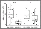
FIG. 4 is a schematic diagram of an exemplary study design showing a spatial analysis workflow to determine the density of LAG-3+ TIL and the proportion of LAG-3+ TIL for MHC II + or MHC II-tumor cells. The distance between LAG-3+ TIL and tumor cells was expressed as >30 μm or <30 μm. "LAG-3-D" is the density (D) of LAG-3+ TIL within 30 μm of MHC II + or MHC II-tumor cells. "LAG-3-P" is the proportion (P) of LAG-3+ TIL within 30 μm of MHC II + vs MHC II-tumor cells.
Figure 5 shows MHC II expression: (A) in IHC stained bladder tumor sample sections, compared to LAG-3, and (B) graphical representation of the percentage expression on bladder and stomach tumor cells.
FIG. 6 is a graph showing LAG-3-D (cells/mm) in bladder and stomach tumor samples as described in FIG. 42) To illustrate (a).
FIG. 7 is a graph showing LAG-3-P as depicted in FIG. 4 as a percentage (%) of conjugated LAG-3+ in bladder and stomach tumor samples.
Detailed Description
The present disclosure provides a method of treating cancer in a human subject in need thereof, comprising administering to the subject a lymphocyte activation gene-3 (LAG-3) antagonist, wherein the subject is identified as having (i) a high LAG-3 density (LAG-3-D) score, (ii) a high LAG-3 proportion (LAG-3-P) score, or (iii) both a high LAG-3-D score and a high LAG-3-P score, in a tumor sample obtained from the subject. The present disclosure also provides a method of treating cancer in a human subject in need thereof, comprising (a) identifying a subject having (i) a high LAG-3-D score, (ii) a high LAG-3-P score, or (iii) both a high LAG-3-D score and a high LAG-3-P score in a tumor sample obtained from the subject, and (b) administering a LAG-3antagonist to the subject. The present disclosure also provides a method of identifying a human subject having a cancer suitable for LAG-3antagonist therapy, comprising calculating (i) a LAG-3-D score, (ii) a LAG-3-P score, or (iii) both a LAG-3-D score and a LAG-3-P score in a tumor sample obtained from a subject in need of LAG-3antagonist therapy. The present disclosure also provides a LAG-3antagonist for treating cancer in a human subject in need thereof, wherein the subject is identified as having a tumor sample obtained from the subject with (i) a high LAG-3-D score, (ii) a high LAG-3-P score, or (iii) both a high LAG-3-D score and a high LAG-3-P score. The present disclosure also provides for use of a LAG-3antagonist for identifying a subject having a cancer suitable for LAG-3antagonist therapy, wherein (i) a LAG-3-D score, (ii) a LAG-3-P score, or (iii) both a LAG-3-D score and a LAG-3-P score are calculated in a tumor sample obtained from the subject.
I. Term(s) for
In order that this disclosure may be more readily understood, certain terms are first defined. As used herein, each of the following terms shall have the meaning set forth below, unless the context clearly dictates otherwise. Additional definitions are set forth throughout this application.
It should be noted that the terms "a" or "an" entity refer to one or more of that entity; for example, "a nucleotide sequence" is understood to mean one or more nucleotide sequences. Thus, the terms "a" (or "an"), "one or more" and "at least one" are used interchangeably herein.
The term "and/or" as used herein should be taken as specifically disclosing each of the two specified features or components, with or without the other. Thus, the term "and/or" as used herein in phrases such as "a and/or B" is intended to include "a and B", "a or B", "a" (alone) and "B" (alone). Also, the term "and/or" as used in phrases such as "A, B and/or C" is intended to encompass each of the following: A. b and C; A. b or C; a or C; a or B; b or C; a and C; a and B; b and C; a (alone); b (alone); and C (alone).
It should be understood that wherever aspects are described herein as "comprising," other similar aspects described as "consisting of … …" and/or "consisting essentially of … …" are also provided.
The terms "about" or "consisting essentially of refer to a value or composition within an acceptable error range for the particular value or composition as determined by one of ordinary skill in the art, which will depend in part on how the value or composition is measured or determined, i.e., the limitations of the measurement system. For example, "about" or "consisting essentially of can mean within 1 or greater than 1 standard deviation, according to practice in the art. Alternatively, "about" or "substantially comprising" may mean a range of up to 10% or 20% (i.e., ± 10% or ± 20%). For example, about 3mg may include any number between 2.7mg and 3.3mg (for 10%) or between 2.4mg and 3.6mg (for 20%). Furthermore, particularly with respect to biological systems or processes, the term may mean values up to an order of magnitude or up to 5-fold. Where a particular value or composition is provided in the application and claims, unless otherwise stated, the meaning of "about" or "consisting essentially of" should be considered to be within an acceptable error range for that particular value or composition.
As described herein, unless otherwise specified, any concentration range, percentage range, ratio range, or integer range is to be understood as including the value of any integer within the range, and where appropriate, including fractions thereof (e.g., tenths and hundredths of integers).
Unless defined otherwise, all technical and scientific terms used herein have the same meaning as commonly understood by one of ordinary skill in the art to which this disclosure pertains. For example, circumcise Dictionary of Biomedicine and Molecular Biology, Juo, Pei-Show, 2 nd edition, 2002, CRC Press; the Dictionary of Cell and Molecular Biology, 3 rd edition, 1999, Academic Press; and Oxford Dictionary of Biochemistry and Molecular Biology, amendment, 2000, Oxford University Press, provides the skilled artisan with a general Dictionary of many of the terms used in this disclosure.
Units, prefixes, and symbols are denoted in their international system of units (SI) accepted form. Numerical ranges include the numbers defining the range.
The headings provided herein are not limitations of the various aspects of the disclosure which can be had by reference to the specification as a whole. Accordingly, the terms defined immediately below are more fully defined by reference to the entire contents of the specification.
An "antagonist" shall include, but is not limited to, any molecule that is capable of blocking, reducing, or otherwise limiting the interaction or activity of a target molecule (e.g., LAG-3). In some aspects, the antagonist is an antibody. In other aspects, the antagonist comprises a small molecule. The terms "antagonist" and "inhibitor" are used interchangeably herein.
An "antibody" (Ab) shall include, without limitation, a glycoprotein immunoglobulin that specifically binds an antigen and comprises at least two heavy (H) chains and two light (L) chains interconnected by disulfide bonds, or an antigen-binding portion thereof. Each H chain comprises a heavy chain variable region (abbreviated herein as VH) and a heavy chain constant region. The heavy chain constant region comprises three constant domains CH1, CH2, and CH 3. Each light chain comprises a light chain variable region (abbreviated herein as VL) and a light chain constant region. The light chain constant region comprises a constant domain CL. The VH and VL regions can be further subdivided into hypervariable regions, termed Complementarity Determining Regions (CDRs), interspersed with regions that are more conserved, termed Framework Regions (FRs). Each VH and VL comprises three CDRs and four FRs, arranged from amino-terminus to carboxy-terminus in the following order: FR1, CDR1, FR2, CDR2, FR3, CDR3 and FR 4. The variable regions of the heavy and light chains contain binding domains that interact with antigens. The constant region of the antibody may mediate the binding of the immunoglobulin to host tissues or factors, including various cells of the immune system (e.g., effector cells) and the first component of the classical complement system (Clq). The heavy chain may or may not have a C-terminal lysine.
The immunoglobulin may be derived from any commonly known isotype, including, but not limited to, IgA, secretory IgA, IgG, and IgM. IgG subclasses are also well known to those skilled in the art and include, but are not limited to, human IgGl, IgG2, IgG3, and IgG 4. "isotype" refers to the antibody class or subclass (e.g., IgM or IgG1) encoded by the heavy chain constant region gene. By way of example, the term "antibody" includes naturally occurring and non-naturally occurring antibodies; monoclonal and polyclonal antibodies; chimeric and humanized antibodies; a human or non-human antibody; fully synthesizing an antibody; a single chain antibody; a monospecific antibody; a bispecific antibody; and multispecific antibodies. Non-human antibodies can be humanized by recombinant methods to reduce their immunogenicity in humans. Unless otherwise stated, and unless the context indicates otherwise, the term "antibody" also includes any of the immunoglobulins described aboveAntigen-binding fragments or antigen-binding portions of a immunoglobulin, and include monovalent and divalent fragments or portions that retain the ability to specifically bind to an antigen bound by the entire immunoglobulin. Examples of "antigen-binding portions" or "antigen-binding fragments" include: (1) fab fragments (fragments from papain cleavage) or similar monovalent fragments consisting of VL, VH, LC and CH1 domains; (2) f (ab')2A fragment (derived from pepsin cleavage) or a similar bivalent fragment comprising two Fab fragments linked by a disulfide bridge at the hinge region; (3) an Fd fragment consisting of the VH and CH1 domains; (4) (ii) an Fv fragment consisting of a single-armed VL and VH domain; (5) single domain antibody (dAb) fragments (Ward et al, (1989) Nature341:544-546), which consist of a VH domain; (6) a dual single domain antibody consisting of two VH domains connected by a hinge (dual affinity retargeting antibody (DART)); or (7) a dual variable domain immunoglobulin. Furthermore, although the two domains of the Fv fragment, VL and VH, are encoded by separate genes, they can be joined using recombinant methods by a synthetic linker that enables them to be made into a single protein chain in which the VL and VH regions pair to form monovalent molecules (known as single chain Fv (scFv); see, e.g., Bird et al (1988) Science 242: 423-. These antigen-binding portions or fragments are obtained using conventional techniques known to those skilled in the art and screened for utility in the same manner as whole antibodies. Antigen-binding portions or fragments may be produced by recombinant DNA techniques or by enzymatic or chemical cleavage of intact immunoglobulins.
An "isolated antibody" refers to an antibody that is substantially free of other antibodies having different antigenic specificities (e.g., an isolated antibody that specifically binds LAG-3 is substantially free of antibodies that specifically bind antigens other than LAG-3). However, an isolated antibody that specifically binds LAG-3 may have cross-reactivity with other antigens (e.g., LAG-3 molecules from different species). Furthermore, the isolated antibody may be substantially free of other cellular material and/or chemicals.
The term "monoclonal antibody" (mAh) refers to a non-naturally occurring preparation of antibody molecules of a single molecular composition, i.e., antibody molecules whose primary sequences are substantially identical and which exhibit a single binding specificity and affinity for a particular epitope. Monoclonal antibodies are examples of isolated antibodies. Monoclonal antibodies can be produced by hybridomas, recombinant, transgenic, or other techniques known to those skilled in the art.
"human antibodies" (HuMAb) refer to antibodies having variable regions in which both the framework and CDR regions are derived from human germline immunoglobulin sequences. Furthermore, if the antibody contains constant regions, the constant regions are also derived from human germline immunoglobulin sequences. The human antibodies of the present disclosure may include amino acid residues not encoded by human germline immunoglobulin sequences (e.g., mutations introduced by random or site-specific mutagenesis in vitro or by somatic mutation in vivo). However, as used herein, the term "human antibody" is not intended to include antibodies in which CDR sequences of an antibody derived from the germline of another mammalian species (e.g., a mouse) have been grafted onto human framework sequences. The terms "human antibody" and "fully human antibody" are used synonymously.
"humanized antibody" refers to an antibody in which some, most, or all of the amino acids other than the CDRs of a non-human antibody are replaced with corresponding amino acids derived from a human immunoglobulin. In one aspect of a humanized form of an antibody, some, most, or all of the amino acids outside of the CDRs have been replaced with amino acids from a human immunoglobulin, while some, most, or all of the amino acids within one or more CDRs are unchanged. Small additions, deletions, insertions, substitutions or modifications of amino acids are permissible as long as they do not abrogate the ability of the antibody to bind to a particular antigen. "humanized antibodies" retain antigen specificity similar to the original antibody.
"chimeric antibody" refers to an antibody in which the variable regions are derived from one species and the constant regions are derived from another species, such as an antibody in which the variable regions are derived from a mouse antibody and the constant regions are derived from a human antibody.
An "anti-antigen antibody" refers to an antibody that specifically binds to an antigen. For example, an anti-LAG-3 antibody specifically binds LAG-3, an anti-PD-1 antibody specifically binds PD-1, an anti-PD-L1 antibody specifically binds PD-L1, and an anti-CTLA-4 antibody specifically binds CTLA-4.
"LAG-3" refers to lymphocyte activation gene-3. The term "LAG-3" includes variants, isoforms, homologs, orthologs, and paralogs. For example, in certain instances, an antibody specific for a human LAG-3 protein may cross-react with LAG-3 proteins from species other than human. In other aspects, an antibody specific for human LAG-3 protein may be completely specific for human LAG-3 protein and does not exhibit species or other types of cross-reactivity, or may cross-react with LAG-3 from some other species but not all others (e.g., cross-react with monkey LAG-3 instead of mouse LAG-3). The term "human LAG-3" refers to the complete amino acid sequence of human sequence LAG-3, such as human LAG-3 having GenBank accession No. NP _ 002277. The term "mouse LAG-3" refers to the complete amino acid sequence of a mouse sequence LAG-3, such as mouse LAG-3 having GenBank accession No. NP _ 032505. LAG-3 is also known in the art as, for example, CD 223. The human LAG-3 sequence may differ from human LAG-3 of GenBank accession No. NP _002277 by having, for example, a conservative mutation or a mutation in a non-conserved region, and LAG-3 has substantially the same biological function as human LAG-3 of GenBank accession No. NP _ 002277. For example, the biological function of human LAG-3 is to have an epitope in the extracellular domain of LAG-3 that is specifically bound by an antibody of the present disclosure, or the biological function of human LAG-3 is to bind MHC class II molecules.
"programmed death-1" (PD-1) refers to an immunosuppressive receptor belonging to the CD28 family. PD-1 is expressed in vivo predominantly on previously activated T cells and binds to two ligands, PD-L1 and PD-L2. The term "PD-1" as used herein includes variants, isoforms, and species homologs of human PD-1(hPD-1), hPD-1, and analogs having at least one common epitope with hPD-1. The complete hPD-1 sequence can be found under GenBank accession No. U64863. "PD-1" and "PD-1 receptor" are used interchangeably herein.
"programmed death ligand-1" (PD-L1) is one of two cell surface glycoprotein ligands of PD-1 (the other being PD-L2) that down-regulates T cell activation and cytokine secretion upon binding to PD-1. The term "PD-L1" as used herein includes variants, isoforms and species homologs of human PD-L1(hPD-L1), hPD-L1, and analogs having at least one common epitope with hPD-L1. The complete hPD-L1 sequence can be found under GenBank accession No. Q9NZQ7. The human PD-L1 protein is encoded by the human CD274 gene (NCBI gene ID: 29126).
The terms "programmed death ligand-2" and "PD-L2" as used herein include variants, isoforms and species homologs of human PD-L2(hPD-L2), hPD-L2, and analogs having at least one common epitope with hPD-L2. The complete hPD-L2 sequence can be found under GenBank accession No. Q9BQ51.
"cytotoxic T-lymphocyte antigen-4" (CTLA-4) refers to an immunosuppressive receptor belonging to the CD28 family. CTLA-4 is exclusively expressed on T cells in vivo and binds to two ligands, CD80 and CD86 (also referred to as B7-1 and B7-2, respectively). The term "CTLA-4" as used herein includes human CTLA-4(hCTLA-4), variants, isoforms and species homologs of hCTLA-4, and analogs having at least one common epitope with hCTLA-4. The complete hCTLA-4 sequence can be found under GenBank accession No. aab59385.
"T cell immunoglobulin and mucin domain-3" (TIM-3), also known as hepatitis a virus cell receptor 2(HAVCR2), refers to a type I transmembrane protein that was originally identified on activated IFN- γ producing T cells (e.g., type 1 helper CD4+ T cells and cytotoxic CD8+ T cells) and was shown to induce T cell death or depletion upon binding to galectin-9 (galectin-9). The term "TIM-3" as used herein includes human TIM-3(hTIM-3), variants, isoforms and species homologs of hTIM-3, and analogs having at least one common epitope with hTIM-3. Two isoforms of hTIM-3 have been identified. Isoform 1(GenBank accession No. np _116171) consists of 301 amino acids and represents the canonical sequence. Isoform 2(GenBank accession No. aah20843) consists of 142 amino acids and is soluble.
The term "LAG-3 positive" or "LAG-3 expression positive" in relation to LAG-3 expression refers to the proportion of cells in a test tissue sample comprising tumor cells and tumor-infiltrating inflammatory cells above which the tissue sample is scored as expressing LAG-3.
"LAG-3 negative" or "LAG-3 expression negative" refers to the proportion of cells that are not LAG-3 positive or LAG-3 expression positive in a test tissue sample comprising tumor cells and tumor-infiltrating inflammatory cells.
The term "PD-L1 positive" or "PD-L1 expression positive" in relation to cell surface PD-L1 expression refers to the proportion of cells in a test tissue sample comprising tumor cells and tumor-infiltrating inflammatory cells above which the sample is scored as expressing cell surface PD-L1.
The terms "PD-L1 negative" or "PD-L1 expression negative" in relation to cell surface PD-L1 expression refer to the proportion of cells in a test tissue sample comprising tumor cells and tumor-infiltrating inflammatory cells that are not PD-L1 positive or PD-L1 expression positive.
The term "tumor mutation burden" (TMB) as used herein refers to the number of somatic mutations in a tumor genome and/or the number of somatic mutations per unit area of a tumor genome. In determining TMB, germline (genetic) variants are excluded because the immune system has a higher probability of recognizing these as self. Tumor Mutation Burden (TMB) may also be used interchangeably with "tumor mutation burden", or "tumor mutation burden".
"subject" includes any human or non-human animal. The term "non-human animal" includes, but is not limited to, vertebrates, such as non-human primates, sheep, dogs, and rodents, such as mice, rats, and guinea pigs. In a preferred aspect, the subject is a human. The terms "subject" and "patient" are used interchangeably herein.
By "administering" is meant physically introducing a composition comprising a therapeutic agent into a subject using any of a variety of methods and delivery systems known to those skilled in the art. Preferred routes of administration for immunotherapy (e.g., LAG-3 antagonists) include intravenous, intramuscular, subcutaneous, intraperitoneal, spinal, or other parenteral routes of administration, e.g., by injection or infusion. The phrase "parenteral administration" as used herein means modes of administration other than enteral and topical administration, typically by injection, and includes, but is not limited to, intravenous, intramuscular, intraarterial, intrathecal, intralymphatic, intralesional, intracapsular, intraorbital, intracardiac, intradermal, intraperitoneal, transtracheal, subcutaneous, subcuticular, intraarticular, subcapsular, subarachnoid, intraspinal, epidural, and intrasternal injection and infusion, and in vivo electroporation. In some aspects, the formulation is administered by a non-parenteral route, and in some aspects, by oral administration. Other non-parenteral routes include topical, epidermal or mucosal routes of administration, such as intranasal, vaginal, rectal, sublingual or topical. Administration may also be performed, for example, once, multiple times, and/or over one or more extended periods of time.
"treatment" or "therapy" of a subject refers to any type of intervention or process performed on the subject, or the administration of an active agent to a subject, with the purpose of reversing, alleviating, ameliorating, inhibiting, slowing the progression, severity, or recurrence of symptoms, complications, or conditions, or biochemical indicators associated with the disease. The solid tumor Response Evaluation Criteria (RECIST) is a measure of the efficacy of a treatment and is an established rule that determines when a tumor responds, stabilizes or progresses during treatment. RECIST 1.1 is a general guideline for the measurement and definition of solid tumors for the objective assessment of tumor size changes in clinical trials of adult and pediatric cancers.
As used herein, "effective treatment" refers to a treatment that produces a beneficial effect, such as ameliorating at least one symptom of a disease or disorder. The beneficial effect may take the form of an improvement over baseline, i.e., an improvement over a measurement or observation made prior to initiation of treatment according to the method. The beneficial effects may also take the form of preventing, slowing, arresting or stabilizing the detrimental progression of solid tumor markers. Effective treatment may refer to alleviating at least one symptom of a solid tumor. Such effective treatment may, for example, reduce pain in the patient, reduce the size and/or number of lesions, may reduce or prevent tumor metastasis, and/or may slow tumor growth.
The term "effective amount" refers to an amount of an agent that provides the desired biological, therapeutic and/or prophylactic result. The result can be a reduction, amelioration, palliation, alleviation, delay and/or remission of one or more signs, symptoms or causes of disease, or any other desired alteration of a biological system. With respect to solid tumors, an effective amount includes an amount sufficient to cause tumor shrinkage and/or to reduce the growth rate of the tumor (e.g., inhibit tumor growth) or to delay other unwanted cell proliferation. In some aspects, an effective amount is an amount sufficient to prevent or delay tumor recurrence. An effective amount may be administered in one or more administrations. The effective amount of the drug or composition may be: (i) reducing the number of cancer cells; (ii) reducing tumor size; (iii) inhibit, delay, slow and may prevent cancer cell infiltration into peripheral organs to some extent; (iv) in one example, an "effective amount" is an amount of anti-LAG-3 antibody alone or in combination with an amount of additional therapeutic agent (e.g., an anti-PD-1 antibody) that clinically demonstrates an effect on the cancer (e.g., advanced solid tumor) that significantly reduces or delays cancer progression.
As used herein, the terms "fixed dose", "flat dose", and "flat-fixed dose" are used interchangeably and refer to a dose that is administered to a patient without regard to the patient's weight or Body Surface Area (BSA). Thus, a fixed or flat dose is not provided in a mg/kg dose, but rather in an absolute amount of the agent (e.g., in μ g or mg).
The use of the term "fixed dose combination" in reference to a composition of the invention means that two or more different inhibitors as described herein (e.g. an anti-LAG-3 antibody and an anti-PD-1 antibody) are present in the composition in a specific (fixed) ratio to each other in a single composition. In some aspects, the fixed dose is based on the weight (e.g., mg) of the inhibitor. In certain aspects, the fixed dose is based on the concentration of the inhibitor (e.g., mg/ml). In some aspects, the ratio is at least about 1:1, about 1:2, about 1:3, about 1:4, about 1:5, about 1:6, about 1:7, about 1:8, about 1:9, about 1:10, about 1:15, about 1:20, about 1:30, about 1:40, about 1:50, about 1:60, about 1:70, about 1:80, about 1:90, about 1:100, about 1:120, about 1:140, about 1:160, about 1:180, about 1:200, about 200:1, about 180:1, about 160:1, about 140:1, about 120:1, about 100:1, about 90:1, about 80:1, about 70:1, about 60:1, about 50:1, about 40:1, about 30:1, about 20: 1. about 5:1, about 4:1, about 3:1, or about 2: 1mg of the first inhibitor to mg of the second inhibitor. For example, a 3:1 ratio of primary and secondary antibodies may mean that the vial may contain about 240mg of primary and 80mg of secondary antibodies or about 3mg/ml of primary and 1mg/ml of secondary antibodies.
The term "weight-based dose" as referred to herein refers to a dose that is administered to a patient calculated based on the patient's weight. For example, where a patient having a weight of 60kg requires 3mg/kg of anti-LAG-3 antibody to bind 3mg/kg of anti-PD-1 antibody, appropriate amounts of anti-LAG-3 antibody (i.e., 180mg) and anti-PD-1 antibody (i.e., 180mg) can be drawn at once from a fixed dose combination of a 1:1 ratio of anti-LAG 3 antibody and anti-PD-1 antibody.
As used herein, "dosing interval" refers to the amount of time that elapses between multiple doses of a formulation disclosed herein administered to a subject. Thus, the dosing interval may be expressed as a range.
The term "dosing frequency" as used herein refers to the frequency with which a dose of a formulation disclosed herein is administered over a given period of time. The frequency of administration may be expressed as the number of doses per given time, e.g., once per week or once every two weeks, etc.
As used herein, the terms "about once a week," "about once every two weeks," or any other similar dosing interval term refer to an approximate number, and "about once a week" or "about once a week" may include every seven days ± two days, i.e., every five days to every nine days. Thus, the frequency of administration "once weekly" may be once every five days, six days, seven days, eight days, or nine days. "about once every three weeks" may include every 21 days ± 3 days, i.e. every 25 days to every 31 days. Similar approximations apply, for example, about once every two weeks, about once every four weeks, about once every five weeks, about once every six weeks, about once every seven weeks, about once every eight weeks, about once every nine weeks, about once every ten weeks, about once every eleven weeks, and about once every twelve weeks. In some aspects, a dosing interval of about once every six weeks or about once every twelve weeks means that a first dose can be administered on any day of the first week, and then the next dose can be administered on any day of the sixth or twelfth weeks, respectively. In other aspects, a dosing interval of about once every six weeks or about once every twelve weeks means that a first dose is administered on a particular day of the first week (e.g., monday) and then the next dose is administered on the same day of the sixth or twelfth week (i.e., monday), respectively.
As used herein, an "adverse event" (AE) is any adverse and often unexpected or undesirable sign (including abnormal laboratory findings), symptom or disease associated with the use of medical treatment. For example, an adverse event can be associated with activation of the immune system or expansion of cells of the immune system (e.g., T cells) in response to a treatment. A medical treatment may have one or more related AEs, and each AE may have the same or different severity. Reference to a method capable of "altering an adverse event" means a treatment regimen that reduces the incidence and/or severity of one or more AEs associated with the use of a different treatment regimen.
The term "tumor" as used herein refers to any tissue mass resulting from excessive cell growth or proliferation, either benign (non-cancerous) or malignant (cancerous), including precancerous lesions.
For example, an "anti-cancer agent" promotes cancer regression in a subject. In a preferred aspect, the therapeutically effective amount of the drug promotes regression of the cancer to the extent that the cancer is eliminated. By "promoting cancer regression" is meant that administration of an effective amount of an anti-cancer agent, alone or in combination with another agent, results in a reduction in tumor growth or size, tumor necrosis, a reduction in the severity of at least one disease symptom, an increase in the frequency and duration of the asymptomatic phase of the disease, or prevention of injury or disability due to the affliction of the disease. Furthermore, the terms "effective" and "effectiveness" with respect to treatment include pharmacological effectiveness and physiological safety. Pharmacological efficacy refers to the ability of a drug to promote cancer regression in a patient. Physiological safety refers to the level of toxicity or other adverse physiological effects (adverse effects) at the cellular, organ, and/or organism level caused by administration of an agent.
As an example of treating a tumor, a therapeutically effective amount of an anti-cancer agent preferably inhibits cell growth or tumor growth by at least about 20%, at least about 40%, at least about 60%, or at least about 80% relative to an untreated subject. In other aspects of the disclosure, tumor regression may be observed for a period of at least about 20 days, at least about 40 days, or at least about 60 days. Despite these final measures of therapeutic efficacy, the evaluation of immunotherapeutic drugs must also take into account immune-related response patterns.
As used herein, "immunooncology" therapy or "I-O" or "IO" therapy refers to therapies that include targeting and treating a tumor in a subject with an immune response. Thus, as used herein, I-O therapy is a class of anti-cancer therapies. In some aspects, the I-O therapy comprises administering to the subject an antibody or antigen-binding fragment thereof. In some aspects, I-O therapy includes administering to a subject an immune cell, e.g., a T cell, e.g., a modified T cell, e.g., a T cell modified to express a chimeric antigen receptor or a specific T cell receptor. In some aspects, the I-O therapy comprises administering a therapeutic vaccine to the subject. In some aspects, the I-O therapy comprises administering a cytokine or chemokine to the subject. In some aspects, the I-O therapy comprises administering an interleukin to the subject. In some aspects, the I-O therapy comprises administering interferon to the subject. In some aspects, the I-O therapy comprises administering colony stimulating factor to the subject.
By "immune response" is meant the action of cells of the immune system (e.g., T lymphocytes, B lymphocytes, Natural Killer (NK) cells, macrophages, eosinophils, mast cells, dendritic cells, and neutrophils) and soluble macromolecules produced by any of these cells or the liver, including antibodies, cytokines, and complements, which results in the selective targeting, binding, damage, destruction, and/or elimination of invading pathogens, pathogen-infected cells or tissues, cancer cells or other abnormal cells in vertebrates, or (in the case of autoimmunity or pathological inflammation) normal human cells or tissues.
A "tumor-infiltrating inflammatory cell" or "tumor-associated inflammatory cell" is any type of cell that is normally involved in an inflammatory response in a subject and infiltrates tumor tissue. Such cells include Tumor Infiltrating Lymphocytes (TILs), macrophages, monocytes, eosinophils, tissue cells and dendritic cells.
The term "tumor sample" as used herein refers to tumor material isolated from a tumor of a subject. The tumor sample may contain any portion of the tumor suitable for determining target protein expression (e.g., LAG-3, MHC class II, and/or tumor antigen), e.g., by Immunohistochemistry (IHC). In one aspect, the tumor sample is a tumor tissue biopsy, such as Formalin Fixed Paraffin Embedded (FFPE) tumor tissue or freshly frozen tumor tissue or the like. In another aspect, a tumor sample can be cut into multiple tumor sections. In other aspects, the tumor sample is serially sectioned into multiple sections.
Various aspects of the disclosure are described in further detail in the following subsections.
Methods of the present disclosure
Provided herein are methods of increasing the efficacy of LAG-3antagonist therapy by identifying or selecting human subjects or populations of human subjects suitable for or responsive to LAG-3antagonist therapy. The methods provided herein relate to quantitative spatial profiling of human subjects suitable for or responsive to LAG-3antagonist therapy. In one aspect, the disclosure relates to a method of treating cancer (e.g., reducing the volume and/or growth of a tumor) in a human subject in need thereof, comprising administering to the subject a lymphocyte activating gene-3 (LAG-3) antagonist, wherein the subject is identified as having (i) a high LAG-3 density (LAG-3-D) score, (ii) a high LAG-3 proportion (LAG-3-P) score, or (iii) both a high LAG-3-D score and a high LAG-3-P score, in a tumor sample obtained from the subject.
In another aspect, the present disclosure relates to a method of treating cancer in a human subject in need thereof, comprising: (a) identifying a subject having (i) a high LAG-3-D score, (ii) a high LAG-3-P score, or (iii) both a high LAG-3-D score and a high LAG-3-P score in a tumor sample obtained from the subject, and (b) administering a LAG-3antagonist to the subject.
In another aspect, the disclosure relates to a method of identifying or selecting a human subject having a cancer suitable for LAG-3antagonist therapy, the method comprising calculating (i) a LAG-3-D score, (ii) a LAG-3-P score, or (iii) both a LAG-3-D score and a LAG-3-P score in a tumor sample obtained from a subject in need of LAG-3antagonist therapy. In some aspects, the subject exhibits a high LAG-3-D score, a high LAG-3-P score, or both a high LAG-3-D score and a high LAG-3-P score. In some aspects, the method further comprises administering to the subject a LAG-3 antagonist. In some aspects, a subject identified or selected by the present methods is responsive to LAG-3antagonist therapy.
In another aspect, the disclosure relates to a LAG-3antagonist for use in treating cancer in a human subject in need thereof, wherein the subject is identified as having (i) a high LAG-3-D score, (ii) a high LAG-3-P score, or (iii) both a high LAG-3-D score and a high LAG-3-P score in a tumor sample obtained from the subject.
In another aspect, the disclosure relates to a LAG-3antagonist for use in identifying or selecting a subject having a cancer suitable for LAG-3antagonist therapy, wherein (i) a LAG-3-D score, (ii) a LAG-3-P score, or (iii) both the LAG-3-D score and the LAG-3-P score are calculated in a tumor sample obtained from the subject. In some aspects, the subject exhibits a high LAG-3-D score, a high LAG-3-P score, or both a high LAG-3-D score and a high LAG-3-P score. In some aspects, a subject identified or selected by the present methods is responsive to LAG-3antagonist therapy.
LAG-3-D and LAG-3-P scores
LAG-3-D scores according to the present disclosure may be determined by measuring the density of LAG-3 expressing T cells in proximity to one or more tumor cells expressing the major histocompatibility complex class II (MHC II) in a tumor sample. In some aspects, the LAG-3-D score is calculated as (i) the number of LAG-3 expressing T cells in proximity to MHC II expressing tumor cells divided by (II) the tumor area (mm) of the tumor sample2)。
LAG-3-P scores according to the present disclosure can be determined by measuring the proportion of LAG-3 expressing T cells in proximity to MHC II expressing one or more tumor cells in a tumor sample. In some aspects, the LAG-3-P score is calculated as (i) the number of LAG-3 expressing T cells in proximity to MHC II expressing tumor cells divided by (II) the total number of LAG-3 expressing T cells in the tumor sample.
In some aspects, the proximity is between the cell surface of LAG-3 expressing T cells and the cell surface of MHC II expressing tumor cells.
In some aspects, the proximity is between the nucleus of a LAG-3 expressing T cell and the nucleus of a MHC II expressing tumor cell.
In some aspects, the proximity is between LAG-3 and MHC class II and/or between LAG-3 and a tumor antigen expressed on a tumor cell.
In some aspects, the proximity is equal to or less than about 50 μm, equal to or less than about 45 μm, equal to or less than about 40 μm, equal to or less than about 35 μm, or equal to or less than about 30 μm. In some aspects, the proximity is equal to or less than about 50 μm. In some aspects, the proximity is equal to or less than about 40 μm. In some aspects, the proximity is equal to or less than about 35 μm. In some aspects, the proximity is equal to or less than about 30 μm.
In some aspects, the tumor sample comprises one or more tumor sections derived from a tumor tissue biopsy or tumor tissue resection. In some aspects, the one or more tumor sections comprise formalin-fixed paraffin-embedded tumor tissue or freshly frozen tumor tissue. In some aspects, the one or more tumor slices comprise serially sliced tumor slices. In some aspects, one or more tumor sections are stained by Immunohistochemistry (IHC). In some aspects, the one or more tumor slices comprise one tumor slice, two tumor slices, three tumor slices, four tumor slices, five tumor slices, six tumor slices, seven tumor slices, eight tumor slices, nine tumor slices, ten tumor slices, 11 tumor slices, 12 tumor slices, 13 tumor slices, 14 tumor slices, 15 tumor slices, 16 tumor slices, 17 tumor slices, 18 tumor slices, 19 tumor slices, 20 tumor slices, 21 tumor slices, 22 tumor slices, 23 tumor slices, 24 tumor slices, 25 tumor slices, 26 tumor slices, 27 tumor slices, 28 tumor slices, 29 tumor slices, or 30 tumor slices. In some aspects, the one or more tumor slices comprise three tumor slices. In some aspects, the one or more tumor slices comprise 15 tumor slices. In some aspects, the one or more tumor slices comprise 18 tumor slices. In some aspects, the one or more tumor slices comprise 20 tumor slices. In some aspects, the one or more tumor slices comprise 21 tumor slices.
In some aspects, one tumor section of the tumor sample is stained for LAG-3 and/or MHC II. In some aspects, the tumor section is further stained for a tumor antigen. In some aspects, the tumor antigen is a Pan-CK. In some aspects, one tumor section of the tumor sample is stained for all three markers, i.e., LAG-3, MHC class II, and tumor antigen (e.g., pan-CK).
In some aspects, the tumor sample comprises a first tumor section stained for LAG-3, a second tumor section stained for MHC II, and a third tumor section stained for a tumor antigen. In some aspects, a first tumor section, a second tumor section, and a third tumor section are serially sectioned from a tumor sample. In some aspects, the tumor sample comprises a first set of tumor slices (e.g., 2,3, 4,5, 6, 7, 8,9, or 10 tumor slices) stained for LAG-3, a second set of tumor slices (e.g., 2,3, 4,5, 6, 7, 8,9, or 10 tumor slices) stained for MHC II, and a third set of tumor slices (e.g., 2,3, 4,5, 6, 7, 8,9, or 10 tumor slices) stained for tumor antigens. In some aspects, the first, second and third sets of tumor sections are serially sectioned from the tumor sample.
In some aspects, the LAG-3-D score and the LAG-3-P score are determined by quantitative spatial profiling. In some aspects, the quantitative spatial profiling is a digital spatial analysis.
In some aspects, a high LAG-3-D score is at least about 5 cells/mm2At least about 6 cells/mm2At least about 7 cells/mm2At least about 8 cells/mm2At least about 9 cells/mm2At least about 10 cells/mm2At least about 11 cells/mm2At least about 12 cells/mm2At least about 13 cells/mm2At least about 14 cells/mm2At least about 15 cells/mm2At least about 16 cells/mm2At least about 17 cells/mm2At least about 18 cells/mm2At least about 19 cells/mm2At least about 20 cells/mm2At least about 25 cells/mm2At least about 30 cells/mm2At least about 35 cells/mm2At least about 40 cells/mm2At least about 45 cells/mm2At least about 50 cells/mm2At least about 55 cells/mm2At least about 60 cells/mm2At least about 65 cells/mm2At least about 70 cells/mm2At least about 75 cells/mm2At least about 80 cells/mm2At least about 85 cells/mm2At least about 90 cells/mm2At least about 95 cells/mm2Or at least about 100 cells/mm2. In some aspects, a high LAG-3-D score of at least about 5 cells/mm2. In some aspects, a high LAG-3-D score of at least about 10 cells/mm2. In some aspects, a high LAG-3-D score of at least about 15 cells/mm2. In some aspects, a high LAG-3-D score of at least about 20 cells/mm2. In some aspects, a high LAG-3-D score is at least about 25 cells/mm 2. In some aspects, a high LAG-3-D score of at least about 30 cells/mm2. In some aspects, a high LAG-3-D score of at least about 35 cells/mm2. In some aspects, a high LAG-3-D score of at least about 40 cells/mm2. In some aspects, a high LAG-3-D score of at least about 45 cells/mm2. In some aspectsMedium, high LAG-3-D score of at least about 50 cells/mm2In some aspects, a high LAG-3-D score of at least about 55 cells/mm2. In some aspects, a high LAG-3-D score of at least about 60 cells/mm2. In some aspects, a high LAG-3-D score of at least about 65 cells/mm2. In some aspects, a high LAG-3-D score of at least about 70 cells/mm2. In some aspects, a high LAG-3-D score of at least about 75 cells/mm2. In some aspects, a high LAG-3-D score of at least about 80 cells/mm2. In some aspects, a high LAG-3-D score of at least about 85 cells/mm2. In some aspects, a high LAG-3-D score of at least about 90 cells/mm2. In some aspects, a high LAG-3-D score of at least about 95 cells/mm2. In some aspects, a high LAG-3-D score of at least about 100 cells/mm2。
In some aspects, a high LAG-3-P score is at least about 40%, at least about 45%, at least about 50%, at least about 55%, at least about 60%, at least about 65%, at least about 70%, at least about 75%, at least about 80%, at least about 85%, at least about 90%, at least about 95%, or at least about 100%. In some aspects, the high LAG-3-P score is between at least about 40% and about 100%. In some aspects, the high LAG-3-P score is at least about 40%. In some aspects, the high LAG-3-P score is at least about 45%. In some aspects, the high LAG-3-P score is at least about 50%. In some aspects, the high LAG-3-P score is at least about 55%. In some aspects, the high LAG-3-P score is at least about 60%. In some aspects, the high LAG-3-P score is at least about 65%. In some aspects, the high LAG-3-P score is at least about 70%. In some aspects, the high LAG-3-P score is at least about 75%. In some aspects, the high LAG-3-P score is at least about 80%. In some aspects, the high LAG-3-P score is at least about 85%. In some aspects, the high LAG-3-P score is at least about 90%. In some aspects, the high LAG-3-P score is at least about 95%. In some aspects, the high LAG-3-P score is at least about 100%. In some aspects, the high LAG-3-P score is between at least about 40% and about 100%. In some aspects, the high LAG-3-P score is between at least about 50% and about 100%. In some aspects, the high LAG-3-P score is between at least about 60% and about 100%. In some aspects, the high LAG-3-P score is between at least about 70% and about 100%. In some aspects, the high LAG-3-P score is between at least about 80% and about 100%. In some aspects, the high LAG-3-P score is between at least about 90% and about 100%.
LAG-3 antagonists and combination therapies
In some aspects, the disclosure relates to methods and uses of treating cancer in a human subject in need thereof comprising administering to the subject a LAG-3 antagonist. In some aspects, the LAG-3antagonist monotherapy is administered to the subject, e.g., wherein the subject is not administered one or more additional therapeutic agents (e.g., anti-cancer agents).
In some aspects, a combination therapy is administered to the subject, e.g., wherein the subject is administered a LAG-3antagonist and one or more additional therapeutic agents (e.g., anti-cancer agents).
In some aspects, the methods and uses for treating cancer further comprise administering to the subject an additional checkpoint inhibitor. In some aspects, the checkpoint inhibitor comprises an anti-PD-1 antibody, an anti-CTLA-4 antibody, an anti-TIM 3 antibody, an anti-PD-L1 antibody, or any combination thereof. In some aspects, the checkpoint inhibitor comprises an anti-PD-1 antibody. In some aspects, the checkpoint inhibitor comprises an anti-PD-L1 antibody.
II.B.1.LAG-3 antagonists
As used herein, LAG-3 antagonists include, but are not limited to, LAG-3 binding agents, such as LAG-3 antibodies, and soluble LAG-3 polypeptides, such as fusion proteins comprising the extracellular portion of LAG-3. The term "LAG-3 antagonist" as used herein is interchangeable with the term "LAG-3 inhibitor".
In some aspects, the LAG-3antagonist is a soluble LAG-3 polypeptide. In some aspects, the soluble LAG-3 polypeptide is a fusion polypeptide, e.g., a fusion protein comprising the extracellular portion of LAG-3. In some aspects, the soluble LAG-3 polypeptide is a LAG-3-Fc fusion polypeptide capable of binding MHC class II. In some aspects, the soluble LAG-3 polypeptide comprises a ligand-binding fragment of the LAG-3 extracellular domain. In some aspects, the soluble LAG-3 polypeptide further comprises a half-life extending moiety. In some aspects, the half-life extending moiety comprises an immunoglobulin constant region or portion thereof, an immunoglobulin-binding polypeptide, an immunoglobulin g (igg), an albumin-binding polypeptide (ABP), a PAS moiety, a HES moiety, an XTEN, a pegylation moiety, an Fc region, or any combination thereof. In some aspects, the soluble LAG-3 polypeptide is IMP321 (fetilagimod alpha). See, e.g., Brignone c. et al, J immunol.2013, 12 months; (2007) (ii) a 179: 4202-4211 and WO 2009/044273.
In some aspects, the LAG-3antagonist is an anti-LAG-3 antibody.
anti-LAG-3 antibodies (or VH/VL domains derived therefrom) suitable for use herein can be generated using methods well known in the art. Alternatively, art-recognized anti-LAG-3 antibodies may be used. Antibodies that bind LAG-3 have been disclosed, for example, in international publication nos. WO/2015/042246; and U.S. publication nos. 2014/0093511 and 2011/0150892, each of which is incorporated by reference herein in its entirety.
An exemplary LAG-3 antibody useful in the present disclosure is 25F7 (described in U.S. publication No. 2011/0150892). Another exemplary LAG-3 antibody that may be used in the present disclosure is BMS-986016 (relatlimab). In some aspects, anti-LAG-3 antibodies useful in the present disclosure cross-compete with 25F7 or BMS-986016(relatlimab) for binding to human LAG-3. In some aspects, anti-LAG-3 antibodies useful in the present disclosure bind the same epitope as 25F7 or BMS-986016 (relatlimab).
Other art-recognized anti-LAG-3 antibodies that may be used in the methods and uses of the present disclosure include IMP731(H5L7BW) as described in US 2011/007023, MK-4280(28G-10) as described in WO2016028672, Burova E, et al, j.immunother.2014; cancer (2016); supplement 1) REGN3767(fianlimab) as described in P195, humanized BAP050 as described in WO2017/019894, GSK2831781, IMP-701 (LAG-525; ieremimab), aLAG3(0414), aLAG3(0416), Sym022, TSR-033, TSR-075, XmAb22841, MGD013, BI754111, FS118, P13B 02-30, AVA-017 and AGEN 1746. These and other anti-LAG-3 antibodies useful in the claimed invention can be found, for example, in: US 10,188,730; WO 2016/028672, WO 2017/106129, WO2017/062888, WO2009/044273, WO2016/069500, WO2016/126858, WO2014/179664, WO2016/200782, WO2015/200119, WO2017/019846, WO2017/198741, WO2017/220555, WO2017/220569, WO2018/071500, WO2017/015560, WO2017/025498, WO2017/087589, WO2017/087901, WO2018/083087, WO2017/149143, WO2017/219995, US2017/0260271, WO2017/086367, WO2017/086419, WO2018/034227, WO2018/185046, WO2018/185043, WO2018/217940, WO19/011306, WO2018/208868, WO 2018/140180, WO2018/201096, WO 2018/2015826 and WO 018730/204374. The contents of each of these references are incorporated by reference in their entirety.
anti-LAG-3 antibodies useful in the methods and uses of the present disclosure also include isolated antibodies that specifically bind human LAG-3 and cross-compete with any of the anti-LAG-3 antibodies disclosed herein (e.g., relatilimab) for binding to human LAG-3. In some aspects, the anti-LAG-3 antibody binds the same epitope as any anti-LAG-3 antibody described herein (e.g., relatlimab).
In some aspects, an antibody that cross-competes for binding to human LAG-3 with any anti-LAG-3 antibody disclosed herein (e.g., relatlmab) or binds to the same epitope region as any anti-LAG-3 antibody disclosed herein (e.g., relatlmab) is a monoclonal antibody. For administration to a human subject, these cross-competing antibodies are chimeric, engineered, or humanized or human antibodies. Such chimeric, engineered, humanized or human monoclonal antibodies can be prepared and isolated by methods well known in the art.
The ability of an antibody to cross-compete for binding to an antigen indicates that the antibody binds to the same epitope region of the antigen and sterically blocks other cross-competing antibodies from binding to that particular epitope region. These cross-competing antibodies are expected to have functional properties very similar to the reference antibody (e.g., relatlimab) due to their binding to the same epitope region. Cross-competing antibodies can be readily identified based on their ability to cross-compete in standard binding assays, such as Biacore analysis, ELISA assays, or flow cytometry (see, e.g., WO 2013/173223).
anti-LAG-3 antibodies useful in the methods and uses of the present disclosure also include antigen-binding portions of any of the above full-length antibodies. It is well established that the antigen binding function of an antibody can be performed by fragments of a full-length antibody.
The biosimics of any of the anti-LAG-3 antibodies disclosed herein may also be used in the methods and uses of the present disclosure.
In other aspects, the anti-LAG-3 antibody has the heavy and light chain CDRs or variable regions of any of the anti-LAG-3 antibodies (e.g., relatlimab) disclosed herein. Thus, in one aspect, an antibody comprises the CDR1, CDR2, and CDR3 domains of the VH region of an anti-LAG-3 antibody (e.g., relatlumab) disclosed herein, and the CDR1, CDR2, and CDR3 domains of the VL region of an antibody (e.g., relatlumab). In another aspect, an anti-LAG-3 antibody comprises the VH and/or VL regions of any of the anti-LAG-3 antibodies (e.g., relatlimab) disclosed herein.
In some aspects, the anti-LAG-3 antibody is a full-length antibody.
In some aspects, the anti-LAG-3 antibody is a monoclonal antibody, a chimeric antibody, a humanized antibody, a human antibody, or a multispecific antibody. In some aspects, the multispecific antibody is a parental strength retargeting antibody (DART), DVD-Ig, or bispecific antibody.
In some aspects, the anti-LAG-3 antibody is F (ab')2A fragment, a Fab' fragment, a Fab fragment, an Fv fragment, a scFv fragment, a dsFv fragment, a dAb fragment, or a single chain binding polypeptide.
In some aspects, the anti-LAG-3 antibody is BMS-986016(relatlimab), LAG-525(IMP-701, ieramimab), MK-4280(28G-10), REGN3767(fianlimab), TSR-033, TSR-075, Sym022, FS-118, IMP731(H5L7BW), GSK2831781, humanized BAP, aLAG3(0414), aLAG3(0416), xlag 22841, MGD013, BI754111, P13B 02-30, AVA-017, 25F7, AGEn1746, or comprises an antigen binding portion thereof.
In certain aspects, anti-LAG-3 antibodies are used to determine LAG-3 expression. In some aspects, an anti-LAG-3 antibody is selected for its ability to bind LAG-3 in a formalin-fixed, paraffin-embedded (FFPE) tissue sample. In other aspects, the anti-LAG-3 antibody is capable of binding LAG-3 in frozen tissue. In other aspects, the anti-LAG-3 antibody is capable of distinguishing between membrane-bound, cytosolic, and/or soluble forms of LAG-3.
In some aspects, an anti-LAG-3 antibody useful for determining, detecting, and/or quantifying LAG-3 expression according to the methods described herein is a 17B4 mouse IgGl anti-human LAG-3 monoclonal antibody or antigen-binding fragment thereof. See, e.g., Matsuzaki, J, etc.; PNAS 107, 7875 (2010).
II.B.2 additional therapeutic agents and therapies
In some aspects, the methods and uses of the present disclosure further comprise administering to the subject an additional therapeutic agent and/or an anti-cancer therapy.
Additional anti-cancer therapies can include any therapy known in the art for treating a tumor in a subject and/or any standard of care therapy as disclosed herein. In some aspects, the additional anti-cancer therapy comprises surgery, radiation therapy, chemotherapy, immunotherapy, or any combination thereof. In some aspects, the additional anti-cancer therapy comprises chemotherapy, including any of the chemotherapeutic agents disclosed herein. In some aspects, the chemotherapy comprises platinum duplex chemotherapy.
In some aspects, the additional therapeutic agent comprises an anti-cancer agent. In some aspects, the anti-cancer agent comprises a tyrosine kinase inhibitor, an anti-angiogenic agent, a checkpoint inhibitor, a checkpoint stimulant, a chemotherapeutic agent, an immunotherapeutic agent, a platinum agent, an alkylating agent, a taxane, a nucleoside analog, an anti-metabolite, a topoisomerase inhibitor, an anthracycline, a vinca alkaloid, or any combination thereof.
In some aspects, the tyrosine kinase inhibitor comprises sorafenib (e.g., sorafenib tosylate, also known as sorafenib tosylate) ) Levatinib (e.g., Levatinib mesylate, also known as
 ) The total weight of regorafenib (e.g.,
 ) Cabozantinib(e.g., cabozantinib S-malate, also known as
 ) Sunitinib (e.g., sunitinib malate, also known as sunitinib malate)
 ) Brivanib, linifinib, erlotinib (e.g., erlotinib hydrochloride, also known as erlotinib hydrochloride)
 ) Pemazotinib (also known as PEMAZYRE)TM) Everolimus (also known as eoverimus)
 Or
Or ) Gefitinib
 Imatinib (e.g., imatinib mesylate), lapatinib (e.g., lapatinib ditosylate, also known as
 ) Pazopanib (e.g. pazopanib hydrochloride, also known as pazopanib hydrochloride)
 ) Temsirolimus (also known as
 ) Or any combination thereof.
In some aspects, the anti-angiogenic agent comprises Vascular Endothelial Growth Factor (VEGF), VEGF receptor (VEGFR), Platelet Derived Growth Factor (PDGF), PDGF receptor (PDGFR), angiogenin (Ang), tyrosine kinase (Tie) receptor with Ig-like and EGF-like domains, Hepatocyte Growth Factor (HGF), tyrosine protein kinase Met (C-Met), C-type lectin family 14 member a (CLEC 14)A) An inhibitor of multimeric protein 2(MMRN2), shock protein 70-1A (HSP70-1A), Epidermal Growth Factor (EGF), EGF receptor (EGFR), or any combination thereof. In some aspects, the anti-angiogenic agent comprises bevacizumab (also referred to as bevacizumab) ) Ramomucirumab (also known as ramucirumab)
 ) Abebispap (also known as Abelson;)
 Or
Or ) Taniluzumab, olaratumab (also known as LARTRUVO)TM) Nesvacumab, AMG780, MEDI3617, vanucizumab, rilotumumab, ficlatuzumab, TAK-701, onartuzumab, emibetuzumab, or any combination thereof.
In some aspects, the checkpoint stimulant includes B7-1, B7-2, CD28, 4-1BB (CD137), 4-1BBL, GITR, inducible T cell costimulatory factor (ICOS), ICOS-L, OX40, OX40L, CD70, CD27, CD40, death receptor 3(DR3), CD28H, or any combination thereof.
In some aspects, the chemotherapeutic agent comprises an alkylating agent, an antimetabolite, an antitumor antibiotic, a mitotic inhibitor, a hormone or hormone modulator, a protein tyrosine kinase inhibitor, an epidermal growth factor inhibitor, a proteasome inhibitor, an additional neoplastic agent, or any combination thereof.
In some aspects, immunotherapeutic agents include antibodies that specifically bind ICOS, CD137(4-1BB), CD134(OX40), NKG2A, CD27, CD96, GITR, Herpes Virus Entry Mediator (HVEM), PD-1, PD-L1, CTLA-4, BTLA, TIM-3, A2aR, lectin-like receptor G1(KLRG-1), natural killer cell receptor 2B4(CD 244), CD160, TIGIT, VISTA, KIR, TGF β, IL-10, IL-8, B7-H4, Fas ligand, CSF1R, CXCR4, mesothelin, CEACAM-1, CD52, HER2, MICA, MICB, or any combination thereof.
In some aspects, the platinum agent comprises cisplatin, carboplatin, oxaliplatin, satraplatin, picoplatin, nedaplatin, triplatin (e.g., triplatin tetranitrate), lipoplatin, phenonthrilatin, or any combination thereof.
In some aspects, the alkylating agent comprises altretamine, bendamustine, busulfan, carboplatin, carmustine, chlorambucil, cisplatin, cyclophosphamide, dacarbazine, ifosfamide, lomustine, mechlorethamine, melphalan, oxaliplatin, procarbazine, streptozocin, temozolomide, thiotepa, or any combination thereof.
In some aspects, the taxane comprises paclitaxel, albumin-bound paclitaxel, docetaxel, cabazitaxel, or any combination thereof.
In some aspects, the nucleoside analog comprises cytarabine, gemcitabine, lamivudine, entecavir, telbivudine, or any combination thereof.
In some aspects, the antimetabolite comprises capecitabine, cladribine, clofarabine, cytarabine, floxuridine, fludarabine, fluorouracil, gemcitabine, mercaptopurine, methotrexate, pemetrexed, pentostatin, pralatrexate, thioguanine, or any combination thereof.
In some embodiments, the topoisomerase inhibitor comprises etoposide, mitoxantrone, doxorubicin, irinotecan, topotecan, camptothecin, or any combination thereof.
In some aspects, the anthracycline is doxorubicin, daunorubicin, epirubicin, idarubicin, or any combination thereof.
In some aspects, the vinca alkaloid is vinblastine, vincristine, vinorelbine, vindesine, vincaminol, vineridine, vinburnine, or any combination thereof.
II.B.3 checkpoint inhibitors
In some aspects, the anti-cancer agent administered as an additional therapeutic agent in the methods of the present disclosure is a checkpoint inhibitor.
In some aspects, checkpoint inhibitors include apoptosis-1 (PD-1) pathway inhibitors, cytotoxic T-lymphocyte-associated protein 4(CTLA-4) inhibitors, T-cell immunoglobulin and ITIM domain (TIGIT) inhibitors, T-cell immunoglobulin and mucin-domain-containing 3(TIM-3) inhibitors, TIM-1 inhibitors, TIM-4 inhibitors, B7-H3 inhibitors, B7-H4 inhibitors, B and T-cell lymphocyte attenuator (BTLA) inhibitors, V-domain Ig inhibitory factor for T-cell activation (VISTA) inhibitors, indoleamine 2, 3-dioxygenase (IDO) inhibitors, nicotinamide adenine dinucleotide phosphate oxidase isoform 2(NOX2) inhibitors, killer cell immunoglobulin-like receptor (KIR) inhibitors, adenosine A2a receptor (A2aR) inhibitors, inhibitors of the receptor for the cell activation, Transforming growth factor beta (TGF-beta) inhibitors, phosphoinositide 3-kinase (PI3K) inhibitors, CD47 inhibitors, CD48 inhibitors, CD73 inhibitors, CD113 inhibitors, sialic acid binding immunoglobulin-like lectin-7 (SIGLEC-7) inhibitors, SIGLEC-9 inhibitors, SIGLEC-15 inhibitors, glucocorticoid-induced TNFR-related protein (GITR) inhibitors, galectin-1 inhibitors, galectin-9 inhibitors, carcinoembryonic antigen-related cell adhesion molecule-1 (CEACAM-1) inhibitors, a G protein-coupled receptor 56(GPR56) inhibitor, a glycoprotein A repeat dominant protein (GARP) inhibitor, a 2B4 inhibitor, a programmed death-1 homolog (PD1H) inhibitor, a leukocyte-associated immunoglobulin-like receptor 1(LAIR1) inhibitor, or any combination thereof.
II.B.4.PD-1 pathway inhibitors
In some aspects, checkpoint inhibitors for use in the methods and uses of the present disclosure include PD-1 pathway inhibitors.
In some aspects, the PD-1 pathway inhibitor is a PD-1 inhibitor and/or a PD-L1 inhibitor.
In some aspects, the PD-1 inhibitor and/or the PD-L1 inhibitor is a small molecule.
In some aspects, the PD-1 inhibitor and/or PD-L1 inhibitor is a millamole.
In some aspects, the PD-1 inhibitor and/or the PD-L1 inhibitor is a macrocyclic peptide
In certain aspects, the PD-1 inhibitor and/or PD-L1 inhibitor is BMS-986189.
In some aspects, the PD-1 inhibitor is an inhibitor disclosed in international publication No. wo2014/151634, which is incorporated herein by reference in its entirety.
In some aspects, the PD-1 inhibitor is INCMGA 00012(Insight Pharmaceuticals).
In some aspects, the PD-1 inhibitor comprises a combination of an anti-PD-1 antibody disclosed herein and a PD-1 small molecule inhibitor.
In some aspects, the PD-L1 inhibitor comprises a millamole having the structural formula shown in formula (I):

wherein R1-R13 are amino acid side chains, Ra-Rn are hydrogen, methyl, or form a ring with an ortho R group, and R14 is-C (O) NHR15, wherein R15 is hydrogen, or a glycine residue, optionally substituted with an additional glycine residue and/or a tail, may improve pharmacokinetic properties. In some aspects, the PD-L1 inhibitor includes a compound disclosed in international publication No. wo2014/151634, which is incorporated herein by reference in its entirety. In some aspects, PD-L1 inhibitors include international publication nos. wo2016/039749, WO2016/149351, WO2016/077518, WO2016/100285, WO2016/100608, WO2016/126646, WO2016/057624, WO2017/151830, WO2017/176608, WO2018/085750, WO2018/237153, or WO2019/070643, each of which is incorporated herein by reference in its entirety.
In some aspects, PD-L1 inhibitors include small molecule PD-L1 inhibitors disclosed in international publication nos. wo2015/034820, WO2015/160641, WO2018/044963, WO2017/066227, WO2018/009505, WO2018/183171, WO2018/118848, WO2019/147662, or WO2019/169123, each of which is incorporated herein by reference in its entirety.
In some aspects, the PD-1 pathway inhibitor is a soluble PD-L2 polypeptide. In some aspects, the soluble PD-L2 polypeptide is a fusion polypeptide. In some aspects, the soluble PD-L2 polypeptide comprises a ligand-binding fragment of the PD-L2 extracellular domain. In some aspects, the soluble PD-L2 polypeptide further comprises a half-life extending moiety. In some aspects, the half-life extending moiety comprises an immunoglobulin constant region or portion thereof, an immunoglobulin-binding polypeptide, an immunoglobulin g (igg), an albumin-binding polypeptide (ABP), a PAS moiety, a HES moiety, an XTEN, a pegylation moiety, an Fc region, or any combination thereof. In some aspects, the soluble PD-L2 polypeptide is AMP-224 (see, e.g., US 2013/0017199).
In some aspects, the PD-1 pathway inhibitor is an anti-PD-1 antibody and/or an anti-PD-LI antibody.
II.B.4.a anti-PD-1 antibody
anti-PD-1 antibodies known in the art can be used in the methods and uses of the present disclosure. Various human monoclonal antibodies that specifically bind to PD-1 with high affinity have been disclosed in U.S. Pat. No.8,008,449. anti-PD-1 human antibodies disclosed in U.S. Pat. No.8,008,449 have been shown to exhibit one or more of the following characteristics: (a) at 1 × 10⁻⁷K of M or lessDBinding to human PD-1 as determined by surface plasmon resonance using a Biacore biosensor system; (b) (ii) does not substantially bind to human CD28, CTLA-4, or ICOS; (c) increasing T cell proliferation in a Mixed Lymphocyte Reaction (MLR) assay; (d) increasing interferon- γ production in an MLR assay; (e) increasing IL-2 secretion in an MLR assay; (f) binds to human PD-1 and cynomolgus monkey PD-1; (g) inhibit the binding of PD-L1 and/or PD-L2 to PD-1; (h) stimulating an antigen-specific memory response; (i) stimulating an antibody response; and (j) inhibiting tumor cell growth in vivo. anti-PD-1 antibodies useful in the present disclosure include monoclonal antibodies that specifically bind human PD-1 and exhibit at least one, and in some aspects, at least five of the foregoing characteristics.
Other anti-PD-1 monoclonal antibodies have been described in, for example, U.S. Pat. Nos. 6,808,710, 7,488,802, 8,168,757 and 8,354,509, U.S. publication No. 2016/0272708, and PCT publications WO 2012/145493, WO2008/156712, WO 2015/112900, WO 2012/145493, WO 2015/112800, WO 2014/206107, WO 2015/35606, WO 2015/085847, WO2014/179664, WO 2017/020291, WO 2017/020858, WO 2016/197367, WO 2017/024515, WO 2017/025051, WO 2017/123557, WO 2016/106159, WO 2014/194302, WO 2017/040790, WO 2017/133540, WO 2017/132827, WO 2017/024465, WO 2017/025016, WO 2017/106061, WO 2017/19846, WO 2017/024465, WO 2017/025016, WO 2017/132825 and WO 2017/133540, each of which is incorporated by reference in its entirety.
In some aspects, the anti-PD-1 antibody is selected from: nivolumab (also known as nivolumab) 5C4, BMS-936558, MDX-1106 and ONO-4538), pembrolizumab (Merck; also known as
 lambrolizumab and MK-3475; see WO2008/156712), PDR001 (Novartis; also known as serpamzumab; see WO 2015/112900), MEDI-0680 (AstraZeneca; also known as AMP-514; see WO 2012/145493), cimicilizumab (Regeneron; also known as
 Or REGN-2810; see WO 2015/112800), JS001 (taizhou jun drug industry; also known as teriepril mab; see Si-Yang Liu et al, j.hematol.oncol.10:136(2017), PF-06801591 (Pfizer; also known as sasanlimab; US 2016/0159905), BGB-a317 (Beigene; also known as tislelizumab; see WO 2015/35606 and US 2015/0079109), BI 754091(Boehringer Ingelheim; see Zettl M et al, cancer.res.2013; 78(13 supplement): abstract 4558), and inc sar 1210 (Jiangsu Hengrui pharmaceutical industry; also known as SHR-1210 or Cerizumab; see WO 2015/085847; Si-Yang Liu et al, J.Hematol.Oncol.10:136(2017)), TSR-042(Tesaro Biopharmacological; also known as ANBOll or dostarimab; see WO2014/179664), GLS-010 (stannless/harbin reputation pharmaceutical; also known as WBP 3055; see Si-Yang Liu et al, J.Hematol. Oncol.10:136(2017)), AM-0001 (armor), STI-1110 (Sorrent's Therapeutics; see WO 2014/194302), age 2034 (Agenus; see WO 2017/040790), MGA012 (macrogenetics, see WO 2017/19846), BCD-100 (Biocad; kaplon et al, mAbs 10(2):183-203(2018), IBI308 (Xinda; also known as Xindilizumab; see WO 2017/024465, WO 2017/025016, WO 2017/132825 and WO 2017/133540) and SSI-361(Lyvgen Biopharma Holdi)ngs Limited,US 2018/0346569)。
anti-PD-1 antibodies useful in the methods and uses of the present disclosure also include isolated antibodies that specifically bind to human PD-1 and cross-compete with any of the anti-PD-1 antibodies disclosed herein (e.g., nivolumab) for binding to human PD-1 (see, e.g., U.S. patent nos. 8,008,449 and 8,779,105; WO 2013/173223). In some aspects, the anti-PD-1 antibody binds to the same epitope as any anti-PD-1 antibody described herein (e.g., nivolumab).
In some aspects, an antibody that cross-competes for binding to human PD-1 with any of the anti-PD-1 antibodies disclosed herein (e.g., nivolumab) or binds to the same epitope region is a monoclonal antibody. For administration to a human subject, these cross-competing antibodies are chimeric, engineered, or humanized or human antibodies. Such chimeric, engineered, humanized or human monoclonal antibodies can be prepared and isolated by methods well known in the art.
anti-PD-1 antibodies useful in the methods of the present disclosure also include antigen-binding portions of any of the above-described full-length antibodies.
anti-PD-1 antibodies useful in the methods of the present disclosure are antibodies that bind to PD-1 with high specificity and affinity, block the binding of PD-L1 and/or PD-L2, and inhibit the immunosuppressive effects of the PD-1 signaling pathway. In any of the compositions or methods disclosed herein, an anti-PD-1 "antibody" includes antigen-binding portions or fragments that bind to the PD-1 receptor and exhibit similar functional properties to those of an intact antibody in terms of inhibiting ligand binding and upregulating the immune system. In certain aspects, the anti-PD-1 antibody or antigen-binding portion thereof cross-competes with nivolumab for binding to human PD-1.
Nivolumab is a fully human IgG4(S228P) PD-1 immune checkpoint inhibitor antibody that selectively prevents interaction with PD-1 ligands (PD-L1 and PD-L2), thereby blocking down-regulation of anti-tumor T cell function (U.S. Pat. No.8,008,449; Wang et al, 2014Cancer immune res.2(9): 846-56).
Pembrolizumab is a humanized monoclonal IgG4(S228P) antibody directed against human cell surface receptor PD-1. Pembrolizumab is described in, for example, U.S. patent nos. 8,354,509 and 8,900,587.
Biomimetics of any of the anti-PD-1 antibodies disclosed herein can also be used in the methods and uses of the present disclosure.
In other aspects, the anti-PD-1 antibody has the heavy and light chain CDRs or variable regions of any of the anti-PD-1 antibodies disclosed herein (e.g., nivolumab). Thus, in one aspect, the antibody comprises the CDR1, CDR2, and CDR3 domains of the VH region of an anti-PD-1 antibody (e.g., nivolumab) disclosed herein, and the CDR1, CDR2, and CDR3 domains of the VL region of an antibody (e.g., nivolumab). In another aspect, the anti-PD-1 antibody comprises a VH and/or VL region of any anti-PD-1 antibody disclosed herein (e.g., nivolumab).
In some aspects, the anti-PD-1 antibody is a full-length antibody.
In some aspects, the anti-PD-1 antibody is a monoclonal antibody, a chimeric antibody, a humanized antibody, a human antibody, or a multispecific antibody. In some aspects, the multispecific antibody is a parental strength retargeting antibody (DART), DVD-Ig, or bispecific antibody.
In some aspects, the anti-PD-1 antibody is F (ab')2A fragment, a Fab' fragment, a Fab fragment, an Fv fragment, a scFv fragment, a dsFv fragment, a dAb fragment, or a single chain binding polypeptide.
In some aspects, the anti-PD-1 antibody cross-competes with nivolumab for binding to human PD-1.
In some aspects, the anti-PD-1 antibody binds to the same epitope as nivolumab.
In some aspects, the anti-PD-1 antibody is a biosimilar of nivolumab.
In some aspects, the anti-PD-1 antibody is nivolumab.
In some aspects, the anti-PD-1 antibody cross-competes with pembrolizumab for binding to human PD-1.
In some aspects, the anti-PD-1 antibody binds the same epitope as pembrolizumab.
In some aspects, the anti-PD-1 antibody is a biomimetic of pembrolizumab.
In some aspects, the anti-PD-1 antibody is pembrolizumab.
In some aspects, the anti-PD-1 antibody is nivolumab, pembrolizumab, PDR001, MEDI-0680, TSR-042, cemiplimab, JS001, PF-06801591, BGB-A317, BI 754091, INCSFR 1210, GLS-010, AM-001, STI-1110, AGEN2034, MGA012, BCD-100, IBI308, SSI-361, or comprises an antigen binding portion thereof.
II.B.4.b anti-PD-L1 antibody
In certain aspects, in any of the methods or uses disclosed herein, the anti-PD-1 antibody is replaced with an anti-PD-L1 antibody.
anti-PD-L1 antibodies known in the art can be used in the methods and uses of the present disclosure. Examples of anti-PD-L1 antibodies that can be used in the compositions and methods of the present disclosure include the antibodies disclosed in U.S. patent No.9,580,507. The anti-PD-L1 human monoclonal antibodies disclosed in U.S. patent No.9,580,507 have been shown to exhibit one or more of the following characteristics: (a) at 1 × 10⁻⁷K of M or lessDBinding to human PD-L1, as determined by surface plasmon resonance using a Biacore biosensor system; (b) increasing T cell proliferation in a Mixed Lymphocyte Reaction (MLR) assay; (c) increasing interferon- γ production in an MLR assay; (d) increasing IL-2 secretion in an MLR assay; (e) stimulating an antibody response; and (f) reversing the effects of T regulatory cells on T cell effector cells and/or dendritic cells. anti-PD-L1 antibodies useful in the present disclosure include monoclonal antibodies that specifically bind human PD-L1 and exhibit at least one, and in some aspects, at least five of the foregoing characteristics.
In certain aspects, the anti-PD-L1 antibody is selected from BMS-936559 (also known as 12A4, MDX-1105; see, e.g., U.S. Pat. No.7,943,743 and WO 2013/173223), attrituximab (Roche; also known as Attapulgite;) MPDL3280A, RG 7446; see US 8,217,149; see also Herbst et al (2013) J Clin Oncol 31 (supplement): 3000) dutifumab (AstraZeneca; also known as ImminziTMMEDI-4736; see WO 2011/066389), avizumab (Pfizer; also known as
 MSB-0010718C; see WO 2013/079174), STI-1014 (Sorrento; see WO2013/181634), CX-072 (Cytomx; see WO2016/149201), KN035(3D Med/Alphamab; see Zhang et al, Cell discov.2013; see, e.g., WO 2017/034916), BGB-A333 (BeiGene; see Desai et al, JCO 36(15 supplement): TPS3113(2018)), ICO 36, and CK-301(Checkpoint Therapeutics; see Gorelik et al, AACR: Abstract 4606 (2016. 4 months)).
anti-PD-L1 antibodies useful in the methods and uses of the present disclosure also include isolated antibodies that specifically bind to human PD-L1 and cross-compete with any of the anti-PD-L1 antibodies disclosed herein (e.g., atuzumab, duvacizumab, and/or avizumab) for binding to human PD-L1. In some aspects, the anti-PD-L1 antibody binds the same epitope as any anti-PD-L1 antibody described herein (e.g., altlizumab, duvallyuzumab, and/or avizumab). In certain aspects, an antibody that cross-competes with any of the anti-PD-L1 antibodies disclosed herein (e.g., atuzumab, duvalulizumab, and/or avizumab) for binding to human PD-L1 or binding to the same epitope region is a monoclonal antibody. For administration to a human subject, these cross-competing antibodies are chimeric, engineered, or humanized or human antibodies. Such chimeric, engineered, humanized or human monoclonal antibodies can be prepared and isolated by methods well known in the art.
anti-PD-L1 antibodies useful in the methods and uses of the present disclosure also include antigen-binding portions of any of the above full-length antibodies.
anti-PD-L1 antibodies useful in the methods and uses of the present disclosure are antibodies that bind to PD-L1 with high specificity and affinity, block the binding of PD-1, and inhibit the immunosuppressive effects of the PD-1 signaling pathway. In any of the methods or uses disclosed herein, an anti-PD-L1 "antibody" includes an antigen-binding portion or fragment that binds to PD-L1 and exhibits similar functional properties as an intact antibody in terms of inhibiting receptor binding and upregulating the immune system. In certain aspects, the anti-PD-L1 antibody or antigen-binding portion thereof cross-competes with atuzumab, dulvaliuzumab, and/or avizumab for binding to human PD-L1.
Biomimetics of any of the anti-PD-L1 antibodies disclosed herein can also be used in the methods and uses of the present disclosure.
In other aspects, the anti-PD-L1 antibody has the heavy and light chain CDRs or variable regions of any of the anti-PD-L1 antibodies disclosed herein (e.g., atelizumab). Thus, in one aspect, the antibody comprises the CDR1, CDR2, and CDR3 domains of the VH region of an anti-PD-L1 antibody (e.g., attritumab) disclosed herein, and the CDR1, CDR2, and CDR3 domains of the VL region of the antibody (e.g., attritumab). In another aspect, the anti-PD-L1 antibody comprises a VH and/or VL region of any anti-PD-L1 antibody disclosed herein (e.g., atelizumab).
In some aspects, the anti-PD-L1 antibody is a full-length antibody.
In some aspects, the anti-PD-L1 antibody is a monoclonal antibody, a chimeric antibody, a humanized antibody, a human antibody, or a multispecific antibody. In some aspects, the multispecific antibody is a parental strength retargeting antibody (DART), DVD-Ig, or bispecific antibody.
In some aspects, the anti-PD-L1 antibody is F (ab')2A fragment, a Fab' fragment, a Fab fragment, an Fv fragment, a scFv fragment, a dsFv fragment, a dAb fragment, or a single chain binding polypeptide.
In some aspects, the anti-PD-L1 antibody is BMS-936559, Attributab, Duvaliuzumab, Avbruzumab, STI-1014, CX-072, KN035, LY3300054, BGB-A333, ICO 36, CK-301, or comprises an antigen binding portion thereof.
anti-CTLA-4 antibodies
In some aspects, the checkpoint inhibitors disclosed herein include CTLA-4 inhibitors. In some aspects, the CTLA-4 inhibitor is an anti-CTLA-4 antibody.
anti-CTLA-4 antibodies known in the art can be used in the methods and uses of the present disclosure. The anti-CTLA-4 antibodies of the present disclosure bind to human CTLA-4 so as to disrupt CTLA-4 interaction with the human B7 receptor. Because the interaction of CTLA-4 with B7 transduces signals that result in the inactivation of CTLA-4 receptor-bearing T cells, disruption of the interaction effectively induces, enhances or prolongs the activation of such T cells, thereby inducing, enhancing or prolonging the immune response.
Human monoclonal antibodies that specifically bind CTLA-4 with high affinity have been disclosed in U.S. patent No.6,984,720. Other anti-CTLA-4 monoclonal antibodies have been described, for example, in U.S. Pat. Nos. 5,977,318, 6,051,227, 6,682,736 and 7,034,121 and International publication Nos. WO 2012/122444, WO 2007/113648, WO2016/196237 and WO2000/037504, each of which is incorporated herein by reference in its entirety. anti-CTLA-4 human monoclonal antibodies disclosed in U.S. patent No.6,984,720 have been shown to exhibit one or more of the following characteristics: (a) specifically binds to human CTLA-4 with a binding affinity reflected by an equilibrium association constant (Ka) of at least about 107M-1, or about 109M-1, or about 1010M-1 to 1011M-1 or higher, as determined by Biacore analysis; (b) a kinetic association constant (Ka) of at least about 103, about 104, or about 105m-1 s-1; (c) a kinetic dissociation constant (kd) of at least about 103, about 104, or about 105m-1 s-1; and (d) inhibits binding of CTLA-4 to B7-1(CD80) and B7-2(CD 86). anti-CTLA-4 antibodies useful in the present disclosure include monoclonal antibodies that specifically bind to human CTLA-4 and exhibit at least one, at least two, or at least three of the foregoing characteristics.
In certain aspects, the CTLA-4 antibody is selected from ipilimumab (also known as ipilimumab) MDX-010,
 10D 1; see U.S. Pat. No.6,984,720), MK-1308(Merck), AGEN-1884(Agenus Inc.; see WO2016/196237) and tremelimumab (AstraZeneca; also known as ticilimumab, CP-675,206; see WO2000/037504 and Ribas, Update Cancer ther.2 (3): 133-39(2007)).
In some aspects, the anti-CTLA-4 antibody specifically binds to human CTLA-4 and cross-competes for binding to human CTLA-4 with any of the anti-CTLA-4 antibodies disclosed herein (e.g., ipilimumab and/or tremelimumab). In some aspects, the anti-CTLA-4 antibody binds the same epitope as any of the anti-CTLA-4 antibodies described herein (e.g., ipilimumab and/or tremelimumab). In some aspects, an antibody that cross-competes for binding to human CTLA-4 with any anti-CTLA-4 antibody disclosed herein (e.g., ipilimumab and/or tremelimumab) or binds to the same epitope region as any anti-CTLA-4 antibody disclosed herein (e.g., ipilimumab and/or tremelimumab) is a monoclonal antibody. For administration to a human subject, these cross-competing antibodies are chimeric, engineered, or humanized or human antibodies.
anti-CTLA-4 antibodies useful in the methods and uses of the present disclosure also include antigen-binding portions of any of the above full-length antibodies.
Biomimetics of any of the anti-CTLA-4 antibodies disclosed herein can also be used in the methods and uses of the present disclosure.
In other aspects, the anti-CTLA-4 antibody has the heavy and light chain CDRs or variable regions of any of the anti-CTLA-4 antibodies disclosed herein (e.g., ipilimumab or tremelimumab). Thus, in one aspect, the antibody comprises the CDR1, CDR2, and CDR3 domains of the VH region of an anti-CTLA-4 antibody (e.g., ipilimumab or tremelimumab) disclosed herein, and the CDR1, CDR2, and CDR3 domains of the VL region of an antibody (e.g., ipilimumab or tremelimumab). In another aspect, the anti-CTLA-4 antibody comprises a VH and/or VL region of any anti-CTLA-4 antibody disclosed herein (e.g., ipilimumab or tremelimumab).
In some aspects, the anti-CTLA-4 antibody is a full-length antibody.
In some aspects, the anti-CTLA-4 antibody is a monoclonal antibody, a human antibody, a humanized antibody, a chimeric antibody, or a multispecific antibody. In some aspects, the multispecific antibody is a DART, DVD-Ig, or bispecific antibody.
In some aspects, the anti-CTLA-4 antibody is F (ab')2A fragment, a Fab' fragment, a Fab fragment, an Fv fragment, a scFv fragment, a dsFv fragment, a dAb fragment, or a single chain binding polypeptide.
In some aspects, the anti-CTLA-4 antibody is ipilimumab, tremelimumab, MK-1308, AGEN-1884, or comprises an antigen-binding portion thereof.
II.B.6. anti-TIM-3 antibodies
In some aspects, the checkpoint inhibitors disclosed herein include TIM-3 inhibitors. In some aspects, the TIM-3 inhibitor is an anti-TIM-3 antibody.
anti-TIM-3 antibodies known in the art may be used in the compositions and methods described so far.
In some aspects, the anti-TIM-3 antibody is an anti-TIM-3 antibody disclosed in TSR-022, LY 3321367, or WO 2018/013818, which are incorporated herein by reference in their entirety.
In some aspects, an anti-TIM-3 antibody specifically binds to human TIM-3 and cross-competes with any anti-TIM-3 antibody disclosed herein for binding to human TIM-3. In some aspects, an anti-TIM-3 antibody binds the same epitope as any anti-TIM-3 antibody described herein. In some aspects, an antibody that cross-competes with any anti-TIM-3 antibody disclosed herein for binding to human TIM-3 or binds to the same epitope region as any anti-TIM-3 antibody disclosed herein is a monoclonal antibody. For administration to a human subject, these cross-competing antibodies are chimeric, engineered, or humanized or human antibodies.
anti-TIM-3 antibodies useful in the methods and uses of the present disclosure also include antigen-binding portions of any of the above full-length antibodies.
Biomimic agents of any of the anti-TIM-3 antibodies disclosed herein may also be used in the methods and uses of the present disclosure.
In other aspects, an anti-TIM-3 antibody has the heavy and light chain CDRs or variable regions of any of the anti-TIM-3 antibodies disclosed herein. Thus, in one aspect, an antibody comprises the CDR1, CDR2, and CDR3 domains of the VH region of an anti-TIM-3 antibody disclosed herein, and the CDR1, CDR2, and CDR3 domains of the VL region of the antibody. In another aspect, anti-TIM-3 comprises the VH and/or VL regions of any of the anti-TIM-3 antibodies disclosed herein.
In some aspects, the anti-TIM-3 antibody is a full length antibody.
In some aspects, the anti-TIM-3 antibody is a monoclonal antibody, a human antibody, a humanized antibody, a chimeric antibody, or a multispecific antibody. In some aspects, the multispecific antibody is a DART, DVD-Ig, or bispecific antibody
In some aspects, the anti-TIM-3 antibody is F (ab')2Fragments, Fab' fragments, Fab fragments, Fv fragments, scFv fragments, dsFv fragmentsA fragment, a dAb fragment, or a single chain binding polypeptide.
II.C. cancer
In some aspects, the cancer disclosed herein is selected from the group consisting of breast cancer, hepatocellular carcinoma, gastroesophageal cancer, melanoma, bladder cancer, gastric cancer, lung cancer, kidney cancer, head and neck cancer, colon cancer, and any combination thereof.
In some aspects, a tumor or tumor sample disclosed herein is associated with a cancer selected from the group consisting of breast cancer, hepatocellular cancer, gastroesophageal cancer, melanoma, bladder cancer, gastric cancer, lung cancer, kidney cancer, head and neck cancer, colon cancer, and any combination thereof.
In some aspects, the cancer is bladder cancer. In some aspects, the cancer is gastric cancer. In some aspects, the cancer is melanoma. In some aspects, the cancer is lung cancer. In some aspects, the cancer is breast cancer. In some aspects, the cancer is hepatocellular carcinoma.
Cancers and benign lesions that can be treated by the methods and uses disclosed herein include, but are not limited to, cancers of the circulatory system and benign lesions, such as the heart (sarcomas [ angiosarcoma, fibrosarcoma, rhabdomyosarcoma, liposarcoma ], myxoma, rhabdomyoma, fibroma, lipoma, and teratoma), mediastinum and pleura, and other intrathoracic organs, vascular tumors, and tumor-associated vascular tissue; respiratory tract, e.g. nasal and middle ear, paranasal sinuses, larynx, trachea, bronchi and lungs, such as Small Cell Lung Cancer (SCLC), non-small cell lung cancer (NSCLC), bronchial carcinomas (squamous cell, undifferentiated small cell, undifferentiated large cell, adenocarcinoma), alveolar carcinoma (bronchiolar) carcinoma, bronchial adenoma, sarcoma, lymphoma, chondromatous hamartoma, mesothelioma; gastrointestinal systems, such as the esophagus (squamous cell carcinoma, adenocarcinoma, leiomyosarcoma, lymphoma), stomach (carcinoma, lymphoma, leiomyosarcoma), stomach, pancreas (ductal adenocarcinoma, insulinoma, glucagonoma, gastrinoma, carcinoid tumor, vasoactive intestinal peptide tumor), small intestine (adenocarcinoma, lymphoma, carcinoid tumor, kaposi's sarcoma, leiomyoma, hemangioma, lipoma, neurofibroma, fibroma), large intestine (adenocarcinoma, tubular adenoma, villous adenoma, hamartoma, leiomyoma); genitourinary tract, such as the kidney (adenocarcinoma, wilms' tumor [ nephroblastoma ], lymphoma, leukemia), bladder and/or urethra (squamous cell carcinoma, transitional cell carcinoma, adenocarcinoma), prostate (adenocarcinoma, sarcoma), testis (seminoma, teratoma, embryonal carcinoma, teratocarcinoma, choriocarcinoma, sarcoma, interstitial cell carcinoma, fibroadenoma, adenomatoid tumors, lipoma); liver, e.g., hepatoma (hepatocellular carcinoma), cholangiocarcinoma, hepatoblastoma, angiosarcoma, hepatocellular adenoma, hemangioma, pancreatic endocrine tumors (e.g., pheochromocytoma, insulinoma, vasoactive intestinal peptide tumor, islet cell tumor, and glucagon tumor); bone, such as osteosarcoma (osteosarcoma), fibrosarcoma, malignant fibrous histiocytoma, chondrosarcoma, ewing's sarcoma, malignant lymphoma (reticulosarcoma), multiple myeloma, malignant giant cell tumor chordoma, osteochondroma (osteochondral exogenic condyloma), benign chondroma, chondroblastoma, cartilage mucofibroma, osteoid osteoma, and giant cell tumor; tumors of the nervous system, such as Central Nervous System (CNS), primary CNS lymphoma, cranial cancers (osteoma, hemangioma, granuloma, xanthoma, osteitis deformans), meninges (meningioma, meningosarcoma, glioma), brain cancers (astrocytoma, medulloblastoma, glioma, ependymoma, germ cell tumor [ pinealoma ], glioblastoma multiforme, oligodendroglioma, schwannoma, retinoblastoma, congenital tumor), spinal neurofibroma, meningioma, glioma, sarcoma); reproductive systems, such as gynaecology, uterus (endometrial carcinoma), cervix (cervical carcinoma, pre-tumoural cervical dysplasia), ovaries (ovarian carcinoma [ serous cystadenocarcinoma, mucinous cystadenocarcinoma, undifferentiated carcinoma ], granulosa-sheath cell tumor, Sertoli-Leydig cell tumor, dysgerminoma, malignant teratoma), vulva (squamous cell carcinoma, intraepithelial carcinoma, adenocarcinoma, fibrosarcoma, melanoma), vagina (clear cell carcinoma, squamous cell carcinoma, botryoid sarcoma (embryonal rhabdomyosarcoma), fallopian tubes (carcinoma) and other sites associated with female reproductive organs, placenta, penis, prostate, testis and other sites associated with male reproductive organs, blood systems, such as myeloid leukemia [ acute and chronic ], acute lymphocytic leukemia, chronic lymphocytic leukemia, myeloproliferative diseases, Multiple myeloma, myelodysplastic syndrome), hodgkin's disease, non-hodgkin's lymphoma [ malignant lymphoma ]; oral cavity, e.g., lips, tongue, gums, floor of mouth, palate and other parts of oral cavity, parotid and other parts of salivary glands, tonsil, oropharynx, nasopharynx, piriformis, hypopharynx and other sites in the lips, oral cavity and pharynx; skin, such as malignant melanoma, cutaneous melanoma, basal cell carcinoma, squamous cell carcinoma, kaposi's sarcoma, dysplastic nevi, lipoma, hemangioma, dermatofibroma, and keloid; adrenal gland: neuroblastoma; and other tissues including connective and soft tissues, retroperitoneum and peritoneum, eyes, intraocular melanoma and adnexa, breast, head or/and neck, anal region, thyroid, parathyroid, adrenal gland and other endocrine glands and related structures, secondary and unspecified malignant lymph node neoplasms, secondary malignant neoplasms of the respiratory and digestive systems, and secondary malignant neoplasms at other sites, or a combination of one or more thereof.
Tumor Mutational Burden (TMB) status
In some aspects, the methods and uses disclosed herein further comprise measuring Tumor Mutational Burden (TMB) status.
TMB is a genetic analysis of the tumor genome and can therefore be measured by applying sequencing methods well known to those skilled in the art. Tumor DNA can be compared to DNA from patient-matched normal tissue to eliminate germline mutations or polymorphisms.
In some aspects, TMB is determined by sequencing tumor DNA using high throughput sequencing techniques (e.g., Next Generation Sequencing (NGS) or NGS-based methods). In some aspects, the NGS-based method is selected from Whole Genome Sequencing (WGS), Whole Exome Sequencing (WES), or Comprehensive Genome Profiling (CGP) of a group of cancer genes, such as CDXTMAnd MSK-IMPACT clinical testing. In some aspects, as herein describedAs used herein, TMB refers to the number of somatic mutations per megabase (Mb) of DNA sequenced. In one aspect, TMB is measured using the total number of non-synonymous mutations, such as missense mutations (i.e., altering a particular amino acid in a protein) and/or nonsense (resulting in premature termination of the protein sequence and thus truncation), identified by normalizing matched tumors to germline samples to exclude any inherited germline genetic alterations. In another aspect, TMB is measured using the total number of missense mutations in the tumor. For the measurement of TMB, a sufficient amount of sample is required. In one aspect, the tissue sample (e.g., a minimum of 10 slides) is used for evaluation. In some aspects, TMB is represented as NsM/megabase (NsM/Mb). 1 megabase represents 1 million bases.
The TMB state may be a numerical or relative value, such as high, medium, or low; within the highest quantile or within the highest tertile of the reference set.
In some aspects, the TMB state is a high TMB.
In some aspects, "high TMB" refers to a number of somatic mutations in the tumor genome that is higher than the normal or average number of somatic mutations. In some aspects, a high TMB has at least 210, at least 215, at least 220, at least 225, at least 230, at least 235, at least 240, at least 245, at least 250, at least 255, at least 260, at least 265, at least 270, at least 275, at least 280, at least 285, at least 290, at least 295, at least 300, at least 305, at least 310, at least 315, at least 320, at least 325, at least 330, at least 335, at least 340, at least 345, at least 350, at least 355, at least 360, at least 365, at least 370, at least 375, at least 380, at least 385, at least 390, at least 395, at least 400, at least 405, at least 410, at least 415, at least 420, at least 425, at least 430, at least 435, at least 440, at least 445, at least 450, at least 455, at least 460, at least 465, at least 470, at least 475, at least 480, at least 240, at least, A score of at least 485, at least 490, at least 495, or at least 500. In some aspects, a high TMB state has a score of at least 221, at least 222, at least 223, at least 224, at least 225, at least 226, at least 227, at least 228, at least 229, at least 230, at least 231, at least 232, at least 233, at least 234, at least 235, at least 236, at least 237, at least 238, at least 239, at least 240, at least 241, at least 242, at least 243, at least 244, at least 245, at least 246, at least 247, at least 248, at least 249, or at least 250. In some aspects, a high TMB state has a score of at least 243.
In some aspects, "high TMB" refers to TMBs within the highest fraction of the reference TMB value. For example, all subjects with evaluable TMB data are grouped according to their quantile distribution, i.e., subjects are ordered from highest to lowest number of genetic alterations and divided into a defined number of groups. In some aspects, all subjects with evaluable TMB data are ranked and divided into three bins, with "high TMB" within the top three bins of the reference TMB value. In some aspects, the tripartite boundary is 0<100 genetic alterations; 100 to 243 genetic alterations; and >243 genetic alterations. It should be understood that once sorted, subjects with evaluable TMB data can be divided into any number of groups, e.g., quartiles, quintiles, etc.
In some aspects, "high TMB" refers to a TMB of at least about 20 mutations/tumor, at least about 25 mutations/tumor, at least about 30 mutations/tumor, at least about 35 mutations/tumor, at least about 40 mutations/tumor, at least about 45 mutations/tumor, at least about 50 mutations/tumor, at least about 55 mutations/tumor, at least about 60 mutations/tumor, at least about 65 mutations/tumor, at least about 70 mutations/tumor, at least about 75 mutations/tumor, at least about 80 mutations/tumor, at least about 85 mutations/tumor, at least about 90 mutations/tumor, at least about 95 mutations/tumor, or at least about 100 mutations/tumor. In some aspects, "high TMB" refers to a TMB of at least about 105 mutations/tumor, at least about 110 mutations/tumor, at least about 115 mutations/tumor, at least about 120 mutations/tumor, at least about 125 mutations/tumor, at least about 130 mutations/tumor, at least about 135 mutations/tumor, at least about 140 mutations/tumor, at least about 145 mutations/tumor, at least about 150 mutations/tumor, at least about 175 mutations/tumor, or at least about 200 mutations/tumor. In certain aspects, a tumor with high TMB has at least about 100 mutations per tumor.
In some aspects, "high TMB" may also refer to the number of mutations per million bases of the sequenced genome, e.g., as determined by mutation (e.g., by mutation assay) CDXTMAssay) was measured. In one aspect, high TMB refers to at least about 9, at least about 10, at least about 11, at least 12, at least about 13, at least about 14, at least about 15, at least about 16, at least about 17, at least about 18, at least about 19, or at least about 20 mutations per megabase of the genome, such as by
 CDXTMThe measured is determined. In a particular aspect, "high TMB" is defined by
 CDXTMAt least 10 mutations per megabase of genome sequenced are determined.
As used herein, the term "medium TMB" refers to a number of somatic mutations in the tumor genome that is at or near a normal or average number of somatic mutations, while the term "low TMB" refers to a number of somatic mutations in the tumor genome that is lower than the normal or average number of somatic mutations. In a particular aspect, a "high TMB" has a score of at least 243, a "medium TMB" has a score between 100 and 242, and a "low TMB" has a score less than 100 (or between 0 and 100). "Medium or Low TMB" means less than 9 mutations per megabase of genome sequenced, e.g., as by CDXTMThe measured is determined.
Microsatellite instability is a condition of genetic hypermutability caused by impaired DNA mismatch repair (MMR). The presence of MSI represents phenotypic evidence of MMR dysfunction. In most cases, the genetic basis for MSI tumor instability is genetic germline alteration of any one of the five human MMR genes MSH2, MLH1, MSH6, PMS2 and PMS 1. In certain aspects, the subject receiving tumor treatment has high microsatellite instability (MSI-H) and has at least one mutation in gene MSH2, MLH1, MSH6, PMS2 or PMS 1. In other aspects, subjects receiving tumor treatment within the control group do not have microsatellite instability (MSS or MSI stabilization) and do not have mutations in genes MSH2, MLH1, MSH6, PMS2, and PMS 1.
Expression of PD-L1 in tumors
In some aspects, the methods and uses disclosed herein further comprise measuring membrane PD-L1 expression in a tumor sample obtained from the subject.
In some aspects, membrane PD-L1 expression in tumors is determined by Immunohistochemistry (IHC), e.g., with mAh 28-8.
In some aspects, the tumor is PD-L1 positive.
In some aspects, a PD-L1-positive tumor or a PD-L1-expression-positive tumor means that at least about 0.01%, at least about 0.5%, at least about 1%, at least about 2%, at least about 3%, at least about 4%, at least about 5%, at least about 6%, at least about 7%, at least about 8%, at least about 9%, at least about 10%, at least about 15%, at least about 20%, at least about 25%, or at least about 30% of the total number of cells express PD-L1. PD-L1-positive tumors or PD-L1-positive tumors may also be referred to herein as PD-L1-expressing tumors. In other aspects, a PD-L1-positive tumor or a PD-L1-expression-positive tumor means that at least about 0.1% to at least about 20% of the total number of cells express PD-L1. In certain aspects, a PD-L1-positive tumor or a PD-L1-expression-positive tumor means that at least about 0.1% to at least about 10% of the total number of cells express PD-L1. In some aspects, a tumor that is PD-L1 positive or positive for PD-L1 expression means that at least about 1% of the total number of cells express PD-L1 on the cell surface. In other aspects, a tumor that is PD-L1 positive or positive for PD-L1 expression means that at least about 5% of the total number of cells express PD-L1 on the cell surface. In a particular aspect, a PD-L1-positive or PD-L1-expression-positive tumor means that at least about 1% or in the range of 1-5% of the total number of cells express PD-L1 on the cell surface.
LAG-3 expression in tumors
In some aspects, the methods and uses disclosed herein further comprise measuring LAG-3 expression in a tumor sample obtained from the subject.
In some aspects, LAG-3 expression in a tumor is determined by Immunohistochemistry (IHC).
In some aspects, the tumor is LAG-3 positive.
In some aspects, a LAG-3 positive tumor or a LAG-3 expression positive tumor means that at least about 0.01%, at least about 0.5%, at least about 1%, at least about 2%, at least about 3%, at least about 4%, at least about 5%, at least about 6%, at least about 7%, at least about 8%, at least about 9%, at least about 10%, at least about 15%, at least about 20%, at least about 25%, at least about 30%, at least about 40%, at least about 50%, at least about 60%, at least about 70%, at least about 80%, at least about 90%, or about 100% of the total number of cells express LAG-3. In other aspects, for LAG-3 expression determined by Immunohistochemistry (IHC) or flow cytometry, a LAG-3 positive tumor or LAG-3 expression positive tumor means that at least about 0.01%, at least about 0.5%, at least about 1%, at least about 2%, at least about 3%, at least about 4%, at least about 5%, at least about 6%, at least about 7%, at least about 8%, at least about 9%, at least about 10%, at least about 15%, at least about 20%, at least about 25%, at least about 30%, at least about 40%, at least about 50%, at least about 60%, at least about 70%, at least about 80%, at least about 90%, or about 100% of the total number of tumor-associated inflammatory cells (e.g., T cells, CD8+ T cells, CD4+ T cells, FOXP3+ cells, NK cells) express LAG-3. LAG-3 positive tumors or LAG-3 expression positive tumors may also be expressed herein as LAG-3 expressing tumors. In some aspects, LAG-3 positive tumors or LAG-3 expression positive tumors mean that at least about 0.1% to at least about 20% of the total number of cells express LAG-3. In some aspects, LAG-3 positive tumors or LAG-3 expression positive tumors mean that at least about 0.1% to at least about 20% of the total number of tumor-associated inflammatory cells (e.g., T cells, CD8+ T cells, CD4+ T cells, FOXP3+ cells, NK cells) express LAG-3. In certain aspects, LAG-3 positive tumors or LAG-3 expression positive tumors mean that at least about 0.1% to at least about 10% of the total number of cells express LAG-3. In certain aspects, LAG-3 positive tumors or LAG-3 expression positive tumors mean that at least about 0.1% to at least about 10% of the total number of tumor infiltrating inflammatory cells (e.g., T cells, CD8+ T cells, CD4+ T cells, FOXP3+ cells, NK cells) express LAG-3. In some aspects, a LAG-3 positive or LAG-3 expression positive tumor means that at least about 1% of the total number of cells express LAG-3 on the cell surface. In some aspects, a LAG-3 positive or LAG-3 expression positive tumor means that at least about 1% of the total number of tumor infiltrating inflammatory cells (e.g., T cells, CD8+ T cells, CD4+ T cells, FOXP3+ cells, NK cells) express LAG-3 on the cell surface. In other aspects, a LAG-3 positive or LAG-3 expression positive tumor means that at least about 5% of the total number of cells express LAG-3 on the cell surface. In other aspects, a LAG-3 positive or LAG-3 expression positive tumor means that at least about 5% of the total number of tumor infiltrating inflammatory cells (e.g., T cells, CD8+ T cells, CD4+ T cells, FOXP3+ cells, NK cells) express LAG-3 on the cell surface. In a particular aspect, a LAG-3 positive or LAG-3 expression positive tumor means at least about 1% or in the range of 1-5% of the total number of cells expressing LAG-3 on the cell surface. In a particular aspect, a LAG-3 positive or LAG-3 expression positive tumor means at least about 1% or in the range of 1-5% of the total number of tumor infiltrating inflammatory cells (e.g., T cells, CD8+ T cells, CD4+ T cells, FOXP3+ cells, NK cells) expressing LAG-3 on the cell surface.
Ii.g. treatment regimens
In some aspects, a suitable treatment regimen for treating cancer in a human subject comprises administering to the patient an effective amount of a LAG-3antagonist (e.g., an anti-LAG-3 antibody, such as relatlimab) as disclosed herein or administering an effective amount of a LAG-3antagonist (e.g., an anti-LAG-3 antibody, such as relatlimab) as disclosed herein and administering an effective amount of a checkpoint inhibitor (e.g., an anti-PD-1 antibody, such as nivolumab) as disclosed herein.
In some aspects, the LAG-3antagonist and/or checkpoint inhibitor is administered at a weight-based dose.
In some aspects, the LAG-3antagonist and/or checkpoint inhibitor are administered in flat doses.
In some aspects, the LAG-3antagonist and/or checkpoint inhibitor is formulated for intravenous administration.
In some aspects, the LAG-3antagonist and checkpoint inhibitor are formulated separately. In some aspects, when the checkpoint inhibitor comprises more than one checkpoint inhibitor, each checkpoint inhibitor is formulated separately. In some aspects, the checkpoint inhibitor is administered prior to the LAG-3 antagonist. In some aspects, the LAG-3antagonist is administered prior to the checkpoint inhibitor.
In some aspects, the LAG-3antagonist and checkpoint inhibitor are formulated together (i.e., as a single composition).
In some aspects, when the checkpoint inhibitor comprises more than one checkpoint inhibitor, two or more checkpoint inhibitors are formulated together.
In some aspects, the LAG-3antagonist and checkpoint inhibitor are administered simultaneously.
In some aspects, the anti-LAG-3 antagonist is an anti-LAG-3 antibody (e.g., relatlimab) and the checkpoint inhibitor is an anti-PD-1 antibody (e.g., nivolumab).
In some aspects, the anti-LAG-3 antibody is administered at a dose of from about 0.0001 to about 100mg/kg or about 0.01 to about 5mg/kg of the subject's body weight. For example, the dose may be about 0.3mg/kg body weight, about 1mg/kg body weight, about 3mg/kg body weight, about 5mg/kg body weight, or about 10mg/kg body weight or in the range of about 1 to about 10mg/kg body weight. In some aspects, the anti-LAG-3 antibody is administered weekly, biweekly, every three weeks, every four weeks, monthly, every 3 months, or every 3 to 6 months. In some aspects, the anti-LAG-3 antibody is administered by intravenous administration at about 1mg/kg body weight or about 3mg/kg body weight, and the antibody is administered using one of the following dosing regimens: (i) once every four weeks for six doses and then once every three months; (ii) once every three weeks; (iii) once at 3mg/kg body weight and then 1mg/kg body weight every three weeks. In some methods, the dose is adjusted to achieve a plasma antibody concentration of about 1-1000 μ g/ml, and in some methods, about 25-300 μ g/ml.
In some aspects, the anti-LAG-3 antibody or combination of anti-LAG-3 antibody and anti-PD-1 antibody or anti-PD-L1 antibody is administered at a dose of about 0.1, about 0.5, about 1, about 2, about 3, about 4, about 5, about 10, about 15, about 20, about 50, about 75, about 80, about 200, about 240, about 300, about 360, about 400, about 480, about 500, about 750, or about 1,500mg of antibody.
In some aspects, the dose of anti-LAG-3 antibody is administered every week, every two weeks, every three weeks, every four weeks, every five weeks, every six weeks, every seven weeks, every eight weeks, every nine weeks, every ten weeks, or every twelve weeks.
In some aspects, the anti-LAG-3 antibody is administered at a dose of about 1, about 3, about 10, about 20, about 50, about 80, about 100, about 120, about 130, about 150, about 160, about 180, about 200, about 240, or about 280mg, while the anti-PD-1 antibody is administered at a dose of about 50, about 80, about 100, about 130, about 150, about 180, about 200, about 240, about 280, about 320, about 360, about 400, about 440, or about 480 mg. In some aspects, the anti-LAG-3 antibody is administered at a dose of about 320, about 360, about 400, about 440, about 480, about 520, about 560, about 600, about 640, about 680, about 720, about 760, about 800, about 840, about 880, about 920, about 960, or about 1000 mg. In some aspects, the anti-LAG-3 antibody is administered at a dose of about 1040, about 1080, about 1120, about 1160, about 1200, about 1240, about 1280, about 1320, about 1360, about 1400, about 1440, about 1480, about 1520, about 1560, about 1600, about 1640, about 1680, about 1720, about 1760, about 1800, about 1840, about 1880, about 1920, about 1960, or about 2000 mg. In some aspects, the anti-LAG-3 antibody is administered at a dose of about 480 mg. In some aspects, the anti-LAG-3 antibody is administered at a dose of about 0.01, about 0.03, about 0.25, about 0.1, about 0.3, about 1, about 3, about 5, about 8, or about 10mg/kg body weight, and the anti-PD-1 antibody is administered at a dose of about 0.1, about 0.3, about 1, about 3, about 5, about 8, or about 10mg/kg body weight.
In some aspects, the anti-LAG-3 antibody and the anti-PD-1 antibody are administered at about 80mg of the anti-LAG-3 antibody and about 240mg of the anti-PD-1 antibody.
In some aspects, the anti-LAG-3 antibody and the anti-PD-1 antibody are administered at about 160mg of the anti-LAG-3 antibody and about 480mg of the anti-PD-1 antibody.
In some aspects, the anti-PD-1 antibody is nivolumab and is administered at a flat dose of about 240mg, about once every 2 weeks. In some aspects, nivolumab is administered at a flat dose of about 240mg, about once every 3 weeks. In some aspects, nivolumab is administered at a flat dose of about 360mg, about once every 3 weeks. In some aspects, nivolumab is administered at a flat dose of about 480mg, about once every 4 weeks.
In some aspects, the checkpoint inhibitor is pembrolizumab and is administered at a flat dose of about 200mg, about once every 2 weeks. In some aspects, pembrolizumab is administered at a flat dose of about 200mg, about once every 3 weeks. In some aspects, pembrolizumab is administered at a flat dose of about 400mg, about once every 4 weeks.
In some aspects, the checkpoint inhibitor is atelizumab and is administered at a flat dose of about 800mg, about once every 2 weeks. In some aspects, the atezumab is administered at a flat dose of about 840mg, about once every 2 weeks.
In some aspects, the checkpoint inhibitor is bevacizumab and is administered at a dose of about 10mg/kg, about once every 2 weeks. In some aspects, the DOVALUOMAB is administered at a flat dose of about 800mg/kg, about once every 2 weeks. In some aspects, the DOVALUOMAB is administered at a flat dose of about 1200mg/kg, about once every 3 weeks.
In some aspects, the checkpoint inhibitor is abamectin, and is administered at a flat dose of about 800mg, about once every 2 weeks.
In some aspects, the checkpoint inhibitor is ipilimumab and is administered at a dose of at least about 3mg/kg, about once every 3 weeks. In some aspects, ipilimumab is administered at a dose of at least about 10mg/kg, about once every 3 weeks. In some aspects, ipilimumab is administered at a dose of at least about 10mg/kg, about once every 12 weeks. In some aspects, ipilimumab is administered in four doses.
II.H. ending
Patients treated according to the methods and uses disclosed herein preferably experience an improvement in at least one sign of cancer. In one aspect, improvement is measured by a measurable reduction in the number and/or size of tumor lesions. In another aspect, the lesion may be measured on a chest X-ray or CT or MRI slice. In another aspect, cytology or histology may be used to assess responsiveness to treatment.
In one aspect, the treated patient exhibits a Complete Response (CR), a Partial Response (PR), a Stable Disease (SD), an immune-related complete response (irCR), an immune-related partial response (irPR), or an immune-related stable disease (irSD). In another aspect, the treated patient experiences a reduction in tumor shrinkage and/or a reduction in growth rate, i.e., inhibition of tumor growth. In another aspect, unwanted cell proliferation is reduced or inhibited. In another aspect, one or more of the following may occur: can reduce the number of cancer cells; tumor size can be reduced; can inhibit, delay, slow or stop cancer cell infiltration into peripheral organs; can slow or inhibit tumor metastasis; can inhibit tumor growth; can prevent or delay the recurrence of tumor; one or more symptoms associated with cancer can be alleviated to some extent.
In other aspects, the methods and uses provided herein produce at least one therapeutic effect selected from the group consisting of a reduction in the size of a tumor, a reduction in the number of metastatic lesions that appear over time, complete remission, partial remission, or stable disease.
In other aspects, the methods and uses provided herein result in an objective response rate (ORR ═ CR + PR) of at least about 15%, at least about 20%, at least about 25%, at least about 30%, at least about 40%, at least about 50%, at least about 60%, at least about 70%, at least about 80%, at least about 90%, or about 100%. In some aspects, the median response duration is greater than or equal to 3 months, greater than or equal to 6 months, greater than or equal to 12 months, or greater than or equal to 18 months. In one aspect, the median response duration is greater than or equal to 6 months. In some aspects, the frequency of patients with a response duration ≧ 6 months is at least about 30%, at least about 40%, at least about 50%, at least about 60%, at least about 70%, at least about 80%, at least about 90%, at least about 95%, at least about 99%, or about 100%.
In other aspects, the methods and uses provided herein result in a rate of disease control (DRR ═ CR + PR + SD) of at least about 20%, at least about 30%, at least about 10%, or at least about 10%. At least about 40%, at least about 50%, at least about 60%, at least about 70%, at least about 80%, at least about 90%, at least about 95%, at least about 99%, or about 100%. In some aspects, the median response duration is greater than or equal to 3 months, greater than or equal to 6 months, greater than or equal to 12 months, or greater than or equal to 18 months. In one aspect, the median response duration is greater than or equal to 6 months. In some aspects, the frequency of patients with a response duration ≧ 6 months is at least about 30%, at least about 40%, at least about 50%, at least about 60%, at least about 70%, at least about 80%, at least about 90%, at least about 95%, at least about 99%, or 100%.
In some aspects, the subject exhibits improved overall survival or progression-free survival compared to non-responders (subjects with low LAG-3D scores, low LAG-3-P scores, or both).
In some aspects, the administration treats cancer.
In some aspects, the administration reduces the size of a tumor associated with the cancer.
In some aspects, the size of the tumor is reduced by at least about 10%, about 20%, about 30%, about 40%, or about 50% compared to the size of the tumor prior to administration.
In some aspects, the subject exhibits progression-free survival of at least about 1 month, at least about 2 months, at least about 3 months, at least about 4 months, at least about 5 months, at least about 6 months, at least about 7 months, at least about 8 months, at least about 9 months, at least about 10 months, at least about 11 months, at least about 1 year, at least about 18 months, at least about 2 years, at least about 3 years, at least about 4 years, or at least about 5 years after initial administration.
In some aspects, the subject exhibits stable disease after administration.
In some aspects, the subject exhibits a partial response after administration.
In some aspects, the subject exhibits a complete response after administration
III. kit
Kits are also within the scope of the present disclosure that include (a) a dose of the LAG-3antagonist disclosed herein, including any dose disclosed herein.
The kit typically includes a label indicating the intended use of the contents of the kit and instructions for use. The term label includes any writing or recording material on or provided with or otherwise accompanying the cartridge. Accordingly, the present disclosure provides a kit for treating a subject having a tumor, the kit comprising: (a) a dose of LAG-3antagonist, including any dose disclosed herein; and (b) instructions for use of the LAG-3antagonist in the methods and uses disclosed herein.
In certain aspects of treating a human patient, the kit further comprises a PD-1 pathway inhibitor. In some aspects, the kit comprises a dose of a PD-1 pathway inhibitor, including any dose of a checkpoint inhibitor disclosed herein. In some aspects, the PD-1 pathway inhibitor is an anti-human PD-1 antibody disclosed herein, e.g., nivolumab or pembrolizumab, and/or an anti-PD-L1 antibody disclosed herein, e.g., atuzumab, bevacizumab, or avizumab. In some aspects, the kit comprises a dose of an anti-PD-1 antibody and/or an anti-PD-L1 antibody.
In some aspects, the kit further comprises an anti-CTLA-4 antibody and/or an anti-TIM-3 antibody.
All references cited above and all references cited herein are incorporated by reference in their entirety.
The following examples are provided for the purpose of illustration and not limitation.
Examples
Example 1
MHC II and LAG-3 interactions were studied in an exemplary tumor sample with quantitative spatial profiling (profiling) based on MHC II ligand binding of LAG-3 receptors, which utilizes a series of Immunohistochemical (IHC) stained slide sections to define the geographic distribution of markers, either alone or in relation to each other.
Assumptions and goals
It is assumed that: localization of the engagement of LAG-3 receptors with MHC II ligands within specific proximity will allow LAG-3 checkpoint engagement and activation and T cell depletion (figure 1).
Thus, the objective of this study was to characterize the spatial association of LAG-3+ Tumor Infiltrating Lymphocytes (TILs) with individual tumor cells of MHC II + or MHC II-using numerical spatial analysis.
Method
Commercially purchased samples of gastric and bladder tumors (each tumor type n-20) were obtained.
Tumor samples were serially sectioned and stained by IHC for the following (fig. 2): (1) LAG-3: use of monoclonal antibody against 17b4, (2) broad spectrum cytokeratin (Pan CK): monoclonal antibodies against AE1-AE3 were used to identify epithelial cell lineages in tumors, and (3) MHC II: monoclonal antibodies against CR3/43 were used.
Digital spatial profiling (profiling) was performed using stained slides (fig. 2-4).
In short, use The AT2 scanner scans the slide using a 20x objective lens.
Using corresponding algorithms for LAG-3, MHC II and Pan CK Software digital alignment and analysis of full-section images of LAG-3, MHC II and Pan CK to generate
 Spatial maps, which are merged for spatial analysis.
The MHC II and Pan CK (tumor) maps were pooled to identify MHC II + and MHC-tumor cells. The LAG-3+ and Pan CK +/MHC II maps are then combined to determine the number of LAG-3+ TILs that localize to Pan CK +/MHC II + or Pan CK +/MHC-tumor cells at ≤ 30 μ M or >30 μ M.
Use of Workflow to record and quantify density, count, and proximity data between markers. The LAG-3 conjugation score for each sample was calculated using R software, which represents the density (D) of LAG-3+ TIL within ≦ 30 μ M of MHC II + vs MHC II-tumor cells (i.e., LAG-3-D) and the ratio (P) of LAG-3+ TIL within ≦ 30 μ M of MHC II + vs MHC II-tumor cells (i.e., LAG-3-P). LAG-3-D (cells/mm)2) Calculated as the number of LAG-3+ TILs within 30. mu.M of MHC II + or MHC II-tumor cells divided by the Pan CK + tumor area. LAG-3-P (%) is calculated as the number of LAG-3+ TILs within 30 μ M of MHC II + or MHC II-tumor cells divided by the total number of LAG-3+ cells on the LAG-3 image, which includes the total tumor area of interest (ROI). Mann-Whitney test was performed to assess the statistical difference between MHC II + and MHC-II tumor cells in the proportion of LAG-3+ TIL within ≦ 30. mu.M.
Results
In the samples studied in this assay, there is a dynamic range for MHC II expression (FIG. 5). In 55% of bladder and 70% of stomach samples, at least 1% of tumor cells express MHC II.
The density of LAG-3 engagement was higher in MHC II + tumor regions (median [ interquartile range) in bladder and gastric cancer]6.53[1.76,24.9]Cells/mm2) Compared to MHC II-tumor cells (0.616[0.213, 2.38)]Cells/mm2[P<0.001]) (FIG. 6).
In bladder and gastric cancers, the proportion of LAG-3+ TIL within ≦ 30 μ M of tumor cells (median [ interquartile distance ]46.7[30.1, 70.4 ]%) was higher in MHC II + tumor cells compared to LAG-3+ TIL within 30 μ M of MHC II-tumor cells (17.5[6.09, 30.1 ]% conjugation [ P <0.001]) (FIG. 7).
Conclusion
These data indicate that LAG-3 expressing TILs are preferentially localized in proximity to MHC II + tumor cells, in which proximity may allow engagement and activation of LAG-3 checkpoints, which contribute to T cell depletion.
Quantitative spatial analysis of tumor cells and TILs in the tumor microenvironment is feasible and captures the cell-cell relationship in tumors with heterogeneous MHC II expression.
Claims (124)
1. A method of treating cancer in a human subject in need thereof, comprising administering to the subject a lymphocyte-activating gene-3 (LAG-3) antagonist, wherein the subject is identified as having (i) a high LAG-3 density (LAG-3-D) score, (II) a high LAG-3 proportion (LAG-3-P) score, or (iii) both a high LAG-3-D score and a high LAG-3-P score, in a tumor sample obtained from the subject, wherein the LAG-3-D score is determined by measuring the density of LAG-3 expressing T cells in proximity to one or more MHC II expressing tumor cells in the tumor sample, and wherein the LAG-3-P score is determined by measuring the proportion of LAG-3 expressing T cells in proximity to the one or more MHC II expressing tumor cells in the tumor sample And (4) determining.
2. A method of treating cancer in a human subject in need thereof, comprising (a) identifying a subject having (i) a high LAG-3-D score, (II) a high LAG-3-P score, or (iii) both a high LAG-3-D score and a high LAG-3-P score in a tumor sample obtained from the subject, and (b) administering a LAG-3antagonist to the subject, wherein the LAG-3-D score is determined by measuring the density of LAG-3 expressing T cells in proximity to one or more MHC II expressing tumor cells in the tumor sample, and wherein the LAG-3-P score is determined by measuring the proportion of LAG-3 expressing T cells in proximity to one or more MHC II expressing tumor cells in the tumor sample.
3. A method of identifying a human subject having a cancer suitable for LAG-3antagonist therapy, comprising calculating (i) a LAG-3-D score, (II) a LAG-3-P score, or (iii) both a LAG-3-D score and a LAG-3-P score in a tumor sample obtained from a subject in need of LAG-3antagonist therapy, wherein the LAG-3-D score is determined by measuring the density of LAG-3 expressing T cells in proximity to one or more MHC II expressing tumor cells in the tumor sample, and wherein the LAG-3-P score is determined by measuring the proportion of LAG-3 expressing T cells in proximity to one or more MHC II expressing tumor cells in the tumor sample.
4. The method of claim 3, wherein the subject exhibits a high LAG-3-D score, a high LAG-3-P score, or both a high LAG-3-D score and a high LAG-3-P score.
5. The method of claim 3 or 4, further comprising administering to the subject a LAG-3 antagonist.
6. A LAG-3antagonist for use in treating cancer in a human subject in need thereof, wherein the subject is identified as having (i) a high LAG-3-D score, (II) a high LAG-3-P score, or (iii) both a high LAG-3-D score and a high LAG-3-P score in a tumor sample obtained from the subject, wherein the LAG-3-D score is determined by measuring the density of LAG-3 expressing T cells in proximity to one or more MHC II expressing tumor cells in the tumor sample, and wherein the LAG-3-P score is determined by measuring the proportion of LAG-3 expressing T cells in proximity to one or more MHC II expressing tumor cells in the tumor sample.
7. A LAG-3antagonist is used to identify a subject having a cancer suitable for LAG-3antagonist therapy, wherein (i) a LAG-3-D score, (II) a LAG-3-P score, or (iii) both a LAG-3-D score and a LAG-3-P score are calculated in a tumor sample obtained from the subject, wherein the LAG-3-D score is determined by measuring the density of LAG-3 expressing T cells in proximity to one or more MHC II expressing tumor cells in the tumor sample, and wherein the LAG-3-P score is determined by measuring the proportion of LAG-3 expressing T cells in proximity to one or more MHC II expressing tumor cells in the tumor sample.
8. The LAG-3antagonist of claim 7 for identifying wherein the subject exhibits a high LAG-3-D score, a high LAG-3-P score, or both a high LAG-3-D score and a high LAG-3-P score.
9. The method of any one of claims 1 to 5 or the LAG-3antagonist of any one of claims 6 to 8 for use, wherein the LAG-3-D score is calculated as (i) the number of LAG-3 expressing T cells in proximity to MHC II expressing tumor cells divided by (II) the tumor face of the tumor sampleVolume (mm)2)。
10. The method of any one of claims 1 to 5 and 9 or the LAG-3antagonist for use of any one of claims 6 to 9, wherein the LAG-3-P score is calculated as (i) the number of LAG-3 expressing T cells in proximity to MHC II expressing tumor cells divided by (II) the total number of LAG-3 expressing T cells in the tumor sample.
11. The method of any one of claims 1 to 5 and 9 and 10 or the LAG-3antagonist of any one of claims 6 to 10 for use, wherein said proximity is between LAG-3 and MHC class II and/or between LAG-3 and a tumor antigen expressed on a tumor cell.
12. The method of any one of claims 1 to 5 and 9 to 11 or the LAG-3antagonist of any one of claims 6 to 11 for use, wherein the proximity is equal to or less than about 50 μ ι η, equal to or less than about 45 μ ι η, equal to or less than about 40 μ ι η, equal to or less than about 35 μ ι η, or equal to or less than about 30 μ ι η.
13. The method of any one of claims 1 to 5 and 9 to 12 or the LAG-3antagonist of any one of claims 6 to 12 for use, wherein the proximity is equal to or less than about 30 μ ι η.
14. The method of any one of claims 1 to 5 and 9 to 13 or the LAG-3antagonist of any one of claims 6 to 13 for use, wherein the tumor sample comprises one or more tumor sections derived from a tumor tissue biopsy or tumor tissue resection.
15. The method of claim 14 or the LAG-3antagonist for use, wherein the one or more tumor sections comprise formalin fixed, paraffin embedded tumor tissue or freshly frozen tumor tissue.
16. The method or LAG-3antagonist for use of claim 14 or 15, wherein the one or more tumor slices comprise serially sectioned tumor slices.
17. The method or LAG-3antagonist for use of any one of claims 14 to 16, wherein one or more tumor sections are stained by Immunohistochemistry (IHC).
18. The method or LAG-3antagonist for use of any one of claims 14 to 17, wherein the one or more tumor slices comprise one tumor slice, two tumor slices, three tumor slices, four tumor slices, five tumor slices, six tumor slices, seven tumor slices, eight tumor slices, nine tumor slices, ten tumor slices, 11 tumor slices, 12 tumor slices, 13 tumor slices, 14 tumor slices, 15 tumor slices, 16 tumor slices, 17 tumor slices, 18 tumor slices, 19 tumor slices, 20 tumor slices, 21 tumor slices, 22 tumor slices, 23 tumor slices, 24 tumor slices, 25 tumor slices, 26 tumor slices, 27 tumor slices, 28 tumor slices, 29 tumor slices, or 30 tumor slices.
19. The method or the LAG-3antagonist for use of any one of claims 14 to 18, wherein one tumor section of the tumor sample is stained for LAG-3 and MHC II.
20. The method of claim 19 or LAG-3antagonist for use, wherein the tumor section is further stained for a tumor antigen.
21. The method or use of a LAG-3antagonist of claim 20, wherein the tumor antigen is a pan Cytokeratin (CK).
22. The method or LAG-3antagonist for use of any one of claims 14 to 21, wherein the tumor sample comprises a first tumor section stained for LAG-3, a second tumor section stained for MHC II, and a third tumor section stained for a tumor antigen.
23. The method of claim 22 or the LAG-3antagonist for use, wherein a first tumor section, a second tumor section, and a third tumor section are serially sectioned from the tumor sample.
24. The method of any one of claims 1 to 5 and 9 to 23 or the LAG-3antagonist of any one of claims 6 to 23 for use, wherein a high LAG-3-D score is at least about 5 cells/mm2At least about 10 cells/mm2At least about 15 cells/mm2At least about 20 cells/mm2At least about 25 cells/mm2At least about 30 cells/mm2At least about 35 cells/mm2At least about 40 cells/mm2At least about 45 cells/mm2At least about 50 cells/mm2At least about 55 cells/mm2At least about 60 cells/mm2At least about 65 cells/mm2At least about 70 cells/mm2At least about 75 cells/mm2At least about 80 cells/mm2At least about 85 cells/mm2At least about 90 cells/mm2At least about 95 cells/mm2Or at least about 100 cells/mm2。
25. The method of any one of claims 1 to 5 and 9 to 24 or the LAG-3antagonist of any one of claims 6 to 24 for use, wherein the high LAG-3-P score is at least about 40%, at least about 45%, at least about 50%, at least about 55%, at least about 60%, at least about 65%, at least about 70%, at least about 75%, at least about 80%, at least about 85%, at least about 90%, at least about 95%, or at least about 100%.
26. The method of any one of claims 1 to 5 and 9 to 25 or the LAG-3antagonist of any one of claims 6 to 25 for use, wherein the subject exhibits improved overall survival or progression-free survival as compared to a non-responder (a subject with a low LAG-3D score, a low LAG-3-P score, or both).
27. The method of any one of claims 1 to 5 and 9 to 26 or the LAG-3antagonist of any one of claims 6 to 26 for use, further comprising measuring Tumor Mutational Burden (TMB) status.
28. The method of any one of claims 1 to 5 and 9 to 27 or the LAG-3antagonist of any one of claims 6 to 27 for use, wherein the subject exhibits high TMB.
29. The method of any one of claims 1 to 5 and 9 to 28 or the LAG-3antagonist of any one of claims 6 to 28 for use, further comprising measuring membrane PD-L1 expression in a tumor.
30. The method of claim 29 or the use of a LAG-3antagonist for wherein the tumor is PD-L1 positive
31. The method of any one of claims 1 to 5 and 9 to 30 or the LAG-3antagonist of any one of claims 6 to 30 for use, wherein the tumor is LAG-3 positive.
32. The method of any one of claims 1 to 5 and 9 to 31 or the LAG-3antagonist for use of any one of claims 6 to 31, wherein the LAG-3antagonist is a soluble LAG-3 polypeptide.
33. The method or use of a LAG-3antagonist of claim 32, wherein the soluble LAG-3 polypeptide is a fusion polypeptide.
34. The method or use of a LAG-3antagonist according to claim 32 or 33, wherein the soluble LAG-3 polypeptide comprises a ligand-binding fragment of the LAG-3 extracellular domain.
35. The method or use of a LAG-3antagonist according to any one of claims 32 to 34, wherein the soluble LAG-3 polypeptide further comprises a half-life extending moiety.
36. The method or use of a LAG-3antagonist of claim 35, wherein the half-life extending moiety comprises an immunoglobulin constant region or portion thereof, an immunoglobulin binding polypeptide, immunoglobulin g (igg), an Albumin Binding Polypeptide (ABP), a PAS moiety, a HES moiety, an XTEN, a pegylated moiety, an Fc region, or any combination thereof.
37. The method or use of an LAG-3antagonist according to any one of claims 32 to 36, wherein the soluble LAG-3 polypeptide is IMP321(eftilagimod alpha).
38. The method of any one of claims 1 to 5 and 9 to 31 or the LAG-3antagonist of any one of claims 6 to 31 for use, wherein the LAG-3antagonist is an anti-LAG-3 antibody.
39. The method or use of a LAG-3antagonist of claim 38, wherein the anti-LAG-3 antibody is a full-length antibody.
40. The method or use of a LAG-3antagonist of claim 38 or 39, wherein the anti-LAG-3 antibody is a monoclonal antibody, a chimeric antibody, a humanized antibody, a human antibody, or a multispecific antibody.
41. The method or LAG-3antagonist for use of claim 40, wherein the multispecific antibody is a parental force retargeting antibody (DART), a DVD-Ig, or a bispecific antibody.
42. The method or use of a LAG-3antagonist according to claim 38, wherein the anti-LAG-3 antibody is F (ab')2A fragment, a Fab' fragment, a Fab fragment, an Fv fragment, a scFv fragment, a dsFv fragment, a dAb fragment, or a single chain binding polypeptide.
43. The method or use of LAG-3antagonist of any one of claims 38 to 42, wherein the anti-LAG-3 antibody cross-competes with BMS-986016(relatlimab) for binding to human LAG-3.
44. The method or use of a LAG-3antagonist of claim 38 or 43, wherein the anti-LAG-3 antibody binds the same epitope as BMS-986016 (relatlimab).
45. The method or use of a LAG-3antagonist of any one of claims 38 to 42, wherein the anti-LAG-3 antibody is BMS-986016(relatlimab), LAG-525(IMP-701, ieramiimab), MK-4280(28G-10), REGN3767(fianlimab), TSR-033, TSR-075, Sym022, FS-118, IMP731(H5L7BW), GSK2831781, humanized BAP050, aLAG3 (4), aLAG3(0416), XmAb22841, MGD013, BI 411751, P13B 02-30, AVA-017, 25F7, AGEN1746, or comprises an antigen-binding portion thereof.
46. The method of any one of claims 1 to 5 and 9 to 45 or the LAG-3antagonist of any one of claims 6 to 45 for use, wherein the LAG-3antagonist is administered in a flat dose.
47. The method of any one of claims 1 to 5 and 9 to 45 or the LAG-3antagonist of any one of claims 6 to 45 for use, wherein the LAG-3antagonist is administered at a weight-based dose.
48. The method or use of a LAG-3antagonist of claim 46 or 47, wherein the dose is administered about once every week, about once every two weeks, about once every three weeks, about once every four weeks, about once every five weeks, about once every six weeks, about once every seven weeks, about once every eight weeks, about once every nine weeks, about once every ten weeks, or about once every twelve weeks.
49. The method of any one of claims 1 to 5 and 9 to 48 or the LAG-3antagonist of any one of claims 6 to 48 for use, further comprising administering to the subject an additional therapeutic agent.
50. The method or use of a LAG-3antagonist of claim 49, wherein the additional therapeutic agent comprises an anti-cancer agent.
51. The method or use of a LAG-3antagonist of claim 50, wherein the anticancer agent comprises a tyrosine kinase inhibitor, an anti-angiogenic agent, a checkpoint inhibitor, a checkpoint stimulant, a chemotherapeutic agent, an immunotherapeutic agent, a platinum agent, an alkylating agent, a taxane, a nucleoside analog, an antimetabolite, a topoisomerase inhibitor, an anthracycline, a vinca alkaloid, or any combination thereof.
52. The method or use of an LAG-3antagonist of claim 51, the tyrosine kinase inhibitor comprising sorafenib, lenvatinib, regorafenib, cabozantinib, sunitinib, brimonib, rilivanib, erlotinib, pemitinib, everolimus, gefitinib, imatinib, lapatinib, nilotinib, pazopanib, temsirolimus, or any combination thereof.
53. The method or use of a LAG-3antagonist of claim 51 or 52, wherein the anti-angiogenic agent comprises Vascular Endothelial Growth Factor (VEGF), VEGF receptor (VEGFR), Platelet Derived Growth Factor (PDGF), PDGF receptor (PDGFR), angiopoietin (Ang), tyrosine kinase (Tie) receptor with Ig-like and EGF-like domains, Hepatocyte Growth Factor (HGF), tyrosine protein kinase Met (C-Met), C-type lectin family 14 member a (CLEC14A), multimeric protein 2(MMRN2), shock protein 70-1A (HSP70-1A), Epidermal Growth Factor (EGF), EGF receptor (EGFR), or any combination thereof.
54. The method or LAG-3antagonist for use of any one of claims 51-53, wherein the anti-angiogenic agent comprises bevacizumab, ramucirumab, aflibercept, tanibirumab, olaratumab, nevacumab (nesvacuumab), AMG780, MEDI3617, Vanucizumab, rituximab (rilotuzumab), daclizumab (tacitumumab), TAK-701, onartuzumab (onartuzumab) mab, emmatuzumab (emibetuzumab), or any combination thereof.
55. The method or use of the LAG-3antagonist of any one of claims 51-54, wherein the checkpoint inhibitor comprises a programmed death-1 (PD-1) pathway inhibitor, a cytotoxic T lymphocyte-associated protein 4(CTLA-4) inhibitor, a T cell immunoglobulin and ITIM domain (TIGIT) inhibitor, a T cell immunoglobulin and mucin-domain-containing-3 (TIM-3) inhibitor, a TIM-1 inhibitor, a TIM-4 inhibitor, a B7-H3 inhibitor, a B7-H4 inhibitor, a B and T cell lymphocyte attenuator (BTLA) inhibitor, a T cell activated V domain Ig inhibitory factor (VISTA) inhibitor, an indoleamine 2, 3-dioxygenase (IDO) inhibitor, a nicotinamide adenine dinucleotide phosphate oxidase isoform 2(NOX2) inhibitor, a, Killer cell immunoglobulin-like receptor (KIR) inhibitors, adenosine A2a receptor (A2aR) inhibitors, transforming growth factor beta (TGF-beta) inhibitors, phosphoinositide 3-kinase (PI3K) inhibitors, CD47 inhibitors, CD48 inhibitors, CD73 inhibitors, CD113 inhibitors, sialic acid binding immunoglobulin-like lectin-7 (SIGLEC-7) inhibitors, SIGLEC-9 inhibitors, SIGLEC-15 inhibitors, glucocorticoid-induced TNFR-related protein (GITR) inhibitors, galectin-1-inhibitors, galectin-9 inhibitors, carcinoembryonic antigen-related cell adhesion molecule-1 (CEACAM-1) inhibitors, G protein-coupled receptor 56(GPR56) inhibitors, glycoprotein A repeat dominant protein (GARP) inhibitors, 2B4 inhibitors, programmed death-1 homolog (PD1H) inhibitors, and the like, An inhibitor of a leukocyte-associated immunoglobulin-like receptor 1(LAIR1), or any combination thereof.
56. The method or LAG-3antagonist for use of any one of claims 51-55, wherein the checkpoint inhibitor comprises a PD-1 pathway inhibitor.
57. The method of claim 55 or 56 or the LAG-3antagonist for use, wherein the PD-1 pathway inhibitor is an anti-PD-1 antibody and/or an anti-PD-L1 antibody.
58. The method of claim 56 or 57 or the LAG-3antagonist for use, wherein the PD-1 pathway inhibitor is an anti-PD-1 antibody.
59. The method or use of a LAG-3antagonist of claim 57 or 58, wherein the anti-PD-1 antibody is a full-length antibody.
60. The method or LAG-3antagonist for use of any one of claims 57 to 59, wherein the anti-PD-1 antibody is a monoclonal antibody, a chimeric antibody, a humanized antibody, a human antibody, or a multispecific antibody.
61. The method or LAG-3antagonist for use of claim 60, wherein the multispecific antibody is a parental strength retargeting antibody (DART), DVD-Ig, or bispecific antibody.
62. The method of claim 57 or 58 or the use of a LAG-3antagonist, wherein the anti-PD-1 antibody is F (ab')2A fragment, a Fab' fragment, a Fab fragment, an Fv fragment, a scFv fragment, a dsFv fragment, a dAb fragment, or a single chain binding polypeptide.
63. The method or LAG-3antagonist for use of any one of claims 57 to 62, wherein the anti-PD-1 antibody cross-competes with nivolumab for binding to human PD-1.
64. The method or LAG-3antagonist for use of any one of claims 57 to 63, wherein the anti-PD-1 antibody binds the same epitope as nivolumab.
65. The method or LAG-3antagonist for use of any one of claims 57 to 62, wherein the anti-PD-1 antibody cross-competes with pembrolizumab for binding to human PD-1.
66. The method or LAG-3antagonist for use of any of claims 57-62 or 65, wherein the anti-PD-1 antibody binds the same epitope as pembrolizumab.
67. The method or LAG-3antagonist for use of any one of claims 57-62, wherein the anti-PD-1 antibody is nivolumab, pembrolizumab, PDR001, MEDI-0680, TSR-042, cimipriflamab (cemipimab), JS001, PF-06801591, BGB-a317, BI 754091, incsrr 1210, GLS-010, AM-001, STI-1110, AGEn2034, MGA012, BCD-100, IBI308, SSI-361, or comprises an antigen-binding portion thereof.
68. The method or use of a LAG-3antagonist of claim 55 or 56, wherein the PD-1 pathway inhibitor is a soluble PD-L2 polypeptide.
69. The method or use of a LAG-3antagonist of claim 68, wherein the soluble PD-L2 polypeptide is a fusion polypeptide.
70. The method of claim 68 or 69 or the LAG-3antagonist for use, wherein the soluble PD-L2 polypeptide comprises a ligand-binding fragment of the PD-L2 extracellular domain.
71. The method or use of a LAG-3antagonist of any one of claims 68-70, wherein the soluble PD-L2 polypeptide further comprises a half-life extending moiety.
72. The method or use of a LAG-3antagonist of claim 71, wherein the half-life extending moiety comprises an immunoglobulin constant region or portion thereof, an immunoglobulin binding polypeptide, immunoglobulin g (igg), an Albumin Binding Polypeptide (ABP), a PAS moiety, a HES moiety, an XTEN, a pegylated moiety, an Fc region, or any combination thereof.
73. The method of claim 72 or use of a LAG-3antagonist, wherein the soluble PD-L2 polypeptide is AMP-224.
74. The method of claim 56 or 57 or the LAG-3antagonist for use, wherein the PD-1 pathway inhibitor is an anti-PD-L1 antibody.
75. The method of claim 57 or 74 or the LAG-3antagonist for use, wherein the anti-PD-L1 antibody is a full-length antibody.
76. The method or use of a LAG-3antagonist of any one of claims 57, 74, or 75, wherein the anti-PD-L1 antibody is a monoclonal antibody, a chimeric antibody, a humanized antibody, a human antibody, or a multispecific antibody.
77. The method or LAG-3antagonist for use of claim 76, wherein the multispecific antibody is a parental strength retargeting antibody (DART), DVD-Ig, or bispecific antibody.
78. The method of claim 57 or 74 or the use of a LAG-3antagonist, wherein the anti-PD-L1 antibody is F (ab')2A fragment, a Fab' fragment, a Fab fragment, an Fv fragment, a scFv fragment, a dsFv fragment, a dAb fragment, or a single chain binding polypeptide.
79. The method or LAG-3antagonist for use of any one of claims 57 or 74-78, wherein the anti-PD-L1 antibody cross-competes with atuzumab for binding to human PD-L1.
80. The method or LAG-3antagonist for use of any one of claims 57 or 74-79, wherein the anti-PD-L1 antibody binds the same epitope as atuzumab.
81. The method or LAG-3antagonist for use of any one of claims 57 or 74-78, wherein the anti-PD-L1 antibody cross-competes with bevacizumab for binding to human PD-L1.
82. The method or LAG-3antagonist for use of any one of claims 57, 74-78, or 81, wherein the anti-PD-L1 antibody binds the same epitope as dulaglutizumab.
83. The method or use of a LAG-3antagonist of any one of claims 57 or 74 to 78, wherein the anti-PD-L1 antibody cross-competes with avilamab for binding to human PD-L1.
84. The method or LAG-3antagonist for use of any one of claims 57, 74-78, or 83, wherein the anti-PD-L1 antibody binds the same epitope as avilamumab (avelumab).
85. The method or use of a LAG-3antagonist for any one of claims 57 or 74 to 78, wherein the anti-PD-L1 antibody is BMS-936559, astuzumab, dutifumab, avizumab, STI-1014, CX-072, KN035, LY3300054, BGB-a333, ICO 36, CK-301, or comprises an antigen-binding portion thereof.
86. The method or use of a LAG-3antagonist of claim 56 or 57, wherein the PD-1 pathway inhibitor is BMS-986189.
87. The method or use of a LAG-3antagonist of any one of claims 51-86, wherein the checkpoint inhibitor comprises a CTLA-4 inhibitor.
88. The method of claim 87 or the use of a LAG-3antagonist for the treatment of a cancer, wherein the CTLA-4 inhibitor is an anti-CTLA-4 antibody.
89. The method or use of a LAG-3antagonist of claim 88, wherein the anti-CTLA-4 antibody is a full-length antibody.
90. The method of claim 88 or 89 or the LAG-3antagonist for use, wherein the anti-CTLA-4 antibody is a monoclonal antibody, a chimeric antibody, a humanized antibody, a human antibody, or a multispecific antibody.
91. The method or LAG-3antagonist for use of claim 90, wherein the multispecific antibody is a parental strength retargeting antibody (DART), DVD-Ig, or bispecific antibody.
92. The method of claim 88 or the use of a LAG-3antagonist, wherein the anti-CTLA-4 antibody is F (ab')2A fragment, a Fab' fragment, a Fab fragment, an Fv fragment, a scFv fragment, a dsFv fragment, a dAb fragment, or a single chain binding polypeptide.
93. The method or use of LAG-3antagonist of any one of claims 88 to 92, wherein the anti-CTLA-4 antibody cross-competes with ipilimumab for binding to human CTLA-4.
94. The method or LAG-3antagonist for use of any one of claims 88 to 93, wherein the anti-CTLA-4 antibody binds the same epitope as ipilimumab.
95. The method or LAG-3antagonist for use of any one of claims 51 to 94, wherein the checkpoint inhibitor is formulated for intravenous administration.
96. The method or use of LAG-3antagonist of any one of claims 51 to 95, wherein the LAG-3antagonist and checkpoint inhibitor are formulated separately.
97. The method or use of a LAG-3antagonist of claim 96, wherein each checkpoint inhibitor is formulated separately when the checkpoint inhibitor comprises more than one checkpoint inhibitor.
98. The method or use of LAG-3antagonist of any one of claims 51 to 95, wherein the LAG-3antagonist and checkpoint inhibitor are formulated together.
99. The method or use of a LAG-3antagonist of claim 98, wherein when the checkpoint inhibitor comprises more than one checkpoint inhibitor, two or more checkpoint inhibitors are formulated together.
100. The method or use of a LAG-3antagonist according to claim 96 or 97, wherein the checkpoint inhibitor is administered prior to the LAG-3 antagonist.
101. The method or use of LAG-3antagonist of claim 96 or 97, wherein the LAG-3antagonist is administered prior to the checkpoint inhibitor.
102. The method or use of LAG-3antagonist of any one of claims 96 to 99, wherein LAG-3antagonist and checkpoint inhibitor are administered simultaneously.
103. The method or LAG-3antagonist for use of any one of claims 51-102, wherein the checkpoint inhibitor is administered in a flat dose.
104. The method or use of a LAG-3antagonist of any one of claims 51 to 102, wherein the checkpoint inhibitor is administered at a weight-based dose.
105. The method or use of a LAG-3antagonist of claim 103 or 104, wherein the dose is administered about once every week, about once every two weeks, about once every three weeks, about once every four weeks, about once every five weeks, about once every six weeks, about once every seven weeks, about once every eight weeks, about once every nine weeks, about once every ten weeks, or about once every twelve weeks.
106. The method of any one of claims 1 to 5 and 9 to 105 or the LAG-3antagonist of any one of claims 6 to 105 for use, wherein the cancer is selected from breast cancer, hepatocellular carcinoma, gastroesophageal cancer, melanoma, bladder cancer, gastric cancer, lung cancer, kidney cancer, head and neck cancer, colon cancer, and any combination thereof.
107. The method of any one of claims 1 to 5 and 9 to 106 or the LAG-3antagonist of any one of claims 6 to 106 for use, wherein the cancer is bladder cancer.
108. The method of any one of claims 1 to 5 and 9 to 106 or the LAG-3antagonist of any one of claims 6 to 106 for use, wherein the cancer is gastric cancer.
109. The method of any one of claims 1 to 5 and 9 to 106 or the LAG-3antagonist of any one of claims 6 to 106 for use, wherein the cancer is melanoma.
110. The method of any one of claims 1 to 5 and 9 to 106 or the LAG-3antagonist of any one of claims 6 to 106 for use, wherein the cancer is lung cancer.
111. The method of any one of claims 1 to 5 and 9 to 106 or the LAG-3antagonist of any one of claims 6 to 106 for use, wherein the cancer is breast cancer.
112. The method of any one of claims 1 to 5 and 9 to 106 or the LAG-3antagonist of any one of claims 6 to 106 for use, wherein the cancer is hepatocellular carcinoma.
113. The method of any one of claims 1 to 5 and 9 to 112 or the LAG-3antagonist of any one of claims 6 to 112 for use, wherein the cancer is unresectable.
114. The method of any one of claims 1 to 5 and 9 to 112 or the LAG-3antagonist of any one of claims 6 to 112 for use, wherein the cancer is locally advanced.
115. The method of any one of claims 1 to 5 and 9 to 112 or the LAG-3antagonist of any one of claims 6 to 112 for use, wherein the cancer is metastatic.
116. The method of any one of claims 1 to 5 and 9 to 115 or the LAG-3antagonist of any one of claims 6 to 115 for use, wherein the administration treats cancer.
117. The method of any one of claims 1 to 5 and 9 to 116 or the LAG-3antagonist of any one of claims 6 to 116 for use, wherein the administration reduces the size of a tumor associated with the cancer.
118. The method of any one of claims 1 to 5 and 9 to 117 or the LAG-3antagonist of any one of claims 6 to 117 for use, wherein the size of the tumor is reduced by at least about 10%, about 20%, about 30%, about 40%, or about 50% as compared to the size of the tumor prior to administration.
119. The method of any one of claims 1 to 5 and 9 to 118 or the LAG-3antagonist for use of any one of claims 6 to 118, wherein the subject exhibits progression-free survival of at least about 1 month, at least about 2 months, at least about 3 months, at least about 4 months, at least about 5 months, at least about 6 months, at least about 7 months, at least about 8 months, at least about 9 months, at least about 10 months, at least about 11 months, at least about 1 year, at least about 18 months, at least about 2 years, at least about 3 years, at least about 4 years, or at least about 5 years after initial administration.
120. The method of any one of claims 1 to 5 and 9 to 119 or the LAG-3antagonist of any one of claims 6 to 119 for use, wherein the subject exhibits stable disease after administration.
121. The method of any one of claims 1 to 5 and 9 to 119, or the LAG-3antagonist of any one of claims 6 to 119, for use, wherein the subject exhibits a partial response after administration.
122. The method of any one of claims 1 to 5 and 9 to 119 or the LAG-3antagonist of any one of claims 6 to 119 for use, wherein the subject exhibits a complete response after administration.
123. A kit for treating a subject having a tumor, the kit comprising:
(a) a dose of LAG-3 antagonist; and
(b) instructions for using the LAG-3antagonist in a method of any one of claims 1 to 5 and 9 to 122 or using the LAG-3antagonist of any one of claims 6 to 122 for use.
124. The kit of claim 123, further comprising a dose of a PD-1 pathway inhibitor.
Applications Claiming Priority (3)
| Application Number | Priority Date | Filing Date | Title |
|---|---|---|---|
| US201962903887P | 2019-09-22 | 2019-09-22 | |
| US62/903,887 | 2019-09-22 | ||
| PCT/US2020/052021 WO2021055994A1 (en) | 2019-09-22 | 2020-09-22 | Quantitative spatial profiling for lag-3 antagonist therapy |
Publications (1)
| Publication Number | Publication Date |
|---|---|
| CN114450028A true CN114450028A (en) | 2022-05-06 |
Family
ID=72811947
Family Applications (1)
| Application Number | Title | Priority Date | Filing Date |
|---|---|---|---|
| CN202080068516.3A Pending CN114450028A (en) | 2019-09-22 | 2020-09-22 | Quantitative spatial profiling for LAG-3antagonist therapy |
Country Status (11)
| Country | Link |
|---|---|
| US (1) | US20220348653A1 (en) |
| EP (1) | EP4031873A1 (en) |
| JP (1) | JP7809635B2 (en) |
| KR (1) | KR102946586B1 (en) |
| CN (1) | CN114450028A (en) |
| AU (1) | AU2020350795A1 (en) |
| BR (1) | BR112022004316A2 (en) |
| CA (1) | CA3153777A1 (en) |
| IL (1) | IL291534A (en) |
| MX (1) | MX2022003197A (en) |
| WO (1) | WO2021055994A1 (en) |
Families Citing this family (4)
| Publication number | Priority date | Publication date | Assignee | Title |
|---|---|---|---|---|
| JP6595458B2 (en) * | 2013-09-20 | 2019-10-23 | ブリストル−マイヤーズ スクイブ カンパニー | Combination of anti-LAG-3 antibody and anti-PD-1 antibody for treating tumor |
| ES2965352T3 (en) | 2017-05-30 | 2024-04-12 | Bristol Myers Squibb Co | Treatment of lymphocyte activation gene 3 (LAG-3) positive tumors |
| AR122043A1 (en) * | 2020-05-12 | 2022-08-03 | Bristol Myers Squibb Co | DOSAGE AND ADMINISTRATION OF ACTIVABLE ANTI-CTLA-4 ANTIBODY |
| KR20230093282A (en) | 2020-10-23 | 2023-06-27 | 브리스톨-마이어스 스큅 컴퍼니 | LAG-3 antagonist therapy for lung cancer |
Family Cites Families (111)
| Publication number | Priority date | Publication date | Assignee | Title |
|---|---|---|---|---|
| US5851795A (en) | 1991-06-27 | 1998-12-22 | Bristol-Myers Squibb Company | Soluble CTLA4 molecules and uses thereof |
| US6051227A (en) | 1995-07-25 | 2000-04-18 | The Regents Of The University Of California, Office Of Technology Transfer | Blockade of T lymphocyte down-regulation associated with CTLA-4 signaling |
| US6682736B1 (en) | 1998-12-23 | 2004-01-27 | Abgenix, Inc. | Human monoclonal antibodies to CTLA-4 |
| CZ302706B6 (en) | 1998-12-23 | 2011-09-14 | Pfizer Inc. | Human monoclonal antibody, pharmaceutical composition containing thereof, cell line producing the antibody, isolated molecule encoding heavy or light chain of said antibody, host cell containing said isolated molecule and use of said antibody |
| PL354286A1 (en) | 1999-08-23 | 2003-12-29 | Dana-Farber Cancer Institutedana-Farber Cancer Institute | Pd-1, a receptor for b7-4, and uses therefor |
| EP1212422B1 (en) | 1999-08-24 | 2007-02-21 | Medarex, Inc. | Human ctla-4 antibodies and their uses |
| JP2003520828A (en) | 2000-01-27 | 2003-07-08 | ジェネティクス インスティテュート,エルエルシー | Antibodies to CTLA4 (CD152), conjugates containing the same, and uses thereof |
| CN101899114A (en) | 2002-12-23 | 2010-12-01 | 惠氏公司 | Anti-PD-1 antibody and uses thereof |
| DK2439273T3 (en) | 2005-05-09 | 2019-06-03 | Ono Pharmaceutical Co | HUMAN MONOCLONAL ANTIBODIES FOR PROGRAMMED DEATH-1 (PD-1) AND PROCEDURES FOR TREATMENT OF CANCER USING ANTI-PD-1 ANTIBODIES ALONE OR IN COMBINATION WITH OTHER IMMUNTER APPLICATIONS |
| PT1907424E (en) | 2005-07-01 | 2015-10-09 | Squibb & Sons Llc | Human monoclonal antibodies to programmed death ligand 1 (pd-l1) |
| WO2007113648A2 (en) | 2006-04-05 | 2007-10-11 | Pfizer Products Inc. | Ctla4 antibody combination therapy |
| BR122017025062B8 (en) | 2007-06-18 | 2021-07-27 | Merck Sharp & Dohme | monoclonal antibody or antibody fragment to human programmed death receptor pd-1, polynucleotide and composition comprising said antibody or fragment |
| EP2044949A1 (en) | 2007-10-05 | 2009-04-08 | Immutep | Use of recombinant lag-3 or the derivatives thereof for eliciting monocyte immune response |
| EP2262837A4 (en) | 2008-03-12 | 2011-04-06 | Merck Sharp & Dohme | BINDING PROTEINS WITH PD-1 |
| AR072999A1 (en) | 2008-08-11 | 2010-10-06 | Medarex Inc | HUMAN ANTIBODIES THAT JOIN GEN 3 OF LYMPHOCYTARY ACTIVATION (LAG-3) AND THE USES OF THESE |
| EP3255060A1 (en) | 2008-12-09 | 2017-12-13 | F. Hoffmann-La Roche AG | Anti-pd-l1 antibodies and their use to enhance t-cell function |
| US20110007023A1 (en) | 2009-07-09 | 2011-01-13 | Sony Ericsson Mobile Communications Ab | Display device, touch screen device comprising the display device, mobile device and method for sensing a force on a display device |
| WO2011066342A2 (en) | 2009-11-24 | 2011-06-03 | Amplimmune, Inc. | Simultaneous inhibition of pd-l1/pd-l2 |
| PL2504364T3 (en) | 2009-11-24 | 2017-12-29 | Medimmune Limited | Targeted binding agents against b7-h1 |
| CA2828940C (en) | 2011-03-10 | 2024-04-16 | Provectus Pharmaceuticals, Inc. | Combination of local and systemic immunomodulative therapies for enhanced treatment of cancer |
| JP6072771B2 (en) | 2011-04-20 | 2017-02-01 | メディミューン,エルエルシー | Antibodies and other molecules that bind to B7-H1 and PD-1 |
| KR101764096B1 (en) | 2011-11-28 | 2017-08-02 | 메르크 파텐트 게엠베하 | Anti-pd-l1 antibodies and uses thereof |
| HK1203971A1 (en) | 2012-05-15 | 2015-11-06 | Bristol-Myers Squibb Company | Cancer immunotherapy by disrupting pd-1/pd-l1 signaling |
| KR20220084444A (en) | 2012-05-31 | 2022-06-21 | 소렌토 쎄라퓨틱스, 인코포레이티드 | Antigen binding proteins that bind pd-l1 |
| UY34887A (en) | 2012-07-02 | 2013-12-31 | Bristol Myers Squibb Company Una Corporacion Del Estado De Delaware | OPTIMIZATION OF ANTIBODIES THAT FIX THE LYMPHOCYTE ACTIVATION GEN 3 (LAG-3) AND ITS USES |
| US9308236B2 (en) | 2013-03-15 | 2016-04-12 | Bristol-Myers Squibb Company | Macrocyclic inhibitors of the PD-1/PD-L1 and CD80(B7-1)/PD-L1 protein/protein interactions |
| US10344088B2 (en) | 2013-03-15 | 2019-07-09 | Glaxosmithkline Intellectual Property Development Limited | Antigen binding proteins |
| SMT202100065T1 (en) | 2013-05-02 | 2021-03-15 | Anaptysbio Inc | Antibodies directed against programmed death-1 (pd-1) |
| WO2014194302A2 (en) | 2013-05-31 | 2014-12-04 | Sorrento Therapeutics, Inc. | Antigen binding proteins that bind pd-1 |
| CN104250302B (en) | 2013-06-26 | 2017-11-14 | 上海君实生物医药科技股份有限公司 | The anti-antibody of PD 1 and its application |
| CN105705489B (en) | 2013-09-04 | 2019-04-26 | 百时美施贵宝公司 | Compounds used as immunomodulators |
| CN112552401B (en) | 2013-09-13 | 2023-08-25 | 广州百济神州生物制药有限公司 | anti-PD 1 antibodies and their use as therapeutic and diagnostic agents |
| JP6595458B2 (en) | 2013-09-20 | 2019-10-23 | ブリストル−マイヤーズ スクイブ カンパニー | Combination of anti-LAG-3 antibody and anti-PD-1 antibody for treating tumor |
| SG10201804945WA (en) | 2013-12-12 | 2018-07-30 | Shanghai hengrui pharmaceutical co ltd | Pd-1 antibody, antigen-binding fragment thereof, and medical application thereof |
| TWI681969B (en) | 2014-01-23 | 2020-01-11 | 美商再生元醫藥公司 | Human antibodies to pd-1 |
| PE20170255A1 (en) | 2014-01-24 | 2017-03-22 | Dana Farber Cancer Inst Inc | ANTIBODY MOLECULES BINDING AND USES OF PD-1 |
| US9850225B2 (en) | 2014-04-14 | 2017-12-26 | Bristol-Myers Squibb Company | Compounds useful as immunomodulators |
| MX2016014434A (en) | 2014-05-13 | 2017-02-23 | Chugai Pharmaceutical Co Ltd | T cell-redirected antigen-binding molecule for cells having immunosuppression function. |
| TWI693232B (en) | 2014-06-26 | 2020-05-11 | 美商宏觀基因股份有限公司 | Covalently bonded diabodies having immunoreactivity with pd-1 and lag-3, and methods of use thereof |
| JO3663B1 (en) | 2014-08-19 | 2020-08-27 | Merck Sharp & Dohme | Anti-lag3 antibodies and antigen-binding fragments |
| WO2016039749A1 (en) | 2014-09-11 | 2016-03-17 | Bristol-Myers Squibb Company | Macrocyclic inhibitors of the pd-1/pd-l1 and cd80 (b7-1)/pd-li protein/protein interactions |
| US9732119B2 (en) | 2014-10-10 | 2017-08-15 | Bristol-Myers Squibb Company | Immunomodulators |
| US9856292B2 (en) | 2014-11-14 | 2018-01-02 | Bristol-Myers Squibb Company | Immunomodulators |
| TWI595006B (en) | 2014-12-09 | 2017-08-11 | 禮納特神經系統科學公司 | Anti-PD-1 antibodies and methods of using same |
| US9861680B2 (en) | 2014-12-18 | 2018-01-09 | Bristol-Myers Squibb Company | Immunomodulators |
| US9944678B2 (en) | 2014-12-19 | 2018-04-17 | Bristol-Myers Squibb Company | Immunomodulators |
| DK3237446T3 (en) | 2014-12-22 | 2021-07-26 | Pd 1 Acquisition Group Llc | Anti-PD-1-antistoffer |
| MA41463A (en) | 2015-02-03 | 2017-12-12 | Anaptysbio Inc | ANTIBODIES DIRECTED AGAINST LYMPHOCYTE ACTIVATION GEN 3 (LAG-3) |
| US20160222060A1 (en) | 2015-02-04 | 2016-08-04 | Bristol-Myers Squibb Company | Immunomodulators |
| CN108112254B (en) | 2015-03-13 | 2022-01-28 | 西托姆克斯治疗公司 | anti-PDL 1 antibodies, activatable anti-PDL 1 antibodies, and methods of use thereof |
| US9809625B2 (en) | 2015-03-18 | 2017-11-07 | Bristol-Myers Squibb Company | Immunomodulators |
| MA53355A (en) | 2015-05-29 | 2022-03-16 | Agenus Inc | ANTI-CTLA-4 ANTIBODIES AND METHODS OF USE THEREOF |
| TWI773646B (en) | 2015-06-08 | 2022-08-11 | 美商宏觀基因股份有限公司 | Lag-3-binding molecules and methods of use thereof |
| WO2016197367A1 (en) | 2015-06-11 | 2016-12-15 | Wuxi Biologics (Shanghai) Co. Ltd. | Novel anti-pd-l1 antibodies |
| CN114853891A (en) | 2015-07-22 | 2022-08-05 | 索伦托药业有限公司 | Antibody therapeutics that bind to LAG3 |
| HRP20211058T8 (en) | 2015-07-29 | 2021-11-26 | Novartis Ag | Combination therapies comprising antibody molecules to lag-3 |
| HUE068868T2 (en) | 2015-07-30 | 2025-02-28 | Macrogenics Inc | Pd-1-binding molecules and methods of use thereof |
| WO2017020291A1 (en) | 2015-08-06 | 2017-02-09 | Wuxi Biologics (Shanghai) Co. Ltd. | Novel anti-pd-l1 antibodies |
| CA2994631A1 (en) | 2015-08-07 | 2017-02-16 | Pieris Pharmaceuticals Gmbh | Novel fusion polypeptide specific for lag-3 and pd-1 |
| WO2017024465A1 (en) | 2015-08-10 | 2017-02-16 | Innovent Biologics (Suzhou) Co., Ltd. | Pd-1 antibodies |
| WO2017024515A1 (en) | 2015-08-11 | 2017-02-16 | Wuxi Biologics (Cayman) Inc. | Novel anti-pd-1 antibodies |
| EP4458417A3 (en) | 2015-08-11 | 2025-02-19 | Wuxi Biologics Ireland Limited | Novel anti-pd-1 antibodies |
| AR105654A1 (en) | 2015-08-24 | 2017-10-25 | Lilly Co Eli | ANTIBODIES PD-L1 (LINKING 1 OF PROGRAMMED CELL DEATH) |
| AU2016317915B2 (en) | 2015-09-01 | 2021-02-18 | Agenus Inc. | Anti-PD-1 antibodies and methods of use thereof |
| TWI756187B (en) | 2015-10-09 | 2022-03-01 | 美商再生元醫藥公司 | Anti-lag3 antibodies and uses thereof |
| US10745382B2 (en) | 2015-10-15 | 2020-08-18 | Bristol-Myers Squibb Company | Compounds useful as immunomodulators |
| CN106699889A (en) | 2015-11-18 | 2017-05-24 | 礼进生物医药科技(上海)有限公司 | PD-1 resisting antibody and treatment application thereof |
| WO2017086419A1 (en) | 2015-11-18 | 2017-05-26 | 中外製薬株式会社 | Method for enhancing humoral immune response |
| IL300122A (en) | 2015-11-18 | 2023-03-01 | Merck Sharp ַ& Dohme Llc | PD1 and/or LAG3 Binders |
| WO2017086367A1 (en) | 2015-11-18 | 2017-05-26 | 中外製薬株式会社 | Combination therapy using t cell redirection antigen binding molecule against cell having immunosuppressing function |
| US20190330336A1 (en) | 2015-11-19 | 2019-10-31 | Sutro Biopharma, Inc. | Anti-lag3 antibodies, compositions comprising anti-lag3 antibodies and methods of making and using anti-lag3 antibodies |
| IL260021B (en) | 2015-12-14 | 2022-09-01 | Macrogenics Inc | Bispecific molecules having immunoreactivity with pd-1 and ctla-4, and methods of use thereof |
| US11045547B2 (en) | 2015-12-16 | 2021-06-29 | Merck Sharp & Dohme Corp. | Anti-LAG3 antibodies and antigen-binding fragments |
| US11814679B2 (en) | 2016-01-11 | 2023-11-14 | Eli Lilly And Company | Interleukin-10 production of antigen-specific CD8+ T cells and methods of use of same |
| WO2017132827A1 (en) | 2016-02-02 | 2017-08-10 | Innovent Biologics (Suzhou) Co., Ltd. | Pd-1 antibodies |
| CN108029076B (en) | 2016-02-02 | 2020-03-10 | 华为技术有限公司 | Method, user equipment and base station for determining transmit power |
| SG10201601719RA (en) | 2016-03-04 | 2017-10-30 | Agency Science Tech & Res | Anti-LAG-3 Antibodies |
| US10143746B2 (en) | 2016-03-04 | 2018-12-04 | Bristol-Myers Squibb Company | Immunomodulators |
| US10358463B2 (en) | 2016-04-05 | 2019-07-23 | Bristol-Myers Squibb Company | Immunomodulators |
| RS61510B1 (en) | 2016-05-18 | 2021-03-31 | Boehringer Ingelheim Int | Anti pd-1 and anti-lag3 antibodies for cancer treatment |
| SG11201811184UA (en) | 2016-06-20 | 2019-01-30 | F Star Beta Ltd | Lag -3 binding members |
| HRP20210602T1 (en) | 2016-06-20 | 2021-05-14 | F-Star Delta Limited | BINDING MOLECULES BIND PD-L1 AND LAG-3 |
| US11155617B2 (en) | 2016-06-23 | 2021-10-26 | Jiangsu Hengrui Medicine Co., Ltd. | LAG-3 antibody, antigen-binding fragment thereof, and pharmaceutical application thereof |
| EA201990221A1 (en) | 2016-07-08 | 2019-06-28 | Бристол-Маерс Сквибб Компани | 1,3-DIHYDROXIPHENYL DERIVATIVES APPLICABLE AS IMMUNOMODULATORS |
| CN109757103B (en) | 2016-07-14 | 2024-01-02 | 百时美施贵宝公司 | Antibodies against TIM3 and their uses |
| JP7054144B2 (en) | 2016-08-15 | 2022-04-13 | 国立大学法人北海道大学 | Anti-LAG-3 antibody |
| US10144706B2 (en) | 2016-09-01 | 2018-12-04 | Bristol-Myers Squibb Company | Compounds useful as immunomodulators |
| JP7066696B2 (en) | 2016-10-11 | 2022-05-13 | アジェナス インコーポレイテッド | Anti-LAG-3 antibody and its usage |
| PH12019500668B1 (en) | 2016-10-13 | 2023-12-06 | Chia Tai Tianqing Pharmaceutical Group Co Ltd | Anti-lag-3 antibodies and compositions |
| WO2018083087A2 (en) | 2016-11-02 | 2018-05-11 | Glaxosmithkline Intellectual Property (No.2) Limited | Binding proteins |
| KR102526034B1 (en) | 2016-11-07 | 2023-04-25 | 브리스톨-마이어스 스큅 컴퍼니 | immunomodulator |
| EP3558970B1 (en) | 2016-12-20 | 2021-09-01 | Bristol-Myers Squibb Company | Compounds useful as immunomodulators |
| CN110461342A (en) * | 2017-02-06 | 2019-11-15 | 诺华股份有限公司 | Methods for predicting response to immunotherapy |
| CN110461829B (en) | 2017-03-27 | 2023-02-28 | 百时美施贵宝公司 | Substituted isoquinoline derivatives as immunomodulators |
| KR102294136B1 (en) | 2017-04-05 | 2021-08-26 | 에프. 호프만-라 로슈 아게 | anti-LAG3 antibody |
| EP4516809A3 (en) | 2017-04-05 | 2025-09-03 | F. Hoffmann-La Roche AG | Bispecific antibodies specifically binding to pd1 and lag3 |
| SG11201909395TA (en) | 2017-04-27 | 2019-11-28 | Tesaro Inc | Antibody agents directed against lymphocyte activation gene-3 (lag-3) and uses thereof |
| JP7382232B2 (en) | 2017-05-02 | 2023-11-16 | メルク・シャープ・アンド・ドーム・エルエルシー | Preparation of anti-LAG3 antibody and co-formulation of anti-LAG3 antibody and anti-PD-1 antibody |
| US11339218B2 (en) | 2017-05-10 | 2022-05-24 | Zhejiang Shimai Pharmaceutical Co., Ltd. | Human monoclonal antibodies against LAG3 and uses thereof |
| WO2018217944A1 (en) | 2017-05-24 | 2018-11-29 | Sutro Biopharma, Inc. | Pd-1/lag3 bi-specific antibodies, compositions thereof, and methods of making and using the same |
| US20200140547A1 (en) * | 2017-05-26 | 2020-05-07 | The Johns Hopkins University | Multifunctional antibody-ligand traps to modulate immune tolerance |
| ES2965352T3 (en) * | 2017-05-30 | 2024-04-12 | Bristol Myers Squibb Co | Treatment of lymphocyte activation gene 3 (LAG-3) positive tumors |
| JP2020522495A (en) * | 2017-05-30 | 2020-07-30 | ブリストル−マイヤーズ スクイブ カンパニーBristol−Myers Squibb Company | Composition comprising a combination of anti-LAG-3 antibody, PD-1 pathway inhibitor and immunotherapeutic agent |
| EP3642220A1 (en) | 2017-06-23 | 2020-04-29 | Bristol-Myers Squibb Company | Immunomodulators acting as antagonists of pd-1 |
| JP6896989B2 (en) | 2017-07-13 | 2021-06-30 | ナンジン リーズ バイオラブズ カンパニー リミテッド | Antibody-bound LAG-3 and its use |
| JP2020527572A (en) | 2017-07-20 | 2020-09-10 | ノバルティス アーゲー | Anti-LAG-3 antibody dosage regimen and its use |
| CN111051332B (en) | 2017-10-03 | 2024-09-06 | 百时美施贵宝公司 | Immunomodulators |
| KR102738303B1 (en) | 2018-01-23 | 2024-12-03 | 브리스톨-마이어스 스큅 컴퍼니 | 2,8-Diacyl-2,8-diazaspiro[5.5]undecane compound useful as immunomodulator |
| CN112041312A (en) | 2018-03-01 | 2020-12-04 | 百时美施贵宝公司 | Compounds used as immunomodulators |
| CN112739371A (en) * | 2018-07-26 | 2021-04-30 | 百时美施贵宝公司 | LAG-3 combination therapy for the treatment of cancer |
| LT4445958T (en) * | 2018-10-19 | 2025-12-10 | Bristol-Myers Squibb Company | Combination therapy for melanoma |
-
2020
- 2020-09-22 WO PCT/US2020/052021 patent/WO2021055994A1/en not_active Ceased
- 2020-09-22 CN CN202080068516.3A patent/CN114450028A/en active Pending
- 2020-09-22 BR BR112022004316A patent/BR112022004316A2/en not_active Application Discontinuation
- 2020-09-22 MX MX2022003197A patent/MX2022003197A/en unknown
- 2020-09-22 US US17/762,576 patent/US20220348653A1/en active Pending
- 2020-09-22 CA CA3153777A patent/CA3153777A1/en active Pending
- 2020-09-22 JP JP2022518264A patent/JP7809635B2/en active Active
- 2020-09-22 KR KR1020227012975A patent/KR102946586B1/en active Active
- 2020-09-22 AU AU2020350795A patent/AU2020350795A1/en not_active Abandoned
- 2020-09-22 EP EP20789320.7A patent/EP4031873A1/en active Pending
-
2022
- 2022-03-20 IL IL291534A patent/IL291534A/en unknown
Also Published As
| Publication number | Publication date |
|---|---|
| CA3153777A1 (en) | 2021-03-25 |
| EP4031873A1 (en) | 2022-07-27 |
| AU2020350795A1 (en) | 2022-03-31 |
| MX2022003197A (en) | 2022-04-11 |
| IL291534A (en) | 2022-05-01 |
| US20220348653A1 (en) | 2022-11-03 |
| KR20220066334A (en) | 2022-05-24 |
| KR102946586B1 (en) | 2026-04-01 |
| WO2021055994A1 (en) | 2021-03-25 |
| JP7809635B2 (en) | 2026-02-02 |
| JP2022549273A (en) | 2022-11-24 |
| BR112022004316A2 (en) | 2022-06-21 |
Similar Documents
| Publication | Publication Date | Title |
|---|---|---|
| US12565528B2 (en) | LAG-3 antagonist therapy for lung cancer | |
| KR102946586B1 (en) | Quantitative spatial profiling of LAG-3 antagonist therapy | |
| JP2026021459A (en) | LAG-3 antagonists for the treatment of melanoma - Patent application | |
| CA3193421A1 (en) | Lag-3 antagonist therapy for hepatocellular carcinoma | |
| CN118765284A (en) | Combination therapy for colorectal cancer | |
| KR20240135661A (en) | Combination therapy for hepatocellular carcinoma | |
| KR20240099362A (en) | LAG-3 antagonist therapy for hematologic malignancies | |
| KR20250123912A (en) | Combination therapy for lung cancer | |
| WO2025245489A1 (en) | Treatment of tumors in subjects having fgl-1 positive samples | |
| CN116568307A (en) | LAG-3 Antagonist Therapy for Lung Cancer |
Legal Events
| Date | Code | Title | Description |
|---|---|---|---|
| PB01 | Publication | ||
| PB01 | Publication | ||
| SE01 | Entry into force of request for substantive examination | ||
| SE01 | Entry into force of request for substantive examination |