CN111936063A - Vascular occlusion device using thin film nitinol foil - Google Patents
Vascular occlusion device using thin film nitinol foil Download PDFInfo
- Publication number
- CN111936063A CN111936063A CN201980023905.1A CN201980023905A CN111936063A CN 111936063 A CN111936063 A CN 111936063A CN 201980023905 A CN201980023905 A CN 201980023905A CN 111936063 A CN111936063 A CN 111936063A
- Authority
- CN
- China
- Prior art keywords
- occlusion device
- support structure
- lattice
- assembly
- aneurysm
- Prior art date
- Legal status (The legal status is an assumption and is not a legal conclusion. Google has not performed a legal analysis and makes no representation as to the accuracy of the status listed.)
- Granted
Links
Images




Classifications
-
- A—HUMAN NECESSITIES
- A61—MEDICAL OR VETERINARY SCIENCE; HYGIENE
- A61B—DIAGNOSIS; SURGERY; IDENTIFICATION
- A61B17/00—Surgical instruments, devices or methods
- A61B17/12—Surgical instruments, devices or methods for ligaturing or otherwise compressing tubular parts of the body, e.g. blood vessels or umbilical cord
- A61B17/12022—Occluding by internal devices, e.g. balloons or releasable wires
- A61B17/12131—Occluding by internal devices, e.g. balloons or releasable wires characterised by the type of occluding device
- A61B17/12168—Occluding by internal devices, e.g. balloons or releasable wires characterised by the type of occluding device having a mesh structure
- A61B17/12172—Occluding by internal devices, e.g. balloons or releasable wires characterised by the type of occluding device having a mesh structure having a pre-set deployed three-dimensional shape
-
- A—HUMAN NECESSITIES
- A61—MEDICAL OR VETERINARY SCIENCE; HYGIENE
- A61B—DIAGNOSIS; SURGERY; IDENTIFICATION
- A61B17/00—Surgical instruments, devices or methods
- A61B17/12—Surgical instruments, devices or methods for ligaturing or otherwise compressing tubular parts of the body, e.g. blood vessels or umbilical cord
- A61B17/12022—Occluding by internal devices, e.g. balloons or releasable wires
- A61B17/12027—Type of occlusion
- A61B17/12031—Type of occlusion complete occlusion
-
- A—HUMAN NECESSITIES
- A61—MEDICAL OR VETERINARY SCIENCE; HYGIENE
- A61B—DIAGNOSIS; SURGERY; IDENTIFICATION
- A61B17/00—Surgical instruments, devices or methods
- A61B17/12—Surgical instruments, devices or methods for ligaturing or otherwise compressing tubular parts of the body, e.g. blood vessels or umbilical cord
- A61B17/12022—Occluding by internal devices, e.g. balloons or releasable wires
- A61B17/12099—Occluding by internal devices, e.g. balloons or releasable wires characterised by the location of the occluder
- A61B17/12109—Occluding by internal devices, e.g. balloons or releasable wires characterised by the location of the occluder in a blood vessel
- A61B17/12113—Occluding by internal devices, e.g. balloons or releasable wires characterised by the location of the occluder in a blood vessel within an aneurysm
-
- A—HUMAN NECESSITIES
- A61—MEDICAL OR VETERINARY SCIENCE; HYGIENE
- A61B—DIAGNOSIS; SURGERY; IDENTIFICATION
- A61B17/00—Surgical instruments, devices or methods
- A61B17/12—Surgical instruments, devices or methods for ligaturing or otherwise compressing tubular parts of the body, e.g. blood vessels or umbilical cord
- A61B17/12022—Occluding by internal devices, e.g. balloons or releasable wires
- A61B17/12131—Occluding by internal devices, e.g. balloons or releasable wires characterised by the type of occluding device
- A61B17/1214—Coils or wires
- A61B17/1215—Coils or wires comprising additional materials, e.g. thrombogenic, having filaments, having fibers, being coated
-
- A—HUMAN NECESSITIES
- A61—MEDICAL OR VETERINARY SCIENCE; HYGIENE
- A61B—DIAGNOSIS; SURGERY; IDENTIFICATION
- A61B17/00—Surgical instruments, devices or methods
- A61B17/12—Surgical instruments, devices or methods for ligaturing or otherwise compressing tubular parts of the body, e.g. blood vessels or umbilical cord
- A61B17/12022—Occluding by internal devices, e.g. balloons or releasable wires
- A61B17/12131—Occluding by internal devices, e.g. balloons or releasable wires characterised by the type of occluding device
- A61B17/12168—Occluding by internal devices, e.g. balloons or releasable wires characterised by the type of occluding device having a mesh structure
- A61B17/12177—Occluding by internal devices, e.g. balloons or releasable wires characterised by the type of occluding device having a mesh structure comprising additional materials, e.g. thrombogenic, having filaments, having fibers or being coated
-
- A—HUMAN NECESSITIES
- A61—MEDICAL OR VETERINARY SCIENCE; HYGIENE
- A61L—METHODS OR APPARATUS FOR STERILISING MATERIALS OR OBJECTS IN GENERAL; DISINFECTION, STERILISATION OR DEODORISATION OF AIR; CHEMICAL ASPECTS OF BANDAGES, DRESSINGS, ABSORBENT PADS OR SURGICAL ARTICLES; MATERIALS FOR BANDAGES, DRESSINGS, ABSORBENT PADS OR SURGICAL ARTICLES
- A61L31/00—Materials for other surgical articles, e.g. stents, stent-grafts, shunts, surgical drapes, guide wires, materials for adhesion prevention, occluding devices, surgical gloves, tissue fixation devices
- A61L31/02—Inorganic materials
- A61L31/022—Metals or alloys
-
- A—HUMAN NECESSITIES
- A61—MEDICAL OR VETERINARY SCIENCE; HYGIENE
- A61L—METHODS OR APPARATUS FOR STERILISING MATERIALS OR OBJECTS IN GENERAL; DISINFECTION, STERILISATION OR DEODORISATION OF AIR; CHEMICAL ASPECTS OF BANDAGES, DRESSINGS, ABSORBENT PADS OR SURGICAL ARTICLES; MATERIALS FOR BANDAGES, DRESSINGS, ABSORBENT PADS OR SURGICAL ARTICLES
- A61L31/00—Materials for other surgical articles, e.g. stents, stent-grafts, shunts, surgical drapes, guide wires, materials for adhesion prevention, occluding devices, surgical gloves, tissue fixation devices
- A61L31/14—Materials characterised by their function or physical properties, e.g. injectable or lubricating compositions, shape-memory materials, surface modified materials
- A61L31/146—Porous materials, e.g. foams or sponges
-
- A—HUMAN NECESSITIES
- A61—MEDICAL OR VETERINARY SCIENCE; HYGIENE
- A61L—METHODS OR APPARATUS FOR STERILISING MATERIALS OR OBJECTS IN GENERAL; DISINFECTION, STERILISATION OR DEODORISATION OF AIR; CHEMICAL ASPECTS OF BANDAGES, DRESSINGS, ABSORBENT PADS OR SURGICAL ARTICLES; MATERIALS FOR BANDAGES, DRESSINGS, ABSORBENT PADS OR SURGICAL ARTICLES
- A61L31/00—Materials for other surgical articles, e.g. stents, stent-grafts, shunts, surgical drapes, guide wires, materials for adhesion prevention, occluding devices, surgical gloves, tissue fixation devices
- A61L31/14—Materials characterised by their function or physical properties, e.g. injectable or lubricating compositions, shape-memory materials, surface modified materials
- A61L31/18—Materials at least partially X-ray or laser opaque
Landscapes
- Health & Medical Sciences (AREA)
- Surgery (AREA)
- Life Sciences & Earth Sciences (AREA)
- Animal Behavior & Ethology (AREA)
- Veterinary Medicine (AREA)
- Vascular Medicine (AREA)
- Public Health (AREA)
- General Health & Medical Sciences (AREA)
- Heart & Thoracic Surgery (AREA)
- Engineering & Computer Science (AREA)
- Medical Informatics (AREA)
- Biomedical Technology (AREA)
- Nuclear Medicine, Radiotherapy & Molecular Imaging (AREA)
- Reproductive Health (AREA)
- Molecular Biology (AREA)
- Epidemiology (AREA)
- Neurosurgery (AREA)
- Chemical & Material Sciences (AREA)
- Inorganic Chemistry (AREA)
- Optics & Photonics (AREA)
- Dispersion Chemistry (AREA)
- Physics & Mathematics (AREA)
- Surgical Instruments (AREA)
- Prostheses (AREA)
Abstract
Description
技术领域technical field
本申请涉及用于治疗神经血管动脉瘤的方法和装置。The present application relates to methods and devices for treating neurovascular aneurysms.
背景技术Background technique
据估计,全世界每年发生中风的病例约为60,000,000例。中风的经济和社会成本巨大。尽管大多数中风是致命的或使人衰弱的,但即使是中度中风也常常导致损害,从而大大降低了生活质量和独立性,同时显著增加医疗保健和日常生活的直接成本。此外,间接成本,诸如失去生产力、直系亲属所提供的护理负担增加、以及将有限的资源分配给康复治疗和康复期,聚集产生了严重未满足的预防中风的需求,超出了当前的护理标准。It is estimated that there are approximately 60,000,000 strokes worldwide each year. The economic and social costs of stroke are enormous. Although most strokes are fatal or debilitating, even moderate strokes often result in impairments that greatly reduce quality of life and independence, while significantly increasing the direct costs of health care and daily living. In addition, indirect costs, such as lost productivity, increased burden of care provided by immediate family members, and allocation of limited resources to rehabilitation and recovery periods, have aggregated to create a significant unmet need for stroke prevention that exceeds current standards of care.
随着时间的推移,虽然中风领域的医学科学、护理标准、预防措施以及对个人生活方式的影响的了解有所进步,但中风的原因却很复杂并且不是在所有情况下都完全理解。中风分为两类:缺血性中风(正常血流的损失)和出血性中风(血管破裂出血)。While medical science, standards of care, preventive measures, and understanding of the impact on an individual's lifestyle have advanced in the field of stroke over time, the causes of stroke are complex and not fully understood in all situations. Strokes are divided into two categories: ischemic strokes (loss of normal blood flow) and hemorrhagic strokes (bleeding from a ruptured blood vessel).
脑(大脑)动脉瘤是在向大脑供血的动脉壁中凸起的薄弱区域。如果脑动脉瘤破裂(蛛网膜下腔出血),它将血液释放到颅骨中,导致中风。根据出血的严重程度,可能会导致脑损伤或死亡。Cerebral (brain) aneurysms are weak areas that bulge in the walls of arteries that supply blood to the brain. If a brain aneurysm ruptures (subarachnoid hemorrhage), it releases blood into the skull, causing a stroke. Depending on the severity of the bleeding, brain damage or death may result.
公认形成动脉瘤的危险因素包括遗传、性别、年龄、种族、血压升高、吸烟和动脉粥样硬化。在许多情况下,只有在检查另一种通常不相关的疾病时才能发现未破裂的脑动脉瘤。在其它情况下,未破裂的脑动脉瘤会通过压迫大脑区域而引起问题。发生这种情况时,个人可能会遭受严重的头痛、视力模糊、言语变化和颈部疼痛,这取决于受影响的大脑区域和动脉瘤的严重程度。Recognized risk factors for aneurysm formation include genetics, sex, age, ethnicity, elevated blood pressure, smoking, and atherosclerosis. In many cases, an unruptured brain aneurysm can only be found when examining another, usually unrelated disorder. In other cases, unruptured brain aneurysms can cause problems by compressing areas of the brain. When this occurs, individuals may experience severe headaches, blurred vision, speech changes, and neck pain, depending on the affected brain area and the severity of the aneurysm.
在美国,估计有6百万人或每50人中就有1人患有未破裂的脑动脉瘤。在美国,每年的破裂率大约是每100,000人中有8-10人,或者每年约有30,000人患有脑动脉瘤破裂。每18分钟就有一例脑动脉瘤破裂。在约40%的病例中,破裂的脑动脉瘤是致命的。在幸存者中,约66%患有某些永久性神经功能缺损。In the United States, an estimated 6 million people, or 1 in 50, have an unruptured brain aneurysm. In the United States, the annual rupture rate is about 8-10 per 100,000 people, or about 30,000 people each year with a ruptured brain aneurysm. A brain aneurysm ruptures every 18 minutes. Ruptured brain aneurysms are fatal in about 40 percent of cases. Of the survivors, about 66% had some permanent neurological deficit.
发明内容SUMMARY OF THE INVENTION
目前,诊断为患有脑动脉瘤的人有三种治疗选择:(1)药物(非手术)治疗;(2)手术治疗或夹闭(clip,夹住);和(3)血管内治疗或绕线(coil,卷绕)。Currently, people diagnosed with a brain aneurysm have three treatment options: (1) medical (non-surgical) treatment; (2) surgical treatment or clipping; and (3) endovascular treatment or wire wrapping (coil, winding).
药物治疗通常只是治疗未破裂颅内动脉瘤的一种选择。策略包括戒烟和控制血压。这些是已显示出对动脉瘤形成、生长和破裂有重大影响的唯一因素。定期的放射成像可用于监测动脉瘤的大小和生长。但是,由于动脉瘤破裂的机制尚不完全清楚,并且由于即使尺寸很小的动脉瘤也可能破裂,因此监测脑动脉瘤并不是满足医疗需求的完整解决方案。Drug therapy is usually only an option for treating unruptured intracranial aneurysms. Strategies include smoking cessation and blood pressure control. These are the only factors that have been shown to have a significant impact on aneurysm formation, growth and rupture. Regular radiographic imaging can be used to monitor the size and growth of the aneurysm. However, because the mechanisms of aneurysm rupture are not fully understood, and because even aneurysms of very small size can rupture, monitoring brain aneurysms is not a complete solution to medical needs.
脑动脉瘤的手术治疗已经存在了150多年,并且80多年来,护理标准已包括使用动脉瘤夹,这种夹已经演变成数百个品种、形状和大小。可用夹子的机械复杂性以及二十世纪六十年代(1960s)手术显微镜的问世,使手术夹闭成为治疗破裂和未破裂脑动脉瘤的金标准。Surgical treatment of brain aneurysms has been around for over 150 years, and for over 80 years, the standard of care has included the use of aneurysm clips, which have evolved into hundreds of varieties, shapes, and sizes. The mechanical complexity of the available clips and the advent of the surgical microscope in the 1960s (1960s) made surgical clipping the gold standard for the treatment of ruptured and unruptured brain aneurysms.
手术夹闭仍然是一种介入性且技术上具有挑战性的程序,其通过颅骨上的开口进入大脑和血管。确定动脉瘤后,将其与周围的脑组织仔细分离。然后将小金属夹固定到动脉瘤的基部。根据动脉瘤的大小和位置选择特定的夹子构造。夹子具有弹簧机构,其允许夹子围绕动脉瘤的任一侧闭合,从而从血管中闭塞动脉瘤。通过从脑循环中排除动脉瘤囊来物理恢复正常的血管解剖结构(anatomy)。Surgical clipping remains an invasive and technically challenging procedure that accesses the brain and blood vessels through openings in the skull. After the aneurysm is identified, it is carefully separated from the surrounding brain tissue. A small metal clip is then secured to the base of the aneurysm. Select a specific clip configuration based on the size and location of the aneurysm. The clip has a spring mechanism that allows the clip to close around either side of the aneurysm, thereby occluding the aneurysm from the vessel. Normal vascular anatomy is physically restored by removing the aneurysm sac from the cerebral circulation.
血管内治疗动脉瘤的技术追溯到二十世纪七十年代(1970s),引入了近端球囊闭塞。美国神经系放射学家Guido Guglielmi发明了可拆卸的铂金微线圈,其在1991年被用于治疗第一个人类。Techniques for endovascular treatment of aneurysms date back to the 1970s (1970s) with the introduction of proximal balloon occlusion. Guido Guglielmi, an American neuroradiologist, invented the detachable platinum microcoil, which was used to treat the first human in 1991.
血管内递送的线圈是最初由铂制成的软线(wire,丝,丝线)螺旋。这些线圈通过微导管部署到动脉瘤中,所述微导管穿过腿部股动脉插入并小心地推进到大脑。将微导管推进到动脉瘤本身中,并按顺序释放微线圈。一旦将线圈释放到动脉瘤中,动脉瘤内的血流模式就会明显减少,从而导致动脉瘤的血栓形成(凝血)。血栓形成的动脉瘤阻止液体血液进入,以类似于夹子的方式提供密封。Coils for intravascular delivery are coils of flexible wire (wire, silk, silk) originally made of platinum. These coils are deployed into the aneurysm via a microcatheter inserted through the femoral artery in the leg and carefully advanced to the brain. The microcatheter is advanced into the aneurysm itself and the microcoils are released sequentially. Once the coil is released into the aneurysm, the blood flow pattern within the aneurysm is significantly reduced, leading to thrombosis (coagulation) of the aneurysm. Thrombosed aneurysms block fluid blood from entering, providing a seal in a clip-like manner.
血管内绕线是治疗动脉瘤的一种有吸引力的选择,因为它不需要打开颅骨,并且通常在较短的时间内完成,从而减轻了身体劳损对患者的影响。绕线的局限性在于,随着时间的流逝,单个线圈的推注物(bolus)最终可能会被压缩,因此可能会重新建立流向动脉瘤的血流。此外,并非所有的动脉瘤都适合绕线:(1)宽颈动脉瘤需要支撑脚手架(通常是支架)作为结构支撑,以防止线圈推注物脱出进入血管;(2)位于神经脉管系统远端的动脉瘤可能超出了当前微导管尺寸的范围;和(3)充满栓塞线圈的微导管并不总是具有足够的柔性来导航神经血管的高度曲折和脆弱的解剖结构。随着绕线经验的增长,适应症和缺陷需继续完善。血管内和线圈技术不断改进:血管内辅助物,例如颅内支架,现在可用于协助绕线程序;原始的铂微线圈经过精制,具有不断改进的特征,例如用于提高部署效率的生物涂层和微工程化。Endovascular wrapping is an attractive option for the treatment of aneurysms because it does not require opening of the skull and is usually done in a shorter period of time, thereby reducing the impact of physical strain on the patient. A limitation of the coils is that over time, the bolus of a single coil may eventually compress, thus potentially re-establishing blood flow to the aneurysm. Furthermore, not all aneurysms are suitable for coiling: (1) wide-necked aneurysms require support scaffolding (usually a stent) as structural support to prevent the coil bolus from protruding into the vessel; (2) located distal to the neurovasculature Aneurysms at the ends may be beyond the size of current microcatheters; and (3) microcatheters filled with embolic coils are not always flexible enough to navigate the highly tortuous and fragile anatomy of neurovasculature. Indications and pitfalls continue to be refined as winding experience grows. Intravascular and coil technology continues to improve: Intravascular aids, such as intracranial stents, are now available to assist in wire-winding procedures; original platinum microcoils have been refined with improved features such as biocoatings for improved deployment efficiency and microengineering.
最近,线圈替代性血管内装置已开始为治疗动脉瘤打开更多的选择。不带线圈的血流转向可以提供更便宜、更有效和更适应性的动脉瘤治疗手段。镍-钛-基(NiTi)流动转向结构为医生和患者提供了更多选择。目前,激光切割海波管或编织线形成了制造流动转向器的结构。激光切割海波管需要复杂的制造,并且可以承受的膨胀变形程度有限。可替代地,编织线形式的制造要简单得多,可以承受大幅的膨胀变形,但是由于在没有机械绑定在一起的线之间允许局部无限制运动,因此对结构孔隙(porosity,气孔,多孔结构,多孔部分)的控制非常有限。More recently, coil-replacement endovascular devices have begun to open up more options for treating aneurysms. Blood flow diversion without coils could provide a cheaper, more effective, and more adaptive approach to aneurysm treatment. Nickel-titanium-based (NiTi) flow-steering structures offer physicians and patients more options. Currently, laser-cut hypotubes or braided wire form the structures for making flow diverters. Laser-cut hypotubes require complex fabrication and have a limited degree of expansion that can be tolerated. Alternatively, braided wire forms are much simpler to manufacture and can withstand large expansion deformations, but are less sensitive to structural porosity by allowing locally unrestricted motion between wires that are not mechanically bound together. structure, porous part) control is very limited.
因此,迫切需要增加微创和具有成本效益的解决方案,以使用通过非常小巧、功能强大且可以可靠生产的介入工具和植入物的手段来控制出血性中风的风险和影响的系统和方法来改善颅内通路。Therefore, there is an urgent need to increase minimally invasive and cost-effective solutions to the use of systems and methods for controlling the risk and effects of hemorrhagic stroke by means of interventional tools and implants that are very small, powerful, and can be produced reliably. Improve intracranial access.
本公开的一些方面提供了通过基于导管的微创介入系统治疗脑动脉瘤的装置和方法,其将血流闭塞植入物放置在动脉瘤囊中。Some aspects of the present disclosure provide devices and methods for the treatment of cerebral aneurysms by a catheter-based minimally invasive interventional system that places a blood flow occluding implant in the aneurysm sac.
在本公开的一些实施方式中,用于治疗动脉瘤的植入物是至少部分包含薄膜NiTi的血流转向器(例如封堵器)。NiTi材料可以处于马氏体(形状记忆)状态、奥氏体(超弹性)状态、两者的混合物,或者可以是几种膜组合物的多层。例如,本公开的已部署的NiTi植入物基本上处于原位奥氏体状态。In some embodiments of the present disclosure, the implant used to treat the aneurysm is a blood flow diverter (eg, an occluder) comprising at least in part thin film NiTi. The NiTi material can be in a martensitic (shape memory) state, an austenitic (superelastic) state, a mixture of the two, or can be a multilayer of several film compositions. For example, the deployed NiTi implants of the present disclosure are substantially in the in-situ austenitic state.
本公开的一些方面涉及一种用于治疗动脉瘤的植入物,其为包含可接受的生物相容性金属的血流转向器(例如封堵器),该可接受的生物相容性金属包括但不限于生物相容性不锈钢、钽、钨、钛、NiTi、铂或它们的组合或多层。Some aspects of the present disclosure relate to an implant for treating an aneurysm that is a blood flow diverter (eg, an occluder) comprising an acceptable biocompatible metal that Including, but not limited to, biocompatible stainless steel, tantalum, tungsten, titanium, NiTi, platinum, or combinations or multilayers thereof.
一些植入物实施方式可以至少部分地包含NiTi薄膜箔,其中膜被形成为基本平面的形式,然后被成型为三维形式,之后再并入导管中。Some implant embodiments may comprise, at least in part, a NiTi thin film foil, wherein the film is formed into a substantially planar form and then shaped into a three-dimensional form before being incorporated into a catheter.
一些植入物实施方式可以包括与动脉瘤的颈部处的开口相符的部分。Some implant embodiments may include a portion that conforms to the opening at the neck of the aneurysm.
一些植入物实施方式可包括将血流从动脉瘤的颈部和囊转移开的底部以及填充动脉瘤囊体积的至少一部分的一个或多个附加部分。Some implant embodiments may include a base that diverts blood flow away from the neck and sac of the aneurysm and one or more additional portions that fill at least a portion of the aneurysm sac volume.
一些植入物实施方式可以至少部分地包含NiTi薄膜,其中膜被形成为部分三维形式,然后进一步成型为最终的三维形式,再并入导管中。Some implant embodiments may comprise, at least in part, a thin film of NiTi, wherein the film is formed into a partial three-dimensional form and then further formed into a final three-dimensional form before being incorporated into a catheter.
一些植入物实施方式可以至少部分地包含NiTi薄膜,其中膜被形成为基本三维形式,之后并入导管中。Some implant embodiments may comprise, at least in part, a thin film of NiTi, wherein the film is formed into a substantially three-dimensional form prior to incorporation into a catheter.
一些实施方式可以至少部分地包含NiTi薄膜,其中膜可以包括网状结构的规则重复图案。例如,网状结构可以由较小的相同子元件形成,每个子元件具有基本上均匀的孔尺寸和/或形状,并且孔之间的间隔基本上均匀。子元件可以按规则的连接模式排列以形成更大的网格。网状结构可以是如图1B所示的任何图案,其优化了膜从高度压缩或包裹状态(如当装入较小直径的导管时)扩展为基本上扩展状态(当从导管释放到脉管系统中)的能力,同时最大程度地减小网格元件承受的局部应力和应变的程度,以免产生局部应力集中点。Some embodiments may comprise, at least in part, a thin film of NiTi, wherein the film may comprise a regularly repeating pattern of network structures. For example, the mesh structure may be formed from smaller identical sub-elements, each sub-element having substantially uniform aperture size and/or shape, and substantially uniform spacing between apertures. Subelements can be arranged in a regular connection pattern to form a larger grid. The mesh structure can be any pattern as shown in Figure IB that optimizes the expansion of the membrane from a highly compressed or wrapped state (such as when loaded into a smaller diameter catheter) to a substantially expanded state (when released from the catheter into the vessel). system), while minimizing the degree of local stress and strain experienced by mesh elements to avoid localized stress concentrations.
一些实施方式可以至少部分地包含NiTi薄膜,其中膜可以包括网状结构的规则重复图案。网状结构可以是优化所述结构的闭塞性能的任何图案孔隙。Some embodiments may comprise, at least in part, a thin film of NiTi, wherein the film may comprise a regularly repeating pattern of network structures. The network structure can be any pattern of pores that optimizes the occlusion properties of the structure.
一些实施方式可以包括附接到支撑结构的一个或多个薄膜NiTi的元件,所述支撑结构诸如引导线或引导线圈,其被用来与导管插入机构对接,以加载和释放所述装置并将薄膜元件定位在动脉瘤中。Some embodiments may include one or more elements of thin film NiTi attached to a support structure, such as a guide wire or guide coil, that is used to interface with a catheterization mechanism to load and release the device and The thin film element is positioned in the aneurysm.
一些实施方式可以包括附接到支撑结构的一个或多个薄膜TiNi的元件,所述支撑结构诸如引导线或引导线圈,并且还可以包括在成型之前或之后附接以提供额外结构支撑的其它激光切割海波管或编织线元件。Some embodiments may include one or more elements of thin film TiNi attached to a support structure, such as guide wires or guide coils, and may also include other lasers attached before or after forming to provide additional structural support Cut hypotube or braided wire elements.
附图说明Description of drawings
为了说明性目的,在附图中描绘了各种实施方式,所述实施方式绝不应该被解释为限制实施方式的范围。另外,可以组合不同公开实施方式的各个特征以形成附加实施方式,其也是本公开的一部分。Various embodiments are depicted in the drawings for illustrative purposes and should in no way be construed as limiting the scope of the embodiments. Additionally, various features of the various disclosed embodiments may be combined to form additional embodiments, which are also a part of this disclosure.
图1A示出了处于平面状态的植入物的薄膜组件。Figure 1A shows the membrane assembly of the implant in a planar state.
图1B示出了图1A所示的植入物的局部放大图。FIG. 1B shows a partial enlarged view of the implant shown in FIG. 1A .
图1C示出了包含在弯曲导管部分中的植入物。Figure 1C shows the implant contained in a curved catheter portion.
图2A示出了植入物,其中图1A的薄膜组件附接在支撑结构上。Figure 2A shows an implant with the membrane assembly of Figure 1A attached to a support structure.
图2B示出了部署(deploy,配置,展开)在动脉瘤中的图2A的植入物。Figure 2B shows the implant of Figure 2A deployed (deployed, deployed) in an aneurysm.
图2C示出了图1A的薄膜组件的端视图,该薄膜组件被包裹以装载到导管组件中。2C shows an end view of the membrane assembly of FIG. 1A wrapped for loading into a catheter assembly.
图3A示出了处于平面状态的植入物的薄膜组件的另一实施方式。Figure 3A shows another embodiment of the membrane assembly of the implant in a planar state.
图3B示出了被包裹以用于导管装载的图3A的薄膜组件。Figure 3B shows the membrane assembly of Figure 3A wrapped for catheter loading.
图4A示出了植入物,其中图4A的薄膜组件附接到支撑结构上。Figure 4A shows an implant with the membrane assembly of Figure 4A attached to a support structure.
图4B示出了部署在动脉瘤中的图4A的植入物。Figure 4B shows the implant of Figure 4A deployed in an aneurysm.
图5示出了具有图4A的薄膜组件的另一植入物,其带有具有盘绕构造的支撑结构。Figure 5 shows another implant having the membrane assembly of Figure 4A with a support structure having a coiled configuration.
为了说明性目的在附图中描绘了各种实施方式,并且所述实施方式绝不应该被解释为限制实施方式的范围。另外,可以组合不同公开实施方式的各个特征以形成附加实施方式,其也是本公开的一部分。Various embodiments are depicted in the drawings for illustrative purposes and should in no way be construed as limiting the scope of the embodiments. Additionally, various features of the various disclosed embodiments may be combined to form additional embodiments, which are also a part of this disclosure.
具体实施方式Detailed ways
如本文前面所述,在脑动脉瘤的微创介入治疗中仍然需要进一步的提升。弯曲的解剖结构、较小的血管直径和神经脉管系统的独特精致解剖结构提供了一组特别具有挑战性的约束条件,其中介入系统必须运行。几乎没有犯错的余地,因为即使是错误的最小意外后果也常常会给患者带来严重的负面后果。As mentioned earlier in this article, further improvements are still needed in the minimally invasive interventional treatment of cerebral aneurysms. The tortuous anatomy, small vessel diameter, and uniquely delicate anatomy of the neurovasculature present a particularly challenging set of constraints within which interventional systems must operate. There is little room for error, as even the smallest unintended consequence of a mistake can often have serious negative consequences for the patient.
导管工作端的尺寸和刚度限制了其对更远端目标的访问,而导管工作端的尺寸和刚度进而可能受到其中容纳的植入物的物理方面的限制。解决此局限性的问题的方案是提供一种植入物结构,该植入物结构同时提供以下能力:能够压缩至非常小的直径(小于0.027英寸或更小的直径),同时在压缩状态下保持柔性,然后能够扩展以固定在动脉瘤内部并在原位安全地进行操作。The size and stiffness of the catheter working end limits its access to more distal targets, which in turn may be limited by the physical aspects of the implant housed therein. A solution to the problem of this limitation is to provide an implant structure that simultaneously provides the ability to compress to very small diameters (less than 0.027 inches or less), while remaining in the compressed state Flexible and then able to expand to secure inside the aneurysm and operate safely in situ.
本申请涉及一种经构造以便被压缩以符合递送系统内径的植入物。植入物包括可以被搭载在刚性、半刚性或完全柔性的脊柱系统(在本文中也称为支撑结构、引导结构或引导线)上的至少一个薄膜组件。植入物包含生物相容性材料,诸如NiTi线、(Pt)铂、(Ta)钽、医用级不锈钢或任何其它长期植入材料。The present application relates to an implant configured to be compressed to conform to the inner diameter of a delivery system. The implant includes at least one membrane component that can be carried on a rigid, semi-rigid, or fully flexible spinal system (also referred to herein as a support structure, guide structure, or guide wire). The implants contain biocompatible materials such as NiTi wire, (Pt) platinum, (Ta) tantalum, medical grade stainless steel or any other long term implant material.
血流转向不需要绝对固体表面即可有效。理想的结果是提供一种足够柔软的结构,从而在闭塞囊内的血液流动的同时避免对动脉瘤囊的内壁施加有害压力,并且还将血流转向流回到天然血管的健康正常路径,并同时具有足够的机械强度以安全地固定到位,从而使人体的自然内皮化或凝血能够凝结并密封主血管中不相称的壁部分。Blood flow diversion does not require an absolutely solid surface to be effective. The ideal result is to provide a structure that is flexible enough to occlude blood flow within the sac while avoiding detrimental pressure on the inner wall of the aneurysm sac, and also to divert blood flow back to the healthy normal path of the native blood vessels, and At the same time have sufficient mechanical strength to be securely held in place, allowing the body's natural endothelialization or coagulation to coagulate and seal disproportionate wall sections in the main vessel.
例如,薄膜组件可以是细网,其中网的孔隙(例如,每个孔的开口区)可以在约50微米至约1500微米的范围内,例如约100微米至约1000微米,例如约100微米至约200微米,约150微米至约250微米,约200微米至约300微米,约250微米至约350微米,约300微米至约400微米,约350微米至约450微米,约400微米至约500微米,约450微米至约550微米,约500微米至约600微米,约550微米至约650微米,约600微米至约700微米,约650微米至约750微米,约700微米至约800微米,约750微米至约850微米,约800微米至约900微米,约850微米至约950微米,或约900微米至约1000微米。本文所述的每个薄膜组件可包括用于血流转向的网格结构,使得网在二维构造和三维构造中具有基本均匀的孔隙。For example, the membrane assembly may be a fine mesh, wherein the pores of the mesh (eg, the open area of each aperture) may be in the range of about 50 microns to about 1500 microns, such as about 100 microns to about 1000 microns, such as about 100 microns to about 100 microns. about 200 microns, about 150 microns to about 250 microns, about 200 microns to about 300 microns, about 250 microns to about 350 microns, about 300 microns to about 400 microns, about 350 microns to about 450 microns, about 400 microns to about 500 microns microns, about 450 microns to about 550 microns, about 500 microns to about 600 microns, about 550 microns to about 650 microns, about 600 microns to about 700 microns, about 650 microns to about 750 microns, about 700 microns to about 800 microns, About 750 microns to about 850 microns, about 800 microns to about 900 microns, about 850 microns to about 950 microns, or about 900 microns to about 1000 microns. Each membrane assembly described herein can include a mesh structure for blood flow diversion such that the mesh has substantially uniform pores in both two-dimensional and three-dimensional configurations.
目前,用于血流转向或闭塞的网是由编织的NiTi线制成的。然而,编织结构固有地允许编织物的各根线彼此移动通过,从而由于在部署之前在成形和/或处理期间自然发生的变形,由各根编织股线形成的单位单元的尺寸不一致(不受控制)。此外,随着线层在压缩和导管编织的植入物中堆叠,刚度的发展可能会导致远端血管通路受限和/或植入物的编织单位单元进一步局部变形。这些问题可以通过用整体材料创建网格结构来改善,其将维持任何子元件的设计形状并且可以包括适于满足上述竞争标准的任何医用级材料(金属、聚合物等)。一种特定的材料是已被形成为膜状厚度并被图案化以在其中具有网格结构的薄膜NiTi。可以进一步通过使用膜沉积和图案化工艺来制造薄膜。Currently, meshes for blood flow diversion or occlusion are made from braided NiTi wires. However, the braided structure inherently allows the individual strands of the braid to move through each other such that the dimensions of the unit cells formed by the individual braided strands are not uniform due to deformations that naturally occur during forming and/or handling prior to deployment (not affected by the control). Furthermore, as wire layers are stacked in compressed and catheter-braided implants, the development of stiffness may lead to restricted distal vascular access and/or further localized deformation of the implant's braided unit cells. These problems can be ameliorated by creating a mesh structure from a monolithic material that will maintain the designed shape of any sub-elements and can include any medical grade material (metals, polymers, etc.) suitable to meet the competitive criteria described above. A specific material is thin film NiTi that has been formed to a film-like thickness and patterned to have a grid structure therein. Thin films can be further fabricated by using film deposition and patterning processes.
此外,诸如支架之类的腔内器械需要积极的抗血小板治疗,并伴随更高的血栓栓塞(TE)并发症发生率。血管内血流阻断器(IFD)是目前设计的编织线装置,可在动脉瘤颈部实现血流阻断,而无需在载瘤血管(parent vessel)中放置材料且不需要抗血小板治疗。In addition, endoluminal devices such as stents require aggressive antiplatelet therapy and are associated with higher rates of thromboembolic (TE) complications. Intravascular flow occlusion devices (IFDs) are currently designed braided wire devices that achieve occlusion of blood flow in the neck of an aneurysm without the need to place material in the parent vessel and without the need for antiplatelet therapy.
通过生产由NiTi薄膜制得的IFD可以实现更佳的系统性能。与编织结构相反,薄膜结构可以被图案化以便网可以对称地重复或优选地以非对称方式被图案化,以提供例如用于三维形状的表面性能优化,相对于与载瘤动脉(parent artery)接触的部分,抵靠动脉瘤囊壁的结构部分具有不同的特性,并且使血流转向。这些薄膜特征的一个或多个可以应用于任何本文描述的植入物实施方式。Better system performance can be achieved by producing IFDs made from NiTi films. In contrast to braided structures, membrane structures can be patterned so that the mesh can be repeated symmetrically or, preferably, in a non-symmetrical manner to provide surface properties optimization, such as for three-dimensional shapes, relative to parent arteries. The part of the structure that contacts, the part of the structure that abuts against the wall of the aneurysm has different properties and diverts blood flow. One or more of these thin film features can be applied to any of the implant embodiments described herein.
本文描述的植入物的薄膜组件可以由连续的或整体的片材(例如薄膜层)形成。连续或整体的片材在基本平面和三维构造下或在压缩和未压缩构造下可具有基本均匀的厚度和/或基本均匀的孔隙。厚度可以小于或等于0.005英寸,小于或等于0.003英寸,小于或等于0.002英寸或小于或等于0.001英寸。The thin film components of the implants described herein can be formed from continuous or integral sheets (eg, thin film layers). The continuous or unitary sheet can have a substantially uniform thickness and/or substantially uniform porosity in substantially planar and three-dimensional configurations or in compressed and uncompressed configurations. The thickness can be less than or equal to 0.005 inches, less than or equal to 0.003 inches, less than or equal to 0.002 inches, or less than or equal to 0.001 inches.
植入物的薄膜组件可以用结构性网图案化,该结构性网在压缩和未压缩构造中保持基本均匀的孔隙率(例如,给定区域中的开口区域的数量)。植入物的初始构造(可以是基本平坦或平面的构造)可以包括圆形、环形、椭圆形、锥形和其它多边形区段。在任何植入物形状变体中,都可以存在没有孔隙的增强部分。增强部分可至少部分地或全部地围绕植入物的外周延伸。增强部分可以至少部分地或全部地跨植入物的宽度或长度延伸(例如,类似于支柱)。增强部分可以包括与植入物的多孔或网状部分相同的厚度。除了薄膜机械特性(例如材料相和相变温度、残余应变和网格结构图案)外,其它机械刚度还可以来自两个或更多个薄膜层的层合膜厚度,以及来自通过将初始平面构造通过弯曲或成型等方式修改为更复杂的3D构造来形成的诸如褶皱或脊柱等加强肋。The membrane components of the implant can be patterned with a structured mesh that maintains a substantially uniform porosity (eg, the number of open areas in a given area) in both compressed and uncompressed configurations. The initial configuration of the implant, which may be a substantially flat or planar configuration, may include circular, annular, elliptical, conical, and other polygonal segments. In any implant shape variant, there may be reinforcements without voids. The reinforcing portion may extend at least partially or fully around the periphery of the implant. The reinforcing portion may extend at least partially or fully across the width or length of the implant (eg, similar to a strut). The reinforcing portion may comprise the same thickness as the porous or mesh portion of the implant. In addition to film mechanical properties (such as material phase and transition temperatures, residual strain, and grid structure patterns), other mechanical stiffnesses can also arise from the laminate film thickness of two or more film layers, as well as from the Reinforcing ribs, such as folds or spines, are formed by bending or shaping them into more complex 3D configurations.
植入物的薄膜组件可以设置形状以实现指定的构造,以便当释放到治疗部位时,它将最佳填充动脉瘤并填充、阻塞或屏蔽颈部过渡(同一血管壁的主动脉至动脉瘤空隙)至动脉瘤空间。在三维构造中,植入物具有足够的结构支撑以在等于或大于高血压水平的液压环境中(例如3/2psig;与高血压的舒张压/收缩压相似,单位为mm HG)保持其形状。The membrane component of the implant can be shaped to achieve a specified configuration so that when released to the treatment site, it will optimally fill the aneurysm and fill, occlude or shield the neck transition (aortic-to-aneurysm void of the same vessel wall). ) to the aneurysm space. In a three-dimensional configuration, the implant has sufficient structural support to maintain its shape in a hydraulic environment equal to or greater than the level of hypertension (eg, 3/2 psig; similar to the diastolic/systolic blood pressure of hypertension, in mm HG) .
完整的封堵器装配可以包括一个或多个薄膜网格组件(网状组件)以及一个或多个支撑结构,该支撑结构可以是线、线圈或激光切割材料。可以在集成之前单独设置组件的形状,也可以将装配设置为完整或部分完整单元的形状。A complete occluder assembly may include one or more membrane mesh assemblies (mesh assemblies) and one or more support structures, which may be wires, coils, or laser cut material. Components can be shaped individually prior to integration, or assemblies can be shaped as complete or partially complete units.
可以通过以原子水平或更大厚度放置金属或非金属材料的惰性微层来增强耐腐蚀性和生物相容性,以确保表面钝化牢固并可以抵抗腐蚀或浸出离子进入血液系统。最终的外表面可以具有最终的材料表面终饰,该材料表面终饰对人体呈惰性并且通过放置(Ti)钛、(Pt)铂、(Pd)钯、(Ir)铱、(Au)金、(Ta)钽或其它生物相容性金属的原子层来抵抗腐蚀,或者可以通过形成表面氧化钛层来钝化。不锈钢薄膜应符合316、316L、316LVM、17/7和任何其它长期植入材料的医疗级ISO标准要求。Corrosion resistance and biocompatibility can be enhanced by placing inert microlayers of metallic or non-metallic materials at atomic level or greater thickness to ensure that the surface is robustly passivated and resistant to corrosion or leaching ions into the blood system. The final outer surface may have a final material surface finish that is inert to the human body and that is produced by placing (Ti) titanium, (Pt) platinum, (Pd) palladium, (Ir) iridium, (Au) gold, Atomic layers of (Ta) tantalum or other biocompatible metals to resist corrosion, or can be passivated by forming a surface titanium oxide layer. The stainless steel membrane shall meet the medical grade ISO standard requirements for 316, 316L, 316LVM, 17/7 and any other long term implant material.
在一些实施方式中,植入物的网基本上或主要处于奥氏体相,从而提供最佳的超弹性和承载强度。体温与植入物和网转变为奥氏体阶段的温度(“Af温度”)之间的差越大,刚度就越大。然而,刚度增加时,会在抗疲劳性方面进行权衡。因此,优化的网格结构可将热机械特性和网格几何学完美结合,从而在部署和原位释放期间、制造操作期间以及通过曲折脉管系统进行导管递送期间允许局部变形。Af温度范围可以为10摄氏度到37摄氏度。膜厚度可以在1微米至50微米的范围内,例如从约6微米至约12微米,其中厚度是向外力和对来自血管的压缩力的受控抵抗力的因数。In some embodiments, the mesh of the implant is substantially or predominantly in the austenitic phase to provide optimal superelasticity and load bearing strength. The greater the difference between body temperature and the temperature at which the implant and mesh transform into austenite ("Af temperature"), the greater the stiffness. However, as stiffness increases, there is a trade-off in fatigue resistance. Therefore, an optimized mesh structure can perfectly combine thermomechanical properties and mesh geometry to allow local deformation during deployment and in situ release, during manufacturing operations, and during catheter delivery through tortuous vasculature. The Af temperature can range from 10 degrees Celsius to 37 degrees Celsius. Membrane thickness may range from 1 micron to 50 microns, eg, from about 6 microns to about 12 microns, where thickness is a factor of outward force and controlled resistance to compressive forces from the blood vessel.
可通过将本文所述的植入物包裹在支撑结构周围而压缩植入物。例如,如图3B所示,可以通过将薄膜组件的一部分包裹在薄膜组件的另一部分上以形成整体锥形来压缩植入物。通过增加重叠的数量或圈数,可以将锥体装入小直径导管中以插入脉管系统。从导管中释放后,该锥体可以扩展到更大的直径以匹配靶动脉瘤的大小。The implants can be compressed by wrapping the implants described herein around a support structure. For example, as shown in Figure 3B, the implant can be compressed by wrapping one portion of the membrane assembly over another portion of the membrane assembly to form an overall cone. By increasing the number of overlaps or turns, the cones can be fitted into small diameter catheters for insertion into the vasculature. After release from the catheter, the cone can expand to a larger diameter to match the size of the target aneurysm.
本文所述的任何实施方式均可包括不透射线的标志物带,其可以由钽、钛或贵金属制成并且放置在封堵器上的任何特定位置,该位置通过薄膜工艺形成有孔眼或结节,或者放置至整个装置的支撑结构(例如直线或具有线圈部分的线)上。基于通过x射线(荧光透视法)识别在体内放置封堵器的最佳位置,可以将标志物卷曲、锻造、融合或粘附到孔眼或框架上。也可以将标志物镀在特定位置上或浸镀中,以确保在荧光透视下可见封堵器特定区域的通畅性。可替代地,可以通过添加高亮度金属作为表面涂层、或通过以不损害将材料成形为所需3维形式或其成功部署所需的机械强度的量将高亮度金属添加到多层结构中,以实现射线不透性。Any of the embodiments described herein may include a radiopaque marker strip, which may be made of tantalum, titanium, or noble metal and placed at any specific location on the occluder that is formed with perforations or junctions through a thin film process sections, or placed onto a support structure for the entire device (eg, a straight line or a wire with coil sections). Markers can be crimped, forged, fused or adhered to the eyelet or frame based on the identification by x-ray (fluoroscopy) of the best place to place the occluder in the body. Markers can also be plated in specific locations or in immersion to ensure patency in specific areas of the occluder visible under fluoroscopy. Alternatively, the high brightness metal can be added to the multilayer structure by adding it as a surface coating, or in an amount that does not compromise the mechanical strength needed to shape the material into the desired 3-dimensional form or its successful deployment , to achieve radiopacity.
封堵器的尺寸应根据神经血管外科医生确定的动脉瘤的荧光镜尺寸来调整。导管装配应预装有特定的封堵器并通过伽玛射线、电子束或ETO进行灭菌,这不影响一次性使用的整体装置功能,并在对递送系统或封堵器没有任何脆弱性的条件下实现对特定位置的可追踪性。一旦神经血管外科医生通过透视检查确认就位,就可以通过扭矩和轴向推动来操纵中心递送线,以确保递送系统尖端位于动脉瘤区域的颈部。释放应通过向远侧移动内递送线、或通过向近侧移动外部鞘、或同时通过两者来完成。封堵器应随着其退出入鞘状态从构造的加载形状部分地改变为最终形状,但一旦从递送系统中完全释放,其将达到最终形状。The size of the occluder should be adjusted according to the fluoroscopic size of the aneurysm as determined by the neurovascular surgeon. The catheter assembly should be preloaded with a specific occluder and sterilized by gamma, electron beam, or ETO, which does not affect the overall device function for single use, and does not have any vulnerability to the delivery system or occluder. Traceability to a specific location under conditions. Once the neurovascular surgeon confirms position by fluoroscopy, the central delivery wire can be manipulated by torque and axial push to ensure that the delivery system tip is positioned at the neck of the aneurysm area. Release should be accomplished by moving the inner delivery wire distally, or by moving the outer sheath proximally, or both. The occluder should partially change from the configured loaded shape to the final shape as it is unsheathed, but will reach the final shape once fully released from the delivery system.
图1A和图1B示出了处于起始二维构造的闭塞装置的薄膜组件100的实施方式(见图1A)。在一些构造中,这一元件可以由薄膜镍钛诺制成。FIGS. 1A and 1B illustrate an embodiment of a
闭塞装置包括具有孔隙120的网格结构,孔隙具有基本均匀或均匀的孔径(参见图1B)。任何单个孔径可以在任何其它孔的孔径或平均孔径的至少约5%内。对于整个薄膜组件,任何两个孔之间的间隔可以在任何其它两个孔之间的间隔或孔之间的平均间隔的至少约5%内。选择穿孔洞的尺寸以及支撑网的尺寸以最大化装置中的闭塞性能,同时保持足够的结构强度以使能够操作和部署装置而不会撕裂。The occlusion device includes a grid structure with
在开始的2D构造中,薄膜组件100处于平坦的平面构造。静止时,薄膜组件100具有基本均匀或均匀的厚度,例如,薄膜元件100的厚度为至少约0.2密耳和/或小于或等于约2.0密耳,例如约0.5密耳至约1.5密耳或约1.0密耳至约2.0密耳。In the initial 2D configuration, the
如图1A所示,薄膜元件100从第一端部102延伸到第二端部104。而元件的主要部分108由网格结构的阵列形成,其还可以包括附加凸片(tab,片)或类似结构,例如通过结合如图1A所示的端部凸片106。As shown in FIG. 1A , the
图1B示出了网格结构的特写,显示了子元件的细节。本文所述的任何实施方式可包括子元件的这种结构。孔隙120由相互连接的环122形成。选择开孔尺寸以优化原位元件的闭塞性能,其中网格的孔隙(例如每个孔的开口区)可以为约50微米至约1500微米,例如约100微米至约1000微米。在所示出的实施方式中,每个环122是圆形的,但是可以使用其它形状,包括但不限于椭圆形或多边形。Figure 1B shows a close-up of the mesh structure showing details of the sub-elements. Any of the embodiments described herein may include such structures of sub-elements. The
环122的实心部分的截面尺寸具有一定宽度和厚度。宽度由用于描绘结构布局的薄膜图案化工艺决定,而膜厚由薄膜沉积工艺中沉积的材料量决定。环的实心环面的宽度w可以为至少约1微米和/或小于或等于约100微米,例如1至20微米或5至10微米。厚度(进入图1B中的页面内)可以为至少约0.2密耳和/或小于或等于约2.0密耳,诸如约0.5密耳至约1.5密耳或约1.0密耳至约2.0密耳。The cross-sectional dimension of the solid portion of the
为了产生网格,每个单独的环122在其外径处在附接点124处连接到周围的环以产生紧密堆积的阵列。To create the grid, each
为了成功地部署到动脉瘤中,限制在导管中的植入物必须能够在不损坏或不可逆地扭曲组件的条件下导航脉管系统的曲折解剖结构。如图1A和1B所示,使用了环的网。环结构可在任何方向上灵活变形,因为圆环没有首选的轴线,并且在所有方向上具有均匀的刚度。作为一个例子,如图1C所示,如果导管140需要被推动穿过脉管系统中的弯曲区域,通过弯曲的外半径148中转的装置区域必须沿弯曲轴线相对于弯曲形状142沿直线段上扩展,并且内半径上的区域146必须收缩。由于导管可能必须在不同方向上移动通过多个弯曲以到达部署位置,因此期望该装置是柔性的,以便于网格元件任意定向拉伸。For successful deployment into an aneurysm, the implant confined in the catheter must be able to navigate the tortuous anatomy of the vasculature without damaging or irreversibly distorting the assembly. As shown in Figures 1A and 1B, a mesh of rings is used. The ring structure can be flexibly deformed in any direction because the torus has no preferred axis and has uniform stiffness in all directions. As an example, as shown in FIG. 1C, if
图2A示出了植入物,该植入物具有在附接位置203处附接到支撑结构202的薄膜组件201。薄膜组件201可从支撑结构202在至少一个方向上径向向外延伸。薄膜组件可以包括薄膜元件100的任何特征。支撑结构202可以是直线或栓塞线圈。附接方法可以是粘合、焊接、软焊或任何其它方式,并且可以利用凸片、索环或薄膜中描绘的其它特征。FIG. 2A shows an implant with a
图2B示出了一个实施方式,其中多个薄膜组件201在附接到支撑结构202之前已经被形状设定为3D形式。图2C显示了植入物的端视图,其显示了如何将薄膜组件201包裹在支撑结构202上以装载到导管管中的示例。当释放到动脉瘤中时,包裹的薄膜组件201将展开为如图2B所示的3D形状设定构造。在一些实施方式中,释放了支撑结构的全长。在一些实施方式中,可以在原位定制支撑结构被释放的长度。FIG. 2B shows an embodiment in which a plurality of
图2B示出了部署的植入物的实施方式,其中在附接到支撑结构260之前,已经将多个薄膜组件201形成设定成3D形式262,其中226是位于由血管段220、222和224形成的y形接合处的动脉瘤。Figure 2B shows an embodiment of a deployed implant in which a plurality of
当前的栓塞绕线治疗基于能够准确放置足够长的线圈或多个线圈以填充动脉瘤的体积至足够高的填充密度的能力,以使流入和流出动脉瘤的血液减少到可发生血栓形成的水平。典型的体积填充因数小于40%。由于线圈至动脉瘤中的详细填充本质上主要是随机的并且可能因患者而异,因此神经外科医生很难预先或甚至在植入程序过程中确切确定需要多长的线圈。线圈长度不足会导致堆积密度不足,而线圈长度过长会在动脉瘤壁上施加较高的径向压力,有破裂的风险,或者使线圈的一部分突入载瘤血管中,使得需要向血管中附加植入支架状元件。Current embolic wrapping therapy is based on the ability to accurately place a coil or coils long enough to fill the volume of the aneurysm to a filling density high enough to reduce blood flow into and out of the aneurysm to levels where thrombosis can occur . Typical volume fill factors are less than 40%. Because the detailed filling of the coil into the aneurysm is largely random in nature and may vary from patient to patient, it is difficult for neurosurgeons to determine exactly how long the coil is needed in advance or even during the implantation procedure. Insufficient coil length can result in insufficient packing density, while excessive coil length can place high radial pressure on the aneurysm wall, risk rupture, or allow a portion of the coil to protrude into the parent vessel, requiring additional Implant stent-like elements.
通过在支撑结构上添加附加薄膜组件,如上所述,相对于光线圈增强了每单位长度支撑结构的闭塞表面积,减少了产生血栓形成所需的总长度并且由此简化了程序。另外,薄膜组件可以构造成具有均匀且最佳尺寸的孔开口,以增强凝血特性。By adding additional membrane components to the support structure, as described above, the occluded surface area per unit length of the support structure is enhanced relative to the optical coil, reducing the overall length required to generate thrombosis and thereby simplifying the procedure. Additionally, the membrane assembly can be configured with uniform and optimally sized pore openings to enhance coagulation properties.
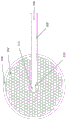
图3A和3B示出了另一实施方式。图3A示出了处于其2D构造的薄膜材料的盘300,其具有均匀孔隙302的区域,该区域被没有孔隙的增强部分或实心边界304包围。盘300可以包括薄膜元件100的任何特征。例如,盘300可以具有带有凸片308的槽306。在盘的中心是孔310,该孔可以具有附加的凸片312。一个或多个薄膜组件300可以包裹在支撑结构(诸如导丝线或线圈)周围,如图3B所示。导丝线穿过中心孔310插入,并且可以使用凸片312或如上文关于图2A所述附接的任何其它附接方法附接在适当位置。槽306使盘300能够如图3B所示包裹,具有重叠的部分,并且包裹直径与导管负载一致,膜的折叠最少。释放之后,盘300可以至少部分地扩展以填充动脉瘤,例如如图4B所示。3A and 3B illustrate another embodiment. FIG. 3A shows a disk 300 of thin film material in its 2D configuration with a region of
图4A和图4B示出了薄膜盘300如何被集成到植入物装配400中。使用任一上述方法可以将一个或多个薄膜盘404附接至支撑结构402。薄膜盘404可以包括薄膜元件100或薄膜盘300的任何特征。薄膜盘404的直径略大于动脉瘤的直径,以便从导管释放时,它们将展开以实现其外边缘与动脉瘤壁的贴合。每个薄膜盘404形成锥形体构造。一个或多个盘404(例如两个、三个或更多个)可以被附接以便每个锥体的顶点朝向支撑结构402的远端并远离动脉瘤颈部(见图4B)以利于插入动脉瘤。可以将直径大于盘404的至少一个附加盘408附接在支撑结构402的近端并且以其锥体的顶点朝向与较小盘404相反的方向并朝向支撑结构402的近端。当盘408退出导管时,它将张开到比动脉瘤的颈部更大的直径,从而有效地在颈部使血流转向。图4B中示出了这一点,其中430是位于由血管段420、422和424形成的y接合处的动脉瘤。植入物装配400被示为插入到动脉瘤中,以盘408阻塞颈部。4A and 4B illustrate how the membrane disc 300 is integrated into the
除了薄膜盘之外,导丝线402还可以具有附接到一端或两端的x射线可观察的夹414或类似元件,并且还可以具有附接的球412以用于与导管推杆分离。In addition to the pellicle disc, the
图5示出了类型400的装配的另一实施方式,其包括一个或多个薄膜盘404和已被形状设置至导丝线402中的螺旋形部分510。可以在插入导管之前将该区域拉直,以在导管约束范围内提供额外的长度,以减少盘404和408之间的任何重叠,从而有助于在植入过程中打开锥体。其还具有提供拉力以将盘408安置到动脉瘤的开口颈部上的作用。FIG. 5 shows another embodiment of an assembly of
除非另有明确说明,或在所用上下文中没有其他理解,条件语言诸如“可以(can)”、“可以(could)”、“可以(might)”或“可以(may)”通常旨在传达某些实施方式包括但其它实施方式不包括某些特征、元件和/或步骤。因此,这样的条件语言通常不意图暗示一个或多个实施方式以任何方式需要所述特征、要素和/或步骤。Unless expressly stated otherwise, or otherwise understood in the context in which it is used, conditional language such as "can," "could," "might," or "may" is generally intended to convey a certain Some embodiments include but other embodiments do not include certain features, elements and/or steps. Thus, such conditional language is generally not intended to imply that one or more embodiments require the described features, elements and/or steps in any way.
术语“包含”、“包括”、“具有”等是同义词,并且以开放式方式广泛使用,并且不排除附加要素、特征、动作、操作等。另外,术语“或”以其包容性含义(而不是其排他性含义)上使用,以便在用来连接一系列要素时,术语“或”是指该列表中要素的一个、一些或全部。The terms "comprising," "including," "having," and the like, are synonymous and are used broadly in an open-ended fashion and do not exclude additional elements, features, acts, operations, and the like. Additionally, the term "or" is used in its inclusive sense (rather than its exclusive sense) so that when used to connect a list of elements, the term "or" refers to one, some, or all of the elements in that list.
尽管本文已经描述了某些实施方式和实施例,但是本领域技术人员可以理解,本公开中示出和描述的递送系统的许多方面可以不同地组合和/或修改以形成其它实施方式或可接受的实施例。所有这样的修改和变型在本文中旨在包括在本公开的范围内。各种设计和方法都是可以的。本文公开的特征、结构或步骤都不是必需的或必不可少的。Although certain embodiments and examples have been described herein, those skilled in the art will appreciate that many aspects of the delivery systems shown and described in this disclosure may be variously combined and/or modified to form other embodiments or acceptable example. All such modifications and variations are intended to be included herein within the scope of the present disclosure. Various designs and approaches are possible. None of the features, structures, or steps disclosed herein are required or essential.
为了本公开的目的,本文描述了某些方面、优点和新的特征。应当理解的是,根据任何特定实施方式不一定可以实现所有这些优点。因此,例如,本领域技术人员将认识到,本公开可以以实现本文所教导的一个优点或一组优点而不必实现本文所教导或建议的其它优点的方式实施或执行。For the purposes of the present disclosure, certain aspects, advantages, and novel features have been described herein. It should be understood that not all of these advantages may be achieved according to any particular implementation. Thus, for example, those skilled in the art will recognize that the present disclosure can be implemented or carried out in a manner that achieves one advantage or group of advantages as taught herein without necessarily achieving other advantages as taught or suggested herein.
另外,尽管本文已经描述了说明性实施方式,但基于本公开,本领域技术人员可以理解具有同等要素、修改、省略、组合(例如各个实施方式的多个方面的组合)、改编和/或变更的任何和所有实施方式的范围。权利要求中的限定应基于权利要求中使用的语言来广义地解释,并且不限于本说明书中或在本申请的进行中描述的实施例,这些实施例应理解为是非排他性的。另外,可以以任何方式修改所公开的工艺和方法的动作,包括通过对动作重新排序和/或插入附加动作和/或删除动作。因此,说明书和实施例旨在仅被认为是说明性的,真正的范围和精神由权利要求及其等同物的全部范围来指示。Additionally, although illustrative embodiments have been described herein, equivalents, modifications, omissions, combinations (eg, combinations of aspects of the various embodiments), adaptations and/or changes will be understood by those skilled in the art based on this disclosure the scope of any and all implementations of The definitions in the claims are to be construed broadly based on the language used in the claims, and are not limited to the embodiments described in this specification or in the course of this application, which are to be construed as non-exclusive. Additionally, the acts of the disclosed processes and methods may be modified in any manner, including by reordering the acts and/or inserting additional acts and/or deleting acts. Therefore, the specification and examples are intended to be regarded as illustrative only, with the true scope and spirit being indicated by the claims and their full scope of equivalents.
如本文所用,术语“大约”、“约”和“基本上”表示接近所述量的量,其仍然执行期望的功能或达到期望的结果。例如,术语“大约”、“约”和“基本上”可以是指处于小于所述量的10%内的量。作为另一示例,在某些实施方式中,术语“大致平行”和“基本平行”是指与精确平行偏离小于或等于15度的值、量或特性。As used herein, the terms "about", "about" and "substantially" mean an amount approximating the stated amount that still performs the desired function or achieves the desired result. For example, the terms "about", "about" and "substantially" can refer to an amount within less than 10% of the stated amount. As another example, in certain embodiments, the terms "substantially parallel" and "substantially parallel" refer to a value, amount or characteristic that deviates from exact parallel by less than or equal to 15 degrees.
示例性实施方式Exemplary Embodiment
以下示例性实施方式确定了本文公开的特征的组合的一些可能排列,但特征的组合的其它排列也是可以的。The following exemplary embodiments identify some possible permutations of combinations of features disclosed herein, although other permutations of combinations of features are possible.
1.一种用于填充动脉瘤的可部署的(deployable,可配置的,可展开的)闭塞装置,闭塞装置包括:1. A deployable (deployable, configurable, deployable) occlusion device for filling an aneurysm, the occlusion device comprising:
包括线的支撑结构;和Support structure including wire; and
包括孔隙的网格组件,网格组件包含第一端部和第二端部,第一端部附接至支撑结构且第二端部为自由端,网格组件从支撑结构径向向外延伸。A mesh assembly including apertures, the mesh assembly including a first end portion and a second end portion, the first end portion being attached to the support structure and the second end portion being a free end, the mesh assembly extending radially outward from the support structure .
2.根据实施方式1的闭塞装置,其中网格组件被构造为在压缩状态和未压缩状态之间转变,网格组件在未压缩状态下形成线圈(coil,卷)。2. The occlusion device of embodiment 1, wherein the mesh assembly is configured to transition between a compressed state and an uncompressed state, the mesh assembly forming a coil in the uncompressed state.
3.根据实施方式2的闭塞装置,其中在压缩状态下,网格组件包裹在支撑结构周围(wrapped around the support structure,围绕支撑结构被包裹)。3. The occlusive device of embodiment 2, wherein, in the compressed state, the mesh assembly is wrapped around the support structure.
4.根据实施方式1至3中任一项的闭塞装置,其中支撑结构包含盘绕区。4. The occlusive device of any one of embodiments 1 to 3, wherein the support structure comprises a coiled region.
5.根据实施方式1至4中任一项的闭塞装置,其中支撑结构包括直线区(straightregion,直区)。5. The occlusive device of any one of embodiments 1 to 4, wherein the support structure comprises a straight region.
6.根据实施方式1至5中任一项的闭塞装置,其中网格组件通过粘合剂、焊接或钎焊而附接至支撑结构。6. The occlusion device of any one of embodiments 1 to 5, wherein the grid assembly is attached to the support structure by adhesive, welding or brazing.
7.根据实施方式1至5中任一项的闭塞装置,其中网格组件被机械地附接至支撑结构上。7. The occlusion device of any one of embodiments 1 to 5, wherein the grid assembly is mechanically attached to the support structure.
8.根据实施方式7的闭塞装置,其中网格组件具有至少一个用于接合支撑结构的凸片。8. The occlusion device of embodiment 7, wherein the grid assembly has at least one tab for engaging the support structure.
9.根据实施方式1至8中任一项的闭塞装置,其中网格组件是整体的(monolithic,单片的)。9. The occlusive device of any one of embodiments 1 to 8, wherein the grid assembly is monolithic.
10.根据实施方式1至9中任一项的闭塞装置,其中网格组件的壁厚不超过0.002英寸。10. The occlusion device of any one of embodiments 1 to 9, wherein the grid assembly has a wall thickness of no more than 0.002 inches.
11.根据实施方式1至10中任一项的闭塞装置,其中网格组件具有基本均匀的孔径。11. The occlusion device of any one of embodiments 1 to 10, wherein the grid assembly has a substantially uniform pore size.
12.根据实施方式1至11中任一项的闭塞装置,其中网格组件具有基本均匀的孔隙。12. The occlusion device of any one of embodiments 1 to 11, wherein the grid assembly has substantially uniform pores.
13.根据实施方式1至12中任一项的闭塞装置,其中第一端部不具有孔隙。13. The occlusive device of any one of embodiments 1 to 12, wherein the first end has no apertures.
14.根据实施方式1至13中任一项的闭塞装置,还包括多个网格组件,每个网格组件从支撑结构延伸。14. The occlusion device of any one of embodiments 1 to 13, further comprising a plurality of grid members, each grid member extending from the support structure.
15.一种用于填充动脉瘤的可部署的装置,闭塞装置包括:15. A deployable device for filling an aneurysm, the occlusion device comprising:
包括线的支撑结构,支撑结构包括近端和远端;和a support structure including a wire, the support structure including a proximal end and a distal end; and
沿支撑结构纵向间隔的多个网格组件,每个网格组件包含孙隙,网格组件被构造为在压缩状态和未压缩状态之间转变。A plurality of grid components spaced longitudinally along the support structure, each grid component containing a grand gap, the grid components are configured to transition between a compressed state and an uncompressed state.
16.根据实施方式15的闭塞装置,其中每个网格组件在未压缩状态下形成锥形(conical shape,圆锥形),使得每个网格组件包含顶点和开口端,每个网格组件的顶点附接到支撑结构。16. The occlusion device of embodiment 15, wherein each mesh component forms a conical shape in an uncompressed state, such that each mesh component includes a vertex and an open end, the The vertex is attached to the support structure.
17.根据实施例15或16的闭塞装置,其中支撑结构包括盘绕区。17. The occlusive device of embodiment 15 or 16, wherein the support structure comprises a coiled region.
18.根据实施方式15至17中任一项的闭塞装置,其中支撑结构包括直线区。18. The occlusive device of any one of embodiments 15 to 17, wherein the support structure comprises a rectilinear region.
19.根据实施方式15至18中任一项的闭塞装置,其中多个网格组件包括第一网格组件和第二网格组件。19. The occlusion device of any one of embodiments 15 to 18, wherein the plurality of mesh components includes a first mesh component and a second mesh component.
20.根据实施方式19的闭塞装置,其中第二网格组件的开口端具有比第一网格组件的开口端更大的直径。20. The occlusion device of embodiment 19, wherein the open end of the second grid member has a larger diameter than the open end of the first grid member.
21.根据实施方式19或20的闭塞装置,其中第一网格组件的顶点在与第二网格组件的顶点相反的方向上取向。21. The occlusion device of embodiment 19 or 20, wherein the vertices of the first mesh component are oriented in an opposite direction to the vertices of the second mesh component.
22.根据实施方式19至21中任一项的闭塞装置,其中第二组件的顶点被构造为位于动脉瘤的颈部中。22. The occlusion device of any one of embodiments 19 to 21, wherein the apex of the second component is configured to be located in the neck of the aneurysm.
23.根据实施方式15至22中任一项的闭塞装置,其中每个网格组件通过粘合剂、焊接或钎焊而附接到支撑结构。23. The occlusion device of any one of embodiments 15 to 22, wherein each grid assembly is attached to the support structure by adhesive, welding or brazing.
24.根据实施方式15至23中任一项的闭塞装置,其中每个网格组件被机械地附接到支撑结构。24. The occlusion device of any one of embodiments 15 to 23, wherein each grid assembly is mechanically attached to the support structure.
25.根据实施方式24的闭塞装置,其中每个网格组件具有至少一个用于接合支撑结构的凸片。25. The occlusion device of embodiment 24, wherein each grid assembly has at least one tab for engaging the support structure.
26.根据实施方式15至25中任一项的闭塞装置,其中每个网格组件是整体的。26. The occlusion device of any one of embodiments 15 to 25, wherein each grid assembly is unitary.
27.根据实施方式15至26中任一项的闭塞装置,其中每个网格组件的壁厚不超过0.002英寸。27. The occlusion device of any one of embodiments 15 to 26, wherein the wall thickness of each grid assembly does not exceed 0.002 inches.
28.根据实施方式15至27中任一项的闭塞装置,其中每个网格组件具有基本均匀的孔径。28. The occlusion device of any one of embodiments 15 to 27, wherein each grid assembly has a substantially uniform pore size.
29.根据实施方式15至28中任一项的闭塞装置,其中每个网格组件具有基本均匀的孔隙。29. The occlusion device of any one of embodiments 15 to 28, wherein each grid assembly has substantially uniform pores.
30.根据实施方式15至29中任一项的闭塞装置,其中每个网格组件的开口端的外周边缘不具有孔隙。30. The occlusion device of any one of embodiments 15 to 29, wherein the peripheral edge of the open end of each grid assembly is free of apertures.
31.根据实施方式15至30中任一项的闭塞装置,其中每个网格组件包括径向延伸的槽。31. The occlusion device of any one of embodiments 15 to 30, wherein each grid assembly includes a radially extending slot.
32.一种将闭塞装置部署到动脉瘤中的方法,该方法包括:32. A method of deploying an occlusive device into an aneurysm, the method comprising:
将携带闭塞装置的递送系统推进到动脉瘤;advancing the delivery system carrying the occlusion device to the aneurysm;
从递送系统部署闭塞装置,以便闭塞装置从约束构造转变到扩展构造,该闭塞装置包含:Deployment of an occlusive device from a delivery system for transition of the occlusive device from a restrained configuration to an expanded configuration, the occlusive device comprising:
支撑结构;和support structure; and
从支撑结构延伸的网格结构;和a grid structure extending from the support structure; and
从递送系统中释放出闭塞装置。The occlusive device is released from the delivery system.
33.根据实施方式32的方法,其中推进递送系统包含推进递送系统通过脉管系统中的弯曲区域,该弯曲区域具有内半径和外半径,网格结构的第一部分沿着沿外半径弯曲的轴线扩展,并且网格结构的第二部分沿着沿内半径弯曲的轴线收缩。33. The method of embodiment 32, wherein advancing the delivery system comprises advancing the delivery system through a curved region in the vasculature, the curved region having an inner radius and an outer radius, the first portion of the mesh structure being along an axis curved along the outer radius Expands and contracts the second portion of the lattice structure along an axis curved along the inner radius.
34.根据实施方式32或33的方法,其中部署闭塞装置包含将闭塞装置的底部定位在动脉瘤的颈部并且将闭塞装置的其余部分定位在动脉瘤的囊腔(sac volume,囊体积)内。34. The method of embodiment 32 or 33, wherein deploying the occlusion device comprises positioning the bottom of the occlusion device in the neck of the aneurysm and positioning the remainder of the occlusion device within the sac volume of the aneurysm .
Claims (34)
Applications Claiming Priority (3)
| Application Number | Priority Date | Filing Date | Title |
|---|---|---|---|
| US201862624672P | 2018-01-31 | 2018-01-31 | |
| US62/624,672 | 2018-01-31 | ||
| PCT/US2019/015716 WO2019152434A1 (en) | 2018-01-31 | 2019-01-29 | Vascular occlusion devices utilizing thin film nitinol foils |
Publications (2)
| Publication Number | Publication Date |
|---|---|
| CN111936063A true CN111936063A (en) | 2020-11-13 |
| CN111936063B CN111936063B (en) | 2024-08-20 |
Family
ID=67478904
Family Applications (1)
| Application Number | Title | Priority Date | Filing Date |
|---|---|---|---|
| CN201980023905.1A Active CN111936063B (en) | 2018-01-31 | 2019-01-29 | Vascular occlusive devices utilizing thin film Nitinol foil |
Country Status (4)
| Country | Link |
|---|---|
| US (2) | US11589872B2 (en) |
| EP (1) | EP3745965B1 (en) |
| CN (1) | CN111936063B (en) |
| WO (1) | WO2019152434A1 (en) |
Cited By (3)
| Publication number | Priority date | Publication date | Assignee | Title |
|---|---|---|---|---|
| CN115399825A (en) * | 2022-08-01 | 2022-11-29 | 上海心玮医疗科技股份有限公司 | Device for treating bifurcation aneurysm |
| US11517321B2 (en) | 2016-05-26 | 2022-12-06 | Nanostructures, Inc. | System and methods for embolized occlusion of neurovascular aneurysms |
| US11589872B2 (en) | 2018-01-31 | 2023-02-28 | Nanostructures, Inc. | Vascular occlusion devices utilizing thin film nitinol foils |
Families Citing this family (7)
| Publication number | Priority date | Publication date | Assignee | Title |
|---|---|---|---|---|
| US11357511B2 (en) | 2008-05-01 | 2022-06-14 | Aneuclose Llc | Intrasacular aneurysm occlusion device with globular first configuration and bowl-shaped second configuration |
| US11583289B2 (en) | 2008-05-01 | 2023-02-21 | Aneuclose Llc | Aneurysm-occluding mesh ribbon with a series of loops or segments having distal-to-proximal variation in size, shape, and/or orientation |
| US11464518B2 (en) | 2008-05-01 | 2022-10-11 | Aneuclose Llc | Proximal concave neck bridge with central lumen and distal net for occluding cerebral aneurysms |
| US11471164B2 (en) | 2008-05-01 | 2022-10-18 | Aneuclose Llc | Methods of occluding a cerebral aneurysm by inserting embolic members or material into an intrasacular implant |
| US11484322B2 (en) | 2018-01-03 | 2022-11-01 | Aneuclose Llc | Aneurysm neck bridge with a closeable opening or lumen through which embolic material is inserted into the aneurysm sac |
| US12004750B2 (en) | 2008-05-01 | 2024-06-11 | Aneuclose Llc | Methods for creating an expandable two-part intrasacular aneurysm occlusion device from a tubular mesh |
| WO2020060932A1 (en) | 2018-09-18 | 2020-03-26 | Nanostructures, Inc. | Catheter based methods and devices for obstructive blood flow restriction |
Citations (14)
| Publication number | Priority date | Publication date | Assignee | Title |
|---|---|---|---|---|
| US20010044634A1 (en) * | 2000-03-10 | 2001-11-22 | Don Michael T. Anthony | Vascular embolism prevention device employing filters |
| US20020169475A1 (en) * | 1999-09-07 | 2002-11-14 | John Gainor | Retrievable septal defect closure device |
| US20030187475A1 (en) * | 1997-05-08 | 2003-10-02 | Scimed Life Systems, Inc | Percutaneous catheter and guidewire having filter and medical device deployment capabilities |
| US20060116713A1 (en) * | 2004-11-26 | 2006-06-01 | Ivan Sepetka | Aneurysm treatment devices and methods |
| US20090112253A1 (en) * | 2007-10-29 | 2009-04-30 | Salviac Limited | Guidewire for embolic device |
| US20090112228A1 (en) * | 2007-10-30 | 2009-04-30 | Boston Scientific Scimed, Inc. | Intravascular filter device with piezoelectric transducer |
| CN103169522A (en) * | 2006-05-24 | 2013-06-26 | 泰科保健集团有限合伙公司 | Flexible vascular occluding device |
| CN104997544A (en) * | 2009-09-04 | 2015-10-28 | 帕尔萨维斯库勒公司 | Systems and methods for enclosing anatomical opening |
| US20150313605A1 (en) * | 2014-04-30 | 2015-11-05 | Cerus Endovascular Limited | Occlusion Device |
| US20160317158A1 (en) * | 2015-05-01 | 2016-11-03 | DePuy Synthes Products, Inc. | Expandable vascular occlusion device with lead framing coil |
| US20170027552A1 (en) * | 2015-07-29 | 2017-02-02 | Cardiac Pacemakers, Inc. | Left atrial appendage implant |
| US20170143359A1 (en) * | 2015-08-06 | 2017-05-25 | Kp Medcure, Inc. | Axial lengthening thrombus capture system |
| US20170156734A1 (en) * | 2015-12-07 | 2017-06-08 | Cerus Endovascular Limited | Occlusion Device |
| US20170165046A1 (en) * | 2013-03-15 | 2017-06-15 | Koninklijke Philips N.V. | Distal protection systems and methods with pressure and ultrasound features |
Family Cites Families (71)
| Publication number | Priority date | Publication date | Assignee | Title |
|---|---|---|---|---|
| DK0791333T3 (en) | 1991-12-12 | 2000-05-01 | Target Therapeutics Inc | Several extruder-carocclusion coil construction with interlocking coupling |
| WO1993013712A1 (en) | 1992-01-21 | 1993-07-22 | Regents Of The University Of Minnesota | Septal defect closure device |
| US5263964A (en) | 1992-05-06 | 1993-11-23 | Coil Partners Ltd. | Coaxial traction detachment apparatus and method |
| US5350397A (en) | 1992-11-13 | 1994-09-27 | Target Therapeutics, Inc. | Axially detachable embolic coil assembly |
| US5624449A (en) | 1993-11-03 | 1997-04-29 | Target Therapeutics | Electrolytically severable joint for endovascular embolic devices |
| US5749894A (en) | 1996-01-18 | 1998-05-12 | Target Therapeutics, Inc. | Aneurysm closure method |
| US6168622B1 (en) | 1996-01-24 | 2001-01-02 | Microvena Corporation | Method and apparatus for occluding aneurysms |
| US5980514A (en) | 1996-07-26 | 1999-11-09 | Target Therapeutics, Inc. | Aneurysm closure device assembly |
| US5928260A (en) | 1997-07-10 | 1999-07-27 | Scimed Life Systems, Inc. | Removable occlusion system for aneurysm neck |
| US7569066B2 (en) | 1997-07-10 | 2009-08-04 | Boston Scientific Scimed, Inc. | Methods and devices for the treatment of aneurysms |
| US6036720A (en) | 1997-12-15 | 2000-03-14 | Target Therapeutics, Inc. | Sheet metal aneurysm neck bridge |
| US7410482B2 (en) | 1998-09-04 | 2008-08-12 | Boston Scientific-Scimed, Inc. | Detachable aneurysm neck bridge |
| US6375668B1 (en) | 1999-06-02 | 2002-04-23 | Hanson S. Gifford | Devices and methods for treating vascular malformations |
| US20020026211A1 (en) * | 1999-12-23 | 2002-02-28 | Farhad Khosravi | Vascular device having emboli and thrombus removal element and methods of use |
| US6689141B2 (en) | 2000-10-18 | 2004-02-10 | Microvention, Inc. | Mechanism for the deployment of endovascular implants |
| US8083768B2 (en) * | 2000-12-14 | 2011-12-27 | Ensure Medical, Inc. | Vascular plug having composite construction |
| US7011094B2 (en) | 2001-03-02 | 2006-03-14 | Emphasys Medical, Inc. | Bronchial flow control devices and methods of use |
| US8715312B2 (en) | 2001-07-20 | 2014-05-06 | Microvention, Inc. | Aneurysm treatment device and method of use |
| JP4429589B2 (en) * | 2001-11-15 | 2010-03-10 | コーディス・ニューロバスキュラー・インコーポレイテッド | Aneurysm embolization device using an occluding member |
| US8075585B2 (en) | 2002-08-29 | 2011-12-13 | Stryker Corporation | Device and method for treatment of a vascular defect |
| US20040153025A1 (en) * | 2003-02-03 | 2004-08-05 | Seifert Paul S. | Systems and methods of de-endothelialization |
| US8777974B2 (en) * | 2004-03-19 | 2014-07-15 | Aga Medical Corporation | Multi-layer braided structures for occluding vascular defects |
| WO2005123171A2 (en) | 2004-06-09 | 2005-12-29 | Stout Medical Group Lp | Three-dimensional coils for treatment of vascular aneurysms |
| US8444668B2 (en) | 2004-09-17 | 2013-05-21 | DePuy Synthes Products, LLC | Expandable vascular occlusion device |
| JP2008513141A (en) | 2004-09-17 | 2008-05-01 | コーディス・ニューロバスキュラー・インコーポレイテッド | Thin-film metal instrument for plugging an aneurysm or blood vessel |
| US20060155323A1 (en) | 2005-01-07 | 2006-07-13 | Porter Stephen C | Intra-aneurysm devices |
| KR101334502B1 (en) | 2005-10-19 | 2013-12-05 | 펄사 배스큘러, 아이엔씨. | Method and systems for endovascularly clipping and repairing lumen and tissue defects |
| US8545530B2 (en) | 2005-10-19 | 2013-10-01 | Pulsar Vascular, Inc. | Implantable aneurysm closure systems and methods |
| US20090112251A1 (en) * | 2007-07-25 | 2009-04-30 | Aga Medical Corporation | Braided occlusion device having repeating expanded volume segments separated by articulation segments |
| US20160220265A1 (en) * | 2007-10-12 | 2016-08-04 | NexGen Medical Systems Incorporated, | Thrombus removal system and process |
| WO2009052432A2 (en) * | 2007-10-19 | 2009-04-23 | Coherex Medical, Inc. | Medical device for modification of left atrial appendange and related systems and methods |
| US8956475B2 (en) | 2007-12-11 | 2015-02-17 | Howard Riina | Method and apparatus for restricting flow through an opening in the side wall of a body lumen, and/or for reinforcing a weakness in the side wall of a body lumen, while still maintaining substantially normal flow through the body lumen |
| JP5610542B2 (en) | 2008-04-21 | 2014-10-22 | コヴィディエン リミテッド パートナーシップ | Blade ball embolization device and delivery system |
| US20160374690A9 (en) | 2010-10-21 | 2016-12-29 | Robert A. Connor | Devices and Methods for Occluding a Cerebral Aneurysm |
| AU2009289488B2 (en) | 2008-09-05 | 2015-09-10 | Pulsar Vascular, Inc. | Systems and methods for supporting or occluding a physiological opening or cavity |
| US9095342B2 (en) | 2009-11-09 | 2015-08-04 | Covidien Lp | Braid ball embolic device features |
| WO2011150118A2 (en) * | 2010-05-25 | 2011-12-01 | The Regents Of The University Of California | Ultra-low fractional area coverage flow diverter for treating aneurysms and vascular diseases |
| US8828051B2 (en) * | 2010-07-02 | 2014-09-09 | Pfm Medical Ag | Left atrial appendage occlusion device |
| US8998947B2 (en) | 2010-09-10 | 2015-04-07 | Medina Medical, Inc. | Devices and methods for the treatment of vascular defects |
| US20120095500A1 (en) * | 2010-10-14 | 2012-04-19 | Heuser Richard R | Concentric wire embolism protection device |
| US10398444B2 (en) | 2011-03-30 | 2019-09-03 | Noha, Llc | Advanced endovascular clip and method of using same |
| US20120330341A1 (en) | 2011-06-22 | 2012-12-27 | Becking Frank P | Folded-Flat Aneurysm Embolization Devices |
| IN2014DN05897A (en) * | 2012-01-06 | 2015-06-05 | Inceptus Medical LLC | |
| US9687245B2 (en) | 2012-03-23 | 2017-06-27 | Covidien Lp | Occlusive devices and methods of use |
| US10537308B2 (en) * | 2012-07-06 | 2020-01-21 | Michael Zhadkevich | Catheter for prevention of stroke and method of use |
| CN105142546B (en) | 2013-03-15 | 2018-11-13 | 柯惠有限合伙公司 | Occlude implant infrastructure |
| US10675039B2 (en) | 2013-03-15 | 2020-06-09 | Embo Medical Limited | Embolisation systems |
| WO2014144980A1 (en) | 2013-03-15 | 2014-09-18 | Covidien Lp | Occlusive device |
| CA2901443A1 (en) | 2013-03-15 | 2014-09-25 | Insera Therapeutics, Inc. | Vascular treatment devices and methods |
| US9629635B2 (en) * | 2014-04-14 | 2017-04-25 | Sequent Medical, Inc. | Devices for therapeutic vascular procedures |
| US9713475B2 (en) | 2014-04-18 | 2017-07-25 | Covidien Lp | Embolic medical devices |
| WO2016014687A1 (en) * | 2014-07-22 | 2016-01-28 | Boston Scientific Scimed, Inc. | Expandable vaso-occlusive devices having shape memory and methods of using the same |
| ES2726910T3 (en) | 2014-07-25 | 2019-10-10 | Incumedx Inc | Covered embolic coils |
| JP7001476B2 (en) * | 2015-02-25 | 2022-02-03 | ギャラクシー セラピューティクス インコーポレイテッド | A device for treating an aneurysm |
| WO2016182949A1 (en) | 2015-05-08 | 2016-11-17 | Stryker European Holdings I, Llc | Vaso-occlusive devices |
| JP2018126173A (en) * | 2015-06-16 | 2018-08-16 | テルモ株式会社 | Medical device and treatment method |
| EP3315091A4 (en) * | 2015-06-29 | 2019-02-20 | Access Point Technologies, Inc. | Emboli capture device |
| CN104958087B (en) * | 2015-07-28 | 2018-09-25 | 杭州诺茂医疗科技有限公司 | A kind of occluder for left auricle |
| WO2017106567A1 (en) | 2015-12-15 | 2017-06-22 | Nsvascular, Inc. | Intrasaccular thin-film flow diverters and related methods |
| CN108472043B (en) | 2015-12-30 | 2022-05-31 | 斯瑞克公司 | Embolization device and method of making same |
| EP3457954A4 (en) * | 2016-05-18 | 2020-01-08 | Microvention, Inc. | EMBOLIC CONTAINMENT |
| EP3463109A4 (en) | 2016-05-26 | 2020-01-08 | Nanostructures, Inc. | SYSTEM AND METHODS FOR EMBOLISED OCCLUSION OF NEUROVASCULAR ANEVRISMS |
| US20170367710A1 (en) * | 2016-06-24 | 2017-12-28 | Cook Medical Technologies Llc | Double sided occlusive device |
| US20180098777A1 (en) * | 2016-10-11 | 2018-04-12 | Shlomo Gabbay | Devices and Methods for Improving Transcatheter Aortic Valve Implantation |
| US11197750B2 (en) * | 2016-11-29 | 2021-12-14 | Lake Region Manufacturing, Inc. | Embolic protection device |
| US20180353279A1 (en) * | 2016-12-05 | 2018-12-13 | Daniel E. Walzman | Exchange wire anchored by a self-expanding retrievable stent and method of use |
| WO2018112203A1 (en) * | 2016-12-14 | 2018-06-21 | eLum Technologies, Inc. | Electrospun stents, flow diverters, and occlusion devices and methods of making the same |
| US10299799B1 (en) * | 2017-07-07 | 2019-05-28 | John S. DeMeritt | Micro-macro endovascular occlusion device and methodology |
| US10342686B2 (en) * | 2017-08-10 | 2019-07-09 | Covidien Lp | Thin film mesh hybrid for treating vascular defects |
| US11185335B2 (en) * | 2018-01-19 | 2021-11-30 | Galaxy Therapeutics Inc. | System for and method of treating aneurysms |
| WO2019152434A1 (en) | 2018-01-31 | 2019-08-08 | Nanostructures, Inc. | Vascular occlusion devices utilizing thin film nitinol foils |
-
2019
- 2019-01-29 WO PCT/US2019/015716 patent/WO2019152434A1/en not_active Ceased
- 2019-01-29 US US16/965,919 patent/US11589872B2/en active Active
- 2019-01-29 CN CN201980023905.1A patent/CN111936063B/en active Active
- 2019-01-29 EP EP19746779.8A patent/EP3745965B1/en active Active
-
2023
- 2023-01-19 US US18/156,735 patent/US20230225737A1/en active Pending
Patent Citations (15)
| Publication number | Priority date | Publication date | Assignee | Title |
|---|---|---|---|---|
| US20030187475A1 (en) * | 1997-05-08 | 2003-10-02 | Scimed Life Systems, Inc | Percutaneous catheter and guidewire having filter and medical device deployment capabilities |
| US20020169475A1 (en) * | 1999-09-07 | 2002-11-14 | John Gainor | Retrievable septal defect closure device |
| US20010044634A1 (en) * | 2000-03-10 | 2001-11-22 | Don Michael T. Anthony | Vascular embolism prevention device employing filters |
| US20060116713A1 (en) * | 2004-11-26 | 2006-06-01 | Ivan Sepetka | Aneurysm treatment devices and methods |
| CN103169522A (en) * | 2006-05-24 | 2013-06-26 | 泰科保健集团有限合伙公司 | Flexible vascular occluding device |
| US20090112253A1 (en) * | 2007-10-29 | 2009-04-30 | Salviac Limited | Guidewire for embolic device |
| US20090112228A1 (en) * | 2007-10-30 | 2009-04-30 | Boston Scientific Scimed, Inc. | Intravascular filter device with piezoelectric transducer |
| CN104997544A (en) * | 2009-09-04 | 2015-10-28 | 帕尔萨维斯库勒公司 | Systems and methods for enclosing anatomical opening |
| US20170165046A1 (en) * | 2013-03-15 | 2017-06-15 | Koninklijke Philips N.V. | Distal protection systems and methods with pressure and ultrasound features |
| US20150313605A1 (en) * | 2014-04-30 | 2015-11-05 | Cerus Endovascular Limited | Occlusion Device |
| CN106456183A (en) * | 2014-04-30 | 2017-02-22 | Cerus血管内设备有限公司 | Occlusion device |
| US20160317158A1 (en) * | 2015-05-01 | 2016-11-03 | DePuy Synthes Products, Inc. | Expandable vascular occlusion device with lead framing coil |
| US20170027552A1 (en) * | 2015-07-29 | 2017-02-02 | Cardiac Pacemakers, Inc. | Left atrial appendage implant |
| US20170143359A1 (en) * | 2015-08-06 | 2017-05-25 | Kp Medcure, Inc. | Axial lengthening thrombus capture system |
| US20170156734A1 (en) * | 2015-12-07 | 2017-06-08 | Cerus Endovascular Limited | Occlusion Device |
Cited By (4)
| Publication number | Priority date | Publication date | Assignee | Title |
|---|---|---|---|---|
| US11517321B2 (en) | 2016-05-26 | 2022-12-06 | Nanostructures, Inc. | System and methods for embolized occlusion of neurovascular aneurysms |
| US12303136B2 (en) | 2016-05-26 | 2025-05-20 | Nanostructures, Inc. | System and methods for embolized occlusion of neurovascular aneurysms |
| US11589872B2 (en) | 2018-01-31 | 2023-02-28 | Nanostructures, Inc. | Vascular occlusion devices utilizing thin film nitinol foils |
| CN115399825A (en) * | 2022-08-01 | 2022-11-29 | 上海心玮医疗科技股份有限公司 | Device for treating bifurcation aneurysm |
Also Published As
| Publication number | Publication date |
|---|---|
| EP3745965A4 (en) | 2021-10-27 |
| EP3745965B1 (en) | 2025-01-01 |
| US20230225737A1 (en) | 2023-07-20 |
| US20210052278A1 (en) | 2021-02-25 |
| EP3745965A1 (en) | 2020-12-09 |
| US11589872B2 (en) | 2023-02-28 |
| CN111936063B (en) | 2024-08-20 |
| WO2019152434A1 (en) | 2019-08-08 |
Similar Documents
| Publication | Publication Date | Title |
|---|---|---|
| US20230225737A1 (en) | Vascular occlusion devices utilizing thin film nitinol foils | |
| US12303136B2 (en) | System and methods for embolized occlusion of neurovascular aneurysms | |
| US12053182B2 (en) | Devices and methods for the treatment of vascular defects | |
| JP5619925B2 (en) | Flexible vascular closure device | |
| EP4039201B1 (en) | Embolic devices and methods of manufacturing same | |
| US8444668B2 (en) | Expandable vascular occlusion device | |
| EP2468349B1 (en) | Thin film metallic devices for plugging aneurysms or vessels | |
| EP3087934B1 (en) | Expandable vascular occlusion device with lead framing coil | |
| EP2938289B1 (en) | Devices for the treatment of vascular defects | |
| EP3668417A1 (en) | Flow attenuation device | |
| CN101472537A (en) | Flexible vascular occluding device | |
| JP7795559B2 (en) | Vascular Occlusion Devices |
Legal Events
| Date | Code | Title | Description |
|---|---|---|---|
| PB01 | Publication | ||
| PB01 | Publication | ||
| SE01 | Entry into force of request for substantive examination | ||
| SE01 | Entry into force of request for substantive examination | ||
| GR01 | Patent grant | ||
| GR01 | Patent grant |