CN111748624A - Biomarkers for predicting recurrence of liver cancer - Google Patents
Biomarkers for predicting recurrence of liver cancer Download PDFInfo
- Publication number
- CN111748624A CN111748624A CN202010514464.XA CN202010514464A CN111748624A CN 111748624 A CN111748624 A CN 111748624A CN 202010514464 A CN202010514464 A CN 202010514464A CN 111748624 A CN111748624 A CN 111748624A
- Authority
- CN
- China
- Prior art keywords
- protein
- recurrence
- liver cancer
- judged
- hnrnpa3
- Prior art date
- Legal status (The legal status is an assumption and is not a legal conclusion. Google has not performed a legal analysis and makes no representation as to the accuracy of the status listed.)
- Granted
Links
- 201000007270 liver cancer Diseases 0.000 title claims abstract description 121
- 208000014018 liver neoplasm Diseases 0.000 title claims abstract description 121
- 239000000090 biomarker Substances 0.000 title claims abstract description 46
- 102000004169 proteins and genes Human genes 0.000 claims abstract description 111
- 108090000623 proteins and genes Proteins 0.000 claims abstract description 111
- 102100035669 Heterogeneous nuclear ribonucleoprotein A3 Human genes 0.000 claims abstract description 40
- 101000854041 Homo sapiens Heterogeneous nuclear ribonucleoprotein A3 Proteins 0.000 claims abstract description 40
- 101000852214 Homo sapiens THO complex subunit 4 Proteins 0.000 claims abstract description 39
- 102100036434 THO complex subunit 4 Human genes 0.000 claims abstract description 37
- 238000000034 method Methods 0.000 claims abstract description 33
- 102100032559 Clathrin light chain B Human genes 0.000 claims abstract description 32
- 101000942271 Homo sapiens Clathrin light chain B Proteins 0.000 claims abstract description 32
- 101000997852 Homo sapiens Protein jagunal homolog 1 Proteins 0.000 claims abstract description 32
- 101001008515 Homo sapiens Ribosomal biogenesis protein LAS1L Proteins 0.000 claims abstract description 32
- 102100033434 Protein jagunal homolog 1 Human genes 0.000 claims abstract description 32
- 102100027433 Ribosomal biogenesis protein LAS1L Human genes 0.000 claims abstract description 32
- 238000004949 mass spectrometry Methods 0.000 claims abstract description 23
- 238000003745 diagnosis Methods 0.000 claims abstract description 19
- 238000001294 liquid chromatography-tandem mass spectrometry Methods 0.000 claims abstract description 15
- 238000012360 testing method Methods 0.000 claims abstract description 4
- 239000000523 sample Substances 0.000 claims description 48
- 239000003153 chemical reaction reagent Substances 0.000 claims description 32
- 230000002980 postoperative effect Effects 0.000 claims description 22
- 238000001356 surgical procedure Methods 0.000 claims description 21
- 108020004707 nucleic acids Proteins 0.000 claims description 13
- 102000039446 nucleic acids Human genes 0.000 claims description 13
- 150000007523 nucleic acids Chemical class 0.000 claims description 13
- 238000011895 specific detection Methods 0.000 claims 6
- 206010028980 Neoplasm Diseases 0.000 abstract description 18
- 201000011510 cancer Diseases 0.000 abstract description 10
- 230000008569 process Effects 0.000 abstract description 4
- 238000004393 prognosis Methods 0.000 abstract description 4
- 230000008901 benefit Effects 0.000 abstract description 3
- 238000002331 protein detection Methods 0.000 abstract 1
- 238000001514 detection method Methods 0.000 description 37
- 230000000694 effects Effects 0.000 description 13
- 206010073071 hepatocellular carcinoma Diseases 0.000 description 11
- 231100000844 hepatocellular carcinoma Toxicity 0.000 description 11
- 239000000243 solution Substances 0.000 description 10
- 238000004422 calculation algorithm Methods 0.000 description 9
- 230000035945 sensitivity Effects 0.000 description 9
- 238000011002 quantification Methods 0.000 description 7
- 238000010586 diagram Methods 0.000 description 6
- 108090000765 processed proteins & peptides Proteins 0.000 description 5
- 238000004445 quantitative analysis Methods 0.000 description 5
- 230000001105 regulatory effect Effects 0.000 description 5
- 238000007619 statistical method Methods 0.000 description 5
- 102000013529 alpha-Fetoproteins Human genes 0.000 description 4
- 108010026331 alpha-Fetoproteins Proteins 0.000 description 4
- 238000007477 logistic regression Methods 0.000 description 4
- 230000037361 pathway Effects 0.000 description 4
- 230000000306 recurrent effect Effects 0.000 description 4
- 238000002271 resection Methods 0.000 description 4
- 238000004885 tandem mass spectrometry Methods 0.000 description 4
- WEVYAHXRMPXWCK-UHFFFAOYSA-N Acetonitrile Chemical compound CC#N WEVYAHXRMPXWCK-UHFFFAOYSA-N 0.000 description 3
- 241000700721 Hepatitis B virus Species 0.000 description 3
- 208000019425 cirrhosis of liver Diseases 0.000 description 3
- 230000003993 interaction Effects 0.000 description 3
- 210000004185 liver Anatomy 0.000 description 3
- 102000004196 processed proteins & peptides Human genes 0.000 description 3
- 238000000729 Fisher's exact test Methods 0.000 description 2
- 206010027476 Metastases Diseases 0.000 description 2
- 102000007079 Peptide Fragments Human genes 0.000 description 2
- 108010033276 Peptide Fragments Proteins 0.000 description 2
- 208000036142 Viral infection Diseases 0.000 description 2
- 238000004458 analytical method Methods 0.000 description 2
- 239000007864 aqueous solution Substances 0.000 description 2
- 230000009286 beneficial effect Effects 0.000 description 2
- 230000008859 change Effects 0.000 description 2
- 238000012937 correction Methods 0.000 description 2
- 238000005516 engineering process Methods 0.000 description 2
- 238000010201 enrichment analysis Methods 0.000 description 2
- 230000007717 exclusion Effects 0.000 description 2
- 208000002672 hepatitis B Diseases 0.000 description 2
- 150000002500 ions Chemical class 0.000 description 2
- 239000007788 liquid Substances 0.000 description 2
- 230000036210 malignancy Effects 0.000 description 2
- 230000009401 metastasis Effects 0.000 description 2
- BDAGIHXWWSANSR-UHFFFAOYSA-N methanoic acid Natural products OC=O BDAGIHXWWSANSR-UHFFFAOYSA-N 0.000 description 2
- 238000002360 preparation method Methods 0.000 description 2
- 238000012216 screening Methods 0.000 description 2
- 230000001225 therapeutic effect Effects 0.000 description 2
- 230000009385 viral infection Effects 0.000 description 2
- OSWFIVFLDKOXQC-UHFFFAOYSA-N 4-(3-methoxyphenyl)aniline Chemical compound COC1=CC=CC(C=2C=CC(N)=CC=2)=C1 OSWFIVFLDKOXQC-UHFFFAOYSA-N 0.000 description 1
- 208000007848 Alcoholism Diseases 0.000 description 1
- 208000000419 Chronic Hepatitis B Diseases 0.000 description 1
- 238000009007 Diagnostic Kit Methods 0.000 description 1
- 241000711549 Hepacivirus C Species 0.000 description 1
- 108010026552 Proteome Proteins 0.000 description 1
- 239000007983 Tris buffer Substances 0.000 description 1
- 102000004142 Trypsin Human genes 0.000 description 1
- 108090000631 Trypsin Proteins 0.000 description 1
- 238000002835 absorbance Methods 0.000 description 1
- 238000010521 absorption reaction Methods 0.000 description 1
- XBJFCYDKBDVADW-UHFFFAOYSA-N acetonitrile;formic acid Chemical compound CC#N.OC=O XBJFCYDKBDVADW-UHFFFAOYSA-N 0.000 description 1
- 230000004913 activation Effects 0.000 description 1
- 239000002115 aflatoxin B1 Substances 0.000 description 1
- OQIQSTLJSLGHID-WNWIJWBNSA-N aflatoxin B1 Chemical compound C=1([C@@H]2C=CO[C@@H]2OC=1C=C(C1=2)OC)C=2OC(=O)C2=C1CCC2=O OQIQSTLJSLGHID-WNWIJWBNSA-N 0.000 description 1
- 229930020125 aflatoxin-B1 Natural products 0.000 description 1
- 201000007930 alcohol dependence Diseases 0.000 description 1
- 239000002246 antineoplastic agent Substances 0.000 description 1
- 229940041181 antineoplastic drug Drugs 0.000 description 1
- 238000003766 bioinformatics method Methods 0.000 description 1
- 239000000872 buffer Substances 0.000 description 1
- 238000004364 calculation method Methods 0.000 description 1
- 239000000969 carrier Substances 0.000 description 1
- 239000013522 chelant Substances 0.000 description 1
- 230000001684 chronic effect Effects 0.000 description 1
- 238000003759 clinical diagnosis Methods 0.000 description 1
- 238000007621 cluster analysis Methods 0.000 description 1
- 238000013211 curve analysis Methods 0.000 description 1
- 230000007812 deficiency Effects 0.000 description 1
- 230000004069 differentiation Effects 0.000 description 1
- 230000029087 digestion Effects 0.000 description 1
- 210000002249 digestive system Anatomy 0.000 description 1
- 201000010099 disease Diseases 0.000 description 1
- 208000037265 diseases, disorders, signs and symptoms Diseases 0.000 description 1
- VHJLVAABSRFDPM-QWWZWVQMSA-N dithiothreitol Chemical compound SC[C@@H](O)[C@H](O)CS VHJLVAABSRFDPM-QWWZWVQMSA-N 0.000 description 1
- 230000003828 downregulation Effects 0.000 description 1
- 230000010102 embolization Effects 0.000 description 1
- 238000002474 experimental method Methods 0.000 description 1
- 238000000605 extraction Methods 0.000 description 1
- 235000019253 formic acid Nutrition 0.000 description 1
- 239000012634 fragment Substances 0.000 description 1
- 230000036541 health Effects 0.000 description 1
- 238000004128 high performance liquid chromatography Methods 0.000 description 1
- 238000003384 imaging method Methods 0.000 description 1
- 208000015181 infectious disease Diseases 0.000 description 1
- 238000002955 isolation Methods 0.000 description 1
- 230000003902 lesion Effects 0.000 description 1
- 238000004895 liquid chromatography mass spectrometry Methods 0.000 description 1
- 238000012317 liver biopsy Methods 0.000 description 1
- 238000013507 mapping Methods 0.000 description 1
- 238000012986 modification Methods 0.000 description 1
- 230000004048 modification Effects 0.000 description 1
- 238000012544 monitoring process Methods 0.000 description 1
- 238000003012 network analysis Methods 0.000 description 1
- 230000008506 pathogenesis Effects 0.000 description 1
- 230000001717 pathogenic effect Effects 0.000 description 1
- 210000003240 portal vein Anatomy 0.000 description 1
- 238000007781 pre-processing Methods 0.000 description 1
- 239000002243 precursor Substances 0.000 description 1
- 238000000751 protein extraction Methods 0.000 description 1
- 238000007674 radiofrequency ablation Methods 0.000 description 1
- 238000011160 research Methods 0.000 description 1
- 238000012827 research and development Methods 0.000 description 1
- 238000012552 review Methods 0.000 description 1
- 238000000926 separation method Methods 0.000 description 1
- 238000002864 sequence alignment Methods 0.000 description 1
- 150000003384 small molecules Chemical class 0.000 description 1
- DAJSVUQLFFJUSX-UHFFFAOYSA-M sodium;dodecane-1-sulfonate Chemical compound [Na+].CCCCCCCCCCCCS([O-])(=O)=O DAJSVUQLFFJUSX-UHFFFAOYSA-M 0.000 description 1
- 239000002689 soil Substances 0.000 description 1
- 238000001228 spectrum Methods 0.000 description 1
- 230000004083 survival effect Effects 0.000 description 1
- 238000002626 targeted therapy Methods 0.000 description 1
- LENZDBCJOHFCAS-UHFFFAOYSA-N tris Chemical compound OCC(N)(CO)CO LENZDBCJOHFCAS-UHFFFAOYSA-N 0.000 description 1
- 239000012588 trypsin Substances 0.000 description 1
- 239000000439 tumor marker Substances 0.000 description 1
- 230000003827 upregulation Effects 0.000 description 1
- 230000009790 vascular invasion Effects 0.000 description 1
Images


Classifications
-
- C—CHEMISTRY; METALLURGY
- C12—BIOCHEMISTRY; BEER; SPIRITS; WINE; VINEGAR; MICROBIOLOGY; ENZYMOLOGY; MUTATION OR GENETIC ENGINEERING
- C12Q—MEASURING OR TESTING PROCESSES INVOLVING ENZYMES, NUCLEIC ACIDS OR MICROORGANISMS; COMPOSITIONS OR TEST PAPERS THEREFOR; PROCESSES OF PREPARING SUCH COMPOSITIONS; CONDITION-RESPONSIVE CONTROL IN MICROBIOLOGICAL OR ENZYMOLOGICAL PROCESSES
- C12Q1/00—Measuring or testing processes involving enzymes, nucleic acids or microorganisms; Compositions therefor; Processes of preparing such compositions
- C12Q1/68—Measuring or testing processes involving enzymes, nucleic acids or microorganisms; Compositions therefor; Processes of preparing such compositions involving nucleic acids
- C12Q1/6876—Nucleic acid products used in the analysis of nucleic acids, e.g. primers or probes
- C12Q1/6883—Nucleic acid products used in the analysis of nucleic acids, e.g. primers or probes for diseases caused by alterations of genetic material
- C12Q1/6886—Nucleic acid products used in the analysis of nucleic acids, e.g. primers or probes for diseases caused by alterations of genetic material for cancer
-
- G—PHYSICS
- G01—MEASURING; TESTING
- G01N—INVESTIGATING OR ANALYSING MATERIALS BY DETERMINING THEIR CHEMICAL OR PHYSICAL PROPERTIES
- G01N30/00—Investigating or analysing materials by separation into components using adsorption, absorption or similar phenomena or using ion-exchange, e.g. chromatography or field flow fractionation
- G01N30/02—Column chromatography
- G01N30/88—Integrated analysis systems specially adapted therefor, not covered by a single one of the groups G01N30/04 - G01N30/86
-
- G—PHYSICS
- G01—MEASURING; TESTING
- G01N—INVESTIGATING OR ANALYSING MATERIALS BY DETERMINING THEIR CHEMICAL OR PHYSICAL PROPERTIES
- G01N33/00—Investigating or analysing materials by specific methods not covered by groups G01N1/00 - G01N31/00
- G01N33/48—Biological material, e.g. blood, urine; Haemocytometers
- G01N33/50—Chemical analysis of biological material, e.g. blood, urine; Testing involving biospecific ligand binding methods; Immunological testing
- G01N33/68—Chemical analysis of biological material, e.g. blood, urine; Testing involving biospecific ligand binding methods; Immunological testing involving proteins, peptides or amino acids
- G01N33/6803—General methods of protein analysis not limited to specific proteins or families of proteins
- G01N33/6848—Methods of protein analysis involving mass spectrometry
-
- G—PHYSICS
- G01—MEASURING; TESTING
- G01N—INVESTIGATING OR ANALYSING MATERIALS BY DETERMINING THEIR CHEMICAL OR PHYSICAL PROPERTIES
- G01N33/00—Investigating or analysing materials by specific methods not covered by groups G01N1/00 - G01N31/00
- G01N33/48—Biological material, e.g. blood, urine; Haemocytometers
- G01N33/50—Chemical analysis of biological material, e.g. blood, urine; Testing involving biospecific ligand binding methods; Immunological testing
- G01N33/68—Chemical analysis of biological material, e.g. blood, urine; Testing involving biospecific ligand binding methods; Immunological testing involving proteins, peptides or amino acids
- G01N33/6893—Chemical analysis of biological material, e.g. blood, urine; Testing involving biospecific ligand binding methods; Immunological testing involving proteins, peptides or amino acids related to diseases not provided for elsewhere
-
- C—CHEMISTRY; METALLURGY
- C12—BIOCHEMISTRY; BEER; SPIRITS; WINE; VINEGAR; MICROBIOLOGY; ENZYMOLOGY; MUTATION OR GENETIC ENGINEERING
- C12Q—MEASURING OR TESTING PROCESSES INVOLVING ENZYMES, NUCLEIC ACIDS OR MICROORGANISMS; COMPOSITIONS OR TEST PAPERS THEREFOR; PROCESSES OF PREPARING SUCH COMPOSITIONS; CONDITION-RESPONSIVE CONTROL IN MICROBIOLOGICAL OR ENZYMOLOGICAL PROCESSES
- C12Q2600/00—Oligonucleotides characterized by their use
- C12Q2600/118—Prognosis of disease development
-
- C—CHEMISTRY; METALLURGY
- C12—BIOCHEMISTRY; BEER; SPIRITS; WINE; VINEGAR; MICROBIOLOGY; ENZYMOLOGY; MUTATION OR GENETIC ENGINEERING
- C12Q—MEASURING OR TESTING PROCESSES INVOLVING ENZYMES, NUCLEIC ACIDS OR MICROORGANISMS; COMPOSITIONS OR TEST PAPERS THEREFOR; PROCESSES OF PREPARING SUCH COMPOSITIONS; CONDITION-RESPONSIVE CONTROL IN MICROBIOLOGICAL OR ENZYMOLOGICAL PROCESSES
- C12Q2600/00—Oligonucleotides characterized by their use
- C12Q2600/158—Expression markers
-
- G—PHYSICS
- G01—MEASURING; TESTING
- G01N—INVESTIGATING OR ANALYSING MATERIALS BY DETERMINING THEIR CHEMICAL OR PHYSICAL PROPERTIES
- G01N30/00—Investigating or analysing materials by separation into components using adsorption, absorption or similar phenomena or using ion-exchange, e.g. chromatography or field flow fractionation
- G01N30/02—Column chromatography
- G01N30/88—Integrated analysis systems specially adapted therefor, not covered by a single one of the groups G01N30/04 - G01N30/86
- G01N2030/8809—Integrated analysis systems specially adapted therefor, not covered by a single one of the groups G01N30/04 - G01N30/86 analysis specially adapted for the sample
- G01N2030/8813—Integrated analysis systems specially adapted therefor, not covered by a single one of the groups G01N30/04 - G01N30/86 analysis specially adapted for the sample biological materials
- G01N2030/8831—Integrated analysis systems specially adapted therefor, not covered by a single one of the groups G01N30/04 - G01N30/86 analysis specially adapted for the sample biological materials involving peptides or proteins
-
- G—PHYSICS
- G01—MEASURING; TESTING
- G01N—INVESTIGATING OR ANALYSING MATERIALS BY DETERMINING THEIR CHEMICAL OR PHYSICAL PROPERTIES
- G01N2800/00—Detection or diagnosis of diseases
- G01N2800/52—Predicting or monitoring the response to treatment, e.g. for selection of therapy based on assay results in personalised medicine; Prognosis
-
- G—PHYSICS
- G01—MEASURING; TESTING
- G01N—INVESTIGATING OR ANALYSING MATERIALS BY DETERMINING THEIR CHEMICAL OR PHYSICAL PROPERTIES
- G01N2800/00—Detection or diagnosis of diseases
- G01N2800/70—Mechanisms involved in disease identification
- G01N2800/7023—(Hyper)proliferation
- G01N2800/7028—Cancer
Landscapes
- Life Sciences & Earth Sciences (AREA)
- Health & Medical Sciences (AREA)
- Engineering & Computer Science (AREA)
- Chemical & Material Sciences (AREA)
- Immunology (AREA)
- Molecular Biology (AREA)
- Physics & Mathematics (AREA)
- Analytical Chemistry (AREA)
- Pathology (AREA)
- Proteomics, Peptides & Aminoacids (AREA)
- Urology & Nephrology (AREA)
- Hematology (AREA)
- Biomedical Technology (AREA)
- Biochemistry (AREA)
- General Health & Medical Sciences (AREA)
- Microbiology (AREA)
- Organic Chemistry (AREA)
- Biotechnology (AREA)
- Bioinformatics & Cheminformatics (AREA)
- General Physics & Mathematics (AREA)
- Medicinal Chemistry (AREA)
- Genetics & Genomics (AREA)
- Bioinformatics & Computational Biology (AREA)
- Zoology (AREA)
- Biophysics (AREA)
- Wood Science & Technology (AREA)
- Cell Biology (AREA)
- Food Science & Technology (AREA)
- Oncology (AREA)
- Hospice & Palliative Care (AREA)
- Spectroscopy & Molecular Physics (AREA)
- General Engineering & Computer Science (AREA)
- Other Investigation Or Analysis Of Materials By Electrical Means (AREA)
- Investigating Or Analysing Biological Materials (AREA)
- Measuring Or Testing Involving Enzymes Or Micro-Organisms (AREA)
Abstract
本发明涉及医学分子诊断领域,具体涉及一种蛋白分子作为肝癌生物标志物及其预后方法;用于预测肝癌复发的生物标志物,包括LAS1L蛋白,CLTB蛋白,JAGN1蛋白,ALYREF蛋白和HNRNPA3蛋白中的一种或几种蛋白分子。本发明采用LC‑MS/MS质谱分析法,将大量临床样本进行质谱分析后,在蛋白质检测中筛选出来5个有代表性的蛋白分子。通过肝癌未复发患者癌组织与肝癌复发患者癌组织相应分子含量的差异倍数(大于2或小于0.5)最终确定5个蛋白分子具有良好的检验效益;将LAS1L蛋白,CLTB蛋白,JAGN1蛋白,ALYREF蛋白和HNRNPA3蛋白作为生物标志物对被试者进行肝癌预后诊断,简单易行、诊断过程安全有效、易为病人所接受、诊断标准统一且受主观因素影响较小。

The invention relates to the field of medical molecular diagnosis, in particular to a protein molecule as a biomarker for liver cancer and a prognosis method thereof; the biomarker for predicting the recurrence of liver cancer includes LAS1L protein, CLTB protein, JAGN1 protein, ALYREF protein and HNRNPA3 protein of one or several protein molecules. The present invention adopts LC-MS/MS mass spectrometry analysis method, after mass spectrometry analysis of a large number of clinical samples, five representative protein molecules are screened out in protein detection. The final determination of five protein molecules by the difference multiples (greater than 2 or less than 0.5) of the corresponding molecular contents in the cancer tissues of patients with non-recurrence of liver cancer and those of patients with recurrence of liver cancer has a good test benefit; LAS1L protein, CLTB protein, JAGN1 protein, ALYREF protein Using HNRNPA3 protein and HNRNPA3 protein as biomarkers to diagnose the prognosis of liver cancer is simple and easy, the diagnosis process is safe and effective, easy to be accepted by patients, the diagnostic standard is unified, and it is less affected by subjective factors.
Description
技术领域technical field
本发明涉及医学分子诊断领域,具体涉及一种用于预测肝癌是否复发的生物标志物。The invention relates to the field of medical molecular diagnosis, in particular to a biomarker for predicting the recurrence of liver cancer.
背景技术Background technique
肝癌是消化系统常见的恶性肿瘤之一,发病率居世界第6位,死亡率居第三位,每年约有25万人死于肝癌。肝癌由于起病隐匿,恶性程度高,治愈率低,是严重威胁人类健康和生活质量的恶性肿瘤之一,且肝癌的发病率每年都在持续地上升。中国是全世界肝癌每年新增病例最多的国家,占全世界总发病率的一半,且欧美国家发病率虽不高但也在逐年增加。肝细胞性肝癌(HCC)是肝癌最主要的组织学类型,占原发性肝恶性肿瘤的70%~85%,在我国更是占到了约90%。迄今为止,HCC的发病机理仍然不明确,但病毒感染、黄曲霉素B1摄入和酗酒等被认为是HCC主要的致病因素。其中10%~40%慢性B型肝炎病毒携带者最终将发展为肝细胞癌,全世界每年估计超过1百万患者死于B型肝炎病毒(hepatitis B virus,HBV)和C型肝炎病毒相关的肝癌。在中国和日本,75%~80%HCC发病与肝脏慢性病毒感染关系密切,其中B型肝炎病毒感染率占50%~55%,而世界范围内80%~90%的HCC病例合并有肝硬化。因此,肝脏HBV感染及肝硬化是HCC最主要的高危因素和癌前病变过程。Hepatocellular carcinoma (HCC) is one of the common malignant tumors in the digestive system. The incidence rate ranks sixth in the world and the mortality rate ranks third. About 250,000 people die of liver cancer every year. Liver cancer is one of the malignant tumors that seriously threaten human health and quality of life due to its insidious onset, high malignancy and low cure rate. The incidence of liver cancer continues to rise every year. China is the country with the largest number of new cases of liver cancer every year in the world, accounting for half of the world's total incidence. Although the incidence in Europe and the United States is not high, it is also increasing year by year. Hepatocellular carcinoma (HCC) is the most important histological type of liver cancer, accounting for 70% to 85% of primary liver malignancies, and about 90% in my country. So far, the pathogenesis of HCC is still unclear, but viral infection, aflatoxin B1 intake, and alcoholism are considered to be the main pathogenic factors of HCC. Among them, 10% to 40% of chronic hepatitis B virus carriers will eventually develop hepatocellular carcinoma, and it is estimated that more than 1 million patients worldwide die each year from hepatitis B virus (HBV) and hepatitis C virus-related diseases. Liver cancer. In China and Japan, 75% to 80% of HCC cases are closely related to chronic viral infection in the liver, of which hepatitis B virus infection accounts for 50% to 55%, while 80% to 90% of HCC cases worldwide are associated with liver cirrhosis. . Therefore, liver HBV infection and liver cirrhosis are the main risk factors and precancerous lesions of HCC.
肝癌的目前治疗手段主要有介入栓塞,手术切除,射频消融,生物靶向治疗等,但是即便如此,肝癌的预后仍旧较差。究其原因,肝癌术后的复发占有相当大的比例。因此,肝癌术后定期随访监测患者肿瘤相关指标,早期发现和诊断肝癌复发转移,是肝癌预后研究的重要课题。临床上常用的肿瘤标记物甲胎蛋白(AFP)敏感性低,部分肝癌术后患者肿瘤复发未能及时检出。肝脏穿刺病理活检用于肝癌诊断及其复发诊断最为可靠,但对患者的损伤较大。影像学手段被广泛应用于术后随访中,根据与甲胎蛋白的结合能够更好地发现复发的肿瘤。上述方法虽都有一定的优势,但是都需要患者定期复查,这对于很多依从性较差的患者来说很难实现。The current treatment methods for liver cancer mainly include interventional embolization, surgical resection, radiofrequency ablation, biological targeted therapy, etc., but even so, the prognosis of liver cancer is still poor. The reason is that the recurrence of liver cancer after surgery occupies a considerable proportion. Therefore, regular follow-up and monitoring of tumor-related indicators in patients after liver cancer surgery, and early detection and diagnosis of liver cancer recurrence and metastasis are important topics in the study of liver cancer prognosis. Alpha-fetoprotein (AFP), a commonly used tumor marker in clinical practice, has low sensitivity, and tumor recurrence cannot be detected in time in some patients after liver cancer surgery. Liver biopsy is the most reliable method for the diagnosis of hepatocellular carcinoma and its recurrence, but it is more harmful to patients. Imaging methods are widely used in postoperative follow-up, and the combination with alpha-fetoprotein can better detect recurrent tumors. Although the above methods have certain advantages, they all require regular review by patients, which is difficult for many patients with poor compliance.
肝癌复发的来源一是多中心发生,即肝癌根治性切除后,由于肝癌生长的土壤(肝硬化)和其它促癌因素的继续存在,再发新的肿瘤;二是单中心发生,即原先切除病灶术前、术中癌细胞经门静脉途径播散,发生肝内复发和肝外转移。目前对影响肝癌术后预后因素的研究,主要集中于对肿瘤大小、数目、分化程度、脉管侵犯、甲胎蛋白水平等临床病理因素的分析,而这些对肝癌肝切除后的复发尚不能进行较准确的预测。因此,针对肝癌复发这一科学难题,如何对切除术后肿瘤复发进行有效的预测,如何预防肝癌术后的肿瘤复发均对提高肝癌的疗效有重大的临床意义,探索和建立简单、快速、敏感性高和特异性强的分子预测指标和新的治疗靶点已成为提高临床疗效的迫切需要。The first source of recurrence of liver cancer is multi-center occurrence, that is, after radical resection of liver cancer, due to the continued existence of the soil in which liver cancer grows (liver cirrhosis) and other cancer-promoting factors, new tumors develop; second, single-center occurrence, that is, the original resection Cancer cells spread through the portal vein before and during the operation, resulting in intrahepatic recurrence and extrahepatic metastasis. At present, the research on the prognostic factors after liver cancer surgery mainly focuses on the analysis of clinicopathological factors such as tumor size, number, degree of differentiation, vascular invasion, and alpha-fetoprotein level. more accurate predictions. Therefore, in view of the scientific problem of liver cancer recurrence, how to effectively predict tumor recurrence after resection and how to prevent tumor recurrence after liver cancer surgery are of great clinical significance for improving the curative effect of liver cancer. The exploration and establishment are simple, rapid and sensitive. Molecular predictors with high sex and specificity and new therapeutic targets have become an urgent need to improve clinical efficacy.
因此,如果在手术切除的标本中加以分析,并且能够准确地预测患者复发的风险,那么就可以让高危复发人群加以重视,提高肝癌生存期。Therefore, if it is analyzed in surgically resected specimens, and the risk of recurrence of patients can be accurately predicted, then the high-risk recurrence population can be paid attention to and the survival time of liver cancer can be improved.
发明内容SUMMARY OF THE INVENTION
(一)解决的技术问题(1) Technical problems solved
针对现有技术的不足,本发明提供了用于预测肝癌是否复发的生物标志物,采用质谱分析的方法检测生物标志物的含量来判断肝癌复发的风险;该方法简单,实用,且通过多种小分子的检测可以更好地提高检测方法的灵敏度与特异性。In view of the deficiencies of the prior art, the present invention provides biomarkers for predicting the recurrence of liver cancer, and uses mass spectrometry to detect the content of the biomarkers to determine the risk of recurrence of liver cancer; the method is simple, practical, and can be achieved through various methods. The detection of small molecules can better improve the sensitivity and specificity of detection methods.
(二)技术方案(2) Technical solutions
为实现以上目的,本发明通过以下技术方案予以实现:To achieve the above purpose, the present invention is achieved through the following technical solutions:
用于预测肝癌复发的生物标志物,包括LAS1L蛋白,CLTB蛋白,JAGN1蛋白,ALYREF蛋白和HNRNPA3蛋白中的一种或几种蛋白分子。The biomarkers used to predict the recurrence of liver cancer include one or several protein molecules of LAS1L protein, CLTB protein, JAGN1 protein, ALYREF protein and HNRNPA3 protein.
用于预测肝癌复发诊断的生物标志物的预后方法,包括以下步骤:采用LC-MS/MS质谱分析法检测待测样本中LAS1L蛋白,CLTB蛋白,JAGN1蛋白,ALYREF蛋白或HNRNPA3蛋白的蛋白质表达量强度值。A prognostic method for biomarkers for predicting the diagnosis of liver cancer recurrence, comprising the following steps: detecting the protein expression levels of LAS1L protein, CLTB protein, JAGN1 protein, ALYREF protein or HNRNPA3 protein in a sample to be tested by LC-MS/MS mass spectrometry intensity value.
优选地,样本中LAS1L蛋白的蛋白质表达量强度值大于52085687.5时,被判为复发患者,否则被判为术后未复发患者,且假阳性率为22.9%。Preferably, when the protein expression intensity value of the LAS1L protein in the sample is greater than 52085687.5, it is judged as a relapsed patient, otherwise it is judged as a postoperative non-relapsed patient, and the false positive rate is 22.9%.
优选地,样本中CLTB蛋白的蛋白质表达量强度值大于941197795时,被判为复发患者,否则被判为术后未复发患者,且假阳性率为22.9%。Preferably, when the protein expression intensity value of the CLTB protein in the sample is greater than 941197795, it is judged as a relapsed patient, otherwise it is judged as a postoperative non-relapsed patient, and the false positive rate is 22.9%.
优选地,样本中JAGN1蛋白的蛋白质表达量强度值大于467852297时,被判为复发患者,否则被判为术后未复发患者,且假阳性率为8.6%。Preferably, when the protein expression intensity value of the JAGN1 protein in the sample is greater than 467852297, it is judged as a relapsed patient, otherwise it is judged as a non-relapsed patient after surgery, and the false positive rate is 8.6%.
优选地,样本中ALYREF蛋白的蛋白质表达量强度值小于149218152时,被判为复发患者,否则被判为术后未复发患者,且假阳性率为22.9%。Preferably, when the protein expression intensity value of ALYREF protein in the sample is less than 149218152, it is judged as a relapsed patient, otherwise it is judged as a postoperative non-relapsed patient, and the false positive rate is 22.9%.
优选地,样本中HNRNPA3蛋白的蛋白质表达量强度值小于4386125605时,被判为复发患者,否则被判为术后未复发患者,且假阳性率为20%。Preferably, when the protein expression intensity value of HNRNPA3 protein in the sample is less than 4386125605, it is judged as a relapsed patient, otherwise it is judged as a postoperative non-relapsed patient, and the false positive rate is 20%.
一种诊断肝癌是否复发的试剂盒,包括特异性检测LAS1L蛋白的试剂、特异性检测CLTB蛋白的试剂、特异性检测JAGN1蛋白的试剂、特异性检测ALYREF蛋白和特异性检测HNRNPA3蛋白的试剂中的一种或几种。A kit for diagnosing the recurrence of liver cancer, comprising a reagent for specifically detecting LAS1L protein, a reagent for specifically detecting CLTB protein, a reagent for specifically detecting JAGN1 protein, a reagent for specifically detecting ALYREF protein and a reagent for specifically detecting HNRNPA3 protein. one or more.
优选地,特异性检测LAS1L蛋白的试剂是特异性识别LAS1L蛋白核酸的引物或探针;特异性检测CLTB蛋白的试剂是特异性识别CLTB蛋白核酸的引物或探针;特异性检测JAGN1蛋白的试剂是特异性识别JAGN1蛋白核酸的引物或探针;特异性检测ALYREF蛋白的试剂是特异性识别ALYREF蛋白核酸的引物或探针;特异性检测HNRNPA3蛋白的试剂是特异性识别HNRNPA3蛋白核酸的引物或探针;试剂可用于检测组织样本。Preferably, the reagent that specifically detects LAS1L protein is a primer or probe that specifically recognizes LAS1L protein nucleic acid; the reagent that specifically detects CLTB protein is a primer or probe that specifically recognizes CLTB protein nucleic acid; the reagent that specifically detects JAGN1 protein It is a primer or probe that specifically recognizes JAGN1 protein nucleic acid; the reagent that specifically detects ALYREF protein is a primer or probe that specifically recognizes ALYREF protein nucleic acid; the reagent that specifically detects HNRNPA3 protein is a primer or probe that specifically recognizes HNRNPA3 protein nucleic acid. Probes; reagents can be used to detect tissue samples.
(三)有益效果(3) Beneficial effects
与现有技术相比,本发明具有如下有益效果:Compared with the prior art, the present invention has the following beneficial effects:
(1)本发明采用LC-MS/MS质谱分析法检测待测样本,将大量临床样本进行质谱分析后,通过将肝癌术后未复发患者癌组织与肝癌术后复发患者癌组织相应分子含量的差异倍数(大于2或小于0.5)确定5个蛋白分子具有良好的检验效益。该5个蛋白分子(即LAS1L蛋白,CLTB蛋白,JAGN1蛋白,ALYREF蛋白和HNRNPA3蛋白)可以作为诊断肝癌复发的生物标志物。(1) The present invention adopts the LC-MS/MS mass spectrometry method to detect the samples to be tested, and after mass spectrometry analysis of a large number of clinical samples, the corresponding molecular content of the cancer tissues of patients with non-recurrence after liver cancer surgery and the cancer tissues of patients with recurrence after liver cancer surgery is calculated. The fold difference (greater than 2 or less than 0.5) to identify 5 protein molecules has good test efficiency. The five protein molecules (ie LAS1L protein, CLTB protein, JAGN1 protein, ALYREF protein and HNRNPA3 protein) can be used as biomarkers for diagnosing liver cancer recurrence.
(2)本发明以LAS1L蛋白,CLTB蛋白,JAGN1蛋白,ALYREF蛋白和HNRNPA3蛋白为生物标志物对被试者进行肝癌是否复发诊断,简单易行、诊断过程安全有效、易为病人所接受、诊断标准统一且受主观因素影响较小。(2) The present invention uses LAS1L protein, CLTB protein, JAGN1 protein, ALYREF protein and HNRNPA3 protein as biomarkers to diagnose the recurrence of liver cancer for the subjects, which is simple and easy to implement, the diagnosis process is safe and effective, and is easily accepted and diagnosed by patients. The standard is uniform and less affected by subjective factors.
(3)本发明通过质谱分析检测出的生物标志物,该方法可以为日后的抗肝癌药物的研发提供新的治疗靶点和思路。(3) The biomarkers detected by mass spectrometry analysis in the present invention can provide new therapeutic targets and ideas for the research and development of anti-cancer drugs in the future.
附图说明Description of drawings
图1为LAS1L蛋白的蛋白质表达量强度值的ROC曲线图;Fig. 1 is the ROC curve diagram of the protein expression intensity value of LAS1L protein;
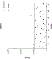
图2为肝癌复发组织和术后未复发组织中LAS1L蛋白的蛋白质表达量强度值图;Figure 2 is a graph showing the protein expression intensity value of LAS1L protein in liver cancer recurrence tissue and postoperative non-recurrence tissue;
图3为CLTB蛋白的蛋白质表达量强度值的ROC曲线图;Fig. 3 is the ROC curve diagram of the protein expression intensity value of CLTB protein;
图4为肝癌复发组织和术后未复发组织中CLTB蛋白的蛋白质表达量强度值图;Figure 4 is a graph showing the protein expression intensity value of CLTB protein in liver cancer recurrence tissue and postoperative non-recurrence tissue;
图5为JAGN1蛋白的蛋白质表达量强度值的ROC曲线图;Fig. 5 is the ROC curve diagram of the protein expression intensity value of JAGN1 protein;
图6为肝癌复发组织和术后未复发组织中JAGN1蛋白的蛋白质表达量强度值图;Figure 6 is a graph showing the protein expression intensity value of JAGN1 protein in liver cancer recurrence tissue and postoperative non-recurrence tissue;
图7为ALYREF蛋白的蛋白质表达量强度值的ROC曲线图;Fig. 7 is the ROC curve diagram of the protein expression intensity value of ALYREF protein;
图8为肝癌复发组织和术后未复发组织中ALYREF蛋白的蛋白质表达量强度值图;Figure 8 is a graph showing the protein expression intensity value of ALYREF protein in liver cancer recurrence tissue and postoperative non-recurrence tissue;
图9为HNRNPA3蛋白的蛋白质表达量强度值的ROC曲线图;Fig. 9 is the ROC curve diagram of the protein expression intensity value of HNRNPA3 protein;
图10为肝癌复发组织和术后未复发组织中HNRNPA3蛋白的蛋白质表达量强度值图;Figure 10 is a graph of the protein expression intensity value of HNRNPA3 protein in liver cancer recurrence tissue and postoperative non-recurrence tissue;
图11为ALYREF蛋白和HNRNPA3蛋白组合的蛋白质表达量强度值的ROC曲线图;Figure 11 is a ROC curve diagram of the protein expression intensity value of the combination of ALYREF protein and HNRNPA3 protein;
图12为肝癌复发组织和术后未复发组织中ALYREF蛋白和HNRNPA3蛋白组合的蛋白质表达量强度值图。Fig. 12 is a graph showing the protein expression intensity value of the combination of ALYREF protein and HNRNPA3 protein in liver cancer recurrence tissue and postoperative non-recurrence tissue.
具体实施方式Detailed ways
为使本发明实施例的目的、技术方案和优点更加清楚,下面将结合本发明实施例,对本发明实施例中的技术方案进行清楚、完整地描述,显然,所描述的实施例是本发明一部分实施例,而不是全部的实施例。基于本发明中的实施例,本领域普通技术人员在没有作出创造性劳动前提下所获得的所有其他实施例,都属于本发明保护的范围。In order to make the purposes, technical solutions and advantages of the embodiments of the present invention clearer, the technical solutions in the embodiments of the present invention will be clearly and completely described below in conjunction with the embodiments of the present invention. Obviously, the described embodiments are part of the present invention. examples, but not all examples. Based on the embodiments of the present invention, all other embodiments obtained by those of ordinary skill in the art without creative efforts shall fall within the protection scope of the present invention.
与肝癌诊断相关的生物标志物的筛选Screening of biomarkers related to the diagnosis of liver cancer
1、实验步骤1. Experimental steps
(1)蛋白质样品信息(1) Protein sample information
样本:分别取自肝癌复发患者癌组织的样本5例和取自术后未复发患者的肝癌组织35例。Samples: 5 samples were taken from the cancer tissue of patients with recurrence of liver cancer and 35 samples of liver cancer tissue were taken from patients without postoperative recurrence.
(2)样本预处理(2) Sample preprocessing
样品采用SDT(4%(w/v)十二烷基磺酸钠,100mM Tris/HCl pH7.6,0.1M二硫苏糖醇)裂解法提取蛋白质,然后采用BCA法进行蛋白质定量;每个样品取适量蛋白质采用Filteraided proteome preparation(FAS)法进行胰蛋白酶酶解,采用C18 Cartridge对肽段进行脱盐,肽段冻干后加入40μL 0.1%甲酸溶液复溶,肽段定量(OD280)。The samples were lysed with SDT (4% (w/v) sodium dodecyl sulfonate, 100 mM Tris/HCl pH7.6, 0.1 M dithiothreitol) for protein extraction, and then BCA was used for protein quantification; each An appropriate amount of protein was taken from the sample for trypsin digestion by the Filteraided proteome preparation (FAS) method, and the peptide fragment was desalted by C18 Cartridge.
BCA法进行蛋白质定量,是根据吸光值可以推算出蛋白浓度,在碱性条件下,蛋白将Cu2+还原为Cu+,Cu+与BCA试剂形成紫颜色的络合物,两分子BCA螯合一个Cu+。将该水溶性复合物在562nm处的吸收值,与标准曲线对比,即可计算待测蛋白的浓度。The BCA method is used for protein quantification, and the protein concentration can be calculated according to the absorbance value. Under alkaline conditions, the protein reduces Cu 2+ to Cu + , Cu + and BCA reagent form a purple complex, and two molecules of BCA chelate a Cu + . The concentration of the protein to be tested can be calculated by comparing the absorption value of the water-soluble complex at 562 nm with the standard curve.
(3)LC-MS/MS数据采集(3) LC-MS/MS data acquisition
每份样品采用纳升流速的HPLC液相系统Easy nLC进行分离。Each sample was separated using the HPLC liquid system Easy nLC at nanoliter flow rates.
其中:缓冲液A液为0.1%甲酸水溶液,B液为0.1%甲酸乙腈水溶液(乙腈为84%)。Among them: buffer A solution is 0.1% formic acid aqueous solution, B solution is 0.1% formic acid acetonitrile aqueous solution (acetonitrile is 84%).
色谱柱以95%的A液平衡,样品由自动进样器上样到上样柱(Thermo ScientificAcclaim PepMap100, 100μm*2cm, nanoViper C18),经过分析柱(Thermo scientificEASY column, 10cm, ID75μm, 3μm, C18-A2)分离,流速为300nL/min。The chromatographic column was equilibrated with 95% liquid A, and the sample was loaded into the sample column (Thermo Scientific Acclaim PepMap100, 100μm*2cm, nanoViper C18) by the autosampler, and passed through the analytical column (Thermo scientific EASY column, 10cm, ID75μm, 3μm, C18 -A2) separation with a flow rate of 300nL/min.
样品经色谱分离后用Q-Exactive质谱仪进行质谱分析。检测方式为正离子,母离子扫描范围300–1800m/z,一级质谱分辨率为70,000 at 200m/z,AGC(Automatic gaincontrol)target为1e6,Maximum IT为50ms,动态排除时间(Dynamic exclusion)为60.0s。多肽和多肽碎片的质量电荷比按照下列方法采集:每次全扫描(full scan)后采集20个碎片图谱(MS2 scan),MS2 Activation Type为HCD,Isolation window为2m/z,二级质谱分辨率17, 500 at 200m/z,Normalized Collision Energy为30eV,Underfill为0.1%。The samples were chromatographically separated and analyzed by mass spectrometry using a Q-Exactive mass spectrometer. The detection method is positive ion, the precursor ion scanning range is 300–1800m/z, the primary mass spectrometry resolution is 70,000 at 200m/z, the AGC (Automatic gaincontrol) target is 1e6, the Maximum IT is 50ms, and the dynamic exclusion time (Dynamic exclusion) is 60.0s. The mass-to-charge ratios of peptides and peptide fragments were collected according to the following methods: 20 fragments were collected after each full scan (MS2 scan), MS2 Activation Type was HCD, Isolation window was 2m/z, MS2 resolution was 17, 500 at 200m/z, Normalized Collision Energy is 30eV, Underfill is 0.1%.
(4)蛋白质鉴定和定量分析(4) Protein identification and quantitative analysis
质谱分析原始数据为RAW文件,用软件MaxQuant软件(版本号1.5.3.17)进行查库鉴定及定量分析。The original data of mass spectrometry analysis were RAW files, and the software MaxQuant software (version number 1.5.3.17) was used for library identification and quantitative analysis.
iBAQ Intensity是基于iBAQ算法得到的样品X中的蛋白质表达量,近似等于该样品中的蛋白质绝对浓度。LFQ Intensity是基于LFQ算法得到的样品X的蛋白相对表达量,常应用于组间比较。一般Labelfree选择其中之一作为定量结果。iBAQ Intensity is the protein expression amount in sample X obtained based on the iBAQ algorithm, which is approximately equal to the absolute concentration of protein in the sample. LFQ Intensity is the relative protein expression of sample X obtained based on the LFQ algorithm, which is often used for comparison between groups. Generally Labelfree chooses one of them as the quantitative result.
iBAQ(Intensity-based absolute quantification)和LFQ属于Maxquant软件提供的两种不同的蛋白质定量算法。iBAQ (Intensity-based absolute quantification) and LFQ belong to two different protein quantification algorithms provided by Maxquant software.
iBAQ一般用于样本的蛋白质绝对定量,主要算法是基于该蛋白质鉴定到的肽段的强度之和与理论肽段个数的比值。iBAQ is generally used for absolute protein quantification of samples. The main algorithm is based on the ratio of the sum of the intensities of the identified peptides to the theoretical number of peptides.
LFQ一般用于多组间的两两定量比较,主要算法是经过肽段和蛋白质多层的pair-wise矫正。本专利采用LFQ进行蛋白质定量。LFQ is generally used for pairwise quantitative comparison between multiple groups, and the main algorithm is pair-wise correction of peptide and protein layers. This patent uses LFQ for protein quantification.
(5)统计学分析(5) Statistical analysis
对符合同组三次重复数据中至少有两个非空值的数据进行比值计算和统计学分析,包含各比较组的LFQ或者iBAQ强度值比值和P-value;初步筛选出各组间的差异物。Ratio calculation and statistical analysis were performed on the data that conformed to three replicate data in the same group with at least two non-null values, including the ratio and P-value of the LFQ or iBAQ intensity values of each comparison group; the differences between the groups were initially screened out .
进一步根据P-value验证差异蛋白物是否具有显著性。选择同时具有多维统计分析Fold change>2或<0.5认为癌组织和癌旁组织之间该蛋白物的含量存在明显的倍数差异,并且筛选出单变量统计分析P value<0.05的蛋白物,作为具有显著性差异的蛋白质;从而得到差异蛋白分子。然后利用SPSS软件作出差异蛋白物ROC曲线,并计算其曲线下面积(AUC),进而判断其诊断价值。具体判断方法为AUC线下面积大于0.7,且P<0.05,利用约登指数最大时候的阈值标准(cut off值)作为判断肿瘤与否的阈值标准(倍数大于2者认为大于阈值为肿瘤检测阳性,倍数小于0.5者认定小于阈值者为肝癌检测阳性),从而得到较高的敏感度和特异度。Further verify whether the difference protein is significant according to P-value. Selecting both with multi-dimensional statistical analysis Fold change>2 or <0.5, it is considered that there is a significant fold difference in the content of the protein between the cancer tissue and the adjacent tissue, and the protein with a univariate statistical analysis P value <0.05 is selected as the protein with a univariate statistical analysis P value <0.05. Significantly different proteins; thus obtaining differential protein molecules. Then use SPSS software to make ROC curve of differential protein, and calculate its area under the curve (AUC), and then judge its diagnostic value. The specific judgment method is that the area under the AUC line is greater than 0.7, and P < 0.05, and the threshold standard (cut off value) when the Youden index is the largest is used as the threshold standard for judging whether the tumor is or not (multiples greater than 2 are considered to be greater than the threshold as positive for tumor detection. , if the multiple is less than 0.5, it is considered as positive for liver cancer detection if it is less than the threshold), so as to obtain higher sensitivity and specificity.
(6)生物信息学分析(6) Bioinformatics analysis
① GO功能注释① GO function comment
利用Blast2GO对目标蛋白质集合进行GO注释,过程大致可以归纳为序列比对(Blast)、GO条目提取(Mapping)、GO注释(Annotation)和InterProScan补充注释(AnnotationAugmentation)等四个步骤。Using Blast2GO to perform GO annotation on the target protein collection, the process can be roughly summarized into four steps: sequence alignment (Blast), GO entry extraction (Mapping), GO annotation (Annotation) and InterProScan supplementary annotation (AnnotationAugmentation).
② KEGG通路注释② KEGG pathway annotation
利用KAAS(KEGG Automatic Annotation Server)软件,对目标蛋白质集合进行KEGG通路注释。Using KAAS (KEGG Automatic Annotation Server) software, KEGG pathway annotation was performed on the target protein set.
③ GO注释和KEGG注释的富集分析③ Enrichment analysis of GO annotation and KEGG annotation
采用Fisher精确检验(Fisher’s Exact Test),比较各个GO分类或KEGG通路在目标蛋白质集合和总体蛋白质集合中的分布情况,对目标蛋白质集合进行GO注释或KEGG通路注释的富集分析。Fisher's Exact Test (Fisher's Exact Test) was used to compare the distribution of each GO classification or KEGG pathway in the target protein set and the overall protein set, and perform GO annotation or KEGG pathway annotation enrichment analysis on the target protein set.
④蛋白质聚类分析④Protein cluster analysis
首先对目标蛋白质集合的定量信息进行归一化处理(归一化到(-1,1)区间)。然后,使用Complexheatmap R包(R Version 3.4)同时对样品和蛋白质的表达量两个维度进行分类(距离算法:欧几里得,连接方式:Average linkage),并生成层次聚类热图。First, the quantitative information of the target protein set is normalized (normalized to the (-1,1) interval). Then, the Complexheatmap R package (R Version 3.4) was used to simultaneously classify the two dimensions of sample and protein expression (distance algorithm: Euclidean, connection method: Average linkage), and generate a hierarchical clustering heat map.
⑤蛋白质相互作用网络分析⑤Protein interaction network analysis
基于STRING(http://string-db.org/)数据库中的信息查找目标蛋白质之间的相互作用关系,并使用CytoScape软件(版本号:3.2.1)生成相互作用网络并对网络进行分析。Based on the information in the STRING (http://string-db.org/) database, the interaction relationship between the target proteins was found, and the CytoScape software (version number: 3.2.1) was used to generate the interaction network and analyze the network.
(7)差异表达蛋白质筛选(7) Screening of differentially expressed proteins
以倍数变化大于2.0倍(上调大于2倍或者下调小于0.5)且P value小于0.05的标准筛选差异表达蛋白质,各比较组的差异表达蛋白质数目。Differentially expressed proteins were screened by the criteria of fold change greater than 2.0 times (up-regulation greater than 2 times or down-regulation less than 0.5) and P value less than 0.05, and the number of differentially expressed proteins in each comparison group.
(8)实验基本原理(8) The basic principle of the experiment
非标记定量蛋白质组学(Label-free)技术近年来已成为重要的质谱定量方法。Label-free技术的定量原理主要有两种:首先,spectrum counts类的非标记定量方法发展较早,也形成了多种定量算法,但核心原理都是根据MS2的鉴定结果作为定量的基础,各种方法的差别在于后期算法对高通量数据的修正;第二种非标记定量方法的原理是以MS1为基础,计算每个肽段信号在LCMS色谱上的积分。本发明采用的Maxquant算法即基于第二种原理。Label-free quantitative proteomics (Label-free) technology has become an important mass spectrometry quantitative method in recent years. There are two main quantitative principles of Label-free technology: First, the non-label quantitative methods of spectrum counts developed earlier, and a variety of quantitative algorithms have also been formed, but the core principles are based on the identification results of MS2 as the basis for quantification. The difference of the first method is the correction of the high-throughput data by the later algorithm; the principle of the second non-labeled quantitative method is based on MS1, and the integral of each peptide signal on the LCMS chromatogram is calculated. The Maxquant algorithm adopted in the present invention is based on the second principle.
2、实验结果2. Experimental results
经质谱数据分析和将复发患者肝癌组织与未复发患者肝癌组织(术后未复发)的蛋白分子进行比较,从而得到5个蛋白分子,可以作为与肝癌复发的生物标志物。After analyzing the mass spectrometry data and comparing the protein molecules of the liver cancer tissue of the recurrence patient with the liver cancer tissue of the non-recurrence patient (no recurrence after surgery), five protein molecules were obtained, which can be used as biomarkers for the recurrence of liver cancer.
为了评估蛋白分子的蛋白质表达量强度值对肝癌复发的诊断效能,本发明采用了ROC曲线分析,AUC为ROC曲线下的面积,是最常用的评价ROC曲线特征的参数,也是重要的试验准确度指标。若AUC在0.7以下,则表示诊断的准确率较低;AUC在0.7以上,则可以满足临床诊断的要求。In order to evaluate the diagnostic efficacy of the protein expression intensity value of the protein molecule on the recurrence of liver cancer, the present invention adopts the ROC curve analysis. AUC is the area under the ROC curve, which is the most commonly used parameter for evaluating the characteristics of the ROC curve, and is also an important test accuracy. index. If the AUC is below 0.7, it means that the accuracy of the diagnosis is low; if the AUC is above 0.7, it can meet the requirements of clinical diagnosis.
具体结果与分析如下:The specific results and analysis are as follows:
(1)采用LC-MS/MS质谱分析法,检测到LAS1L蛋白在肝癌复发组织与术后未复发组织中存在差异。(1) Using LC-MS/MS mass spectrometry, it was detected that the LAS1L protein was different in the recurrence tissue of liver cancer and the non-recurrence tissue after surgery.
研究发现,LAS1L蛋白在肝癌复发样本显著性上调了11.68倍,p值<0.05。The study found that the LAS1L protein was significantly up-regulated by 11.68 times in the recurrence of liver cancer samples, p value < 0.05.
由图1可知,LAS1L蛋白的AUC为0.806>0.7,说明LAS1L蛋白具有较好的判断效果,可以作为肝癌是否复发的生物标志物。It can be seen from Figure 1 that the AUC of LAS1L protein is 0.806>0.7, indicating that LAS1L protein has a good judgment effect and can be used as a biomarker for the recurrence of liver cancer.
在LAS1L蛋白的cut off值为52085687.5时,灵敏度为80%,特异度为77.1%。当进行个体检测时,LAS1L蛋白的蛋白质表达量强度值大于52085687.5时,被判为复发患者,否则被判为未复发患者(假阳性率为22.9%)。When the cut off value of LAS1L protein was 52085687.5, the sensitivity was 80% and the specificity was 77.1%. When the individual detection was performed, when the protein expression intensity value of LAS1L protein was greater than 52085687.5, it was judged as a relapsed patient, otherwise it was judged as a non-relapsed patient (false positive rate was 22.9%).
由图2可知,肝癌复发组织样本主要分布在检测阈值(图2中实线)以上,术后未复发组织主要分布在检测阈值以下,说明肝癌复发组织和术后未复发组织的蛋白质表达量强度值相差甚大,该检测阈值检测效果良好。It can be seen from Figure 2 that the liver cancer recurrence tissue samples are mainly distributed above the detection threshold (solid line in Figure 2), and the postoperative non-recurrence tissue samples are mainly distributed below the detection threshold, indicating the intensity of protein expression in the liver cancer recurrence tissue and postoperative non-recurrence tissue. The value is very different, the detection threshold detection effect is good.
综上所述,LAS1L蛋白可以作肝癌复发的生物标志物。In conclusion, LAS1L protein can be used as a biomarker for liver cancer recurrence.
(2)采用LC-MS/MS质谱分析法,检测到CLTB蛋白在肝癌复发组织与术后未复发组织中存在差异。(2) Using LC-MS/MS mass spectrometry, it was detected that the CLTB protein was different in the recurrence tissue of liver cancer and the non-recurrence tissue after surgery.
研究发现,CLTB蛋白在肝癌复发样本显著性上调了6.68倍,p值<0.05。The study found that CLTB protein was significantly up-regulated by 6.68 times in liver cancer recurrence samples, p value < 0.05.
由图3可知,CLTB蛋白质的AUC为0.806>0.7,说明CLTB蛋白具有较好的判断效果,可以作为肝癌是否复发的生物标志物。It can be seen from Figure 3 that the AUC of CLTB protein is 0.806>0.7, indicating that CLTB protein has a good judgment effect and can be used as a biomarker for the recurrence of liver cancer.
在CLTB蛋白的蛋白质表达量强度值为941197795时,灵敏度为80%,特异度为77.1%。当进行个体检测时,CLTB蛋白的蛋白质表达量强度值大于941197795时,被判为复发患者,否则被判为未复发患者(假阳性率为22.9%)。When the protein expression intensity value of CLTB protein was 941197795, the sensitivity was 80% and the specificity was 77.1%. When the individual detection was performed, when the protein expression intensity value of CLTB protein was greater than 941197795, it was judged as a relapsed patient, otherwise it was judged as a non-relapsed patient (false positive rate was 22.9%).
由图4可知,肝癌复发组织样本主要分布在检测阈值(图4中实线)以上,术后未复发组织主要分布在检测阈值以下,说明肝癌复发组织和术后未复发组织的蛋白质表达量强度值相差甚大,该检测阈值检测效果良好。It can be seen from Figure 4 that the liver cancer recurrence tissue samples are mainly distributed above the detection threshold (solid line in Figure 4), and the postoperative non-recurrence tissue samples are mainly distributed below the detection threshold, indicating the intensity of protein expression in the liver cancer recurrence tissue and postoperative non-recurrence tissue. The value is very different, the detection threshold detection effect is good.
综上所述,CLTB蛋白可以作为肝癌复发的生物标志物。In conclusion, CLTB protein can be used as a biomarker for liver cancer recurrence.
(3)采用LC-MS/MS质谱分析法,检测到JAGN1蛋白在肝癌复发组织与术后未复发组织中存在差异。(3) Using LC-MS/MS mass spectrometry analysis, it was detected that the JAGN1 protein was different in the recurrence tissue of liver cancer and the non-recurrence tissue after surgery.
研究发现,JAGN1蛋白在肝癌复发样本显著性上调了2.45倍,p值<0.05。The study found that JAGN1 protein was significantly up-regulated by 2.45 times in the recurrence of liver cancer samples, p value < 0.05.
由图5可知,JAGN1蛋白的AUC为0.840>0.7,说明JAGN1蛋白具有较好的判断效果,可以作肝癌是否复发的生物标志物。It can be seen from Figure 5 that the AUC of JAGN1 protein is 0.840>0.7, indicating that JAGN1 protein has a good judgment effect and can be used as a biomarker for the recurrence of liver cancer.
在JAGN1蛋白的蛋白质表达量强度值为467852297时,灵敏度为80%,特异度为91.4%。JAGN1蛋白的当进行个体检测时,蛋白质表达量强度值大于467852297时,被判为复发患者,否则被判为未复发患者(假阳性率为8.6%)。When the protein expression intensity value of JAGN1 protein was 467852297, the sensitivity was 80% and the specificity was 91.4%. When the JAGN1 protein was detected individually, when the protein expression intensity value was greater than 467852297, it was judged as a relapsed patient, otherwise it was judged as a non-relapsed patient (false positive rate was 8.6%).
由图6可知,肝癌复发组织样本主要分布在检测阈值(图6中实线)以上,术后未复发组织主要分布在检测阈值以下,说明肝癌复发组织和术后未复发组织的蛋白质表达量强度值相差甚大,该检测阈值检测效果良好。It can be seen from Figure 6 that the liver cancer recurrence tissue samples are mainly distributed above the detection threshold (solid line in Figure 6), and the postoperative non-recurrence tissue samples are mainly distributed below the detection threshold, indicating the intensity of protein expression in the liver cancer recurrence tissue and postoperative non-recurrence tissue. The value is very different, the detection threshold detection effect is good.
综上所述,JAGN1蛋白可以作为肝癌复发的生物标志物。In conclusion, JAGN1 protein can be used as a biomarker for liver cancer recurrence.
(4)采用LC-MS/MS质谱分析法,检测到ALYREF蛋白在复发组织与未复发组织中存在差异。(4) Using LC-MS/MS mass spectrometry, it was detected that the ALYREF protein was different in the relapsed tissue and the non-relapsed tissue.
研究发现,ALYREF蛋白在肝癌复发样本显著性下调了0.19倍,p值<0.05。The study found that ALYREF protein was significantly down-regulated by 0.19 times in liver cancer recurrence samples, p value < 0.05.
由图7可知,ALYREF蛋白的AUC为0.937>0.7,说明ALYREF蛋白具有较好的判断效果,可以作为肝癌是否复发的生物标志物。It can be seen from Fig. 7 that the AUC of ALYREF protein is 0.937>0.7, indicating that ALYREF protein has a good judgment effect and can be used as a biomarker for the recurrence of liver cancer.
在ALYREF蛋白的cut off值为149218152时,灵敏度为100%,特异度为77.1%。当进行个体检测时,ALYREF蛋白的蛋白质表达量强度值小于149218152时,被判为复发患者,否则被判为术后未复发患者(假阳性率为22.9%)。When the cut off value of ALYREF protein was 149218152, the sensitivity was 100% and the specificity was 77.1%. When the individual detection was performed, when the protein expression intensity value of ALYREF protein was less than 149218152, it was judged as a recurrence patient, otherwise it was judged as a non-relapsed patient after surgery (false positive rate was 22.9%).
由图8可知,肝癌复发组织样本主要分布在检测阈值(图8中实线)以下,未复发组织主要分布在检测阈值以上,说明肝癌复发组织和未复发组织的蛋白质表达量强度值相差甚大,该检测阈值检测效果良好。It can be seen from Figure 8 that the samples of recurrent liver cancer tissue are mainly distributed below the detection threshold (solid line in Figure 8), and the non-recurrence tissue samples are mainly distributed above the detection threshold, indicating that the protein expression intensity values of recurrent liver cancer tissue and non-recurrence tissue are very different. The detection threshold detection effect is good.
综上所述,ALYREF蛋白可以作为肝癌复发的生物标志物。In conclusion, ALYREF protein can be used as a biomarker for liver cancer recurrence.
(5)采用LC-MS/MS质谱分析法,检测到HNRNPA3蛋白在复发组织与未复发组织中存在差异。(5) Using LC-MS/MS mass spectrometry, it was detected that the HNRNPA3 protein was different in the relapsed and non-recurrent tissues.
研究发现,HNRNPA3蛋白在肝癌复发样本显著性下调了0.49倍,p值<0.05。The study found that HNRNPA3 protein was significantly down-regulated by 0.49 times in liver cancer recurrence samples, p value <0.05.
由图9可知,HNRNPA3蛋白的AUC为0.920>0.7,说明HNRNPA3蛋白具有较好的判断效果,可以作为肝癌复发是否的生物标志物。It can be seen from Figure 9 that the AUC of HNRNPA3 protein is 0.920>0.7, indicating that HNRNPA3 protein has a good judgment effect and can be used as a biomarker for liver cancer recurrence.
在HNRNPA3蛋白的蛋白质表达量强度值为4386125605时,灵敏度为100%,特异度为80%。当进行个体检测时,HNRNPA3蛋白的蛋白质表达量强度值小于4386125605时,被判为复发患者,否则被判为术后未复发患者(假阳性率为20%)。When the protein expression intensity value of HNRNPA3 protein was 4386125605, the sensitivity was 100% and the specificity was 80%. When the individual detection was performed, when the protein expression intensity value of HNRNPA3 protein was less than 4386125605, it was judged as a relapsed patient, otherwise it was judged as a postoperative non-relapsed patient (false positive rate was 20%).
由图10可知,肝癌复发组织样本主要分布在检测阈值(图10中实线)以下,未复发组织主要分布在检测阈值以上,说明肝癌复发组织和未复发组织的蛋白质表达量强度值相差甚大,该检测阈值检测效果良好。It can be seen from Figure 10 that the samples of liver cancer recurrence tissue are mainly distributed below the detection threshold (solid line in Figure 10), and the non-recurrence tissue samples are mainly distributed above the detection threshold, indicating that the protein expression intensity values of liver cancer recurrence tissue and non-recurrence tissue are very different. The detection threshold detection effect is good.
综上所述,HNRNPA3蛋白可以作为肝癌复发的生物标志物。In conclusion, HNRNPA3 protein can be used as a biomarker for liver cancer recurrence.
(6)ALYREF蛋白和HNRNPA3蛋白组合在制备早期肝癌诊断试剂盒中的应用。(6) Application of the combination of ALYREF protein and HNRNPA3 protein in the preparation of early-stage liver cancer diagnostic kit.
本发明还提供一种肝癌的诊断方法,步骤为:采用二元逻辑回归分析计算P(肝癌术后复发概率)的数值,SPSS软件二元逻辑回归后得到的公式为:The present invention also provides a method for diagnosing liver cancer. The steps are: adopting binary logistic regression analysis to calculate the value of P (probability of recurrence of liver cancer after operation), and the formula obtained after the binary logistic regression of SPSS software is:

其中a为ALYREF蛋白的蛋白质表达量强度值,b为HNRNPA3蛋白的蛋白质表达量强度值;若检测P(肝癌术后复发概率)大于0.0694114则被判为会复发患者,否则被判为未复发患者。Among them, a is the protein expression intensity value of ALYREF protein, and b is the protein expression intensity value of HNRNPA3 protein; if the detected P (probability of recurrence after liver cancer surgery) is greater than 0.0694114, it is judged as a patient with recurrence, otherwise it is judged as a patient without recurrence .
如图11所示,组合蛋白的AUC为0.977>0.7,说明具有较好的判断效果,即组合蛋白可以作为肝癌是否复发的生物标志物。As shown in Figure 11, the AUC of the combined protein was 0.977>0.7, indicating that it has a good judgment effect, that is, the combined protein can be used as a biomarker for the recurrence of liver cancer.
在组合蛋白的蛋白质表达量强度值为0.0694114时,灵敏度为100%,特异度为88.6%。当进行个体检测时,组合蛋白的蛋白质表达量强度值大于0.0694114时,被判为复发患者,否则被判为术后未复发患者(假阳性率为11.4%)。When the protein expression intensity value of the combined protein was 0.0694114, the sensitivity was 100% and the specificity was 88.6%. When the individual detection was performed, when the protein expression intensity value of the combined protein was greater than 0.0694114, it was judged as a patient with recurrence, otherwise it was judged as a patient without recurrence after surgery (false positive rate was 11.4%).
由图12可知,肝癌复发组织样本主要分布在检测阈值(图12中实线)以上,未复发组织主要分布在检测阈值以上,说明肝癌复发组织和未复发组织的蛋白质表达量强度值相差甚大,该检测阈值检测效果良好。It can be seen from Figure 12 that the samples of liver cancer recurrence tissue are mainly distributed above the detection threshold (solid line in Figure 12), and the non-recurrence tissue samples are mainly distributed above the detection threshold, indicating that the protein expression intensity values of liver cancer recurrence tissue and non-recurrence tissue are very different. The detection threshold detection effect is good.
鉴于上述结果,ALYREF蛋白和HNRNPA3蛋白组成的组合蛋白可以作为肝癌复发的生物标志物。In view of the above results, the combined protein composed of ALYREF protein and HNRNPA3 protein can be used as a biomarker of liver cancer recurrence.
实施例1Example 1
用于预测肝癌复发的生物标志物,包括LAS1L蛋白。Biomarkers for predicting liver cancer recurrence, including LAS1L protein.
用于预测肝癌复发肝癌诊断的生物标志物的预后方法,包括以下步骤:采用LC-MS/MS质谱分析法检测待测样本中LAS1L蛋白的蛋白质表达量强度值;样本中LAS1L蛋白的蛋白质表达量强度值大于52085687.5时,被判为复发患者,否则被判为未复发患者(假阳性率为22.9%)。A prognostic method for biomarkers for predicting the recurrence of liver cancer and liver cancer diagnosis, comprising the following steps: detecting the protein expression intensity value of LAS1L protein in a sample to be tested by LC-MS/MS mass spectrometry; the protein expression level of LAS1L protein in the sample When the intensity value is greater than 52085687.5, it is judged as a relapsed patient, otherwise it is judged as a non-relapsed patient (false positive rate 22.9%).
一种诊断肝癌是否复发的试剂盒,包括特异性检测LAS1L蛋白的试剂,特异性检测LAS1L蛋白的试剂是特异性识别LAS1L蛋白核酸的探针。A kit for diagnosing the recurrence of liver cancer comprises a reagent for specifically detecting LAS1L protein, and the reagent for specifically detecting LAS1L protein is a probe for specifically recognizing nucleic acid of LAS1L protein.
实施例2Example 2
用于预测肝癌复发的生物标志物,包括CLTB蛋白。Biomarkers for predicting liver cancer recurrence, including CLTB protein.
用于预测肝癌复发肝癌诊断的生物标志物的预后方法,包括以下步骤:采用LC-MS/MS质谱分析法检测待测样本中CLTB蛋白的蛋白质表达量强度值;样本中CLTB蛋白的蛋白质表达量强度值大于941197795时,被判为复发患者,否则被判为未复发患者(假阳性率为22.9%)。A prognostic method for biomarkers for predicting liver cancer recurrence and liver cancer diagnosis, comprising the following steps: detecting the protein expression intensity value of CLTB protein in a sample to be tested by LC-MS/MS mass spectrometry; the protein expression level of CLTB protein in the sample When the intensity value is greater than 941197795, it is judged as a relapsed patient, otherwise it is judged as a non-relapsed patient (false positive rate 22.9%).
一种诊断肝癌是否复发的试剂盒,包括特异性检测CLTB蛋白的试剂,特异性检测CLTB蛋白的试剂是特异性识别CLTB蛋白核酸的探针。A kit for diagnosing the recurrence of liver cancer includes a reagent for specifically detecting CLTB protein, and the reagent for specifically detecting CLTB protein is a probe for specifically recognizing nucleic acid of CLTB protein.
实施例3Example 3
用于预测肝癌复发的生物标志物,包括JAGN1蛋白。Biomarkers for predicting liver cancer recurrence, including JAGN1 protein.
用于预测肝癌复发肝癌诊断的生物标志物的预后方法,包括以下步骤:采用LC-MS/MS质谱分析法检测待测样本中JAGN1蛋白的蛋白质表达量强度值;样本中JAGN1蛋白的蛋白质表达量强度值大于467852297时,被判为复发患者,否则被判为未复发患者(假阳性率为8.6%)。A prognostic method for biomarkers for predicting the recurrence of liver cancer and liver cancer diagnosis, comprising the following steps: detecting the protein expression intensity value of JAGN1 protein in a sample to be tested by LC-MS/MS mass spectrometry; the protein expression level of JAGN1 protein in the sample When the intensity value is greater than 467852297, it is judged as a relapsed patient, otherwise it is judged as a non-relapsed patient (the false positive rate is 8.6%).
一种诊断肝癌是否复发的试剂盒,包括特异性检测JAGN1蛋白的试剂,特异性检测JAGN1蛋白的试剂是特异性识别JAGN1蛋白核酸的探针。A kit for diagnosing the recurrence of liver cancer includes a reagent for specifically detecting JAGN1 protein, and the reagent for specifically detecting JAGN1 protein is a probe for specifically recognizing nucleic acid of JAGN1 protein.
实施例4Example 4
用于预测肝癌复发的生物标志物,包括ALYREF蛋白。Biomarkers for predicting liver cancer recurrence, including ALYREF protein.
用于预测肝癌复发肝癌诊断的生物标志物的预后方法,包括以下步骤:采用LC-MS/MS质谱分析法检测待测样本中ALYREF蛋白的蛋白质表达量强度值;样本中ALYREF蛋白的蛋白质表达量强度值小于149218152时,被判为复发患者,否则被判为术后未复发患者(假阳性率为22.9%)。A prognostic method for biomarkers used to predict the recurrence of liver cancer and liver cancer diagnosis, comprising the following steps: detecting the protein expression intensity value of ALYREF protein in a sample to be tested by LC-MS/MS mass spectrometry; the protein expression level of ALYREF protein in the sample When the intensity value is less than 149218152, it is judged as a recurrence patient, otherwise it is judged as a postoperative non-recurrence patient (false positive rate is 22.9%).
一种诊断肝癌是否复发的试剂盒,包括特异性检测ALYREF蛋白的试剂,特异性检测ALYREF蛋白的试剂是特异性识别ALYREF蛋白核酸的探针。A kit for diagnosing the recurrence of liver cancer comprises a reagent for specifically detecting ALYREF protein, and the reagent for specifically detecting ALYREF protein is a probe for specifically recognizing nucleic acid of ALYREF protein.
实施例5Example 5
用于预测肝癌复发的生物标志物,包括HNRNPA3蛋白。Biomarkers for predicting liver cancer recurrence, including HNRNPA3 protein.
用于预测肝癌复发肝癌诊断的生物标志物的预后方法,包括以下步骤:采用LC-MS/MS质谱分析法检测待测样本中HNRNPA3蛋白的蛋白质表达量强度值;样本中HNRNPA3蛋白的蛋白质表达量强度值小于4386125605时,被判为复发患者,否则被判为术后未复发患者(假阳性率为20%)。A prognostic method for biomarkers for predicting liver cancer recurrence and liver cancer diagnosis, comprising the following steps: detecting the protein expression intensity value of HNRNPA3 protein in a sample to be tested by LC-MS/MS mass spectrometry; the protein expression level of HNRNPA3 protein in the sample When the intensity value is less than 4386125605, it is judged as a recurrence patient, otherwise it is judged as a postoperative non-recurrence patient (false positive rate is 20%).
一种诊断肝癌是否复发的试剂盒,包括特异性检测HNRNPA3蛋白的试剂,特异性检测HNRNPA3蛋白的试剂是特异性识别HNRNPA3蛋白核酸的探针。A kit for diagnosing the recurrence of liver cancer includes a reagent for specifically detecting HNRNPA3 protein, and the reagent for specifically detecting HNRNPA3 protein is a probe for specifically recognizing nucleic acid of HNRNPA3 protein.
实施例6Example 6
肝癌诊断的生物标志物,包括ALYREF蛋白和HNRNPA3蛋白。Biomarkers for liver cancer diagnosis, including ALYREF protein and HNRNPA3 protein.
肝癌诊断的生物标志物的预后方法,包括以下步骤:采用二元逻辑回归分析计算P(肝癌术后复发概率)的数值,SPSS软件二元逻辑回归后得到的公式为:The prognostic method of biomarkers for liver cancer diagnosis includes the following steps: using binary logistic regression analysis to calculate the value of P (probability of recurrence after liver cancer operation), the formula obtained after binary logistic regression in SPSS software is:
其中:a为ALYREF蛋白的蛋白质表达量强度值,b为HNRNPA3蛋白的蛋白质表达量强度值;若检测P(肝癌术后复发概率)大于0.0694114则判为会复发患者,否则判为未复发患者。Among them: a is the protein expression intensity value of ALYREF protein, and b is the protein expression intensity value of HNRNPA3 protein; if the detection P (probability of recurrence after liver cancer surgery) is greater than 0.0694114, it is judged as a patient with recurrence, otherwise it is judged as a patient without recurrence.
当进行个体检测时,组合蛋白的蛋白质表达量强度值大于0.0694114时,被判为复发患者,否则被判为术后未复发患者(假阳性率为11.4%)。When the individual detection was performed, when the protein expression intensity value of the combined protein was greater than 0.0694114, it was judged as a patient with recurrence, otherwise it was judged as a patient without recurrence after surgery (false positive rate was 11.4%).
本发明以LAS1L蛋白,CLTB蛋白,JAGN1蛋白,ALYREF蛋白和HNRNPA3蛋白中至少一种蛋白作为生物标志物对被试者进行肝癌诊断,简单易行、诊断过程安全有效、易为病人所接受、诊断标准统一受个人主观因素影响较小。The present invention uses at least one protein among LAS1L protein, CLTB protein, JAGN1 protein, ALYREF protein and HNRNPA3 protein as biomarkers to diagnose liver cancer for subjects, which is simple and easy to implement, safe and effective in the diagnosis process, and easy to be accepted and diagnosed by patients. Standard unification is less affected by individual subjective factors.
以上实施例仅用以说明本发明的技术方案,而非对其限制;尽管参照前述实施例对本发明进行了详细的说明,本领域的普通技术人员应当理解:其依然可以对前述各实施例所记载的技术方案进行修改,或者对其中部分技术特征进行等同替换;而这些修改或者替换,并不使相应技术方案的本质脱离本发明各实施例技术方案的精神和范围。The above embodiments are only used to illustrate the technical solutions of the present invention, but not to limit them; although the present invention has been described in detail with reference to the foregoing embodiments, those of ordinary skill in the art should understand that: The recorded technical solutions are modified, or some technical features thereof are equivalently replaced; and these modifications or replacements do not make the essence of the corresponding technical solutions deviate from the spirit and scope of the technical solutions of the embodiments of the present invention.
Claims (9)
Priority Applications (1)
| Application Number | Priority Date | Filing Date | Title |
|---|---|---|---|
| CN202010514464.XA CN111748624B (en) | 2020-06-08 | 2020-06-08 | Biomarker for predicting whether liver cancer is recurrent |
Applications Claiming Priority (1)
| Application Number | Priority Date | Filing Date | Title |
|---|---|---|---|
| CN202010514464.XA CN111748624B (en) | 2020-06-08 | 2020-06-08 | Biomarker for predicting whether liver cancer is recurrent |
Publications (2)
| Publication Number | Publication Date |
|---|---|
| CN111748624A true CN111748624A (en) | 2020-10-09 |
| CN111748624B CN111748624B (en) | 2022-11-04 |
Family
ID=72675139
Family Applications (1)
| Application Number | Title | Priority Date | Filing Date |
|---|---|---|---|
| CN202010514464.XA Active CN111748624B (en) | 2020-06-08 | 2020-06-08 | Biomarker for predicting whether liver cancer is recurrent |
Country Status (1)
| Country | Link |
|---|---|
| CN (1) | CN111748624B (en) |
Cited By (2)
| Publication number | Priority date | Publication date | Assignee | Title |
|---|---|---|---|---|
| CN113528670A (en) * | 2021-08-23 | 2021-10-22 | 郑州大学第一附属医院 | A biomarker and detection kit for predicting the risk of late postoperative recurrence in patients with liver cancer |
| CN115873950A (en) * | 2022-11-17 | 2023-03-31 | 浙江大学医学院附属第一医院 | Application of binding protein ALYREF based on m5C methylation in liver cancer |
Citations (8)
| Publication number | Priority date | Publication date | Assignee | Title |
|---|---|---|---|---|
| US20140045915A1 (en) * | 2010-08-31 | 2014-02-13 | The General Hospital Corporation | Cancer-related biological materials in microvesicles |
| US20160320395A1 (en) * | 2013-11-13 | 2016-11-03 | Electrophoretics Limited | Materials and methods for diagnosis and prognosis of liver cancer |
| CN106282321A (en) * | 2015-05-26 | 2017-01-04 | 中山大学 | The liver cancer recurrence risk profile mark being made up of tissue snoRNA and test kit |
| WO2019140387A1 (en) * | 2018-01-12 | 2019-07-18 | Kymera Therapeutics, Inc. | Crbn ligands and uses thereof |
| CN110446790A (en) * | 2016-11-30 | 2019-11-12 | 外来体诊断公司 | Methods and compositions for detection of mutations in plasma using exosomal RNA and cell-free DNA from non-small cell lung cancer patients |
| CN110499364A (en) * | 2019-07-30 | 2019-11-26 | 北京凯昂医学诊断技术有限公司 | A kind of probe groups and its kit and application for detecting the full exon of extended pattern hereditary disease |
| WO2020010177A1 (en) * | 2018-07-06 | 2020-01-09 | Kymera Therapeutics, Inc. | Tricyclic crbn ligands and uses thereof |
| CN111187839A (en) * | 2020-01-17 | 2020-05-22 | 郑州大学第一附属医院 | Application of m5C methylation related regulatory gene in liver cancer prognosis prediction |
-
2020
- 2020-06-08 CN CN202010514464.XA patent/CN111748624B/en active Active
Patent Citations (9)
| Publication number | Priority date | Publication date | Assignee | Title |
|---|---|---|---|---|
| US20140045915A1 (en) * | 2010-08-31 | 2014-02-13 | The General Hospital Corporation | Cancer-related biological materials in microvesicles |
| US20160153053A1 (en) * | 2010-08-31 | 2016-06-02 | The General Hospital Corporation | Cancer-related biological materials in microvesicles |
| US20160320395A1 (en) * | 2013-11-13 | 2016-11-03 | Electrophoretics Limited | Materials and methods for diagnosis and prognosis of liver cancer |
| CN106282321A (en) * | 2015-05-26 | 2017-01-04 | 中山大学 | The liver cancer recurrence risk profile mark being made up of tissue snoRNA and test kit |
| CN110446790A (en) * | 2016-11-30 | 2019-11-12 | 外来体诊断公司 | Methods and compositions for detection of mutations in plasma using exosomal RNA and cell-free DNA from non-small cell lung cancer patients |
| WO2019140387A1 (en) * | 2018-01-12 | 2019-07-18 | Kymera Therapeutics, Inc. | Crbn ligands and uses thereof |
| WO2020010177A1 (en) * | 2018-07-06 | 2020-01-09 | Kymera Therapeutics, Inc. | Tricyclic crbn ligands and uses thereof |
| CN110499364A (en) * | 2019-07-30 | 2019-11-26 | 北京凯昂医学诊断技术有限公司 | A kind of probe groups and its kit and application for detecting the full exon of extended pattern hereditary disease |
| CN111187839A (en) * | 2020-01-17 | 2020-05-22 | 郑州大学第一附属医院 | Application of m5C methylation related regulatory gene in liver cancer prognosis prediction |
Cited By (2)
| Publication number | Priority date | Publication date | Assignee | Title |
|---|---|---|---|---|
| CN113528670A (en) * | 2021-08-23 | 2021-10-22 | 郑州大学第一附属医院 | A biomarker and detection kit for predicting the risk of late postoperative recurrence in patients with liver cancer |
| CN115873950A (en) * | 2022-11-17 | 2023-03-31 | 浙江大学医学院附属第一医院 | Application of binding protein ALYREF based on m5C methylation in liver cancer |
Also Published As
| Publication number | Publication date |
|---|---|
| CN111748624B (en) | 2022-11-04 |
Similar Documents
| Publication | Publication Date | Title |
|---|---|---|
| CN107064508A (en) | Aid in colorectal cancer early diagnosis and molecular marker and its application of Prognosis scoveillance | |
| CN105572355B (en) | Detect the biomarker of the cancer of the esophagus | |
| CN105277718A (en) | Product for malignant tumor related screening and assessing, and application and method thereof | |
| WO2023179263A1 (en) | System, model and kit for evaluating malignancy grade or probability of thyroid nodules | |
| CN115032395B (en) | Plasma protein marker for identifying lung adenocarcinoma and benign pulmonary nodules and application thereof | |
| EP3257953A1 (en) | Methods of identification, assessment, prevention and therapy of lung diseases and kits thereof including gender-based disease identification, assessment, prevention and therapy | |
| CN114032284A (en) | Esophageal cancer detection reagent and application thereof in esophageal cancer detection | |
| CN118362666A (en) | Metabolic marker composition for distinguishing health, non-colorectal cancer disease and colorectal cancer and its application | |
| CN111748624B (en) | Biomarker for predicting whether liver cancer is recurrent | |
| Song et al. | MALDI‐TOF‐MS analysis in low molecular weight serum peptidome biomarkers for NSCLC | |
| Wan et al. | Retinol-binding protein 4 as a promising serum biomarker for the diagnosis and prognosis of hepatocellular Carcinoma | |
| CN111751550B (en) | Biomarkers for the diagnosis of liver cancer and their prognostic methods | |
| CN101451975A (en) | Method for detecting cancer of stomach prognosis and staging blood serum protein | |
| CN115684451B (en) | Metabolic-study-based esophageal squamous carcinoma lymph node metastasis diagnosis marker and application thereof | |
| Langbein et al. | Protein profiling of bladder cancer using the 2D-PAGE and SELDI-TOF-MS technique | |
| CN107273717A (en) | A kind of detection model of Sera of Lung Cancer gene and its construction method and application | |
| CN114428172A (en) | Marker for predicting postoperative distant metastasis risk of colorectal cancer patient and application thereof | |
| CN116106453B (en) | Application of D-sorbitol in screening of esophageal squamous cell carcinoma | |
| CN111748623B (en) | Predictive marker and kit for recurrence of liver cancer patient | |
| CN116359374A (en) | Metabolic group marker and kit for early screening of esophageal cancer | |
| CN101210929A (en) | Method for detecting endometriosis plasma marker protein | |
| Liu et al. | Potential biomarkers for esophageal carcinoma detected by matrix-assisted laser desorption/ionization time-of-flight mass spectrometry. | |
| CN111751551B (en) | Protein molecule as biomarker for diagnosing liver cirrhosis and prognosis method thereof | |
| CN113447586B (en) | Marker for cardiac cancer screening and detection kit | |
| CN114578060A (en) | A method based on SAMHD1 protein as a predictor of efficacy in stage II colorectal cancer |
Legal Events
| Date | Code | Title | Description |
|---|---|---|---|
| PB01 | Publication | ||
| PB01 | Publication | ||
| SE01 | Entry into force of request for substantive examination | ||
| SE01 | Entry into force of request for substantive examination | ||
| GR01 | Patent grant | ||
| GR01 | Patent grant | ||
| TR01 | Transfer of patent right |
Effective date of registration: 20230329 Address after: Room 102, Management Office, Floor 1, Building 3, the Taihu Lake New Town Science and Technology Innovation Park, No. 18, Suzhou River Road, East the Taihu Lake Ecological Tourism Resort (the Taihu Lake New Town), Wujiang District, Suzhou City, Jiangsu Province, 215200 Patentee after: Phoenix Intelligent Pharmaceutical Biotechnology (Suzhou) Co.,Ltd. Address before: 450000 East Construction Road No. 50, Erqi District, Zhengzhou City, Henan Province Patentee before: THE FIRST AFFILIATED HOSPITAL OF ZHENGZHOU University |
|
| TR01 | Transfer of patent right |