CN110069798B - Dental jaw reference model for light-cured three-dimensional printing precision evaluation and evaluation method - Google Patents
Dental jaw reference model for light-cured three-dimensional printing precision evaluation and evaluation method Download PDFInfo
- Publication number
- CN110069798B CN110069798B CN201810927498.4A CN201810927498A CN110069798B CN 110069798 B CN110069798 B CN 110069798B CN 201810927498 A CN201810927498 A CN 201810927498A CN 110069798 B CN110069798 B CN 110069798B
- Authority
- CN
- China
- Prior art keywords
- tooth
- teeth
- model
- width
- simulated
- Prior art date
- Legal status (The legal status is an assumption and is not a legal conclusion. Google has not performed a legal analysis and makes no representation as to the accuracy of the status listed.)
- Active
Links
Images


Classifications
-
- A—HUMAN NECESSITIES
- A61—MEDICAL OR VETERINARY SCIENCE; HYGIENE
- A61C—DENTISTRY; APPARATUS OR METHODS FOR ORAL OR DENTAL HYGIENE
- A61C19/00—Dental auxiliary appliances
- A61C19/04—Measuring instruments specially adapted for dentistry
-
- G—PHYSICS
- G06—COMPUTING OR CALCULATING; COUNTING
- G06F—ELECTRIC DIGITAL DATA PROCESSING
- G06F30/00—Computer-aided design [CAD]
- G06F30/20—Design optimisation, verification or simulation
-
- Y—GENERAL TAGGING OF NEW TECHNOLOGICAL DEVELOPMENTS; GENERAL TAGGING OF CROSS-SECTIONAL TECHNOLOGIES SPANNING OVER SEVERAL SECTIONS OF THE IPC; TECHNICAL SUBJECTS COVERED BY FORMER USPC CROSS-REFERENCE ART COLLECTIONS [XRACs] AND DIGESTS
- Y02—TECHNOLOGIES OR APPLICATIONS FOR MITIGATION OR ADAPTATION AGAINST CLIMATE CHANGE
- Y02P—CLIMATE CHANGE MITIGATION TECHNOLOGIES IN THE PRODUCTION OR PROCESSING OF GOODS
- Y02P10/00—Technologies related to metal processing
- Y02P10/25—Process efficiency
Landscapes
- Engineering & Computer Science (AREA)
- Health & Medical Sciences (AREA)
- Theoretical Computer Science (AREA)
- Physics & Mathematics (AREA)
- Life Sciences & Earth Sciences (AREA)
- Biomedical Technology (AREA)
- Oral & Maxillofacial Surgery (AREA)
- General Engineering & Computer Science (AREA)
- Geometry (AREA)
- Evolutionary Computation (AREA)
- Computer Hardware Design (AREA)
- Biophysics (AREA)
- General Physics & Mathematics (AREA)
- Dentistry (AREA)
- Epidemiology (AREA)
- Animal Behavior & Ethology (AREA)
- General Health & Medical Sciences (AREA)
- Public Health (AREA)
- Veterinary Medicine (AREA)
- Dental Tools And Instruments Or Auxiliary Dental Instruments (AREA)
Abstract
本发明涉及一种光固化三维打印精度评价用牙颌参考模型及评价方法,包括:上颌牙颌的第11牙和第21牙,近远中向宽8mm,颊舌向宽7mm,


The present invention relates to a dento-jaw reference model and an evaluation method for light-cured three-dimensional printing accuracy evaluation, comprising: the 11th tooth and the 21st tooth of the maxillary dentition, with a width of 8 mm in the mesio-distal direction and a width of 7 mm in the buccolingual direction,
Description
Technical Field
The invention relates to a simulated dental reference model, in particular to a dental reference model for light-cured three-dimensional printing precision evaluation and an evaluation method.
Background
The plaster dental model is an important tool for clinical practice of traditional stomatology, can be used for analyzing, diagnosing and recording the state of illness of patients, so as to carry out treatment design and curative effect evaluation, and in the disciplines of oral restoration, orthodontics, orthognathic surgery and the like, some key treatment steps also need to be completed by depending on the plaster dental model. However, the plaster dental model has the defects of easy damage, large storage burden, high density, difficult realization of remote data sharing and the like. With the development of digital technology in recent years, various three-dimensional imaging and optical scanning technologies enrich data acquisition paths, and the application of three-dimensional printing technology in stomatology is a hot spot for researches of students. Three-dimensional printing (three-dimensional printing), also known as additive manufacturing (additive manufacturing), or rapid prototyping (rapid prototyping), is a technique for materializing three-dimensional data by accumulating material layer by layer based on three-dimensional digital files. The photocuring 3D printing technology has the characteristics of high molding speed, material saving and high molding precision, can show the detailed characteristic of complex dental model, and simultaneously provides possibility for remote data sharing and information long-term storage. Currently, photo-curing 3D printing techniques have been applied to the fabrication of dental diagnostic models, prosthetic substitutes, implant guides, wax-type and temporary restorations.
Whether the precision of the photo-curing 3D printing dental model can meet the clinical requirements of the oral cavity is a concern in the field of stomatology. In recent years, students have initially explored the field of 3D printing precision evaluation, and evaluated for different printing technologies and printing objects. However, the research results are inconsistent due to different principles of 3D printing technology, different printing objects, different measuring methods and other factors. Reviewing the previous studies, a method of measuring a specific measurement index by a vernier caliper and evaluating the accuracy of a 3D printed dental model using a plaster dental model as a standard is more commonly used, but errors in model scanning in this method may affect the results. The learner tries to measure on the digital model obtained by scanning the original model through the commercial software, so as to compare with the measurement result of the vernier caliper on the 3D printing dental model, thereby avoiding the influence of scanning errors. However, subjective influence of selecting measurement points exists in the process, and consistent standards are difficult to achieve by selecting mark points on an irregular physical dental model and a digital model in software, so that high requirements are put on the operation of experimenters, and certain influence is also caused on the evaluation result.
Disclosure of Invention
First, the technical problem to be solved
The invention aims to provide a dental reference model for light-cured three-dimensional printing precision evaluation and an evaluation method, and the core is that a dental reference model is established, the natural dental crown size and dental characteristics are simulated by using a simplified standard geometrical body combination model, the three-dimensional feature size precision of a light-cured 3D printing technology is evaluated by adopting an inherent feature measurement method influenced by non-mark points, the three-dimensional morphological precision of the printing model is evaluated by combining a three-dimensional morphological analysis method, and the precision expression of the 3D printing dental model is comprehensively evaluated; the invention solves the problem of establishing a dental reference model for evaluating the precision of a photocuring 3D printing dental model and a matched evaluation method thereof so as to provide reference and guidance for oral clinical application of the photocuring 3D printing technology.
(II) technical scheme
The invention relates to a dental jaw reference model for light-cured three-dimensional printing precision evaluation, which simulates the dimensions of a natural dental crown and the characteristics of dental jaws by using a simplified standard geometrical body combination model, and specifically comprises the following steps:
1) The 11 th tooth and the 21 st tooth of the maxillary dental reference model have a mesial-distal width of 8mm and a buccal lingual width of 7mm, gingival height 10mm;
 teeth 12 and 22, width of near to far and middle 7mm, width of buccal and lingual 6mm,/o> Gingival height 9mm;
 teeth 13 and 23, width of near to far and middle 8mm, width of buccal and lingual 8mm,/and-> The gingival height is 11mm;
 tooth 14, tooth 24, tooth 15, tooth 25, width of near-far middle 7mm, cheek-tongue width 9mm,> gingival height 10mm; 16 th and 26 th teeth, 10mm in width in the mesial-distal direction, 11mm in width in the buccal-lingual direction,>
 the gingival height is 8mm; 17 th and 27 th teeth, width of near to far to middle 9mm, width of cheek to tongue 11mm,/o>
 Gingival height 5mm; the distance between adjacent parallel surfaces of each tooth is 2mm;
2) The near-far direction of the 12 th tooth, the 11 th tooth, the 21 st tooth and the 22 nd tooth of the maxillary dental reference model is the horizontal direction, and the central axes are on the same horizontal line; the proximal and distal directions of the 16 th tooth, the 17 th tooth, the 26 th tooth and the 27 th tooth are vertical directions, and the central axes are respectively on the same vertical line; the central axes of the 13 th tooth, the 14 th tooth and the 15 th tooth are on the same straight line, and form an included angle of 100 degrees with the central axes of the 12 th tooth, the 11 th tooth, the 21 st tooth and the 22 nd tooth; the central axes of the 23 th tooth, the 24 th tooth and the 25 th tooth are on the same straight line, and form 100-degree included angles with the central axes of the 12 th tooth, the 11 th tooth, the 21 st tooth and the 22 nd tooth; the closest point distances between the 12 th tooth and the 13 th tooth, between the 15 th tooth and the 16 th tooth, between the 22 nd tooth and the 23 rd tooth, and between the 25 th tooth and the 26 th tooth are all 1mm;
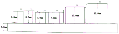
3) The 31 st tooth and the 41 st tooth of the mandibular teeth reference model, the mesial-distal width is 5mm, the cheek-lingual width is 6mm, the gingival height is 8mm; 32 th tooth and 32 nd tooth, width of near-far middle 6mm, width of buccal tongue 6mm,/o>
 The gingival height is 8mm; 33 rd teeth and 43 rd teeth, width of near-far middle 7mm, width of buccal tongue 7mm,/o>
 The gingival height is 8mm; 34 th, 44 th, 35 th, 45 th, width of near-far middle 7mm, cheek-lingual width 8mm,>
 gingival height 7mm; 36 th tooth and 46 th tooth, width of near-far middle 11mm, width of buccal tongue 10mm,/o>
 Gingival height 10mm;
 teeth 37 and 47, width 11mm in the mesial-distal direction, width 10mm in the buccal-lingual direction,> gingival height 12mm; the distance between adjacent parallel surfaces of each tooth is 2mm;
4) The 32 rd, 31 st, 41 st and 42 th mesial-distal directions of the mandibular dental reference model are horizontal directions, and the central axes are on the same horizontal line; the proximal and distal directions of the 36 th tooth, the 37 th tooth, the 46 th tooth and the 47 th tooth are vertical directions, and the central axes are respectively on the same vertical line; the central axes of the 33 th tooth, the 34 th tooth and the 35 th tooth are on the same straight line, and form 130-degree included angles with the central axes of the 32 nd tooth, the 31 st tooth, the 41 st tooth and the 42 th tooth; the central axes of the 43 th tooth, the 44 th tooth and the 45 th tooth are on the same straight line, and form an included angle of 130 degrees with the central axes of the 32 nd tooth, the 31 st tooth, the 41 st tooth and the 42 th tooth; the closest point distances between the 32 th tooth and the 33 th tooth, between the 35 th tooth and the 36 th tooth, between the 42 th tooth and the 43 th tooth, and between the 45 th tooth and the 46 th tooth are all 1mm.
The invention relates to an evaluation method of dental jaw reference model accuracy for light-cured three-dimensional printing accuracy evaluation, which comprises the following steps:
1) Three-dimensional printing of dental reference model:
storing the designed dental reference model data in a triangular mesh data format, inputting the data into three-dimensional printer software to be evaluated, slicing, placing dental model bases in parallel on a printing chassis, and simulating The plane being parallel to the x-y plane, +.>
 The gum direction is consistent with the z axis, and a three-dimensional printer matched model resin material is used for printing; printing 1 pair of upper and lower jaw models, and 2 models in total; model post-processing and the following measurement operations are completed on the same day as model printing; the x-y plane is a plane parallel to the ground, and the z axis is perpendicular to the x-y plane;
2) Three-dimensional morphological error measurement:
scanning the printing model by using a high-precision dental model three-dimensional scanner in a full dental arch scanning mode, storing the printing model as an STL format file, and calling the STL format file into geomic Studio 2012 software;
registering the upper and lower jaw scanning models with the original design model by using a global registration function in the geomatic Studio 2012 software, and calculating the overall 3D deviation and standard deviation of the upper and lower jaw models respectively;
using the feature-plane-best fit function in the geomic Studio 2012 software, selecting the corresponding scan area data of the simulated crown to fit each tooth position The surfaces PO, PB and PL are respectively recorded with the maximum positive and negative error spacing between each fitting plane and the scanned data, and are defined as the planeness of the characteristic plane, and the total number of measured values is 84, and the unit is mm;
using a characteristic-plane-best fitting function in the geomic Studio 2012 software to respectively fit reference planes P of upper and lower jaw model bases, respectively calculating included angles of PO and P of each simulated dental crown, defining a parallelism error of a printing form, wherein the unit is degree, and the total number of the measured values is 28; calculating the included angles of each simulated dental crown PB and PL and P respectively, defining the included angles as perpendicularity errors of printing forms, wherein the units are degrees, and 56 measured values are obtained;
and calculating the average value and standard deviation of the 86 flatness errors, the 28 parallelism errors and the 56 perpendicularity errors, and comprehensively defining the three-dimensional form errors of three-dimensional printing.
3) Plane relative error measurement:
the following measurement analyses were performed on each of the printed dental models by a trained experimenter using electronic digital vernier calipers:
defining the near-far pitch diameter and the cheek-tongue diameter of each dental position simulated dental crown as MD and BL, measuring and averaging the MD and BL of each simulated dental crown five times, and calculating to obtain relative percentage error values of the near-far pitch diameter, the cheek-tongue diameter and the designed size of 28 simulated dental crowns, wherein the calculation formula is as follows:

the true value in the above formula is the design size of the dental reference model, the calculated relative error is expressed by percentage, the positive value represents the size enlarging proportion, and the negative value represents the size reducing proportion;
measuring a characteristic dimension of a simulated dentition segment length, comprising: the distance L1 from the 17 th tooth far-middle surface to the 16 th tooth near-middle surface, the distance L2 from the 15 th tooth far-middle surface to the 13 th tooth near-middle surface, the distance L3 from the 12 th tooth far-middle surface to the 22 th tooth far-middle surface, the distance L4 from the 23 rd tooth far-middle surface to the 25 th tooth far-middle surface, the distance L5 from the 26 th tooth near-middle surface to the 27 th tooth far-middle surface, the distance L6 from the 37 th tooth far-middle surface to the 36 th tooth near-middle surface, the distance L7 from the 35 th tooth far-middle surface to the 33 th tooth near-middle surface, the distance L8 from the 32 th tooth far-middle surface to the 42 th tooth far-middle surface, the distance L9 from the 43 th tooth far-middle surface to the 45 th tooth far-middle surface, and the distance L10 from the 46 th tooth near-middle surface to the 47 th tooth far-middle surface. Measuring a characteristic dimension of a simulated dentition arch width, comprising: distances L11 and L12 from the buccal side of the crown of teeth 16 to 26 and 36 to 46. Calculating to obtain the relative percentage error values of the characteristic sizes of the 12 simulated dentition compared with the design size, wherein a calculation formula is the same as the formula (1);
the average value and standard deviation of the calculated relative percentage error values of the 40 measurement indexes are defined as three-dimensional printing In-layer molding dimensional percentage error in the plane direction;
4) gingival relative error measurement:
definition of each simulated dental crown The gingival height value is H, five measurements are carried out on each simulated crown height H, the average value is taken as a measured value, the relative percentage error value of 28 simulated crown heights compared with the design size (namely, true value) is obtained through calculation, and the calculation formula is the same as the formula (1).
Calculating the average value and standard deviation of the 28 height relative percentage error values, and defining the average value and standard deviation as a printing model Percentage error in the overall dimension of the layer height in the gingival direction;
the three-dimensional form error is integrated, In-layer percentage error in planar direction and +.>
 The layer height percentage error in the gingival direction, the accuracy of three-dimensional printing is obtained.
The invention relates to a method for evaluating precision of a dental reference model with photo-curing three-dimensional printing precision, which comprises the following steps:
printing the reference dental model 1 pair each time every day for 5 continuous days, and printing 5 pairs of 10 models in total; each printed model completed measurement in the same day:
1) Three-dimensional morphological error measurement:
scanning the printing model by using a high-precision dental model three-dimensional scanner in a full dental arch scanning mode, storing the printing model as an STL format file, and calling the STL format file into geomic Studio 2012 software;
registering the upper and lower jaw scanning models with the original design model by using a global registration function in the geomatic Studio 2012 software, and calculating the overall 3D deviation and standard deviation of the upper and lower jaw models respectively;
using the feature-plane-best fit function in the geomic Studio 2012 software, selecting the corresponding scan area data of the simulated crown to fit each tooth position The surfaces PO, PB and PL are respectively recorded with the maximum positive and negative error spacing between each fitting plane and the scanned data, and are defined as the planeness of the characteristic plane, and the total number of measured values is 84, and the unit is mm;
using a characteristic-plane-best fitting function in the geomic Studio 2012 software to respectively fit reference planes P of upper and lower jaw model bases, respectively calculating included angles of PO and P of each simulated dental crown, defining a parallelism error of a printing form, wherein the unit is degree, and the total number of the measured values is 28; calculating the included angles of each simulated dental crown PB and PL and P respectively, defining the included angles as perpendicularity errors of printing forms, wherein the units are degrees, and 56 measured values are obtained;
and calculating the average value and standard deviation of the 86 flatness errors, the 28 parallelism errors and the 56 perpendicularity errors, and comprehensively defining the three-dimensional form errors of three-dimensional printing.
2) Plane relative error measurement:
the following measurement analyses were performed on each of the printed dental models by a trained experimenter using electronic digital vernier calipers:
defining the near-far pitch diameter and the cheek-tongue diameter of each dental position simulated dental crown as MD and BL, measuring and averaging the MD and BL of each simulated dental crown five times, and calculating to obtain relative percentage error values of the near-far pitch diameter, the cheek-tongue diameter and the designed size of 28 simulated dental crowns, wherein the calculation formula is as follows:

the true value in the above formula is the design size of the dental reference model, the calculated relative error is expressed by percentage, the positive value represents the size enlarging proportion, and the negative value represents the size reducing proportion;
measuring a characteristic dimension of a simulated dentition segment length, comprising: the distance L1 from the 17 th tooth far-middle surface to the 16 th tooth near-middle surface, the distance L2 from the 15 th tooth far-middle surface to the 13 th tooth near-middle surface, the distance L3 from the 12 th tooth far-middle surface to the 22 th tooth far-middle surface, the distance L4 from the 23 rd tooth far-middle surface to the 25 th tooth far-middle surface, the distance L5 from the 26 th tooth near-middle surface to the 27 th tooth far-middle surface, the distance L6 from the 37 th tooth far-middle surface to the 36 th tooth near-middle surface, the distance L7 from the 35 th tooth far-middle surface to the 33 th tooth near-middle surface, the distance L8 from the 32 th tooth far-middle surface to the 42 th tooth far-middle surface, the distance L9 from the 43 th tooth far-middle surface to the 45 th tooth far-middle surface, and the distance L10 from the 46 th tooth near-middle surface to the 47 th tooth far-middle surface. Measuring a characteristic dimension of a simulated dentition arch width, comprising: distances L11 and L12 from the buccal side of the crown of teeth 16 to 26 and 36 to 46. Calculating to obtain the relative percentage error values of the characteristic sizes of the 12 simulated dentition compared with the design size, wherein a calculation formula is the same as the formula (1);
the average value and standard deviation of the calculated relative percentage error values of the 40 measurement indexes are defined as three-dimensional printing In-layer molding dimensional percentage error in the plane direction;
3) gingival relative error measurement:
definition of each simulated dental crown The gingival height value is H, five measurements are carried out on each simulated crown height H, the average value is taken as a measured value, the relative percentage error value of 28 simulated crown heights compared with the design size (namely, true value) is obtained through calculation, and the calculation formula is the same as the formula (1).
Calculating the average value and standard deviation of the 28 height relative percentage error values, and defining the average value and standard deviation as a printing model Percentage error in the overall dimension of the layer height in the gingival direction; three-dimensional morphological errors of each model obtained by calculation, < >>
 Plane relative error, < >>
 Gingival direction relative error; and respectively calculating the average value and standard deviation of the errors of the 5 models, and evaluating the repeatability and reliability of three-dimensional printing to obtain precision evaluation data.
(III) beneficial effects
The invention has the advantages that: 1. establishing a dental reference model, simulating the natural dental crown size and dental characteristics by using a simplified standard geometrical body combined model, evaluating the three-dimensional characteristic size precision of a photocuring 3D printing technology by adopting an inherent characteristic measurement method influenced by non-mark points, evaluating the three-dimensional morphological precision of a printing model by combining a three-dimensional morphological analysis method, and comprehensively evaluating the precision performance of the 3D printing dental model; 2. the invention aims at establishing a dental reference model for evaluating the precision of a photocuring 3D printing dental model and a matched evaluation method thereof so as to provide reference and guidance for oral clinical application of the photocuring 3D printing technology.
Drawings
FIG. 1 is a view of a reference model of the maxillary dental system of the present invention Schematic view of the face view;
FIG. 2 is a schematic illustration of a buccal view of a maxillary dental reference model of the present invention;
FIG. 3 is a schematic illustration of a lingual view of a maxillary dental reference model of the present invention;
FIG. 4 is a mandibular dental reference model of the present invention Schematic view of the face view;
FIG. 5 is a schematic illustration of a buccal view of a mandibular dental reference model of the present invention;
FIG. 6 is a schematic diagram of a lingual view of a mandibular dental reference model of the present invention;
FIG. 7 is an enlarged schematic view of the near-far mesial and facial lingual dimensions of a simulated crown of the invention;
FIG. 8 is a schematic representation of simulated dentition segment length and arch width feature sizes of the present invention;
FIG. 9 is a simulated dental crown of the present invention An enlarged schematic view of gingival elevation;
in the figure: 11. tooth 11; 12. tooth 12; 13. tooth 13; 14. tooth 14; 15. tooth 15; 16. tooth 16; 17. tooth 17; 21. 21 st tooth; 22. tooth 22; 23. 23 rd tooth; 24. 24 th tooth; 25. 25 th tooth; 26. tooth 26; 27. tooth 27; 31. tooth 31; 32. tooth 32; 33. tooth 33; 34. 34 th tooth; 35. tooth 35; 36. tooth 36; 37. tooth 37; 41. tooth 41; 42. tooth 42; 43. tooth 43; 44. tooth 44; 45. tooth 45; 46. 46 th tooth; 47. tooth 47; MD: each tooth position simulates the near-far pitch diameter of the dental crown; BL: each tooth position simulates the cheek-tongue diameter of the dental crown; l1: the distance from the distal surface of the 17 th tooth to the proximal surface of the 16 th tooth; l2: 15 th tooth mesial-distal distance to 13 th tooth mesial distance, L3: first, theThe distance from the far and middle surface of the 12 th tooth to the far and middle surface of the 22 nd tooth; l4: the distance from the mesial surface of the 23 rd tooth to the distal surface of the 25 th tooth; l5: the distance from the mesial surface of the 26 th tooth to the distal surface of the 27 th tooth; l6: the distance from the distal surface of the 37 th tooth to the proximal surface of the 36 th tooth; l7: the distance from the distal surface of the 35 th tooth to the proximal surface of the 33 th tooth; l8: the distance from the 32 th tooth mesial surface to the 42 th tooth mesial surface; l9: the distance from the mesial surface of the 43 rd tooth to the distal surface of the 45 th tooth; l10: the distance from the mesial surface of the 46 th tooth to the distal surface of the 47 th tooth; l11: distance from the 17 th buccal side to the 27 th buccal side; l12: distance from the 37 th buccal side to the 47 th buccal side; h: each simulated dental crown A gingival height value;
Detailed Description
The following examples are illustrative of the invention and are not intended to limit the scope of the invention.
The invention relates to a dental reference model for light-cured three-dimensional printing precision evaluation, which simulates the dimensions of a natural dental crown and the characteristics of dental jaw by using a simplified standard geometrical body combination model, and specifically comprises the following steps:
1) The 11 th tooth and the 21 st tooth of the maxillary dental reference model have a mesial-distal width of 8mm and a buccal lingual width of 7mm, gingival height 10mm;
 teeth 12 and 22, width of near to far and middle 7mm, width of buccal and lingual 6mm,/o> Gingival height 9mm;
 teeth 13 and 23, width of near to far and middle 8mm, width of buccal and lingual 8mm,/and-> The gingival height is 11mm;
 tooth 14, tooth 24, tooth 15, tooth 25, width of near-far middle 7mm, cheek-tongue width 9mm,> gingival height 10mm;
 teeth 16 and 26, 10mm in width in the mesial-distal direction, 11mm in width in the buccal lingual direction, the gingival height is 8mm; 17 th and 27 th teeth, width of near to far to middle 9mm, width of cheek to tongue 11mm,/o>
 Gingival height 5mm; the distance between adjacent parallel surfaces of each tooth is 2mm;
2) The near-far direction of the 12 th tooth, the 11 th tooth, the 21 st tooth and the 22 nd tooth of the maxillary dental reference model is the horizontal direction, and the central axes are on the same horizontal line; the proximal and distal directions of the 16 th tooth, the 17 th tooth, the 26 th tooth and the 27 th tooth are vertical directions, and the central axes are respectively on the same vertical line; the central axes of the 13 th tooth, the 14 th tooth and the 15 th tooth are on the same straight line, and form an included angle of 100 degrees with the central axes of the 12 th tooth, the 11 th tooth, the 21 st tooth and the 22 nd tooth; the central axes of the 23 th tooth, the 24 th tooth and the 25 th tooth are on the same straight line, and form 100-degree included angles with the central axes of the 12 th tooth, the 11 th tooth, the 21 st tooth and the 22 nd tooth; the closest point distances between the 12 th tooth and the 13 th tooth, between the 15 th tooth and the 16 th tooth, between the 22 nd tooth and the 23 rd tooth, and between the 25 th tooth and the 26 th tooth are all 1mm;
3) The 31 st tooth and the 41 st tooth of the mandibular teeth reference model, the mesial-distal width is 5mm, the cheek-lingual width is 6mm, the gingival height is 8mm; 32 th tooth and 32 nd tooth, width of near-far middle 6mm, width of buccal tongue 6mm,/o>
 The gingival height is 8mm; 33 rd teeth and 43 rd teeth, width of near-far middle 7mm, width of buccal tongue 7mm,/o>
 The gingival height is 8mm; 34 th, 44 th, 35 th, 45 th, width of near-far middle 7mm, cheek-lingual width 8mm,>
 gingival height 7mm;
 teeth 36 and 46, mesial-distal11mm wide, 10mm buccal-lingual width, < >> Gingival height 10mm;
 teeth 37 and 47, width 11mm in the mesial-distal direction, width 10mm in the buccal-lingual direction,> gingival height 12mm; the distance between adjacent parallel surfaces of each tooth is 2mm;
4) The 32 rd, 31 st, 41 st and 42 th mesial-distal directions of the mandibular dental reference model are horizontal directions, and the central axes are on the same horizontal line; the proximal and distal directions of the 36 th tooth, the 37 th tooth, the 46 th tooth and the 47 th tooth are vertical directions, and the central axes are respectively on the same vertical line; the central axes of the 33 th tooth, the 34 th tooth and the 35 th tooth are on the same straight line, and form 130-degree included angles with the central axes of the 32 nd tooth, the 31 st tooth, the 41 st tooth and the 42 th tooth; the central axes of the 43 th tooth, the 44 th tooth and the 45 th tooth are on the same straight line, and form an included angle of 130 degrees with the central axes of the 32 nd tooth, the 31 st tooth, the 41 st tooth and the 42 th tooth; the closest point distances between the 32 th tooth and the 33 th tooth, between the 35 th tooth and the 36 th tooth, between the 42 th tooth and the 43 th tooth, and between the 45 th tooth and the 46 th tooth are all 1mm.
The invention relates to an evaluation method of dental jaw reference model accuracy for light-cured three-dimensional printing accuracy evaluation, which comprises the following steps:
1) Three-dimensional printing of dental reference model:
storing the designed dental reference model data in a triangular mesh data format, inputting the data into three-dimensional printer software to be evaluated, slicing, placing dental model bases in parallel on a printing chassis, and simulating The plane being parallel to the x-y plane, +.>
 The gum direction is consistent with the z axis, and a three-dimensional printer matched model resin material is used for printing; printing 1 pair of upper and lower jaw models, and 2 models in total; model post-processing and the following measurement operations are all in the modelPrinting is completed on the same day;
2) Three-dimensional morphological error measurement:
scanning the printing model by using a high-precision dental model three-dimensional scanner in a full dental arch scanning mode, storing the printing model as an STL format file, and calling the STL format file into geomic Studio 2012 software;
registering the upper and lower jaw scanning models with the original design model by using a global registration function in the geomatic Studio 2012 software, and calculating the overall 3D deviation and standard deviation of the upper and lower jaw models respectively;
using the feature-plane-best fit function in the geomic Studio 2012 software, selecting the corresponding scan area data of the simulated crown to fit each tooth position The surfaces PO, PB and PL are respectively recorded with the maximum positive and negative error spacing between each fitting plane and the scanned data, and are defined as the planeness of the characteristic plane, and the total number of measured values is 84, and the unit is mm;
using a characteristic-plane-best fitting function in the geomic Studio 2012 software to respectively fit reference planes P of upper and lower jaw model bases, respectively calculating included angles of PO and P of each simulated dental crown, defining a parallelism error of a printing form, wherein the unit is degree, and the total number of the measured values is 28; calculating the included angles of each simulated dental crown PB and PL and P respectively, defining the included angles as perpendicularity errors of printing forms, wherein the units are degrees, and 56 measured values are obtained;
and calculating the average value and standard deviation of the 86 flatness errors, the 28 parallelism errors and the 56 perpendicularity errors, and comprehensively defining the three-dimensional form errors of three-dimensional printing.
3) Plane relative error measurement:
the following measurement analyses were performed on each of the printed dental models by a trained experimenter using electronic digital vernier calipers:
defining the near-far pitch diameter and the cheek-tongue diameter of each dental position simulated dental crown as MD and BL, measuring and averaging the MD and BL of each simulated dental crown five times, and calculating to obtain relative percentage error values of the near-far pitch diameter, the cheek-tongue diameter and the designed size of 28 simulated dental crowns, wherein the calculation formula is as follows:

the true value in the above formula is the design size of the dental reference model, the calculated relative error is expressed by percentage, the positive value represents the size enlarging proportion, and the negative value represents the size reducing proportion;
measuring a characteristic dimension of a simulated dentition segment length, comprising: the distance L1 from the 17 th tooth far-middle surface to the 16 th tooth near-middle surface, the distance L2 from the 15 th tooth far-middle surface to the 13 th tooth near-middle surface, the distance L3 from the 12 th tooth far-middle surface to the 22 th tooth far-middle surface, the distance L4 from the 23 rd tooth far-middle surface to the 25 th tooth far-middle surface, the distance L5 from the 26 th tooth near-middle surface to the 27 th tooth far-middle surface, the distance L6 from the 37 th tooth far-middle surface to the 36 th tooth near-middle surface, the distance L7 from the 35 th tooth far-middle surface to the 33 th tooth near-middle surface, the distance L8 from the 32 th tooth far-middle surface to the 42 th tooth far-middle surface, the distance L9 from the 43 th tooth far-middle surface to the 45 th tooth far-middle surface, and the distance L10 from the 46 th tooth near-middle surface to the 47 th tooth far-middle surface. Measuring a characteristic dimension of a simulated dentition arch width, comprising: distances L11 and L12 from the buccal side of the crown of teeth 16 to 26 and 36 to 46. Calculating to obtain the relative percentage error values of the characteristic sizes of the 12 simulated dentition compared with the design size, wherein a calculation formula is the same as the formula (1);
the average value and standard deviation of the calculated relative percentage error values of the 40 measurement indexes are defined as three-dimensional printing In-layer molding dimensional percentage error in the plane direction;
4) gingival relative error measurement:
definition of eachSimulated dental crowns The gingival height value is H, five measurements are carried out on each simulated crown height H, the average value is taken as a measured value, the relative percentage error value of 28 simulated crown heights compared with the design size (namely, true value) is obtained through calculation, and the calculation formula is the same as the formula (1).
Calculating the average value and standard deviation of the 28 height relative percentage error values, and defining the average value and standard deviation as a printing model Percentage error in the overall dimension of the layer height in the gingival direction;
the three-dimensional form error is integrated, In-layer percentage error in planar direction and +.>
 The layer height percentage error in the gingival direction, the accuracy of three-dimensional printing is obtained.
The three-dimensional form error is integrated, In-layer percentage error in planar direction and +.>
 And the absolute error of the layer height in the gingival direction is used for obtaining the accuracy of three-dimensional printing.
The invention relates to a method for evaluating precision of a dental reference model with photo-curing three-dimensional printing precision, which comprises the following steps:
printing the reference dental model 1 pair each time every day for 5 continuous days, and printing 5 pairs of 10 models in total; each printed model completed measurement in the same day:
1) Three-dimensional morphological error measurement:
scanning the printing model by using a high-precision dental model three-dimensional scanner in a full dental arch scanning mode, storing the printing model as an STL format file, and calling the STL format file into geomic Studio 2012 software;
registering the upper and lower jaw scanning models with the original design model by using a global registration function in the geomatic Studio 2012 software, and calculating the overall 3D deviation and standard deviation of the upper and lower jaw models respectively;
using the feature-plane-best fit function in the geomic Studio 2012 software, selecting the corresponding scan area data of the simulated crown to fit each tooth position The surfaces PO, PB and PL are respectively recorded with the maximum positive and negative error spacing between each fitting plane and the scanned data, and are defined as the planeness of the characteristic plane, and the total number of measured values is 84, and the unit is mm;
using a characteristic-plane-best fitting function in the geomic Studio 2012 software to respectively fit reference planes P of upper and lower jaw model bases, respectively calculating included angles of PO and P of each simulated dental crown, defining a parallelism error of a printing form, wherein the unit is degree, and the total number of the measured values is 28; calculating the included angles of each simulated dental crown PB and PL and P respectively, defining the included angles as perpendicularity errors of printing forms, wherein the units are degrees, and 56 measured values are obtained;
and calculating the average value and standard deviation of the 86 flatness errors, the 28 parallelism errors and the 56 perpendicularity errors, and comprehensively defining the three-dimensional form errors of three-dimensional printing.
2) Plane relative error measurement:
the following measurement analyses were performed on each of the printed dental models by a trained experimenter using electronic digital vernier calipers:
defining the near-far pitch diameter and the cheek-tongue diameter of each dental position simulated dental crown as MD and BL, measuring and averaging the MD and BL of each simulated dental crown five times, and calculating to obtain relative percentage error values of the near-far pitch diameter, the cheek-tongue diameter and the designed size of 28 simulated dental crowns, wherein the calculation formula is as follows:

the true value in the above formula is the design size of the dental reference model, the calculated relative error is expressed by percentage, the positive value represents the size enlarging proportion, and the negative value represents the size reducing proportion;
measuring a characteristic dimension of a simulated dentition segment length, comprising: the distance L1 from the 17 th tooth far-middle surface to the 16 th tooth near-middle surface, the distance L2 from the 15 th tooth far-middle surface to the 13 th tooth near-middle surface, the distance L3 from the 12 th tooth far-middle surface to the 22 th tooth far-middle surface, the distance L4 from the 23 rd tooth far-middle surface to the 25 th tooth far-middle surface, the distance L5 from the 26 th tooth near-middle surface to the 27 th tooth far-middle surface, the distance L6 from the 37 th tooth far-middle surface to the 36 th tooth near-middle surface, the distance L7 from the 35 th tooth far-middle surface to the 33 th tooth near-middle surface, the distance L8 from the 32 th tooth far-middle surface to the 42 th tooth far-middle surface, the distance L9 from the 43 th tooth far-middle surface to the 45 th tooth far-middle surface, and the distance L10 from the 46 th tooth near-middle surface to the 47 th tooth far-middle surface. Measuring a characteristic dimension of a simulated dentition arch width, comprising: distances L11 and L12 from the buccal side of the crown of teeth 16 to 26 and 36 to 46. Calculating to obtain the relative percentage error values of the characteristic sizes of the 12 simulated dentition compared with the design size, wherein a calculation formula is the same as the formula (1);
the average value and standard deviation of the calculated relative percentage error values of the 40 measurement indexes are defined as three-dimensional printing In-layer molding dimensional percentage error in the plane direction;
3) gingival relative error measurement:
definition of each simulated dental crown The gingival height value is H, and each simulated crown height H is coincidedFive measurements are performed, an average value is taken as a measured value, the relative percentage error values of 28 simulated crown heights and the designed size (namely, the true value) are obtained through calculation, and the calculation formula is the same as the formula (1).
Calculating the average value and standard deviation of the 28 height relative percentage error values, and defining the average value and standard deviation as a printing model Percentage error in the overall dimension of the layer height in the gingival direction;
the three-dimensional form error is integrated, In-layer percentage error in planar direction and +.>
 The layer height percentage error in the gingival direction, the accuracy of three-dimensional printing is obtained.
The three-dimensional form error is integrated, In-layer percentage error in planar direction and +.>
 And the absolute error of the layer height in the gingival direction is used for obtaining the accuracy of three-dimensional printing.
Calculating the three-dimensional form error of each model, Plane relative error, < >>
 Gingival direction relative error; and respectively calculating the average value and standard deviation of the errors of the 5 models, and evaluating the repeatability and reliability of three-dimensional printing to obtain precision evaluation data.
P0: each tooth position A noodle;
PB: the cheek side of each tooth position;
PL: sides of the tooth position tongue;
p is as follows: a reference plane of the maxillary model base;
the following steps: a reference plane of the mandibular model base.
As described above, the present invention can be more fully realized. The foregoing is merely a more reasonable embodiment of the present invention, and the scope of the present invention is not limited thereto, and any insubstantial modifications of the present invention based on the technical solution of the present invention are included in the scope of the present invention.
Claims (3)


Priority Applications (1)
| Application Number | Priority Date | Filing Date | Title |
|---|---|---|---|
| CN201810927498.4A CN110069798B (en) | 2018-08-15 | 2018-08-15 | Dental jaw reference model for light-cured three-dimensional printing precision evaluation and evaluation method |
Applications Claiming Priority (1)
| Application Number | Priority Date | Filing Date | Title |
|---|---|---|---|
| CN201810927498.4A CN110069798B (en) | 2018-08-15 | 2018-08-15 | Dental jaw reference model for light-cured three-dimensional printing precision evaluation and evaluation method |
Publications (2)
| Publication Number | Publication Date |
|---|---|
| CN110069798A CN110069798A (en) | 2019-07-30 |
| CN110069798B true CN110069798B (en) | 2023-06-27 |
Family
ID=67365827
Family Applications (1)
| Application Number | Title | Priority Date | Filing Date |
|---|---|---|---|
| CN201810927498.4A Active CN110069798B (en) | 2018-08-15 | 2018-08-15 | Dental jaw reference model for light-cured three-dimensional printing precision evaluation and evaluation method |
Country Status (1)
| Country | Link |
|---|---|
| CN (1) | CN110069798B (en) |
Families Citing this family (1)
| Publication number | Priority date | Publication date | Assignee | Title |
|---|---|---|---|---|
| CN114330027B (en) * | 2022-01-26 | 2024-07-26 | 华侨大学 | Structure optimization design method of molar prosthesis crown |
Citations (2)
| Publication number | Priority date | Publication date | Assignee | Title |
|---|---|---|---|---|
| CN105982747A (en) * | 2015-03-06 | 2016-10-05 | 北京大学口腔医学院 | Designing and manufacturing method of tooth supporting type implant surgery guide plate for restoration |
| CN105982753A (en) * | 2015-02-04 | 2016-10-05 | 北京大学口腔医学院 | Method for improving positioning precision of edentulous personalized tray in oral cavity |
Family Cites Families (1)
| Publication number | Priority date | Publication date | Assignee | Title |
|---|---|---|---|---|
| DE102007030768A1 (en) * | 2007-07-02 | 2009-01-08 | Sirona Dental Systems Gmbh | Measuring device and method for 3D measurement of tooth models |
-
2018
- 2018-08-15 CN CN201810927498.4A patent/CN110069798B/en active Active
Patent Citations (2)
| Publication number | Priority date | Publication date | Assignee | Title |
|---|---|---|---|---|
| CN105982753A (en) * | 2015-02-04 | 2016-10-05 | 北京大学口腔医学院 | Method for improving positioning precision of edentulous personalized tray in oral cavity |
| CN105982747A (en) * | 2015-03-06 | 2016-10-05 | 北京大学口腔医学院 | Designing and manufacturing method of tooth supporting type implant surgery guide plate for restoration |
Non-Patent Citations (3)
| Title |
|---|
| "3D打印牙模与传统石膏牙模的比较研究";张江涛等;《增材制造技术》;20171031(第10期);第35-40页 * |
| "口腔用光固化三维打印精度评价方法的建立及";萧宁等;《北京大学学报(医学版)》;20190228;第51卷(第1期);第120-130页 * |
| 三维打印牙颌模型的计算机辅助测量与精确度研究;陈一炜等;《计算机测量与控制》;20180425;第26卷(第04期);第28-31页 * |
Also Published As
| Publication number | Publication date |
|---|---|
| CN110069798A (en) | 2019-07-30 |
Similar Documents
| Publication | Publication Date | Title |
|---|---|---|
| Revilla-León et al. | Comparison of conventional, photogrammetry, and intraoral scanning accuracy of complete-arch implant impression procedures evaluated with a coordinate measuring machine | |
| Ries et al. | Three-dimensional analysis of the accuracy of conventional and completely digital interocclusal registration methods | |
| Cheng et al. | Accuracy of edentulous full‐arch implant impression: An in vitro comparison between conventional impression, intraoral scan with and without splinting, and photogrammetry | |
| Nestler et al. | Dimensional accuracy of extrusion-and photopolymerization-based 3D printers: In vitro study comparing printed casts | |
| Hassan et al. | Comparison of reconstructed rapid prototyping models produced by 3-dimensional printing and conventional stone models with different degrees of crowding | |
| Garino et al. | Comparison of dental arch measurements between stone and digital casts. | |
| US10206757B2 (en) | Method and system for dental planning and production | |
| Huang et al. | Effect of distance between the abutment and the adjacent teeth on intraoral scanning: An in vitro study | |
| Jeon et al. | Three-dimensional evaluation of the repeatability of scanned conventional impressions of prepared teeth generated with white-and blue-light scanners | |
| Revilla‐León et al. | Classification of Complete‐Arch Implant Scanning Techniques Recorded by Using Intraoral Scanners | |
| WO2016110855A1 (en) | System, device, and method for improved intraoral scanning accuracy | |
| Yuan et al. | Accuracy evaluation of a new three-dimensional reproduction method of edentulous dental casts, and wax occlusion rims with jaw relation | |
| CN103908352A (en) | Method for generating digital virtual jaw rack, and system thereof | |
| García-Martínez et al. | Accuracy of digitization obtained from scannable and nonscannable elastomeric impression materials | |
| Mourouzis et al. | Beyond the surface: A comparative study of intraoral scanners in subgingival configuration scanning | |
| Boehm et al. | Effect of a calibration aid and the intraoral scanner on the registration of a partially edentulous maxilla: An in vitro study | |
| CN110069798B (en) | Dental jaw reference model for light-cured three-dimensional printing precision evaluation and evaluation method | |
| Bai et al. | Comparison of four CAD-CAM guides for preparing guiding planes of removable partial dentures | |
| CN105796203B (en) | A kind of multi-functional correction measurement component | |
| Sierra et al. | Accuracy of anterior denture tooth arrangements of CAD-CAM complete removable dental prostheses made with a tooth mold template | |
| CN112137749B (en) | Method for measuring vertical position relation between tooth and jaw | |
| CN117745795A (en) | CBCT image processing and measuring method for alveolar bone | |
| KR20160095754A (en) | DEntal XYZ articulator and ZIG FOR DENTAL CAD CAM SYSTEM | |
| Šimunić et al. | Analysis of surface curvature influence on 3D scanning accuracy of dental castings | |
| Mohite et al. | Comparative Evaluation of Accuracy of Reconstructed 3D Printed Rapid Prototyping Models and Conventional Stone Models with Different Ranges of Crowding: An In-vitro Study. |
Legal Events
| Date | Code | Title | Description |
|---|---|---|---|
| PB01 | Publication | ||
| PB01 | Publication | ||
| SE01 | Entry into force of request for substantive examination | ||
| SE01 | Entry into force of request for substantive examination | ||
| GR01 | Patent grant | ||
| GR01 | Patent grant |