CN109679970B - Preparation method for rapidly detecting feline herpes type I virus - Google Patents
Preparation method for rapidly detecting feline herpes type I virus Download PDFInfo
- Publication number
- CN109679970B CN109679970B CN201811386302.1A CN201811386302A CN109679970B CN 109679970 B CN109679970 B CN 109679970B CN 201811386302 A CN201811386302 A CN 201811386302A CN 109679970 B CN109679970 B CN 109679970B
- Authority
- CN
- China
- Prior art keywords
- virus
- feline herpes
- sequence
- recombinant protein
- recombinant
- Prior art date
- Legal status (The legal status is an assumption and is not a legal conclusion. Google has not performed a legal analysis and makes no representation as to the accuracy of the status listed.)
- Active
Links
- 241000700605 Viruses Species 0.000 title abstract description 47
- 241000282324 Felis Species 0.000 title abstract description 42
- XQFRJNBWHJMXHO-RRKCRQDMSA-N IDUR Chemical compound C1[C@H](O)[C@@H](CO)O[C@H]1N1C(=O)NC(=O)C(I)=C1 XQFRJNBWHJMXHO-RRKCRQDMSA-N 0.000 title abstract description 41
- 238000002360 preparation method Methods 0.000 title abstract description 13
- 102000007056 Recombinant Fusion Proteins Human genes 0.000 claims abstract description 38
- 108010008281 Recombinant Fusion Proteins Proteins 0.000 claims abstract description 38
- 239000002773 nucleotide Substances 0.000 claims abstract description 12
- 125000003729 nucleotide group Chemical group 0.000 claims abstract description 12
- 108090000623 proteins and genes Proteins 0.000 claims description 16
- 239000013600 plasmid vector Substances 0.000 claims description 2
- 125000003275 alpha amino acid group Chemical group 0.000 claims 1
- 239000000427 antigen Substances 0.000 abstract description 25
- 102000036639 antigens Human genes 0.000 abstract description 25
- 108091007433 antigens Proteins 0.000 abstract description 25
- PCHJSUWPFVWCPO-UHFFFAOYSA-N gold Chemical compound [Au] PCHJSUWPFVWCPO-UHFFFAOYSA-N 0.000 abstract description 24
- 208000015181 infectious disease Diseases 0.000 abstract description 19
- 238000000034 method Methods 0.000 abstract description 16
- 238000003745 diagnosis Methods 0.000 abstract description 14
- 230000001154 acute effect Effects 0.000 abstract description 11
- 230000014509 gene expression Effects 0.000 abstract description 11
- 208000032420 Latent Infection Diseases 0.000 abstract description 9
- 208000037771 disease arising from reactivation of latent virus Diseases 0.000 abstract description 9
- 239000013604 expression vector Substances 0.000 abstract description 9
- 241000588724 Escherichia coli Species 0.000 abstract description 8
- 230000009465 prokaryotic expression Effects 0.000 abstract description 6
- 108020004705 Codon Proteins 0.000 abstract description 5
- 238000003259 recombinant expression Methods 0.000 abstract description 5
- 238000002405 diagnostic procedure Methods 0.000 abstract description 3
- 230000000694 effects Effects 0.000 abstract description 3
- 238000010353 genetic engineering Methods 0.000 abstract description 3
- 230000001939 inductive effect Effects 0.000 abstract description 2
- 210000001236 prokaryotic cell Anatomy 0.000 abstract description 2
- BPHPUYQFMNQIOC-NXRLNHOXSA-N isopropyl beta-D-thiogalactopyranoside Chemical compound CC(C)S[C@@H]1O[C@H](CO)[C@H](O)[C@H](O)[C@H]1O BPHPUYQFMNQIOC-NXRLNHOXSA-N 0.000 abstract 1
- 239000000243 solution Substances 0.000 description 37
- 210000002966 serum Anatomy 0.000 description 27
- 241000282326 Felis catus Species 0.000 description 22
- 238000012360 testing method Methods 0.000 description 22
- 241000701087 Felid alphaherpesvirus 1 Species 0.000 description 21
- RAXXELZNTBOGNW-UHFFFAOYSA-N imidazole Natural products C1=CNC=N1 RAXXELZNTBOGNW-UHFFFAOYSA-N 0.000 description 18
- KRKNYBCHXYNGOX-UHFFFAOYSA-N citric acid Chemical compound OC(=O)CC(O)(C(O)=O)CC(O)=O KRKNYBCHXYNGOX-UHFFFAOYSA-N 0.000 description 12
- 229910021642 ultra pure water Inorganic materials 0.000 description 12
- 239000012498 ultrapure water Substances 0.000 description 12
- 238000005406 washing Methods 0.000 description 12
- 239000007788 liquid Substances 0.000 description 11
- 210000004027 cell Anatomy 0.000 description 10
- 239000011248 coating agent Substances 0.000 description 10
- 238000000576 coating method Methods 0.000 description 10
- 239000012528 membrane Substances 0.000 description 10
- 238000001514 detection method Methods 0.000 description 9
- 239000000203 mixture Substances 0.000 description 9
- 238000002965 ELISA Methods 0.000 description 8
- PEDCQBHIVMGVHV-UHFFFAOYSA-N glycerol Substances OCC(O)CO PEDCQBHIVMGVHV-UHFFFAOYSA-N 0.000 description 8
- 229930027917 kanamycin Natural products 0.000 description 8
- SBUJHOSQTJFQJX-NOAMYHISSA-N kanamycin Chemical compound O[C@@H]1[C@@H](O)[C@H](O)[C@@H](CN)O[C@@H]1O[C@H]1[C@H](O)[C@@H](O[C@@H]2[C@@H]([C@@H](N)[C@H](O)[C@@H](CO)O2)O)[C@H](N)C[C@@H]1N SBUJHOSQTJFQJX-NOAMYHISSA-N 0.000 description 8
- 229960000318 kanamycin Drugs 0.000 description 8
- 229930182823 kanamycin A Natural products 0.000 description 8
- 239000000020 Nitrocellulose Substances 0.000 description 7
- 230000001580 bacterial effect Effects 0.000 description 7
- 239000010931 gold Substances 0.000 description 7
- 229910052737 gold Inorganic materials 0.000 description 7
- 229920001220 nitrocellulos Polymers 0.000 description 7
- 102000004169 proteins and genes Human genes 0.000 description 7
- 239000006228 supernatant Substances 0.000 description 7
- DHMQDGOQFOQNFH-UHFFFAOYSA-N Glycine Chemical compound NCC(O)=O DHMQDGOQFOQNFH-UHFFFAOYSA-N 0.000 description 6
- 241000283973 Oryctolagus cuniculus Species 0.000 description 6
- 230000035945 sensitivity Effects 0.000 description 6
- 238000007865 diluting Methods 0.000 description 5
- FAPWRFPIFSIZLT-UHFFFAOYSA-M Sodium chloride Chemical compound [Na+].[Cl-] FAPWRFPIFSIZLT-UHFFFAOYSA-M 0.000 description 4
- 210000004369 blood Anatomy 0.000 description 4
- 239000008280 blood Substances 0.000 description 4
- 238000013461 design Methods 0.000 description 4
- 238000001976 enzyme digestion Methods 0.000 description 4
- 230000003053 immunization Effects 0.000 description 4
- 239000002244 precipitate Substances 0.000 description 4
- 238000005507 spraying Methods 0.000 description 4
- QKNYBSVHEMOAJP-UHFFFAOYSA-N 2-amino-2-(hydroxymethyl)propane-1,3-diol;hydron;chloride Chemical compound Cl.OCC(N)(CO)CO QKNYBSVHEMOAJP-UHFFFAOYSA-N 0.000 description 3
- 241001198387 Escherichia coli BL21(DE3) Species 0.000 description 3
- 239000004471 Glycine Substances 0.000 description 3
- 239000012880 LB liquid culture medium Substances 0.000 description 3
- 208000037581 Persistent Infection Diseases 0.000 description 3
- 150000001413 amino acids Chemical group 0.000 description 3
- 238000007664 blowing Methods 0.000 description 3
- 238000005119 centrifugation Methods 0.000 description 3
- 238000006243 chemical reaction Methods 0.000 description 3
- 239000000499 gel Substances 0.000 description 3
- 230000006698 induction Effects 0.000 description 3
- 210000004379 membrane Anatomy 0.000 description 3
- 108020004707 nucleic acids Proteins 0.000 description 3
- 102000039446 nucleic acids Human genes 0.000 description 3
- 150000007523 nucleic acids Chemical class 0.000 description 3
- 238000000746 purification Methods 0.000 description 3
- 238000011084 recovery Methods 0.000 description 3
- 238000006748 scratching Methods 0.000 description 3
- 230000002393 scratching effect Effects 0.000 description 3
- 230000000405 serological effect Effects 0.000 description 3
- 239000013598 vector Substances 0.000 description 3
- 108091003079 Bovine Serum Albumin Proteins 0.000 description 2
- 241000984690 Catherpes Species 0.000 description 2
- 108090000790 Enzymes Proteins 0.000 description 2
- 102000004190 Enzymes Human genes 0.000 description 2
- 239000006137 Luria-Bertani broth Substances 0.000 description 2
- 108091028043 Nucleic acid sequence Proteins 0.000 description 2
- 239000007983 Tris buffer Substances 0.000 description 2
- 238000002835 absorbance Methods 0.000 description 2
- 239000002253 acid Substances 0.000 description 2
- 239000002671 adjuvant Substances 0.000 description 2
- 230000000903 blocking effect Effects 0.000 description 2
- 229940098773 bovine serum albumin Drugs 0.000 description 2
- 239000007853 buffer solution Substances 0.000 description 2
- 239000001110 calcium chloride Substances 0.000 description 2
- 229910001628 calcium chloride Inorganic materials 0.000 description 2
- 238000010276 construction Methods 0.000 description 2
- 210000004087 cornea Anatomy 0.000 description 2
- 230000000120 cytopathologic effect Effects 0.000 description 2
- 238000000502 dialysis Methods 0.000 description 2
- 201000010099 disease Diseases 0.000 description 2
- 208000037265 diseases, disorders, signs and symptoms Diseases 0.000 description 2
- 238000001962 electrophoresis Methods 0.000 description 2
- 239000003480 eluent Substances 0.000 description 2
- 238000005516 engineering process Methods 0.000 description 2
- 238000009472 formulation Methods 0.000 description 2
- 239000012634 fragment Substances 0.000 description 2
- 238000007602 hot air drying Methods 0.000 description 2
- 238000002649 immunization Methods 0.000 description 2
- 238000010166 immunofluorescence Methods 0.000 description 2
- 239000000411 inducer Substances 0.000 description 2
- 238000002955 isolation Methods 0.000 description 2
- 125000001449 isopropyl group Chemical group [H]C([H])([H])C([H])(*)C([H])([H])[H] 0.000 description 2
- 238000002372 labelling Methods 0.000 description 2
- 238000011068 loading method Methods 0.000 description 2
- 239000006166 lysate Substances 0.000 description 2
- 239000002609 medium Substances 0.000 description 2
- 238000002156 mixing Methods 0.000 description 2
- 238000006386 neutralization reaction Methods 0.000 description 2
- 238000011587 new zealand white rabbit Methods 0.000 description 2
- 239000002245 particle Substances 0.000 description 2
- 239000013612 plasmid Substances 0.000 description 2
- 239000000843 powder Substances 0.000 description 2
- 239000000047 product Substances 0.000 description 2
- 238000007790 scraping Methods 0.000 description 2
- 238000007789 sealing Methods 0.000 description 2
- 238000000926 separation method Methods 0.000 description 2
- 239000011780 sodium chloride Substances 0.000 description 2
- LENZDBCJOHFCAS-UHFFFAOYSA-N tris Chemical compound OCC(N)(CO)CO LENZDBCJOHFCAS-UHFFFAOYSA-N 0.000 description 2
- 230000009385 viral infection Effects 0.000 description 2
- 208000030507 AIDS Diseases 0.000 description 1
- 229920000936 Agarose Polymers 0.000 description 1
- 241000894006 Bacteria Species 0.000 description 1
- 108091026890 Coding region Proteins 0.000 description 1
- 208000003322 Coinfection Diseases 0.000 description 1
- 208000035473 Communicable disease Diseases 0.000 description 1
- 206010011224 Cough Diseases 0.000 description 1
- 208000020401 Depressive disease Diseases 0.000 description 1
- KCXVZYZYPLLWCC-UHFFFAOYSA-N EDTA Chemical compound OC(=O)CN(CC(O)=O)CCN(CC(O)=O)CC(O)=O KCXVZYZYPLLWCC-UHFFFAOYSA-N 0.000 description 1
- 241000620209 Escherichia coli DH5[alpha] Species 0.000 description 1
- LFQSCWFLJHTTHZ-UHFFFAOYSA-N Ethanol Chemical compound CCO LFQSCWFLJHTTHZ-UHFFFAOYSA-N 0.000 description 1
- 241000700586 Herpesviridae Species 0.000 description 1
- 102000003960 Ligases Human genes 0.000 description 1
- 108090000364 Ligases Proteins 0.000 description 1
- 238000012408 PCR amplification Methods 0.000 description 1
- 108091000080 Phosphotransferase Proteins 0.000 description 1
- 208000035415 Reinfection Diseases 0.000 description 1
- 206010051497 Rhinotracheitis Diseases 0.000 description 1
- 101710120037 Toxin CcdB Proteins 0.000 description 1
- 241000223997 Toxoplasma gondii Species 0.000 description 1
- 239000002250 absorbent Substances 0.000 description 1
- 230000002745 absorbent Effects 0.000 description 1
- 239000011543 agarose gel Substances 0.000 description 1
- 238000000246 agarose gel electrophoresis Methods 0.000 description 1
- 230000003321 amplification Effects 0.000 description 1
- 210000001367 artery Anatomy 0.000 description 1
- 238000003556 assay Methods 0.000 description 1
- 238000001574 biopsy Methods 0.000 description 1
- 230000015572 biosynthetic process Effects 0.000 description 1
- 230000036760 body temperature Effects 0.000 description 1
- 230000001684 chronic effect Effects 0.000 description 1
- 238000010367 cloning Methods 0.000 description 1
- 238000013329 compounding Methods 0.000 description 1
- 239000013078 crystal Substances 0.000 description 1
- 238000012258 culturing Methods 0.000 description 1
- 238000005520 cutting process Methods 0.000 description 1
- 238000011161 development Methods 0.000 description 1
- 230000004069 differentiation Effects 0.000 description 1
- 238000013399 early diagnosis Methods 0.000 description 1
- 238000010828 elution Methods 0.000 description 1
- 230000002327 eosinophilic effect Effects 0.000 description 1
- 210000003743 erythrocyte Anatomy 0.000 description 1
- GNBHRKFJIUUOQI-UHFFFAOYSA-N fluorescein Chemical compound O1C(=O)C2=CC=CC=C2C21C1=CC=C(O)C=C1OC1=CC(O)=CC=C21 GNBHRKFJIUUOQI-UHFFFAOYSA-N 0.000 description 1
- 230000004927 fusion Effects 0.000 description 1
- 239000003365 glass fiber Substances 0.000 description 1
- 230000035931 haemagglutination Effects 0.000 description 1
- 238000010438 heat treatment Methods 0.000 description 1
- 238000010211 hemagglutination inhibition (HI) assay Methods 0.000 description 1
- 229910052900 illite Inorganic materials 0.000 description 1
- 230000005847 immunogenicity Effects 0.000 description 1
- 238000011534 incubation Methods 0.000 description 1
- 230000002458 infectious effect Effects 0.000 description 1
- 230000005764 inhibitory process Effects 0.000 description 1
- 238000011081 inoculation Methods 0.000 description 1
- 206010023332 keratitis Diseases 0.000 description 1
- 201000010666 keratoconjunctivitis Diseases 0.000 description 1
- 210000003292 kidney cell Anatomy 0.000 description 1
- 238000011031 large-scale manufacturing process Methods 0.000 description 1
- 210000001165 lymph node Anatomy 0.000 description 1
- 238000004519 manufacturing process Methods 0.000 description 1
- 201000003102 mental depression Diseases 0.000 description 1
- 244000000010 microbial pathogen Species 0.000 description 1
- 238000012986 modification Methods 0.000 description 1
- 230000004048 modification Effects 0.000 description 1
- 238000007857 nested PCR Methods 0.000 description 1
- VGIBGUSAECPPNB-UHFFFAOYSA-L nonaaluminum;magnesium;tripotassium;1,3-dioxido-2,4,5-trioxa-1,3-disilabicyclo[1.1.1]pentane;iron(2+);oxygen(2-);fluoride;hydroxide Chemical compound [OH-].[O-2].[O-2].[O-2].[O-2].[O-2].[F-].[Mg+2].[Al+3].[Al+3].[Al+3].[Al+3].[Al+3].[Al+3].[Al+3].[Al+3].[Al+3].[K+].[K+].[K+].[Fe+2].O1[Si]2([O-])O[Si]1([O-])O2.O1[Si]2([O-])O[Si]1([O-])O2.O1[Si]2([O-])O[Si]1([O-])O2.O1[Si]2([O-])O[Si]1([O-])O2.O1[Si]2([O-])O[Si]1([O-])O2.O1[Si]2([O-])O[Si]1([O-])O2.O1[Si]2([O-])O[Si]1([O-])O2 VGIBGUSAECPPNB-UHFFFAOYSA-L 0.000 description 1
- 238000003199 nucleic acid amplification method Methods 0.000 description 1
- 210000001328 optic nerve Anatomy 0.000 description 1
- 238000005457 optimization Methods 0.000 description 1
- 210000002741 palatine tonsil Anatomy 0.000 description 1
- 230000007918 pathogenicity Effects 0.000 description 1
- 102000020233 phosphotransferase Human genes 0.000 description 1
- 229920002401 polyacrylamide Polymers 0.000 description 1
- 229920000136 polysorbate Polymers 0.000 description 1
- TYJJADVDDVDEDZ-UHFFFAOYSA-M potassium hydrogencarbonate Chemical compound [K+].OC([O-])=O TYJJADVDDVDEDZ-UHFFFAOYSA-M 0.000 description 1
- 230000037452 priming Effects 0.000 description 1
- 238000011027 product recovery Methods 0.000 description 1
- 238000003753 real-time PCR Methods 0.000 description 1
- 210000001533 respiratory mucosa Anatomy 0.000 description 1
- 210000002345 respiratory system Anatomy 0.000 description 1
- 108091008146 restriction endonucleases Proteins 0.000 description 1
- 238000012216 screening Methods 0.000 description 1
- 230000028327 secretion Effects 0.000 description 1
- 235000020183 skimmed milk Nutrition 0.000 description 1
- 229910000030 sodium bicarbonate Inorganic materials 0.000 description 1
- UIIMBOGNXHQVGW-UHFFFAOYSA-M sodium bicarbonate Substances [Na+].OC([O-])=O UIIMBOGNXHQVGW-UHFFFAOYSA-M 0.000 description 1
- 230000002269 spontaneous effect Effects 0.000 description 1
- 238000003756 stirring Methods 0.000 description 1
- 239000012089 stop solution Substances 0.000 description 1
- 239000000126 substance Substances 0.000 description 1
- 208000024891 symptom Diseases 0.000 description 1
- 230000002194 synthesizing effect Effects 0.000 description 1
- 230000009897 systematic effect Effects 0.000 description 1
- 229940104230 thymidine Drugs 0.000 description 1
- 230000001131 transforming effect Effects 0.000 description 1
- 210000003901 trigeminal nerve Anatomy 0.000 description 1
- SWGJCIMEBVHMTA-UHFFFAOYSA-K trisodium;6-oxido-4-sulfo-5-[(4-sulfonatonaphthalen-1-yl)diazenyl]naphthalene-2-sulfonate Chemical compound [Na+].[Na+].[Na+].C1=CC=C2C(N=NC3=C4C(=CC(=CC4=CC=C3O)S([O-])(=O)=O)S([O-])(=O)=O)=CC=C(S([O-])(=O)=O)C2=C1 SWGJCIMEBVHMTA-UHFFFAOYSA-K 0.000 description 1
- 210000001944 turbinate Anatomy 0.000 description 1
- 238000011144 upstream manufacturing Methods 0.000 description 1
- XLYOFNOQVPJJNP-UHFFFAOYSA-N water Substances O XLYOFNOQVPJJNP-UHFFFAOYSA-N 0.000 description 1
Images


Classifications
-
- C—CHEMISTRY; METALLURGY
- C07—ORGANIC CHEMISTRY
- C07K—PEPTIDES
- C07K14/00—Peptides having more than 20 amino acids; Gastrins; Somatostatins; Melanotropins; Derivatives thereof
- C07K14/005—Peptides having more than 20 amino acids; Gastrins; Somatostatins; Melanotropins; Derivatives thereof from viruses
-
- C—CHEMISTRY; METALLURGY
- C12—BIOCHEMISTRY; BEER; SPIRITS; WINE; VINEGAR; MICROBIOLOGY; ENZYMOLOGY; MUTATION OR GENETIC ENGINEERING
- C12N—MICROORGANISMS OR ENZYMES; COMPOSITIONS THEREOF; PROPAGATING, PRESERVING, OR MAINTAINING MICROORGANISMS; MUTATION OR GENETIC ENGINEERING; CULTURE MEDIA
- C12N15/00—Mutation or genetic engineering; DNA or RNA concerning genetic engineering, vectors, e.g. plasmids, or their isolation, preparation or purification; Use of hosts therefor
- C12N15/09—Recombinant DNA-technology
- C12N15/63—Introduction of foreign genetic material using vectors; Vectors; Use of hosts therefor; Regulation of expression
- C12N15/70—Vectors or expression systems specially adapted for E. coli
-
- G—PHYSICS
- G01—MEASURING; TESTING
- G01N—INVESTIGATING OR ANALYSING MATERIALS BY DETERMINING THEIR CHEMICAL OR PHYSICAL PROPERTIES
- G01N33/00—Investigating or analysing materials by specific methods not covered by groups G01N1/00 - G01N31/00
- G01N33/48—Biological material, e.g. blood, urine; Haemocytometers
- G01N33/50—Chemical analysis of biological material, e.g. blood, urine; Testing involving biospecific ligand binding methods; Immunological testing
- G01N33/53—Immunoassay; Biospecific binding assay; Materials therefor
- G01N33/569—Immunoassay; Biospecific binding assay; Materials therefor for microorganisms, e.g. protozoa, bacteria, viruses
- G01N33/56983—Viruses
- G01N33/56994—Herpetoviridae, e.g. cytomegalovirus, Epstein-Barr virus
-
- C—CHEMISTRY; METALLURGY
- C12—BIOCHEMISTRY; BEER; SPIRITS; WINE; VINEGAR; MICROBIOLOGY; ENZYMOLOGY; MUTATION OR GENETIC ENGINEERING
- C12N—MICROORGANISMS OR ENZYMES; COMPOSITIONS THEREOF; PROPAGATING, PRESERVING, OR MAINTAINING MICROORGANISMS; MUTATION OR GENETIC ENGINEERING; CULTURE MEDIA
- C12N2710/00—MICROORGANISMS OR ENZYMES; COMPOSITIONS THEREOF; PROPAGATING, PRESERVING, OR MAINTAINING MICROORGANISMS; MUTATION OR GENETIC ENGINEERING; CULTURE MEDIA dsDNA viruses
- C12N2710/00011—Details
- C12N2710/16011—Herpesviridae
- C12N2710/16021—Viruses as such, e.g. new isolates, mutants or their genomic sequences
-
- G—PHYSICS
- G01—MEASURING; TESTING
- G01N—INVESTIGATING OR ANALYSING MATERIALS BY DETERMINING THEIR CHEMICAL OR PHYSICAL PROPERTIES
- G01N2333/00—Assays involving biological materials from specific organisms or of a specific nature
- G01N2333/005—Assays involving biological materials from specific organisms or of a specific nature from viruses
- G01N2333/01—DNA viruses
- G01N2333/03—Herpetoviridae, e.g. pseudorabies virus
Landscapes
- Health & Medical Sciences (AREA)
- Life Sciences & Earth Sciences (AREA)
- Chemical & Material Sciences (AREA)
- Engineering & Computer Science (AREA)
- Genetics & Genomics (AREA)
- Virology (AREA)
- Biomedical Technology (AREA)
- Molecular Biology (AREA)
- Organic Chemistry (AREA)
- Biotechnology (AREA)
- Immunology (AREA)
- General Health & Medical Sciences (AREA)
- Biochemistry (AREA)
- General Engineering & Computer Science (AREA)
- Urology & Nephrology (AREA)
- Wood Science & Technology (AREA)
- Microbiology (AREA)
- Bioinformatics & Cheminformatics (AREA)
- Medicinal Chemistry (AREA)
- Biophysics (AREA)
- Zoology (AREA)
- Physics & Mathematics (AREA)
- Hematology (AREA)
- Cell Biology (AREA)
- Tropical Medicine & Parasitology (AREA)
- Food Science & Technology (AREA)
- Plant Pathology (AREA)
- Analytical Chemistry (AREA)
- General Physics & Mathematics (AREA)
- Pathology (AREA)
- Gastroenterology & Hepatology (AREA)
- Proteomics, Peptides & Aminoacids (AREA)
- Peptides Or Proteins (AREA)
- Micro-Organisms Or Cultivation Processes Thereof (AREA)
Abstract
The invention belongs to the technical field of biological engineering, and is characterized in that a feline herpes I virus antigen sequence is analyzed, one section of dominant antigen epitope sequence is selected, the sequence is repeated in order to increase the expression effect of the feline herpes I virus antigen sequence in prokaryotic cells, and a corresponding nucleotide sequence is synthesized by adopting an escherichia coli most-addictive codon. Inserting the nucleotide sequence into a prokaryotic expression vector by adopting a genetic engineering method, constructing a recombinant expression strain, inducing the expression strain by IPTG to generate recombinant protein FHV51, and finally establishing the feline herpes type I virus colloidal gold diagnostic method based on the recombinant protein. The invention mainly relates to preparation of a recombinant antigen of a feline herpes type I virus, preparation of a polyclonal antibody of the recombinant antigen and a rapid diagnosis method for acute infection and latent infection of the feline herpes type I virus established based on the recombinant protein.
Description
Technical Field
The invention belongs to the technical field of biological engineering, and is characterized in that a dominant antigen epitope sequence is selected by analyzing a feline herpes I virus antigen sequence, the dominant antigen epitope sequence is repeated in order to increase the expression effect of the dominant antigen epitope sequence in prokaryotic cells, and a corresponding nucleotide sequence is synthesized by adopting an escherichia coli most-addictive codon. Inserting the nucleotide sequence into a prokaryotic expression vector by adopting a genetic engineering method, constructing a recombinant expression strain, inducing the expression strain by IPTG to generate recombinant protein FHV51, and finally establishing the feline herpes type I virus colloidal gold diagnostic method based on the recombinant protein. The invention mainly relates to preparation of a recombinant antigen of a feline herpes type I virus, preparation of a polyclonal antibody of the recombinant antigen and a rapid diagnosis method for acute infection and latent infection of the feline herpes type I virus established based on the recombinant protein.
Background
Feline herpesvirus type 1 (FHV-1) belongs to the family of herpesviridae, members of the subfamily alpha-herpesviridae, and is primarily responsible for infectious rhinotracheitis in felines, an acute infectious disease of cats with very strong acute upper respiratory tract infectivity. FHV-1 mainly infects young cats, after infection, the upper respiratory mucosa is replicated to cause the clinical symptoms of the cats such as high body temperature, mental depression, cough, sneeze and the like and the complications such as chronic nasosinusitis, keratoconjunctivitis and the like, the morbidity is 100 percent, and the fatality rate of the young cats can reach 50 percent. After the infected cat recovers, FHV-1 is latent in trigeminal nerve, optic nerve, tonsil, submandibular lymph node, turbinates and oral-nasal-ocular secretions to cause persistent infection and latent infection, and can be reactivated to cause secondary morbidity of the infected cat, so the method is particularly important for early diagnosis of the feline herpes I virus and diagnosis and treatment of the latent infection.
Currently, the commonly used diagnostic methods for FHV-1 include virus isolation and identification, serological diagnosis and molecular biological diagnosis.
1. Isolation and identification of viruses
Because FHV-1 type virus can rapidly replicate in cells and generate typical cytopathic effect, the typical cytopathic effect appears after 2 days of inoculation, which is represented by focal shrinkage of cells, formation of syncytial and multinucleated giant cells, by collecting eye, throat swab (acute infection) or conjunctival and cornea scrapings (chronic infection), inoculating cat kidney cells (FK-81) after sample treatment. Although FHV-1 infection can be accurately diagnosed by virus separation, FHV-1 has weak resistance to the external environment, can only proliferate on FK-81 cells, is very easy to inactivate viruses in the transportation process, takes time for detection, and generally does not suggest virus separation for diagnosis clinically.
2. Serological diagnosis
Mainly comprises an immunofluorescence test (FA), an enzyme-linked immunosorbent assay (ELISA), a neutralization test and a hemagglutination-hemagglutination inhibition test (HA-HI).
Immunofluorescence assay: after cornea, conjunctival scrapings and biopsy of the diseased cat are taken and treated, the diseased cat is combined with a fluorescein-labeled antibody, and specific fluorescence is observed under a fluorescence microscope for diagnosis. The method has low sensitivity, can only diagnose sick cats in an acute infection stage, and has low detection success rate for spontaneous chronic infection cats.
Enzyme-linked immunosorbent assay (ELISA): the antigen or antibody is detected by enzyme-labeled color development by utilizing the principle that the soluble antigen is specifically combined with the antibody. The ELISA method can detect the FHV-1 serum antibody, but the detection is time-consuming and complicated to operate, and the large-scale production and putting of the antibody are limited.
Neutralization test: because the antibody titer of an organism is low during the primary infection of FHV-1 and the serum antibody titer is greatly increased after the recurrent infection, the kit can be used for the detection of the antibody titer of FHV-1 and the differentiation of the primary infection and the secondary infection of FHV-1. However, the detection of antibody titer is not related to the clinical manifestations of the diseased cats, and the method is not generally used for diagnosis.
Hemagglutination-hemagglutination inhibition assay (HA-HI): the detection of FHV-1 infection is realized by utilizing the characteristic that FHV-1 can agglutinate cat erythrocytes, but the hemagglutination of FHV-1 is still controversial at present, and a systematic HA-HI diagnosis method is not established.
3. Molecular biological diagnostics
According to the highly conserved sequence-Thymidine Kinase (TK) gene of FHV-1, the detection of FHV-1 infection can be realized by common PCR, nested PCR, real-time fluorescent quantitative PCR and other technologies. The molecular biological diagnosis technology has the characteristics of high sensitivity and strong specificity, but the operation is complex and time-consuming. Therefore, the method for rapidly and simply detecting FHV-1 acute infection and latent infection is significant.
The invention analyzes dominant epitope of feline herpes I virus antigen, gives consideration to the characteristics of specificity and the like, selects the dominant epitope sequence for repeating, and adopts the most eosinophilic codon of escherichia coli to synthesize the corresponding nucleotide sequence. The nucleotide sequence is inserted into a prokaryotic expression vector by adopting a genetic engineering method, a recombinant expression strain is constructed, the expression strain is induced by IPTG to generate recombinant protein FHV51, and finally, a colloidal gold rapid diagnosis method for acute infection and latent infection of the feline herpes type I virus is established based on the recombinant protein.
Disclosure of Invention
One of the objects of the present invention is to provide a DNA sequence encoding the recombinant protein FHV51 of feline herpes type I virus.
The invention also aims to provide the recombinant escherichia coli capable of specifically expressing the recombinant protein FHV51 of the feline herpesvirus I. The strain can specifically express recombinant protein FHV51 of the feline herpes I virus after being induced by isopropyl thio-beta-D-galactoside (IPTG), and provides specific antigen for serological diagnosis.
It is another object of the present invention to provide a polyclonal antibody that specifically recognizes the recombinant protein.
The fourth purpose of the invention is to provide a rapid and simple-operated method for diagnosing acute infection and latent infection of the feline herpes type I virus.
The invention is realized by the following experimental scheme:
1) comparing the gene sequence of the known feline herpes type I virus with the characteristic epitopes of other homologous pathogenic microorganisms, simulating the dominant epitope of the feline herpes type I virus by a computer, taking specificity and sensitivity and high-efficiency expression effect of escherichia coli into consideration, finally selecting a segment of dominant antigen epitope sequence of the feline herpes type I virus as a target sequence, and synthesizing a corresponding nucleotide sequence by adopting the most-preferred codon of the escherichia coli.
2) The synthesized target nucleotide sequence is directionally inserted between the multiple cloning sites BamHI and EcoRI of the prokaryotic expression vector pET-28a (+) to construct and obtain a recombinant prokaryotic expression vector pET-28a (+) -FHV-1A.
3) Transforming the recombinant prokaryotic expression vector pET-28a (+) -FHV-1A obtained in the step 2) into escherichia coli BL21(DE3) competent cells, and screening a monoclonal strain by utilizing the characteristic that the strain has kanamycin resistance.
4) Selecting the monoclonal strains obtained in the step 3) to respectively culture, identifying the bacterial liquid after IPTG induction, and selecting the strains capable of specifically expressing the recombinant protein FHV 51.
5) Amplifying and culturing the specific strain selected in the step 4), and purifying the expressed recombinant protein FHV 51.
6) Immunizing a New Zealand white rabbit with the FHV51 recombinant protein purified in the step 5), preparing a polyclonal antibody, and purifying the prepared polyclonal antibody.
7) Labeling the FHV51 recombinant protein obtained by purifying in the step 5) with colloidal gold particles.
8) Coating the polyclonal antibody purified in the step 6) as a C line (nitrocellulose membrane).
9) The mouse anti-cat IgM monoclonal antibody (Hangzhouxian to Biotech limited) and the mouse anti-cat IgG monoclonal antibody (Hangzhouxian to Biotech limited) were used as T1Wire, T2The thread was coated (nitrocellulose membrane).
10) And assembling the colloidal gold test strip, and detecting the serum to be detected by using the colloidal gold test strip. The clinical use of the colloidal gold is determined according to the color change of the colloidal gold.
The plasmid vector pET-28a (+) and the Escherichia coli strain BL21(DE3) related by the invention are the most widely applied vector tools and expression strains in the field of molecular biology, are convenient and easy to obtain, have no potential safety hazard to environment and operators, express the fusion antigen obtained, have no pathogenicity and infectivity, and improve the immunogenicity in the preparation process of the polyclonal antibody because the dominant antigen epitope is repeated for many times. Meanwhile, by respectively coating the cat IgM and IgG monoclonal antibodies, the double diagnosis of acute infection and latent infection of the cat herpes I virus can be realized, and the clinical omission ratio is greatly reduced.
Drawings
FIG. 1 is a graph showing the condition of feline herpes type I virus infection at day 7 for test samples;
figure 2 is a graph showing the status of feline herpes type I virus infection at day 45 for the test samples.
Detailed Description
Although the following embodiments describe the design concept of the present invention in more detail, these descriptions are only simple words for describing the design concept of the present invention, and are not intended to limit the design concept of the present invention, and any combination, addition or modification without departing from the scope of the design concept of the present invention will fall within the scope of the present invention.
Example 1: feline herpes type I virus dominant antigen epitope sequence selection
The feline herpes type I virus is taken as a target antigen, the biological software DNAssist2.0 is utilized to analyze the hydrophilicity and antigenicity of an antigen epitope sequence, the specificity and the sensitivity are considered, and finally a dominant antigen epitope sequence is selected, wherein the dominant antigen epitope sequence is a sequence specific to all feline herpes type I virus proteins and has no obvious homology with other protein sequences.
Example 2: cascade and optimization of dominant antigen epitope sequences of feline herpes type I virus
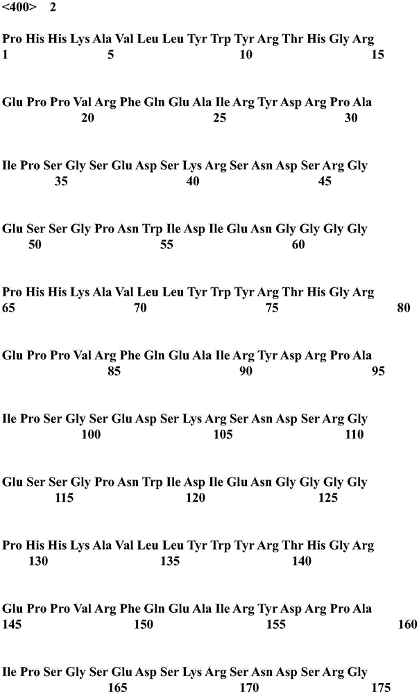
According to the requirements of the preparation titer of the protein polyclonal antibody, the dominant antigen epitope sequence of the feline herpes type I virus is repeated for 3 times, in order to improve the expression amount of the recombinant protein in escherichia coli, the most-addictive codon of the escherichia coli is adopted to synthesize a corresponding nucleotide sequence, and the amino acid sequence of the recombinant protein is obtained, and the specific sequence is shown as the sequence table SEQ ID No. 2. The connected recombinant protein amino acid sequence is converted into a corresponding nucleotide sequence, the specific coding sequence is shown as a sequence table SEQ ID No. 1, nucleotide sequences corresponding to enzyme cutting sites BamHI and EcoRI are respectively added at the upstream and downstream of the sequence, and the sequence is synthesized by Nanjing Kingsry Biotech Co.
Example 3: construction of recombinant protein expression vectors
And carrying out PCR amplification on the synthesized target gene, detecting an amplification product through 1% agarose gel electrophoresis, and recovering a target band which is named as pET-28a (+) -FHV-1A.
The recovered target gene and pET-28a (+) vector (Novagen, Germany) are respectively subjected to double enzyme digestion for 12 hours in water bath at 37 ℃ by restriction enzymes BamHI and EcoRI, and the enzyme digestion product is subjected to gel recovery after electrophoresis by 1% agarose gel (the target gene recovery and the enzyme digestion product recovery both adopt Ningbo Zhongding biotechnology, Inc. gel recovery kit).
The recovered objective gene fragment and pET-28a (+) vector fragment were ligated at 4 ℃ for 12 hours using T4 ligase (Bao bioengineering, Dalian Co., Ltd.) to transform DH 5. alpha. competent cells, which were plated on LB plates containing kanamycin resistance (50. mu.g/mL) and incubated at 37 ℃ overnight. The next day, a monoclonal strain is picked from the plate and inoculated into LB liquid culture medium containing kanamycin resistance (50 mug/mL), after shaking culture at a constant temperature of 37 ℃ for 5 hours, strain plasmids are extracted (plasmid purification kit of Ningbo Zhongding biotechnology Limited company), and a correct recombinant expression vector pET-28a (+) -FHV-1A is obtained after double enzyme digestion identification of BamHI and EcoRI.
Preparation of DH5 α competent cells: adding 100uL of escherichia coli DH5 alpha bacterial liquid into 2mL of LB liquid culture medium, carrying out constant temperature shaking culture at 37 ℃ for 3-4 hours until OD600 is 0.6, adding 100uL of activated bacterial liquid into 6mL of LB liquid culture medium, carrying out constant temperature shaking culture at 37 ℃ for 1.5 hours until OD600 is 0.6, taking 1mL of bacterial liquid, centrifuging at low temperature, removing supernatant, adding 160uL of CaCl-containing bacterial liquid into precipitate, and removing supernatant2Glycerol solution, blowing evenly, centrifuging at 5000rpm and 4 deg.C for 3min, discarding supernatant, adding 160uL CaCl2Glycerol solution, and then DH5 alpha competent cells were obtained by blowing. Wherein CaCl2-glycerol solution formulation: CaCl2The final concentration is 1mmol/L, and the final volume of glycerol is 10%.
Example 4: construction of recombinant protein expression Strain
The constructed recombinant expression vector pET-28a (+) -FHV-1A is transformed into competent cells of Escherichia coli BL21(DE3), and the competent cells are spread on an LB plate containing kanamycin resistance (50 mug/mL) and cultured overnight at 37 ℃. The next day, the monoclonal strain on the plate is picked and inoculated into LB liquid medium (2mL) containing kanamycin resistance (50 mug/mL), after the monoclonal strain is cultured for 4 hours at 37 ℃ by a constant temperature shaking table, an inducer isopropyl thio-beta-D-galactoside (IPTG) with the final concentration of 1.0mmol/L is added for induction, after the monoclonal strain is cultured for 1.5 hours at 37 ℃ by the constant temperature shaking table, the result of electrophoresis identification by 12 percent polyacrylamide gel shows that the recombinant protein FHV51 is successfully expressed, and the recombinant protein expression strain is obtained.
Example 5: large-scale expression and purification of recombinant protein FHV51
pET-28a (+) -FHV-1A monoclonal recombinant strains were picked from kanamycin-resistant LB plates, added to kanamycin-containing (50. mu.g/mL) LB broth (6mL), incubated at 37 ℃ for 1.5 hours with shaking at an OD600 of 0.6, and cultured using kanamycin-containing LB broth at a final concentration of 50. mu.g/mL in a manner of 1: diluting at a ratio of 100, subpackaging into bacteria culture bottles, placing into a shaker at 37 ℃ for constant temperature overnight culture until OD600 is 0.6, adding an inducer isopropylthio-beta-D-galactoside (IPTG) until the final concentration is 1.0mmol/L, and continuing to perform induction culture for 5 hours. And (3) collecting the precipitate (thallus) after low-temperature centrifugation, resuspending the precipitate by using a bacterial lysate, carrying out ultrasonic disruption for 3 minutes, and taking the supernatant after low-temperature centrifugation. The supernatant was passed through a 0.45 μm filter, passed through a Ni column (Hezhou Tiandi and Biotech Co., Ltd.) at room temperature to remove hetero-proteins with 20mM imidazole, eluted with 300mM imidazole to elute the objective protein, collected, allowed to stand at 4 ℃ for 30 minutes, transferred into a dialysis bag having a molecular weight cut-off of 10kDa to 12kDa, and dialyzed overnight in PBS (10mmol/L, pH 7.4). Immediately taking out after dialysis and subpackaging, and storing at-20 ℃ for later use.
The formula of the bacterial lysate comprises: Tris-HCl (50mmol/L, pH8.0), EDTA (1mmol/L), NaCl (100 mmol/L).
20mM imidazole preparation: imidazole 1.36g, add 10mmol/L, pH7.4PBS solution to dissolve to 1000 mL.
300mM imidazole preparation: 10.2g of imidazole, 10mmol/L of PBS solution with pH value of 7.4 is dissolved to be 500 mL.
Example 6: ELISA detection
Diluting the purified FHV51 with coating solution to a final concentration of 1 mug/mL, adding 100 mug/well into an ELISA plate (Stannless China, Biotech, Inc.), coating overnight at 4 ℃, washing 1 time with washing solution by a DEM-3 plate washing machine (Daan Gene, Inc. of Zhongshan university), adding 200 mug L of blocking solution for blocking after plate washing, and washing 1 time with washing solution after incubation for 1 hour at 37 ℃. And then adding clinically confirmed cat herpes type I virus negative serum, IgM positive serum and IgG positive serum (primary antibody) into the mixture, incubating the mixture for 35 minutes at 37 ℃, washing the mixture for 3 times by using a washing solution, adding an HRP-labeled rabbit anti-cat IgM antibody (from Hangzhouxian to Biotechnology Co., Ltd.) and an HRP-labeled rabbit anti-cat IgG antibody (from Hangzhouxian to Biotechnology Co., Ltd.) into the mixture, incubating the mixture for 35 minutes at 37 ℃, and washing the mixture for 4 times by using a washing solution. And adding the chromogenic solution A and the chromogenic solution B, then adding the stop solution, and measuring the OD value after zero calibration of the blank control hole, wherein the result shows that the OD value of the IgM positive serum hole is 2.5 times that of the negative serum hole, and the OD value of the IgG positive serum hole is 3.2 times that of the negative serum hole.
Coating liquid: na (Na)2CO3 1.59g,NaHCO32.93g, and adding ultrapure water to a volume of 1000mL (pH 9.6).
Sealing liquid: 3g of illite skim milk powder, 10mmol/L of PBS solution with pH of 7.4 is added to dissolve and fix the volume to 100 mL.
Washing liquid: na (Na)2HPO4.12H2O 2.68g,NaH2PO4.2H20.39g of O, 8.5g of NaCl, and 200.5 mL of Tween, and adding ultrapure water to the volume of 1000mL (pH7.4).
Color developing solution A: 200mg of TMB is dissolved in 100mL of absolute ethyl alcohol, and ultrapure water is added to the solution to reach the volume of 1000 mL.
Color developing solution B: citric acid 2.1g, Na2HPO4.12H2O71 g, and adding ultrapure water to the solution to make the volume reach 1000 mL.
When in use: 1mL of developing solution A +1mL of developing solution B + 0.4. mu.L of 30% H2O2
Stopping liquid: 2M H2SO421.7mL of concentrated H2SO4Adding ultrapure water to the solution until the volume is 1000 mL.
Example 7: preparation of recombinant protein FHV51 rabbit polyclonal antibody
FHV51 recombinant protein 300. mu.g was emulsified in Freund's complete adjuvant (total 1mL) and injected intradermally into male New Zealand white rabbits at multiple points for primary priming. After 20 days, a booster immunization was performed, and 150ug of FHV51 recombinant protein was emulsified with Freund's incomplete adjuvant (total 1mL) and injected into the skin at multiple points. And then boosting once every 15 days, wherein the method is the same as the second boosting, and 10 days after the fourth boosting, 5mL of blood is collected from the ear artery, the blood is purified to obtain FHV51 polyclonal antibody, and the titer of the antibody is detected by ELISA. The specific procedure was the same as in example 6, except that FHV51 polyclonal antibody was added as a primary antibody, similarly to IgG positive serum of feline herpesvirus type I in example 6. 10 days after the fifth booster immunization, about 100mL of carotid blood was collected and the white rabbits were sacrificed.
Example 8: purification of recombinant protein FHV51 rabbit polyclonal antibody
The agarose affinity medium Protein G column (Nanjing King Shirui Biotech Co., Ltd.) was equilibrated to room temperature, the computer nucleic acid Protein detector (Shanghai West analytical Instrument Co., Ltd.) was preheated for 20 minutes, and the column was washed with 10mmol/L of PBS solution (pH7.4) until the absorbance A of the computer nucleic acid Protein detector became 0. After centrifugation at 12000rpm for 5 minutes, the supernatant was applied to a 0.45 μm filter, and then 10mmol/L PBS (pH7.4) was added thereto, and the mixture was subjected to column washing until the absorbance A in a computer nucleic acid protein detector showed 0. Elution was performed with 0.1mol/L glycine solution at pH 3.0. And collecting the eluent, and adding 0.5mol/L Tris-HCl buffer solution with the pH value of 8.5 to neutralize the eluent to the pH value of 7.0, so as to obtain the recombinant protein FHV51 rabbit polyclonal antibody.
Preparing a glycine solution: 7.5g of glycine and ultrapure water are dissolved to a constant volume of 800mL, and 0.5M HCl is added to adjust the pH value to 3.0. Preparing a Tris-HCl buffer solution: 75.4g of Tris, stirring and dissolving with ultrapure water to reach the constant volume of 1000mL, and adding 0.5M HCl to adjust the pH value to 8.5.
Example 9: colloidal gold particles labeled by recombinant protein FHV51
Adding 10 mu L of 0.2mol/L potassium carbonate solution into 5mL of 0.01% colloidal gold solution, fully mixing uniformly, adding 50 mu g of recombinant protein FHV51 of the feline herpes type I virus, mixing uniformly, standing at room temperature for 2 hours, adding 500 mu L of 10% BSA (bovine serum albumin) solution for sealing, standing at room temperature for 1 hour, centrifuging (6500rpm, 20min), removing supernatant, and fully dissolving precipitate with 500 mu L of redissolving solution. The dissolved gold solution was uniformly sprayed on a 6mm wide glass fiber using a gold spraying and film drawing instrument (Shanghai gold-labeled Biotech Co., Ltd.) in an amount of 10. mu.L/cm, and then dried by blowing at 37 ℃ for 1 hour in an electric hot air drying oven (Shanghai-Hengscientific Instrument Co., Ltd.).
The relevant solution formulation is as follows:
0.01% colloidal gold solution: 1mL of 1% chloroauric acid solution, 1.4mL of 1% citric acid solution, and heating ultrapure water to dissolve the reaction and fixing the volume to 100 mL.
1% chloroauric acid solution: AuCL3.HCl.4H2O powder (1 g) was dissolved in ultrapure water and the volume was adjusted to 100 mL.
1% citric acid solution: 1g of citric acid crystal and ultrapure water are dissolved and the volume is 100 mL.
Compounding the solution: 6.057g of Tris base is dissolved in 800mL of ultrapure water, the pH is adjusted to 8.0 by using a proper amount of HCl, and the volume of the ultrapure water is adjusted to 1000 mL.
Example 10: preparation of test paper strip for diagnosing feline herpes type I virus
The feline herpes type I virus recombinant protein FHV51 polyclonal antibody is diluted by coating liquid to the final concentration of 1mg/mL, and is uniformly coated on a nitrocellulose membrane (Sartorius, CN140) by a gold spraying and membrane scratching instrument (Shanghai gold-labeled Biotech Co., Ltd.) according to 1 muL/cm to form a C line. Diluting mouse anti-cat IgM monoclonal antibody (Hangzhou xian till Biotechnology Co., Ltd.) with coating solution to final concentration of 1mg/mL, and uniformly coating the IgM monoclonal antibody on a nitrocellulose membrane (Sartorius, CN140) by a gold spraying and membrane scratching instrument (Shanghai gold mark Biotechnology Co., Ltd.) according to 1 μ L/cm to obtain T1A wire. Diluting mouse anti-cat IgG monoclonal antibody (Hangzhou Xianzhi Biotech Co., Ltd.) with coating solution to final concentration of 1mg/mL, and uniformly coating the IgG monoclonal antibody on a nitrocellulose membrane (Sartorius, CN140) by a gold spraying and membrane scratching instrument (Shanghai gold labeling Biotech Co., Ltd.) according to 1 μ L/cm to obtain T2A wire. After the completion of the film-scribing, the nitrocellulose membrane was air-dried at 37 ℃ for 30 minutes in an electric hot air drying oven (Shanghai-Hengyu scientific instruments Co., Ltd.). The nitrocellulose membrane, the colloidal gold pad, the sample pad and the absorbent paper are assembled on a PVC pad in sequence, then cut into strips with the width of 4mm, and then packed into a card shell and compressed.
Example 11: operation steps and standard of test strip for diagnosing feline herpes type I virus
Immunizing normal domestic cat with standard substance of feline herpesvirus type I, collecting serum A7 days later, diluting with gradient, loading into 100 μ L/well, standing at room temperature for 15min, and showing purple red reaction strip (T) with increased concentration of test sample as shown in FIG. 11Line) gradually darkens in color. Boosting normal domestic cats once on day 20, boosting domestic cats again 35 days later, obtaining serum B on day 45, and performing gradient dilutionAfter loading 100. mu.L/well, and standing at room temperature for 15min, as shown in FIG. 2, as the concentration of the test sample increased, a purple reaction band (T)2Line) gradually darkens in color. The colloidal gold test strip established on the basis of the recombinant protein FHV51 prepared by the invention can quickly and effectively identify and diagnose acute infection and latent infection of the feline herpes type I virus.
Example 12: sensitivity and specificity test of test paper
And (3) measuring the sensitivity: according to the operation method of the test strip for the feline herpes type I virus, clinically confirmed feline herpes type I virus negative serum, IgM positive serum, IgG positive serum, IgM and IgG double positive serum are respectively diluted in a gradient manner by 1 part and then are detected by using the test strip. Reading the values respectively by a colloidal gold reading instrument (Hangzhou Weizan science and technology Co., Ltd.), (T)1the/C line cut-off value is 0.10, T2the/C line cut-off value is 0.10, T1C line and T2The arbitrary ratio of the/C line is positive when the cut-off value is larger than the corresponding cut-off value, and is negative when the cut-off value is smaller than the cut-off value), the coincidence rate is 100%, and the results are shown in the attached table 1.
Attached table 1

Serum a: clinically established feline herpes type I virus negative sera
Serum B: clinically-established feline herpes type I virus IgM positive serum
Serum C: clinically established IgG positive serum for feline herpes type I virus
Serum x D: clinically determined feline herpes type I virus IgM and IgG double-positive blood
And (3) specific determination: according to the operation method of the test strip for the feline herpes type I virus, 10 cat negative serums (determined clinically) are detected by using the test strip, false positive does not appear, and the coincidence rate is 100%. According to the operation method of the test strip for the feline herpes type I virus, 10 cat toxoplasma gondii positive serum samples, 10 cat AIDS positive serum samples, 10 cat calicivirus positive serum samples and the like are respectively detected by using the test strip, and no positive production exists. The result shows that the colloidal gold rapid detection test strip prepared by utilizing the feline herpes type I virus recombinant protein FHV51 basically does not cause false positive with the basic diseases and other diseases of cats and has good specificity.
SEQ ID NO 1: a nucleotide sequence encoding a recombinant protein;
SEQ ID NO 2: amino acid sequence of a recombinant protein.


Claims (3)
1. A gene for coding recombinant protein, the nucleotide sequence of which is shown as SEQ ID No: l is shown.
2. A recombinant protein has an amino acid sequence shown as SEQ ID No. 2.
3. A recombinant plasmid vector comprising the gene of claim 1.
Priority Applications (1)
| Application Number | Priority Date | Filing Date | Title |
|---|---|---|---|
| CN201811386302.1A CN109679970B (en) | 2018-11-20 | 2018-11-20 | Preparation method for rapidly detecting feline herpes type I virus |
Applications Claiming Priority (1)
| Application Number | Priority Date | Filing Date | Title |
|---|---|---|---|
| CN201811386302.1A CN109679970B (en) | 2018-11-20 | 2018-11-20 | Preparation method for rapidly detecting feline herpes type I virus |
Publications (2)
| Publication Number | Publication Date |
|---|---|
| CN109679970A CN109679970A (en) | 2019-04-26 |
| CN109679970B true CN109679970B (en) | 2020-12-08 |
Family
ID=66185466
Family Applications (1)
| Application Number | Title | Priority Date | Filing Date |
|---|---|---|---|
| CN201811386302.1A Active CN109679970B (en) | 2018-11-20 | 2018-11-20 | Preparation method for rapidly detecting feline herpes type I virus |
Country Status (1)
| Country | Link |
|---|---|
| CN (1) | CN109679970B (en) |
Families Citing this family (3)
| Publication number | Priority date | Publication date | Assignee | Title |
|---|---|---|---|---|
| CN112457414B (en) * | 2020-12-11 | 2023-09-19 | 杭州爱谨生物科技有限公司 | A kind of feline herpes virus type I gB-gD recombinant protein and its preparation method and application |
| CN113943354B (en) * | 2021-10-11 | 2022-09-06 | 中国农业科学院上海兽医研究所(中国动物卫生与流行病学中心上海分中心) | Recombinant feline herpesvirus gB protein antigen and application thereof in antibody diagnosis and vaccine preparation |
| CN116693665B (en) * | 2023-06-30 | 2024-11-05 | 深圳赫兹生命科学技术有限公司 | Antibodies for detecting feline herpesvirus based on gB protein |
Family Cites Families (5)
| Publication number | Priority date | Publication date | Assignee | Title |
|---|---|---|---|---|
| AU672372B2 (en) * | 1991-11-08 | 1996-10-03 | Pharmacia & Upjohn Company | Feline leukemia virus vaccines |
| US6010703A (en) * | 1993-07-26 | 2000-01-04 | Board Of Trustees Operating Michigan State University | Recombinant poxvirus vaccine against feline rhinotracheitis |
| JPH09267A (en) * | 1995-06-23 | 1997-01-07 | Kyoritsu Shoji Kk | Recombinant feline herpesvirus type 1 having mutation in thymidine kinase gene and vaccine containing the same |
| JP2008283921A (en) * | 2007-05-18 | 2008-11-27 | Univ Of Tokyo | Veterinary vaccine |
| EP2611460B1 (en) * | 2010-08-31 | 2016-10-05 | Merial, Inc. | Newcastle disease virus vectored herpesvirus vaccines |
-
2018
- 2018-11-20 CN CN201811386302.1A patent/CN109679970B/en active Active
Also Published As
| Publication number | Publication date |
|---|---|
| CN109679970A (en) | 2019-04-26 |
Similar Documents
| Publication | Publication Date | Title |
|---|---|---|
| CN106279410B (en) | Two type dengue fever virus NS1 albumen multivalence nano antibodies of one kind and preparation method | |
| CN109679970B (en) | Preparation method for rapidly detecting feline herpes type I virus | |
| CN114276445A (en) | Rotavirus recombinant protein specific antibody, plasmid vector and method | |
| CN110133284A (en) | Protein antigens and their coding genes and their application in the identification of Mycoplasma hyopneumoniae inactivated vaccine antibodies and natural infection antibodies | |
| CN106442981B (en) | A kind of 1 type antibody indirect ELISA diagnostic kit of human bocavirus | |
| CN117054650B (en) | A kit for detecting feline herpesvirus type I antibodies | |
| CN112500494B (en) | Antigen for detecting novel coronavirus and preparation method thereof | |
| CN116333060B (en) | Porcine reproductive and respiratory syndrome virus GP5 protein, preparation method and application thereof, gene, kit and detection method | |
| CN117700539A (en) | Anti-canine distemper virus monoclonal antibody, plasmid vector and preparation method | |
| CN111621506B (en) | Mycoplasma bovis secreted protein MbovP0145 and its application | |
| CN106146627B (en) | Fc Specific binding proteins, IgG affinity chromatography medium and the preparation method and application thereof | |
| CN112521462B (en) | An equine infectious anemia virus p26-gp90 recombinant protein and its preparation method and application | |
| CN105137076B (en) | Small-fragment gG antigen and application of small-fragment gG antigen in detecting pseudorabies virus gG antibody | |
| CN105277713B (en) | Rapid detection method and kit for streptococcus pneumoniae based on magnetic separation and quantum dot labeling | |
| CN115785286B (en) | Fusion protein of porcine reproductive and respiratory syndrome virus and application thereof | |
| WO2025092709A1 (en) | Application of c5orf24 protein in tuberculosis diagnostic reagent | |
| CN114085274B (en) | FIPV N recombinant protein and colloidal gold test strip for rapidly detecting FIPV infection | |
| CN116751264A (en) | A recombinant protein and its application in differential diagnosis of LSDV | |
| CN112521461B (en) | Preparation of hepatitis A virus recombinant protein and rapid detection method thereof | |
| CN108165569A (en) | A kind of recombinant protein of quick detection Infection of Toxoplasma Gondii and its preparation method and application | |
| CN115975052B (en) | Fusion protein of swine fever virus and application thereof | |
| CN110540598B (en) | Haemophilus influenzae Elisa detection kit based on surface protein antibody of haemophilus influenzae and preparation method | |
| CN105277714B (en) | Rapid detection method and kit for human parainfluenza virus based on magnetic separation and quantum dot labeling | |
| CN115856298B (en) | A recombinant Treponema pallidum antigen and its preparation method | |
| RU2754791C1 (en) | Mosaic recombinant polypeptide containing fragments of hepatitis e virus proteins of genotypes 1 and 3 in one polypeptide chain, intended for use in test systems used in the serodiagnosis of hepatitis e |
Legal Events
| Date | Code | Title | Description |
|---|---|---|---|
| PB01 | Publication | ||
| PB01 | Publication | ||
| SE01 | Entry into force of request for substantive examination | ||
| SE01 | Entry into force of request for substantive examination | ||
| GR01 | Patent grant | ||
| GR01 | Patent grant |