CN102762990A - Devices, systems, methods and computer readable media for acoustic flow cytometry - Google Patents
Devices, systems, methods and computer readable media for acoustic flow cytometry Download PDFInfo
- Publication number
- CN102762990A CN102762990A CN2010800617745A CN201080061774A CN102762990A CN 102762990 A CN102762990 A CN 102762990A CN 2010800617745 A CN2010800617745 A CN 2010800617745A CN 201080061774 A CN201080061774 A CN 201080061774A CN 102762990 A CN102762990 A CN 102762990A
- Authority
- CN
- China
- Prior art keywords
- flow
- fluid
- sample
- particles
- sheath
- Prior art date
- Legal status (The legal status is an assumption and is not a legal conclusion. Google has not performed a legal analysis and makes no representation as to the accuracy of the status listed.)
- Granted
Links
Images




Classifications
-
- G—PHYSICS
- G01—MEASURING; TESTING
- G01N—INVESTIGATING OR ANALYSING MATERIALS BY DETERMINING THEIR CHEMICAL OR PHYSICAL PROPERTIES
- G01N21/00—Investigating or analysing materials by the use of optical means, i.e. using sub-millimetre waves, infrared, visible or ultraviolet light
- G01N21/17—Systems in which incident light is modified in accordance with the properties of the material investigated
- G01N21/41—Refractivity; Phase-affecting properties, e.g. optical path length
- G01N21/45—Refractivity; Phase-affecting properties, e.g. optical path length using interferometric methods; using Schlieren methods
- G01N21/453—Holographic interferometry
-
- G—PHYSICS
- G01—MEASURING; TESTING
- G01N—INVESTIGATING OR ANALYSING MATERIALS BY DETERMINING THEIR CHEMICAL OR PHYSICAL PROPERTIES
- G01N15/00—Investigating characteristics of particles; Investigating permeability, pore-volume or surface-area of porous materials
- G01N15/10—Investigating individual particles
- G01N15/14—Optical investigation techniques, e.g. flow cytometry
- G01N15/1404—Handling flow, e.g. hydrodynamic focusing
-
- G—PHYSICS
- G01—MEASURING; TESTING
- G01N—INVESTIGATING OR ANALYSING MATERIALS BY DETERMINING THEIR CHEMICAL OR PHYSICAL PROPERTIES
- G01N15/00—Investigating characteristics of particles; Investigating permeability, pore-volume or surface-area of porous materials
- G01N15/10—Investigating individual particles
- G01N15/14—Optical investigation techniques, e.g. flow cytometry
- G01N15/1404—Handling flow, e.g. hydrodynamic focusing
- G01N2015/142—Acoustic or ultrasonic focussing
Landscapes
- Chemical & Material Sciences (AREA)
- Biochemistry (AREA)
- Physics & Mathematics (AREA)
- Health & Medical Sciences (AREA)
- Life Sciences & Earth Sciences (AREA)
- Analytical Chemistry (AREA)
- General Health & Medical Sciences (AREA)
- General Physics & Mathematics (AREA)
- Immunology (AREA)
- Pathology (AREA)
- Dispersion Chemistry (AREA)
- Investigating, Analyzing Materials By Fluorescence Or Luminescence (AREA)
- Investigating Or Analyzing Materials By The Use Of Ultrasonic Waves (AREA)
Abstract
Description
相关申请的交叉引用Cross References to Related Applications
本申请要求于2009年12月4日提交的美国临时申请第61/266,907号、于2010年2月12日提交的美国临时申请第61/303,938号以及于2010年6月28日提交的美国临时申请第61/359,310号的优先权,上述每一申请的全部内容通过引用被合并于本文中。This application claims U.S. Provisional Application No. 61/266,907, filed December 4, 2009, U.S. Provisional Application No. 61/303,938, filed February 12, 2010, and U.S. Provisional Application No. 61/303,938, filed June 28, 2010 The priority of application Ser. No. 61/359,310, the entire contents of each of which is incorporated herein by reference.
技术领域 technical field
本申请一般地涉及流式细胞计量术,尤其涉及使用声学流式细胞计量术检测罕见事件的装置、系统、方法和计算机可读介质。The present application relates generally to flow cytometry, and more particularly to devices, systems, methods and computer readable media for detecting rare events using acoustic flow cytometry.
背景技术 Background technique
在传统流式细胞计量术中,通过使鞘液以极高的容积流率(大约是样本流体的容积流率的100-1000倍)围绕样本流体流动而将样本流体聚焦到大约10-50μm的小核心直径。样本流体中的颗粒以极快的线速度(以米/秒的数量级)流动并且因此仅需要极短的时间穿过探询点(通常仅1-10μs)。这具有明显的缺点。首先,颗粒不能被重定向到探询点,原因是流动不能被反向。其次,颗粒不能保持在探询点,原因是在没有鞘液的情况下聚焦丢失。再次,较短的通过时间限制了灵敏度和分辨率,这导致罕见事件检测困难并且耗时。In traditional flow cytometry, the sample fluid is focused to approximately 10-50 μm by flowing the sheath fluid around the sample fluid at a very high volumetric flow rate (approximately 100-1000 times that of the sample fluid). Small core diameter. Particles in the sample fluid flow at extremely fast linear velocities (on the order of m/s) and therefore only require an extremely short time to pass through the interrogation point (typically only 1-10 μs). This has obvious disadvantages. First, particles cannot be redirected to the interrogation point because the flow cannot be reversed. Second, the particle cannot remain at the interrogation point due to loss of focus in the absence of sheath fluid. Again, short transit times limit sensitivity and resolution, which makes detection of rare events difficult and time-consuming.
解决这些缺点的以前尝试不令人满意。可以增加样本流体中的颗粒浓度以补偿这些缺点中的一些,但是这可能不总是可行的并且可能是成本很高的。同样地,可以增加探询点处的光子通量以提取更多的信号,但是这可能常常光漂白(即,激发到非辐射状态)用于生成信号的荧光团并且可能增加背景瑞利散射、拉曼散射和荧光。因此,需要用于流式细胞计量术的新装置、系统、方法和计算机可读介质,其在避免或最小化这些缺点中的一个或多个的同时,允许颗粒的高吞吐量分析以及快速高效的罕见事件检测。Previous attempts to address these shortcomings have been unsatisfactory. It is possible to increase the particle concentration in the sample fluid to compensate for some of these disadvantages, but this may not always be possible and may be costly. Likewise, the photon flux at the interrogation site can be increased to extract more signal, but this may often photobleach (i.e., excite to a non-radiative state) the fluorophore used to generate the signal and may increase background Rayleigh scattering, pulling Mann scattering and fluorescence. Accordingly, there is a need for new devices, systems, methods, and computer-readable media for flow cytometry that allow high-throughput analysis of particles as well as rapid and efficient rare event detection.
发明内容 Contents of the invention
根据在本申请中具体化的原理,提供了用于流式细胞计量术的新装置、系统、方法和计算机可读介质,其在避免或最小化这些缺点中的一个或多个的同时,允许颗粒的高吞吐量分析以及快速高效的罕见事件检测。In accordance with the principles embodied in this application, new devices, systems, methods and computer-readable media for flow cytometry are provided which, while avoiding or minimizing one or more of these disadvantages, allow High-throughput analysis of particles and fast and efficient detection of rare events.
根据本发明的实施例,提供了一种流式细胞计数器,其包括:(1)包括样本通道的毛细管;(2)耦合至所述毛细管的至少一个振动产生换能器,所述至少一个振动产生换能器被配置成产生声信号,所述声信号引起所述样本通道内的声辐射压力以声学地集中在所述样本通道中的流体样本流内流动的颗粒;以及(3)包括紫光激光器和蓝光激光器的探询源,所述紫光激光器和所述蓝光激光器被配置成与所述经声学集中的颗粒中的至少一些相互作用以产生输出信号。According to an embodiment of the present invention, there is provided a flow cytometer comprising: (1) a capillary including a sample channel; (2) at least one vibration generating transducer coupled to the capillary, the at least one vibration a generating transducer configured to generate an acoustic signal that induces an acoustic radiation pressure within the sample channel to acoustically concentrate particles flowing within the fluid sample stream in the sample channel; and (3) comprising violet light An interrogation source of a laser and a blue laser configured to interact with at least some of the acoustically concentrated particles to generate an output signal.
根据本发明的另一个实施例,提供了一种流式细胞计数器,其包括:(1)毛细管,被配置成允许包括颗粒的样本流体在其中流动;(2)第一聚焦机构,被配置成将所述样本流体中的所述颗粒中的至少一些声学聚焦在所述毛细管内的第一区域中;(3)第二聚焦机构,被配置成将包括所述至少一些经声学聚焦的颗粒的样本流体流体动力学地聚焦在所述毛细管内位于所述第一区域下游的第二区域中;(4)在所述毛细管中或下游的探询区,所述经声学和流体动力学聚焦颗粒中的至少一些可以流动通过所述探询区;以及(5)至少一个检测器,被配置成检测在所述探询区获得的关于所述经声学和流体动力学聚焦的颗粒中的至少一些的至少一个信号。According to another embodiment of the present invention, there is provided a flow cytometer, which includes: (1) a capillary configured to allow a sample fluid including particles to flow therein; (2) a first focusing mechanism configured to acoustically focusing at least some of the particles in the sample fluid in a first region within the capillary; (3) a second focusing mechanism configured to focus The sample fluid is hydrodynamically focused in the capillary in a second region downstream of the first region; (4) in the interrogation region in or downstream of the capillary, the acoustically and hydrodynamically focused particles At least some of the interrogation zone may flow through the interrogation zone; and (5) at least one detector configured to detect at least one of the at least some of the acoustically and hydrodynamically focused particles obtained in the interrogation zone Signal.
根据本发明的另一个实施例,提供了一种使用流式细胞计数器检测罕见事件的方法,包括:(1)使包括颗粒的样本流体流入通道;(2)通过将声辐射压力施加到包含在所述通道内的第一区域而将所述样本流体中的所述颗粒的至少一些声学聚焦在所述第一区域中;(3)通过使鞘液围绕所述样本流体流动而将包括所述至少一些经声学聚焦的颗粒的样本流体流体动力学地聚焦在位于所述第一区域下游的第二区域中;(4)调节所述鞘液与所述样本流体的容积比以保持位于所述第二区域内或下游的探询区中的大致恒定的总颗粒速度;(5)分析所述探询区中的所述经声学和流体动力学聚焦的颗粒中的至少一些;以及(6)基于在所述探询区检测到的至少一个信号检测一个或多个罕见事件,所述一个或多个罕见事件选自下列:一个或多个罕见荧光事件、一个或多个罕见细胞类型以及一个或多个死亡细胞。According to another embodiment of the present invention, there is provided a method for detecting rare events using a flow cytometer, comprising: (1) flowing a sample fluid including particles into a channel; (2) applying acoustic radiation pressure to the (3) by causing sheath fluid to flow around the sample fluid will include the The sample fluid of at least some of the acoustically focused particles is hydrodynamically focused in a second region downstream of the first region; (4) adjusting the volume ratio of the sheath fluid to the sample fluid to maintain the a substantially constant total particle velocity in the interrogation zone within or downstream of the second region; (5) analyzing at least some of the acoustically and hydrodynamically focused particles in the interrogation zone; and (6) based on The at least one signal detected by the interrogation region detects one or more rare events selected from the group consisting of: one or more rare fluorescent events, one or more rare cell types, and one or more dead cells.
在附图和以下描述中阐述了本发明的这些和其它实施例的附加细节,其仅仅是示例性的和解释性的并且不以任何方式限制本发明。本发明的其它实施例、特征、目的和优点将从描述和附图并且从权利要求中显见。Additional details of these and other embodiments of the invention are set forth in the drawings and the following description, which are exemplary and explanatory only and do not limit the invention in any way. Other embodiments, features, objects and advantages of the invention will be apparent from the description and drawings, and from the claims.
附图说明 Description of drawings
包含在说明书中并且形成说明书的一部分的附图示出了本发明的一个或多个实施例,并且与描述一起用于解释本发明的各实施例的原理。附图仅仅是示例性的和解释性的并且不应当被理解为以任何方式限制或约束本发明。The accompanying drawings, which are incorporated in and form a part of the specification, illustrate one or more embodiments of the invention and together with the description serve to explain the principles of the various embodiments of the invention. The drawings are exemplary and explanatory only and should not be construed as limiting or restricting the invention in any way.
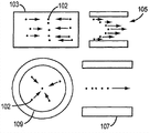
图1示出了平面和线驱动毛细管聚焦的比较。Figure 1 shows a comparison of planar and line-driven capillary focusing.
图2A和2B示出了线驱动声学聚焦装置。Figures 2A and 2B illustrate a wire-driven acoustic focusing device.
图3示出了流动越过线驱动声学聚焦装置中的层流线的经声学聚焦的颗粒。Figure 3 shows acoustically focused particles flowing across a laminar flow line in a line driven acoustic focusing device.
图4A和4B示出了声学聚焦装置中的经声学再定向的层流。Figures 4A and 4B illustrate acoustically redirected laminar flow in an acoustically focusing device.
图5A-5C示出了声学聚焦装置中的微米级聚苯乙烯荧光橙色/红色颗粒与纳米级绿色颗粒的背景的分离。图5D示出了流动越过声学聚焦装置中的层流线的声学聚焦颗粒。5A-5C illustrate the separation of micron-sized polystyrene fluorescent orange/red particles from a background of nano-sized green particles in an acoustic focusing device. Figure 5D shows acoustically focusing particles flowing across laminar flow lines in an acoustically focusing device.
图6A-6C示出了越过层流边界的颗粒的声学分离。Figures 6A-6C illustrate the acoustic separation of particles across a laminar boundary.
图7A-7C示出了若干声学聚焦装置。Figures 7A-7C illustrate several acoustic focusing devices.
图8示出了与声学流式细胞计数器组合的声学聚焦流动室的示意图。Figure 8 shows a schematic diagram of an acoustically focused flow cell combined with an acoustical flow cytometer.
图9示出了声学聚焦系统的流程图。Figure 9 shows a flow diagram of the acoustic focusing system.
图10示出了修改成包括线内分层冲洗的图9的示图。Figure 10 shows the diagram of Figure 9 modified to include in-line stratified flushing.
图11示出了分层冲洗流体的声学聚焦。Figure 11 shows acoustic focusing of stratified irrigation fluid.
图12示出了并行流体声学切换装置的示意图。Fig. 12 shows a schematic diagram of a parallel hydroacoustic switching device.
图13A和13B示出了未溶解全血的切换的示意图。13A and 13B show a schematic diagram of switching of unlysed whole blood.
图14示出了声学流切换颗粒计数装置的示意图。Figure 14 shows a schematic diagram of an acoustic flow switching particle counting device.
图15示出了阴性对比载体颗粒与血液样本核心的分离。Figure 15 shows the separation of negative control carrier particles from blood sample cores.
图16示出了声学冲洗系统中的多路免疫测定。Figure 16 shows a multiplex immunoassay in an acoustic irrigation system.
图17示出了使用声学聚焦的高吞吐量筛选的流程图。Figure 17 shows a flowchart for high-throughput screening using acoustic focusing.
图18示出了使用声学冲洗的双室培养/收获皿。Figure 18 shows a dual chamber culture/harvest dish using acoustic irrigation.
图19A-19C示出了来自库的适体选择。Figures 19A-19C show selection of aptamers from libraries.
图20示出了双级声学阀分拣器。Figure 20 shows a dual stage acoustic valve sorter.
图21A和21B示出了经声学再定位的颗粒和介质的光学分析。Figures 21A and 21B show optical analysis of acoustically repositioned particles and media.
图22示出了具有不同参数的颗粒分组的示图。Figure 22 shows a diagram of particle groupings with different parameters.
图23示出了由成像器成像的经声学再定位的颗粒。Figure 23 shows acoustically relocated particles imaged by an imager.
图24示出了颗粒的声学融合。Figure 24 shows the acoustic fusion of particles.
图25示出了颗粒的声学聚焦和分离。Figure 25 illustrates the acoustic focusing and separation of particles.
图26A-26F示出了使用各种激光器和灵敏度设置在非声学流式细胞计数器上和在声学聚焦细胞计数器上运行的荧光微球的比较输出绘图。26A-26F show plots of comparative output of fluorescent microspheres run on a non-acoustic flow cytometer and on an acoustic focusing cytometer using various laser and sensitivity settings.
图27A和27B是直方图绘图,示出了与声学细胞计量术关联的通过时间的8倍增加对细胞周期分析的影响。Figures 27A and 27B are histogram plots showing the effect of an 8-fold increase in transit time associated with acoustic cytometry on cell cycle analysis.
图28A是细胞被声学集中以形成在声学细胞计数器中流动的绳状结构的血液的照片。图28B是细胞被声学集中在声学细胞计数器中的单行线中的更为稀释的血液的照片。Figure 28A is a photograph of blood with cells acoustically concentrated to form rope-like structures flowing in an acoustic cytometer. Figure 28B is a photograph of more diluted blood with cells acoustically concentrated in a single line in an acoustic cytometer.
图29是光谱图,显示了紫光激发荧光团Pacific BlueTM的激发和发射光谱。Figure 29 is a spectral graph showing the excitation and emission spectra of the violet-excited fluorophore Pacific Blue ™ .
图30示出了将0.07%CD34阳性细胞的罕见事件群体检测作为活CD45阳性细胞的亚群。Figure 30 shows detection of a rare event population of 0.07% CD34 positive cells as a subpopulation of live CD45 positive cells.
图31A和31B分别显示了声学聚焦系统中和单独流体动力学聚焦系统中的溶解全血的FSC相对于SSC的绘图。31A and 31B show plots of FSC versus SSC for lysed whole blood in an acoustic focusing system and in a hydrodynamic focusing system alone, respectively.
图32A和32B显示了使用与图31A和31B中相同的系统和参数获得的Jurkat细胞的FSC相对于SSC的绘图。Figures 32A and 32B show plots of FSC versus SSC for Jurkat cells obtained using the same system and parameters as in Figures 31A and 31B.
图33示出了声学流式细胞计量系统的示意图。Figure 33 shows a schematic diagram of an acoustic flow cytometry system.
图34示出了声学流式细胞计数器中的声学聚焦毛细管的示意图。Figure 34 shows a schematic diagram of an acoustic focusing capillary in an acoustic flow cytometer.
图35示出了声学流式细胞计数器中的光学收集块的一部分。Figure 35 shows a portion of an optical collection block in an acoustic flow cytometer.
图36示出了声学流式细胞计数器中的光学数据收集块的示意图。Figure 36 shows a schematic diagram of the optical data collection block in an acoustic flow cytometer.
图37示出了声学流式细胞计数器中的射流系统的示意图。Figure 37 shows a schematic diagram of the fluidics system in an acoustic flow cytometer.
图38示出了具有下游流体动力学聚焦的单换能器声学聚焦毛细管的示意图。Figure 38 shows a schematic of a single transducer acoustic focusing capillary with downstream hydrodynamic focusing.
图39示出了可以调节声学流式细胞计数器中的前向散射孔径的阻挡杆装置的示意图。Figure 39 shows a schematic diagram of a barrier rod arrangement that can adjust the forward scatter aperture in an acoustic flow cytometer.
图40A-40F示出了将0.050%和0.045%CD34阳性细胞的罕见事件群体检测作为活CD45阳性细胞的亚群。Figures 40A-40F show detection of rare event populations of 0.050% and 0.045% CD34 positive cells as subpopulations of viable CD45 positive cells.
图41A-41D示出了在非声学流式细胞计数器上和声学聚焦细胞计数器上运行的细胞检测的比较输出绘图。Figures 41A-41D show comparative output plots for cell assays run on a non-acoustic flow cytometer and an acoustic focusing cytometer.
图42示出了声学聚焦细胞计数器的各部件的示意图。Figure 42 shows a schematic diagram of the components of an acoustic focusing cytometer.
图中的相似标记指示相似元件。Like numbers in the figures indicate like elements.
具体实施方式 Detailed ways
当在本文中使用时,“声学对比度”表示两对象的材料性质在用声辐射压力操纵其位置的能力方面的相对差异,并且例如可以包括密度和压缩率的差异;“测定”表示用于探询一个或多个颗粒或一个或多个流体的方法;“测定物”表示产品,例如包括测定套件、数据和/或报告;“流动室”表示通道、室或毛细管,具有选自矩形、方形、椭圆形、扁圆形、圆形、圈形(round)、八边形、七边形、六边形五边形和三角形的内部形状;并且“通道”表示至少具有入口并且优选地具有出口的路线、路径或管道,其可以包含一定量的流体,并且具有选自矩形、方形、椭圆形、扁圆形、圆形、圈形、八边形、七边形、六边形五边形和三角形的内部形状。As used herein, "acoustic contrast" means the relative difference in the material properties of two objects in their ability to manipulate their position with acoustic radiation pressure, and may include, for example, differences in density and compressibility; method of one or more particles or one or more fluids; "assay" means a product, including, for example, assay kits, data and/or reports; "flow cell" means a channel, chamber, or capillary with features selected from the group consisting of rectangular, square, oval, oblate, circular, round, octagonal, heptagonal, hexagonal, pentagonal, and triangular internal shapes; and "channel" means a channel having at least an inlet and preferably an outlet A route, path, or conduit that may contain a volume of fluid and has a shape selected from the group consisting of rectangles, squares, ellipses, oblates, circles, circles, octagons, heptagons, hexagons, pentagons, and The inner shape of the triangle.
当在本文中使用时,“声学聚焦”、“经声学聚焦”、“经声学聚焦的”以及“经声学聚焦地”表示借助于声场定位流动室内的颗粒的动作。颗粒的声学聚焦的例子是沿着通道的轴线对准颗粒。颗粒所在的聚焦区域的空间范围可以由流动室几何形状、声场和声学对比度确定。在流动室的横截面中观察,观察到的聚焦区域的形状可以类似于规则几何形状(例如点、线、弧、椭圆等)或者它可以是任意的。用于定位对象的主要的力是声辐射压力。As used herein, "acoustically focused", "acoustically focused", "acoustically focused" and "acoustically focused" refer to the act of localizing particles within a flow chamber by means of an acoustic field. An example of acoustic focusing of particles is aligning the particles along the axis of the channel. The spatial extent of the focal region where the particles reside can be determined by the flow chamber geometry, acoustic field, and acoustic contrast. Viewed in cross-section of the flow cell, the shape of the observed focal region can resemble a regular geometric shape (eg point, line, arc, ellipse, etc.) or it can be arbitrary. The main force used to position objects is acoustic radiation pressure.
当在本文中使用时,“声学再定向的”和“声学再定向”表示用声辐射压力再定位装置内的流体或介质的可混溶、部分可混溶或不可混溶层流的位置的动作。该技术利用的是流动通道中的独立层流的机械性质(声学对比度)的差异。当使两个流体接触时,会由于它们的分子组成的差异而存在大浓度梯度,导致界面密度和/或压缩率梯度(各流之间的声学对比度)。在声场的作用下,各个流可以基于其声学对比度在流动室内再定向。As used herein, "acoustically redirected" and "acoustically redirected" mean the use of acoustic radiation pressure to reposition the position of miscible, partially miscible or immiscible laminar flow of a fluid or medium within a device. action. The technique exploits the differences in the mechanical properties (acoustic contrast) of individual laminar flows in the flow channel. When two fluids are brought into contact, large concentration gradients exist due to differences in their molecular composition, resulting in interfacial density and/or compressibility gradients (acoustic contrast between the streams). Under the influence of the sound field, individual flows can be redirected within the flow chamber based on their acoustic contrast.
当在本文中使用时,“颗粒”表示物质的小单位,例如包括生物细胞(例如真核细胞和原核细胞)、古生菌、细菌、霉菌、植物细胞、酵母菌、原生动物、变形虫、原生生物、动物细胞;细胞器;有机/无机元素或分子;微球;以及不可混溶流体的小滴(例如水包油)。As used herein, "particle" means a small unit of matter, including, for example, biological cells (such as eukaryotic and prokaryotic cells), archaea, bacteria, molds, plant cells, yeasts, protozoa, amoebae, Protists, animal cells; organelles; organic/inorganic elements or molecules; microspheres; and droplets of immiscible fluids (such as oil-in-water).
当在本文中使用时,“分析物”表示待分析的物质或材料;“探针”表示物质,其有标签或以另外方式被标记并且用于检测或识别流体或样本中的另一种物质;“靶部”表示探针的结合部分;并且“试剂”表示已知以特定方式反应的物质。As used herein, "analyte" means a substance or material to be analyzed; "probe" means a substance, which is tagged or otherwise marked and used to detect or identify another substance in a fluid or sample ; "target portion" means the binding portion of the probe; and "reagent" means a substance known to react in a particular manner.
当在本文中使用时,“微球”或“小珠”表示具有声学对比度的颗粒,其可以与球形一样是对称的、与哑铃形一样是不对称的或者是不具有对称性的大分子。微球或小珠的例子例如包括硅石、玻璃和中空玻璃、乳胶、硅橡胶、聚合物,例如聚苯乙烯、聚甲基丙烯酸甲酯、聚亚甲基三聚氰胺、聚丙烯腈、聚甲基丙烯腈、聚(偏二氯乙烯共丙烯腈)和聚交酯。As used herein, "microsphere" or "bead" means a particle of acoustic contrast, which may be symmetric like a sphere, asymmetric like a dumbbell, or a macromolecule with no symmetry. Examples of microspheres or beads include, for example, silica, glass and insulating glass, latex, silicone rubber, polymers such as polystyrene, polymethylmethacrylate, polymethylenemelamine, polyacrylonitrile, polymethacrylic Nitrile, poly(vinylidene chloride coacrylonitrile), and polylactide.
当在本文中使用时,“标签”表示可识别物质,例如染料或放射性同位素,其被引入系统(例如生物系统)中并且可以跟随流动室或通道的路线,提供关于流动室或通道中的颗粒或靶部的信息;并且“信号分子”表示可识别物质,例如染料或放射性同位素,其被引入系统(例如生物系统)中并且可以用作颗粒的信号。As used herein, "label" means an identifiable substance, such as a dye or a radioactive isotope, which is introduced into a system (such as a biological system) and which can follow the course of a flow cell or channel, providing information about the particle in the flow cell or channel. or target moiety; and "signal molecule" means an identifiable substance, such as a dye or a radioactive isotope, which is introduced into a system (eg, a biological system) and which can serve as a signal for the particle.
图1示出了根据本发明的示例性实施例的平面驱动和线驱动毛细管聚焦的比较。在平面聚焦103/105中,可能包括一个或多个罕见事件颗粒的颗粒102可被聚焦为二维片并且具有沿着流动方向的可变速度(参见不同箭头)。在线驱动毛细管聚焦107/109中,颗粒102可被聚焦到毛细管的中心轴线并且具有沿流动方向的共同速度。毛细管可以具有例如圆形、扁圆形或椭圆形横截面。作为替换,颗粒102也可被聚焦到毛细管内的另一轴线或沿着毛细管的壁。Figure 1 shows a comparison of planar-driven and line-driven capillary focusing according to an exemplary embodiment of the present invention. In planar focusing 103/105,
图2A和2B示出了根据本发明的示例性实施例的线驱动声学聚焦装置的侧视图和轴视图。可能包括一个或多个罕见事件颗粒的颗粒203可以使用换能器205被声学聚焦到管201(例如可以是圆柱形管)的中心209中的压力最小值。颗粒203可以形成单线轨迹211,其可以允许具有类似尺寸和声学对比度的颗粒的均匀停留时间,这又可以允许颗粒的高吞吐量系列分析而不损害灵敏度和分辨率。2A and 2B show side and axial views of a wire-driven acoustic focusing device according to an exemplary embodiment of the present invention.
图3示出了流经根据本发明的示例性实施例的线驱动声学聚焦装置中的层流线的经声学聚焦的颗粒。可能包括一个或多个罕见事件颗粒的颗粒305可以使用换能器303从样本流309被声学聚焦到线驱动毛细管301中的流动流体/冲洗流315的中心307。颗粒305可以越过层流线移动并且于是可以作为单行线移动并且在分析点311处被分析。Figure 3 illustrates acoustically focused particles flowing through a laminar flow line in a line driven acoustic focusing device according to an exemplary embodiment of the present invention. Particles 305 , which may include one or more rare event particles, can be acoustically focused from sample stream 309 to center 307 of flowing fluid/flush stream 315 in wire driven capillary 301 using transducer 303 . Particles 305 may move across the laminar flow lines and may then move as a single file and be analyzed at analysis point 311 .
图4A和4B示出了根据本发明的示例性实施例的声学聚焦装置中的声学再定向层流。在图4A中,没有声场并且层流403和407彼此平行地在声驱动毛细管401中流动。在图4B中,声场由换能器405施加并且因此流403和407可以基于它们的声学对比度被声学再定向。具有较大声学对比度的流403b可以被再定向到毛细管401的中心,而具有较低声学对比度的流407b可被再定向为靠近毛细管401的壁。如果在偶极模式中启动声场,则流403b与毛细管401的中心轴线重合地移动,部分地移位流407b,如图4B中所示。流可以是不可混溶的、部分可混溶的或可混溶的。大浓度梯度可能由于它们的不同分子组成而存在于流之间。为了声压力,浓度梯度可以被视为密度和/或压缩率梯度,并且流可以被视为可以用声辐射压力作用的具有不同密度和压缩率(声学对比度)的孤立实体。4A and 4B illustrate acoustically redirected laminar flow in an acoustically focusing device according to an exemplary embodiment of the present invention. In Figure 4A, there is no acoustic field and
图5A-5C示出了根据本发明的示例性实施例的声学聚焦装置中的微米级聚苯乙烯荧光橙色/红色颗粒与纳米级绿色颗粒的背景的分离。图5D示出了流动越过根据本发明的示例性实施例的声学聚焦装置中的层流线的经声学聚焦的颗粒。分离可以基于尺寸和声学对比度,原因是时间平均声力与颗粒的体积成比例。如果中心冲洗流具有比外部样本流更高的特定比重和/或更低的压缩率,则初始在具有比中心冲洗流更大的声学对比度的外部样本流中的颗粒将继续聚焦到毛细管轴线,而较小对比度的颗粒将被排除。图5A显示了当声场关闭时与绿色200nm颗粒混合的红色5.7μm颗粒在落射荧光照明下流动通过毛细管。图5B显示了当声场打开时5.7μm颗粒(其在蓝光照明下发出黄色荧光)被声学聚焦到中心线,而200nm颗粒保持在它们的初始流中。图5C显示了5.7μm颗粒在具有红色带通滤光器的绿光照明下发出红色荧光,而200nm颗粒未被激发。图5D示出了在同轴流505的旁边引入的清洁核心流507,所述同轴流包含在毛细管501中流动的荧光背景流体。当换能器503产生声驻波(未显示)时,可能包括一个或多个罕见事件颗粒的颗粒509可以被声学聚焦并且从同轴流505移动到核心流507,在那里它们以单行朝着分析点511流动。5A-5C illustrate the separation of micron-sized polystyrene fluorescent orange/red particles from a background of nano-sized green particles in an acoustic focusing device according to an exemplary embodiment of the present invention. Figure 5D shows acoustically focused particles flowing across a laminar flow line in an acoustic focusing device according to an exemplary embodiment of the present invention. Separation can be based on size and acoustic contrast, since the time-averaged acoustic force is proportional to the volume of the particle. If the central flushing stream has a higher specific gravity and/or lower compressibility than the outer sample stream, particles initially in the outer sample stream with greater acoustic contrast than the central flushing stream will continue to focus to the capillary axis, Particles with lesser contrast will be excluded. Figure 5A shows red 5.7 μm particles mixed with green 200 nm particles flowing through a capillary under epi-fluorescent illumination when the sound field is turned off. Figure 5B shows that 5.7 μm particles (which fluoresce yellow under blue light illumination) are acoustically focused to the centerline when the acoustic field is turned on, while 200 nm particles remain in their initial flow. Figure 5C shows that 5.7 μm particles fluoresce red under illumination with green light with a red bandpass filter, while 200 nm particles are not excited. FIG. 5D shows
图6A-6C示出了越过根据本发明的示例性实施例的层流边界的颗粒的声学分离。介质或流体可被声学再定向,同时可能包括一个或多个罕见事件颗粒的介质或流体中的颗粒可被声学操纵或聚焦。图6A显示了当声场关闭时耦合到250μm声学聚焦室609的端部的光室的荧光图像。白线601和611指示流动室的边缘。掺有25μg/ml的藻红蛋白(R-Phycoerythrin)荧光蛋白(橙色荧光)的PBS缓冲液中的10%全血的混合物流动通过流动室的下半部605(白血细胞DNA用
图7A-7C示出了根据本发明的示例性实施例的若干声学聚焦装置。图7A显示了流式细胞计量系统700a,其中包括颗粒712(可能包括一个或多个罕见事件颗粒)的样本715a和冲洗缓冲液713a被引入毛细管703。线驱动器701(例如,PZT驱动器或能够产生声驻波的其它机构)在用户定义模式(例如偶极模式)下引入声驻波(未显示)。因此,样本715a和冲洗缓冲液713a可以被声学再定向(如715b和713b)并且颗粒712可以基于它们的声学对比度被声学聚焦(如717)。照明源709(例如激光器或一组激光器,或任何合适的照明源,例如发光二极管)在探询点716照明颗粒717。照明源可以是紫光激光器(例如405nm激光器)、蓝光激光器(例如488nm激光器)、红光激光器(例如640nm激光器)或它们的组合。来自被测样本的光信号719可以由检测器或检测器阵列705(例如PMT阵列、光电倍增管、雪崩光电二极管(APD)、多像素APD装置、硅PMT等)检测。图7B显示了流式细胞计量系统700b,其中清洁流713a可以独立地流动通过光室。可能包括一个或多个罕见事件颗粒的颗粒702可以被声学聚焦以如线717流动,样本缓冲液715b可以被丢弃到废物721,并且线717可以传递到第二声波引起机构714。图7C显示了流式细胞计量系统700c,其中样本715a可被喷射为稍偏向中心的一侧并在毛细管壁703附近流动,同时缓冲液713a贴靠相对壁流动。换能器701可以声学再定向样本715a(如715b),可以声学聚焦可能包括一个或多个罕见事件颗粒的颗粒702(如714/717),并且可以声学地再定向缓冲液713a。7A-7C illustrate several acoustic focusing devices according to exemplary embodiments of the present invention. FIG. 7A shows a
图8示出了与根据本发明的示例性实施例的用于声学定向颗粒和流的声学流式细胞计数器组合的声学聚焦流动室的示意图。包括颗粒803、807和809且可能包括一个或多个罕见事件颗粒的样本801被引入包含换能器811的流动室810,所述换能器将颗粒803、807和809声学聚焦为在收集/培育部位819收集的颗粒815。来自冲洗容器802的冲洗或其它试剂805作为侧向离开的背景流813被引入流动室810。另一冲洗流821从冲洗容器817引入具有声场发生器822的聚焦细胞计数器850的流动室810b中,在进入另一个流动室851中的入口829之前所述声场发生器将颗粒823声学聚焦为颗粒825/827。在由探询光833在探询点852进行探询之前换能器831还将颗粒825/827声学聚焦为颗粒832。在收集点837收集被测量颗粒之前,来自被探询颗粒的信号854被发送到检测器835进行分析。Figure 8 shows a schematic diagram of an acoustically focusing flow cell combined with an acoustic flow cytometer for acoustically orienting particles and flow according to an exemplary embodiment of the present invention. A sample 801 comprising
图9示出了根据本发明的示例性实施例的声学聚焦系统的流程图。在步骤901中,包括颗粒的样本被收集并且引导到可控流动泵。在步骤903中,可控流动泵将样本泵送到声学聚焦设备。在步骤905中,声学聚焦设备将可能包括一个或多个罕见事件颗粒的颗粒中的至少一些聚焦成线或平面,并且然后将颗粒引导到探询区进行光学激发和检测。在步骤907中,光学激发颗粒中的至少一些并且检测来自激发颗粒的至少一些信号,并且随后引导颗粒进行进一步分析或者引导到废物或进行某些其它处理。在步骤909中,可以由更长通过时间数据收集和分析部分进一步分析颗粒。在步骤911中,颗粒可以由废物或附加处理部分提取为废物或者受到附加处理。可控泵可以为颗粒的期望线速度而被调节到期望颗粒流率,其可以在例如大约0m/s至大约10m/s、大约0m/s至大约0.3m/s或大约0.3m/s至大约3m/s的范围内。激发/检测可以是脉动的或经调制的,并且可以使用在模拟/数字电子学和/或光学中已知的任何合适的激发/检测方法进行,包括使用瑞利散射检测器。Fig. 9 shows a flowchart of an acoustic focusing system according to an exemplary embodiment of the present invention. In
颗粒速度的控制具有许多优点。首先,它可以通过增加由荧光/发光标签发出的光子的数量来改善信号,原因是标签可以被照明更长时间。在0.3m/s的线速度下,光子的数量可以增加大约10倍并且然后当使用声学聚焦时每秒可以分析大约3,000个颗粒(假设颗粒中心之间的平均距离为100微米)。并且在0.03m/s的线速度下,该数量可以增加大约100倍并且每秒可以分析300个颗粒。其次,典型地由于快速通过时间在常规流式细胞计量术中不使用的标记物(例如镧系元素、镧系元素螯合物、使用铕的纳米颗粒、半导体纳米晶体(例如量子点)、诸如细胞染色剂和台盼蓝的吸收性染料等可以变得可使用。再次,其它标记物(例如具有长寿命和/或低量子产率/消光系数的荧光团或发光团;多数化学-生物发光物种;寿命大于大约10ns、在大约10nm至大约1μs之间、在大约1μs至大约10μs之间、在大约10μs至大约100μs之间和在大约100μs至大约1ms之间的标签)可以受益于减小光漂白的更低激光功率并且受益于通过线速度的控制成为可能的更长通过时间。对于例如10ms的通过时间,在每秒一千次的速率以10μs脉冲进行的脉冲调制允许激发和发光收集的10个周期,其中实际上例如铕螯合物的所有发光衰减可以被监测。在该脉冲速率下若没有通过本发明的实施例提供的线速度的控制而成为可能的更长通过时间的益处,90%或以上的颗粒可能在未被探询的情况下通过。如果脉冲速率增加到100kHz并且具有1μs脉冲,则可能仍然有将近9μs监测镧系元素发光(原因是多数荧光团具有1-2ns寿命并且多数自发荧光在10ns内衰减)。Control of particle velocity has many advantages. First, it can improve the signal by increasing the number of photons emitted by the fluorescent/luminescent label, since the label can be illuminated for a longer period of time. At a linear velocity of 0.3 m/s, the number of photons can be increased by a factor of about 10 and then about 3,000 particles per second can be analyzed when using acoustic focusing (assuming an average distance between particle centers of 100 microns). And at a line speed of 0.03m/s, the number can be increased approximately 100 times and 300 particles per second can be analyzed. Second, labels that are typically not used in conventional flow cytometry due to fast transit times (e.g. lanthanides, lanthanide chelates, nanoparticles using europium, semiconductor nanocrystals (e.g. quantum dots), such as Absorbing dyes such as cell stains and trypan blue may become available. Again, other labels such as fluorophores or luminophores with long lifetimes and/or low quantum yields/extinction coefficients; most chemi-bioluminescent species; tags with lifetimes greater than about 10 ns, between about 10 nm to about 1 μs, between about 1 μs to about 10 μs, between about 10 μs to about 100 μs, and between about 100 μs to about 1 ms) may benefit from reduced Lower laser power for photobleaching and benefit from longer transit times made possible by control of the pass line velocity. For a transit time of e.g. 10 ms, pulse modulation with 10 μs pulses at a rate of a thousand times per second allows excitation and
图10示出了根据本发明的示例性实施例的修改成包括线内分层冲洗的图9的示图。在步骤1001中,包括颗粒的样本被收集并引导到可控样本流动泵。在步骤1005中,可控样本流动泵将样本泵送到声学装置1019的分层冲洗装置部分中(其可以基于颗粒和流体的声学聚焦和/或再定向)。与此同时,在步骤1003中,冲洗流体被收集并引导到可控冲洗流体泵。在步骤1007中,可控冲洗流体泵将冲洗流体泵送到分层冲洗装置中。在步骤1009中,分层冲洗装置线内冲洗样本/颗粒。在步骤1011中,收集清洁流体,并且然后将可能包括一个或多个罕见事件颗粒的已冲洗样本/颗粒引导到探询区以进行光学激发和检测。在步骤1013中,光学地激发颗粒中的至少一些,检测来自经激发的颗粒的至少一些信号,并且然后引导颗粒进行进一步分析或将其引导到废物或进行某些其它处理。在步骤1015中,所述颗粒可由更长通过时间数据收集和分析部分进一步分析。在步骤1017中,所述颗粒可由废物或附加处理部分提取为废物或者受到附加处理。FIG. 10 shows the diagram of FIG. 9 modified to include in-line stratified flushing, according to an exemplary embodiment of the invention. In
图11示出了根据本发明的示例性实施例的分层冲洗流体的声学聚焦。包含颗粒1109(可能包括一个或多个罕见事件颗粒)的样本连同分层冲洗流体1107一起被引入平面声学流动室1101中。换能器1103基于声学对比度生成声波1105,所述声波将颗粒1109声学聚焦到穿过节点1111的轨迹。平面声学流动室1101也可以具有位于外部的声学节点,在该情况下颗粒1109可以声学聚焦到流动室的顶部。Figure 11 illustrates acoustic focusing of stratified irrigation fluid according to an exemplary embodiment of the present invention. A sample containing particles 1109 (possibly including one or more rare event particles) is introduced into planar
图12示出了根据本发明的示例性实施例的并行流体声学切换装置的示意图。包含第一颗粒1202和第二颗粒1204(可能包括一个或多个罕见事件颗粒)的第一、最外侧样本介质1205被引入毛细管1201。第二、中间介质1213连同第三、最内侧介质1209一起被引入毛细管1201。线驱动器1203可以声学地再定向第一、第二和第三介质并且可以基于它们的声学对比度而声学聚焦第一颗粒和第二颗粒。所述颗粒然后可以流到毛细管之外。当切换时,颗粒中的一些可以从第一介质穿过第二介质(其可以是试剂流)声学聚焦到第三介质,或者它们可以从第二介质声学聚焦到第三介质。Fig. 12 shows a schematic diagram of a parallel hydroacoustic switching device according to an exemplary embodiment of the present invention. A first,
图13A和13B示出了根据本发明的示例性实施例的未溶解全血的切换。血液样本1309和冲洗缓冲液1307在不同位置被引入毛细管1302。当启动换能器1304时,可能包括一个或多个罕见事件红/白血细胞的红血细胞1303和白血细胞1305被声学聚焦,并且样本1309和冲洗缓冲液1307被声学再定向。由于它们的数量较小,所以白血细胞在经聚焦血液的绳状结构中保持分离。13A and 13B illustrate switching of unlysed whole blood according to an exemplary embodiment of the present invention.
图14示出了根据本发明的示例性实施例的声学流切换颗粒计数装置的示意图。所述装置1400允许与未知或不可用导电缓冲液1403一起流动的具有颗粒1409(可能包括一个或多个罕见事件颗粒)的样本1405的线内分析。颗粒1409可以使用换能器1407声学聚焦到缓冲液1403,而样本介质可以在废物出口1411被丢弃。当颗粒移动经过第二换能器1413到达具有孔径大小1419b的检测点1419时颗粒可以由检测电极1415处的信号的任何合适的电子检测器1417分析并且计数。Fig. 14 shows a schematic diagram of an acoustic flow switching particle counting device according to an exemplary embodiment of the present invention. The
图15示出了根据本发明的示例性实施例的阴性对比度载体颗粒与血液样本核心的分离。其中,换能器1507可以声学聚焦可能包括一个或多个罕见事件颗粒的阴性对比度载体颗粒1505,这些颗粒1505从初始包括它们和血细胞1503的血液核心样本1511横越血液核心样本1511和清洁缓冲液1513之间的界面1502,朝着毛细管壁1501移动。在其它声学模式中,血细胞1503可以被驱动到壁,而阴性对比度载体颗粒1505可以被驱动到中心轴线。Figure 15 shows the separation of negative contrast carrier particles from a blood sample core according to an exemplary embodiment of the invention. Wherein,
图16示出了根据本发明的示例性实施例的声学冲洗系统中的多路免疫测定。在声学冲洗系统1600中,可以通过使分析物1609/1611/1613在中心流1607中流动并且将预先结合有荧光抗原的小珠1603/1605/1621从外部流1601推入中心流1607中而快速地执行竞争免疫测定。特定化学制品可被置于在单反应容器中混合并且在流动中处理的每个群体上。离开容器的群体1615(可能包括一个或多个罕见事件群体)可以在分析点1617经由尺寸和或荧光颜色和/或荧光而加以区分。Figure 16 shows a multiplexed immunoassay in an acoustic irrigation system according to an exemplary embodiment of the present invention. In the
图17示出了根据本发明的示例性实施例的使用声学流体的高吞吐量筛选的流程图。在步骤1701中,可能包括一个或多个罕见事件颗粒的细胞/小珠型颗粒被收集和/或培养以供试验。在步骤1703中,用感兴趣的标签和/或药物候选培育这些颗粒并将其引导到声学聚焦器/流切换器。与此同时,在步骤1709中,(一种或多种)其它药物和/或(一种或多种)附加反应物可以被引入声学聚焦器/流切换器。在步骤1705中,声学聚焦器/流切换器聚焦和/或切换颗粒和/或流,并且可以使颗粒从过量的药物/配体中分离。在步骤1707中,立即或在附加切换之后使用声学聚焦器/流切换器收集清洁流体。在步骤1711中,可以识别和/或分拣颗粒。然后,在步骤1713中,可以将不需要的颗粒送往废物,并且在步骤1715中,可以将选定颗粒送往附加分析或处理。在步骤1717中,可以执行包括药物结合的确定、闪烁计数、存活/凋亡确定和基因表达分析的附加处理。Figure 17 shows a flow diagram of high throughput screening using acoustic fluids according to an exemplary embodiment of the present invention. In
图18示出了根据本发明的示例性实施例的使用声学冲洗的双室培养/收获皿。其中,可能包括一个或多个罕见事件细胞的细胞在室1801中培养并且可被定期发送以声学聚焦到通道1805中,在那里它们可由光学检测器1817检查细胞密度/生长。当满足生长目标并且室1801中的培养基用完时,可以启动阀以允许来自容器1803的新鲜培养基沿着通道1805流动并且在室1811中收获用过的培养基。细胞然后可被声学聚焦到新鲜培养基中并被转移到第二培育室1809。于是可以反向重复相同过程,以使得细胞在室1809中培养并被转移到室1801内的新鲜培养基中。Figure 18 shows a dual chamber culture/harvest dish using acoustic irrigation according to an exemplary embodiment of the invention. Therein, cells, possibly including one or more rare event cells, are cultured in
图19A-19C示出了根据本发明的示例性实施例的来自库的适体选择。图19A显示了具有用适体库1901培育的靶分子的多重小珠/细胞1903。图19B示出了使用线内声学介质切换使小珠/细胞1903和1907从未结合适体1904分离。可以调节冲洗核心(中心圆)的盐和/或pH以选择更高亲和力适体,并且可以执行连续冲洗以增加纯度。图19C示出了经分选的小珠1911。19A-19C illustrate selection of aptamers from libraries according to exemplary embodiments of the invention. Figure 19A shows multiplexed beads/
图20示出了根据本发明的示例性实施例的允许罕见细胞群体的线内非稀释高速分选的双级声学阀分选器。其中,包括颗粒2007的样本被引入声学阀分选器2000的部分2001。第一换能器2002引起通道2004中的声波,并且探询源2013在探询点2006探询颗粒2007。在分选点2009检测到的不需要的颗粒可以被引导通过废物阀2010a,而选定颗粒可以沿着通道2004被引导到下游处理2011以供第二换能器2002进一步聚焦、由光源2015探询并且对于不需要的颗粒朝着废物阀2010b适当地分选或者对于选定颗粒2019从通道2004离开。对于罕见细胞,上述分选提供捕获感兴趣细胞的高速初始阀分选,因此增加了分选部分中的期望细胞的比率。随后,上述分选可以在更低速率下再次进行以用于提高纯度。如果例如以每秒30,000个细胞的速率分析细胞并且阀分选能够以每秒300个细胞的速度进行分选,则每个初始分选决定应当包含平均大约100个细胞。如果这些100个细胞随后以更慢的流速转移到第二分选器(或在初始分选之后的相同分选器),则可以显著地纯化感兴趣的细胞。Figure 20 shows a dual-stage acoustic valve sorter allowing in-line non-dilution high-speed sorting of rare cell populations according to an exemplary embodiment of the present invention. Therein, a
图21A和21B示出了根据本发明的示例性实施例的声学再定位颗粒和介质的光学分析。其中,样本2103中的颗粒2102(可能包括一个或多个罕见事件颗粒)基于声学对比度由线驱动器2105声学聚焦(作为颗粒2115)。颗粒2115进入光室2117并且探询源2111探询这些颗粒。检测器阵列2107随后收集来自每个颗粒的光信号2113,并且如果信号满足某个用户确定的标准,则相应颗粒(或颗粒组)由光源2119(例如闪光LED(宽带或UV))照明并且由成像器2109成像。在图21A中,不采集图像并且颗粒2125的流速2129保持不变。然而在图21B中,颗粒2125的流速2127减小到适合于所需成像分辨率的值以采集颗粒的图像。21A and 21B illustrate optical analysis of acoustically repositioned particles and media according to exemplary embodiments of the present invention. Therein, particles 2102 (possibly including one or more rare event particles) in
图22示出了具有不同参数的颗粒分组的示图,所述参数例如可以在如图21A和21B中所示的系统中被分析。颗粒组2202内的每个颗粒关于参数1和参数2(各自例如可以是前向散射、侧向散射或荧光)是类似的。用户定义的阈值2201基于参数1和参数2的值识别满足该阈值的用于成像的颗粒。如果颗粒满足用户定义的阈值,则流动可以减小到合适速率以供成像器捕获颗粒的焦内图像。也可以建立其它检测阈值2205、2209和2215。当然,不需要成像每个颗粒。相反地,可以构造来自选通亚群的颗粒的采样矩阵以基于它们的散射和荧光标记来限定待捕获的一组颗粒图像,这可以允许高颗粒分析速率(例如超过每秒2000)。图像可以捕获细胞形态、定向和内部结构(例如细胞核的位置和数量),并且可使用本领域中已知的任何合适的成像装置获得,包括电子CCD摇拍技术。成像可以相对较慢(达300细胞/秒),但是更慢的流动可以允许保持高灵敏度的长探询时间并实现良好的空间分辨率(达0.5微米)。Fig. 22 shows a diagram of particle groupings with different parameters that can be analyzed, for example, in a system as shown in Figs. 21A and 21B. Each particle within
图23示出了根据本发明的示例性实施例的由成像器成像的声学再定位颗粒。它显示了从声学再定向流2305捕获的血细胞2303的照片,其中为了血细胞2303的焦内图像捕获而减慢光室2307中的流。为创建图像,例如内径410μm的线驱动毛细管可以用光室(其例如可以是具有内部圆柱形通道的硼硅玻璃立方体,所述内部圆柱形通道具有与线驱动毛细管的内径相同的直径)截断。激发频率为大约2.1MHz并且声学装置的功耗为125毫瓦。线驱动毛细管可以在超过5ml/min的容积流速下产生5μm乳胶颗粒和血细胞的精细聚焦。线驱动毛细管可以附连到方形横截面石英光室。光室的内腔可以在横截面上为圆形,并且可以具有与线驱动毛细管相同的内径以延长流体柱的共振条件并且由此将声学聚焦力延伸到光室中。Figure 23 shows acoustically repositioned particles imaged by an imager according to an exemplary embodiment of the invention. It shows a photograph of a
图24示出了根据本发明的示例性实施例的颗粒的声学融合。包含第一颗粒类型的第一样本2401被泵送通过由PZT换能器2404驱动的第一声学聚焦器2402并且所述颗粒被声学聚焦成线2408。包含第二颗粒类型的第二样本2403类似地被泵送并且聚焦成由PZT换能器2407驱动的第二声学聚焦器2405中的线2409。样本流动到由PZT换能器2411驱动的第三声学聚焦器2410中使得颗粒的线被聚焦以形成颗粒可以相互作用的单线。在下游,颗粒穿过使用融合颗粒2412的电极2413产生的电场,可能形成一个或多个罕见事件颗粒。Fig. 24 illustrates acoustic fusion of particles according to an exemplary embodiment of the present invention. A
图25示出了根据本发明的示例性实施例的颗粒的声学聚焦和分离。可能包括一个或多个罕见事件颗粒的颗粒2503移动到第一声学聚焦器2505,所述第一声学聚焦器用第一换能器2507将它们聚焦成单行线2509。线2509随后可以被给馈入基于尺寸和声学性质中的一个或多个进行分离和收集的声学分离器2513中,所述声学分离器配备有第二换能器2512和多个出口2519a、2519b和2519c。当进入声学分离器2513中时可以通过拉走流体或以另外方式例如通过侧向通道2511去除流体而调节线2509的位置。Figure 25 illustrates acoustic focusing and separation of particles according to an exemplary embodiment of the invention.
根据本发明的示例性实施例,通过使用能够控制颗粒速度并且允许长通过时间的声学流式细胞计数器执行这样的测定,如本文中所述,可以减小对单个患者的血液进行测定的临床免疫表型板中的测定量,这可以增加一次测定的标记物的数量。可以一次执行例如4、6或更多抗体的更大无补偿板。例如,在AIDS进展监测中用于T细胞的CD4阳性枚举的anti-CD45、CD4和CD8抗体的板中,例如可以加入或代用CD3以有助于识别T细胞。可以使用蓝色(例如488nm)和红色(例如635nm)激光细胞计数器进行测定,每个抗体具有不同的荧色物(例如FITC、PE、PE-Cy5和APC)。例如可以使用用于白血病/淋巴瘤分类的许多四抗体测定组合,例如可以包括(1)CD3、CD14、HLADr和CD45;(2)CD7、CD13、CD2和CD19;(3)CD5、Lambda、CD19和Kappa;(4)CD20、CD11c、CD22和CD25;(5)CD5、CD19、CD10和CD34;以及(6)CD15、CD56、CD19和CD34。此外,在Sutherland等人的“Enumeration ofCD34+Hematopoietic Stem and Progenitor Cells”,Current Protocolsin Cytometry,6.4.1-6.4.23(2003)中描述的方案可以有利地用于本文中所述的本发明的示例性实施例中的一个或多个,上述文献通过引用完整地被合并于本文中。According to an exemplary embodiment of the present invention, by performing such assays using an acoustic flow cytometer capable of controlling particle velocity and allowing for long transit times, as described herein, the clinical immunological burden of assaying blood from a single patient can be reduced. Assay volume in phenotyping plates, which can increase the number of markers assayed at one time. Larger uncompensated plates of
也可以使用用于白血病/淋巴瘤分类的许多六抗体测定组合,包括表1中所示的例子(左列指示测定编号并且顶栏指示用于每个抗体的荧色物;从左到右在其相应的荧色物标记物之下列出了每个抗体的特异性)。通过用长寿命试剂和窄带试剂代替荧色物,最小补偿抗体板是可能的。在表2中显示了可以实现不需要补偿控制的最小补偿结果的标记物的更多几个例子。测定例如可以使用405nm和635nm脉冲二极管激光器。Many six-antibody assay combinations for leukemia/lymphoma classification are also available, including the examples shown in Table 1 (left column indicates assay number and top column indicates fluorochrome used for each antibody; left to right in The specificity of each antibody is listed under its corresponding fluorochrome label). By replacing fluorochromes with long-lived and narrow-band reagents, minimally compensated antibody panels are possible. A few more examples of markers that can achieve minimal compensation results that do not require compensation controls are shown in Table 2. Assays can use, for example, 405 nm and 635 nm pulsed diode lasers.
表1Table 1

表2Table 2

可以通过将快速红细胞溶解试剂并入中心冲洗流来溶解流动分离器中的线内红细胞而用红细胞溶解执行血液中的免疫表型。在溶解之后,未溶解白细胞可被快速转移到后续分离器中的淬灭缓冲液。这可以在很短的时间内执行,最小化白细胞的损伤或损失,并且还可以用于排除包括溶解红细胞“鬼影”的碎片,所述“鬼影”具有由溶解过程导致的降低的声学对比度。可以在溶解之前在小体积血液内进行用于免疫表型的白血细胞的染色,或者可以在溶解之后进行(同时小心地控制样本体积和白细胞数量以保证恰当的免疫反应)。如本文中所述的声学冲洗系统可以用于将靶细胞或颗粒集中成小体积以进行恰当的免疫染色,这对具有低浓度靶细胞的样本是有用的。例如,这样的系统可以用于减小对于AIDS进展监测有价值的CD4阳性T细胞的测定的成本。Immunophenotyping in blood can be performed with erythrolysis by incorporating a rapid erythrolysis reagent into the central flush flow to lyse in-line erythrocytes in the flow separator. After lysis, unlysed leukocytes can be quickly transferred to quenching buffer in a subsequent separator. This can be performed in a very short time, minimizing leukocyte damage or loss, and can also be used to exclude debris including lysed red blood cell "ghosts" with reduced acoustic contrast caused by the lysis process . Staining of white blood cells for immunophenotyping can be performed in a small volume of blood prior to lysis, or can be performed after lysis (while carefully controlling sample volume and number of white blood cells to ensure an appropriate immune response). Acoustic flushing systems as described herein can be used to concentrate target cells or particles into small volumes for proper immunostaining, which is useful for samples with low concentrations of target cells. For example, such a system could be used to reduce the cost of assaying CD4 positive T cells valuable for AIDS progression monitoring.
血液免疫表型还可以通过触发荧光信号而不是散射信号的检测在没有红细胞溶解的情况下执行。全血随后可用适当的抗体染色并在没有溶解的情况下给馈入细胞计数器,在一些情况下实际上不进行稀释。根据本发明的实施例的声学细胞计数器可以对大约每分钟100-500μl的全血执行该类型的测定,原因是血细胞可以被集中到具有很小间质间隙的中心核心内。由于正常患者体内的白血细胞通常占全血中的细胞总数的1%以下,因此密集血液核心中的白血细胞的重合是罕见的。流体动力学聚焦的唯一用途似乎不是产生这样的固体核心,这限制了穿过指定横截面区域的细胞的数量。还可以执行远离自由抗体将血细胞转移到清洁缓冲液中的声学冲洗步骤,这可以减小荧光背景并且增加灵敏度。Blood immunophenotyping can also be performed without red blood cell lysis by triggering the detection of fluorescent signals rather than scatter signals. Whole blood can then be stained with the appropriate antibody and fed into the cytometer without lysis, and in some cases virtually no dilution. Acoustic cytometers according to embodiments of the present invention can perform this type of assay on whole blood at approximately 100-500 μl per minute, since blood cells can be concentrated into a central core with very little interstitial space. Since white blood cells in normal patients typically make up less than 1% of the total number of cells in whole blood, superposition of white blood cells in the dense blood core is rare. The only use of hydrodynamic focusing does not appear to be to generate such solid cores, which limit the number of cells that pass through a given cross-sectional area. An acoustic wash step that transfers blood cells away from free antibody into a cleaning buffer can also be performed, which reduces fluorescent background and increases sensitivity.
图26A-26F示出了使用各种激光器和灵敏度设置在非声学流式细胞计数器上和在声学聚焦细胞计数器上运行的荧光微球的比较输出绘图以显示使用根据本发明的示例性实施例的声学细胞计数器可以实现的增加的通过时间。荧光微球(可从Spherotech,Libertyville,IL获得,商标为Rainbow RCP-30-5A,3.2μm)使用488nm蓝光激光器(顶行,图26A-26C)和405nm紫光激光器(底行,图26D-26F)在仅仅使用流体动力学聚焦的非声学流式细胞计数器上(图26C和26F)和使用上游声学聚焦及随后使用下游流体动力学聚焦的声学聚焦细胞计数器上(图26A、26B、26D和26E)运行。非声学流式细胞计数器以其具有15μl/min的样本输入速率的最高灵敏度设置运行(右列,图26C和26F)。声学聚焦细胞计数器以其具有100μl/min的样本输入速率的标准灵敏度设置(中间列,图26B和26E)和以其具有100μl/min的样本输入速率的最高灵敏度设置并且颗粒被激光器照射的时间增加4倍(左列,图26A和26D)运行。考虑两个总流速(2.4ml/min和0.6ml/min)。鞘和样本输入速率相对于彼此调节以针对2.4ml/min总速率允许25μl/min至1000μl/min并且针对0.6ml/min的总速率允许25μl/min至200μl/min的样本输入速率。图26A-26F显示了8峰荧光rainbow微球(其由不同荧光强度水平的8个群体组成)由声学聚焦细胞计数器更清楚地分辨,这可以通过该组8峰小珠内各峰值之间的更大和更清楚分离看到,尤其在图26A和26D中,这得益于颗粒被激光器照射时间的4倍增加(例如从大约10μs增加到大约40μs)。这证明荧光群体的更好分辨率由减慢流动并且增加通过时间导致。26A-26F show plots of comparative output of fluorescent microspheres run on a non-acoustic flow cytometer and on an acoustic focusing cytometer using various lasers and sensitivity settings to show the Increased transit times achievable with acoustic cell counters. Fluorescent microspheres (available from Spherotech, Libertyville, IL under the trademark Rainbow RCP-30-5A, 3.2 μm) using a 488 nm blue laser (top row, Figures 26A-26C ) and a 405 nm violet laser (bottom row, Figures 26D-26F ) on non-acoustic flow cytometers using only hydrodynamic focusing (Figs. 26C and 26F) and on acoustic focusing cytometers using upstream acoustic focusing followed by downstream hydrodynamic focusing (Figs. 26A, 26B, 26D and 26E )run. The non-acoustic flow cytometer was run at its highest sensitivity setting with a sample input rate of 15 μl/min (right column, Figures 26C and 26F). Acoustic focusing cytometer at its standard sensitivity setting with a sample input rate of 100 μl/min (middle column, Figures 26B and 26E) and at its highest sensitivity setting with a sample input rate of 100 μl/min and increased time for particles to be illuminated by the laser 4x (left column, Figures 26A and 26D) runs. Consider two total flow rates (2.4ml/min and 0.6ml/min). The sheath and sample input rates were adjusted relative to each other to allow sample input rates of 25 μl/min to 1000 μl/min for a total rate of 2.4 ml/min and 25 μl/min to 200 μl/min for a total rate of 0.6 ml/min. Figures 26A-26F show that 8-peak fluorescent rainbow microspheres (which consist of 8 populations of different fluorescence intensity levels) are more clearly resolved by an acoustic focusing cytometer, which can be seen by the distance between peaks within the set of 8-peak beads. Larger and clearer separations are seen, especially in FIGS. 26A and 26D , which benefit from a 4-fold increase in the time the particles are illuminated by the laser (eg, from about 10 μs to about 40 μs). This demonstrates that better resolution of fluorescent populations results from slower flow and increased transit time.
图27A和27B是直方图绘图,示出了根据本发明的示例性实施例的与声学细胞计量术关联的通过时间的约8倍增加对细胞周期分析的影响。图27A显示了当使用紫光激光器和低样本速率设置(通过时间为大约5μs)通过非声学(仅流体动力学)流式细胞计数器运行用紫染色(可从加利福尼亚州Carlsbad的Life Technologies Corp.获得,商标为FxCycleTM)标记的ST486B淋巴球时获得的数据。图27B显示了相同类型的数据,但是使用紫光激光器和25μl/min样本输入速率(通过时间为大约40μs)由具有上游声学聚焦和下游流体动力学聚焦的声学聚焦细胞计数器获得。在两种情况下采集到总共大约15,000个事件。使用可从Verity Software House获得的、商标为ModFit LTv.3.2.1的曲线拟合软件执行的数据分析产生底层细胞周期相分布和软件定义的G0G1峰值的变化的百分比系数(%CV)和G2/G1比率。%CV是落入G0G1峰值的细胞的精度的量度(%CV越低,测量精度越高)。图27A显示了当使用声学聚焦时的更不同群体和更低%CV(2.81%相对于5.84%)。27A and 27B are histogram plots showing the effect of an approximately 8-fold increase in transit time associated with acoustic cytometry on cell cycle analysis, according to an exemplary embodiment of the invention. FIG. 27A shows staining with violet (available from Life Technologies Corp., Carlsbad, Calif., Data obtained when ST486B lymphocytes were labeled with the trademark FxCycle ™ . Figure 27B shows the same type of data, but obtained from an acoustically focused cytometer with upstream acoustic focusing and downstream hydrodynamic focusing using a violet laser and a sample input rate of 25 μl/min (pass time approximately 40 μs). A total of approximately 15,000 events were collected in both cases. Data analysis performed using curve fitting software available from Verity Software House under the trademark ModFit LTv.3.2.1 yielded the underlying cell cycle phase distribution and the percent coefficient of variation (%CV) of the software-defined G 0 G 1 peak and the G 2 /G 1 ratio. %CV is a measure of the precision of cells falling into the G0G1 peak (the lower the %CV, the higher the precision of the measurement). Figure 27A shows a more diverse population and lower %CV (2.81% vs. 5.84%) when using acoustic focusing.
其它类似细胞周期分析实验显示尽管仅使用流体动力学聚焦会在样本速率增加时使得数据质量和%CV降低,但是使用声学聚焦的情况下在样本速率增加时数据质量和%CV可以几乎不变化。具体地,对于仅在1x10⁶细胞/ml的浓度下的流体动力学聚焦,12μl/min、35μl/min和60μl/min的样本速率的%CV值分别为4.83%、6.12%和7.76%,并且S相数据从12μl/min低速率的37.83%变化到60μl/min高速率的26.17%。但是对于下游流体动力学聚焦已经声学聚焦的样本,25μl/min、100μl/min、200μl/min、500μl/min和1000μl/min的样本速率的%CV值分别为3.22%、3.16%、3.17%、4.16%和4.21%,并且S相数据仅从25μl/min低速率的40.29%变化到1000μl/min高速率的38.55%。因此,即使在远超过非声学聚焦系统的样本速率下,声学系统也可以显著地改善性能。Other similar cell cycle analysis experiments showed that while hydrodynamic focusing alone degrades data quality and %CV as sample rate increases, acoustic focusing can be used with little change in data quality and %CV as sample rate increases. Specifically, for hydrodynamic focusing only at a concentration of 1x10⁶ cells/ml, the %CV values for sample rates of 12 μl/min, 35 μl/min and 60 μl/min were 4.83%, 6.12% and 7.76%, respectively, and The S-phase data changed from 37.83% at the low rate of 12 μl/min to 26.17% at the high rate of 60 μl/min. But for samples that have been acoustically focused by downstream hydrodynamic focusing, the %CV values for sample rates of 25 μl/min, 100 μl/min, 200 μl/min, 500 μl/min and 1000 μl/min were 3.22%, 3.16%, 3.17%, 4.16% and 4.21%, and the S-phase data only changed from 40.29% at the low rate of 25 μl/min to 38.55% at the high rate of 1000 μl/min. Therefore, even at sample rates far exceeding those of non-acoustically focused systems, acoustic systems can significantly improve performance.
根据本发明的示例性实施例,声学细胞计数器可以允许在极大缩短的时间段内统计学地采集相当数量的罕见事件,原因是这样的细胞计数器可以输送几乎高一个量级的样本输入速率。例如,非声学流式细胞计数器通常具有10-150μl/min的样本输送速率,这可以导致在5x105细胞/ml的浓度下运行2ml细胞超过13分钟的估计运行时间,而声学聚焦细胞计数器可以具有25-1000μl/min的样本输入速率,这可以导致在5x105细胞/ml的浓度下运行2ml细胞的约2分钟的估计运行时间。表3显示了样本浓度和样本流速的各种组合可以获得的事件的数量。当然可能通过增加浓度而增加事件的数量。但是通过用声学聚焦使用尽可能高的容积样本输入速率,可以在较低浓度下获得相同数量,即,高样本输入速率允许高数据率而不需要增加与非声学系统关联的样本浓度。According to an exemplary embodiment of the present invention, an acoustic cytometer may allow a statistically significant number of rare events to be collected over a greatly reduced period of time since such a cytometer can deliver almost an order of magnitude higher sample input rate. For example, a non-acoustic flow cytometer typically has a sample delivery rate of 10-150 μl/min, which can result in an estimated run time of over 13 minutes for running 2ml of cells at a concentration of 5x10⁵ cells/ml, whereas an acoustic focusing cytometer can have With a sample input rate of 25-1000 μl/min, this can result in an estimated run time of approximately 2 minutes for 2 ml cells run at a concentration of 5x10⁵ cells/ml. Table 3 shows the number of events that can be obtained for various combinations of sample concentration and sample flow rate. It is of course possible to increase the number of events by increasing the concentration. But by using as high a volumetric sample input rate as possible with acoustic focusing, the same amount can be obtained at lower concentrations, ie a high sample input rate allows high data rates without the need for increased sample concentration associated with non-acoustic systems.
表3table 3


需要时由声学聚焦细胞计数器提供的样本输入速率的宽范围允许高容积样本吞吐量与低鞘或无鞘或高容积鞘组合。对于低浓度样本,高容积吞吐量转化为快得多的颗粒分析速率,这又转化为更短的测定时间,特别是对于罕见事件分析,其中必须被处理以获得统计学显著结果的体积在毫升或更大量级上。对于适度高浓度样本该容积吞吐量也可以转化为超高颗粒速率。例如如果声学细胞计数器将使用6百万细胞/ml的样本浓度并且样本输入速率为1000μl/min,则细胞将以100,000细胞/s的速率穿过器械激光器。在2.4ml/min的总流速下,该浓度将导致极高的重合事件率,但是器械例如可以使用快得多的总流速,例如24ml/min。这样的性能明显优于其中通过探询激光器的通过时间通常仅为约1-6μs的常规细胞计数器。对于单位时间0.1的平均事件率,10μs对应于大约10,000颗粒/s的分析速率。对于可接受的重合和1000颗粒/s的事件率,本发明的声学系统可以适应100μs的通过时间,该范围大大改善了光子统计并且开启了更长效光探针的应用领域。声学聚焦细胞计数器中的颗粒分析的速率可以达到70,000颗粒/s,并且当定期调节聚焦流的速度时可达100,000细胞/min以上。The wide range of sample input rates provided by the Acoustic Focusing Cytometer allows high volume sample throughput in combination with low or no sheaths or high volume sheaths when required. For low concentration samples, the high volumetric throughput translates into a much faster rate of particle analysis, which in turn translates into shorter assay times, especially for rare event analysis where the volumes that must be processed to obtain statistically significant results are in milliliters or more orders of magnitude. This volumetric throughput also translates into ultra-high particle rates for moderately concentrated samples. For example if the acoustic cytometer will use a sample concentration of 6 million cells/ml and a sample input rate of 1000 μl/min, cells will pass through the instrument laser at a rate of 100,000 cells/s. At a total flow rate of 2.4ml/min, this concentration would result in a very high coincidence event rate, but the instrument could for example use a much faster total flow rate, eg 24ml/min. Such performance is significantly better than conventional cytometers where the transit time through the interrogation laser is typically only about 1-6 μs. For an average event rate of 0.1 per unit time, 10 μs corresponds to an analysis rate of approximately 10,000 particles/s. For acceptable coincidences and event rates of 1000 particles/s, the acoustic system of the present invention can accommodate transit times of 100 μs, a range that greatly improves photon statistics and opens the field of application for longer-lasting optical probes. The rate of particle analysis in an acoustic focusing cytometer can reach 70,000 particles/s and more than 100,000 cells/min when the speed of the focusing flow is periodically adjusted.
对于300μm直径的声学聚焦毛细管、通过探询激光器的10μs通过时间和10,000颗粒/s的颗粒速率,需要大约2.8x105细胞/ml或更小的浓度来实现小于每十个时间窗口中一个的平均事件率。根据泊松统计,这对应于时间窗口将包含一个以上事件的大约1%的概率,意味着大约10%的事件将是重合的。该10,000颗粒/s速率的例子所需的容积流速为大约2.1ml/min。对于这类的300μm直径毛细管,大约2.8x105细胞/ml的浓度对于具有大约10%重合事件的最大吞吐量是最佳的。对于更大颗粒或更大激光束,或者如果期望更少的重合事件,可以通过减小浓度来降低重合事件。在比以10μl/min的样本速率运行的非声学细胞计数器需要更多时间来处理样本之前,以2.1ml/min的流速在声学细胞计数器上运行的样本可以稀释达到210倍。因此,使用简单的提前稀释,对于达到大约6x107细胞/ml的浓度,声学细胞计数器可以在比非声学细胞计数器更高的吞吐量下操作。6x107细胞/ml浓度的样本可以在1000细胞/s的最大速率下进行常规处理。大约10μl/min的输入速率典型地稀释大约20倍以达到用于声学细胞计数器的最佳浓度。通过在2ml/min下运行,可以使用声学聚焦细胞计数器以非声学细胞计数器的速率的近10倍速率分析颗粒。在一些实施例中,可以在非声学细胞计数器的速率的至少2倍、至少4倍、至少5倍、至少8倍或至少15倍的速率下分析颗粒。如果用户偏好利用通过激光器的更长通过时间,则样本可以减慢到0.2ml/min,其中它将具有类似于非声学细胞计数器的颗粒分析速率,但是具有更长通过时间,而这则开启了更长效光探针的应用领域。For an acoustically focused capillary of 300 μm diameter, a 10 μs transit time by the interrogating laser, and a particle velocity of 10,000 particles/s, a concentration of approximately 2.8x10⁵ cells/ml or less is required to achieve an average event of less than one in every ten time windows Rate. According to Poisson statistics, this corresponds to about a 1% probability that the time window will contain more than one event, meaning that about 10% of the events will be coincident. The volumetric flow rate required for this example of a 10,000 particle/s rate is about 2.1 ml/min. For such 300 μm diameter capillaries, a concentration of approximately 2.8x10⁵ cells/ml is optimal for maximum throughput with approximately 10% coincident events. For larger particles or larger laser beams, or if fewer coincidence events are desired, the coincidence events can be reduced by reducing the concentration. Samples run on an acoustic cytometer at a flow rate of 2.1 ml/min can be diluted up to 210-fold before requiring more time to process the sample than a non-acoustic cytometer running at a sample rate of 10 μl/min. Therefore, using a simple pre-dilution, the acoustic cytometer can be operated at a higher throughput than the non-acoustic cytometer for concentrations up to approximately 6x10⁷ cells/ml. Samples at a concentration of 6x10 7 cells/ml can be routinely processed at a maximum rate of 1000 cells/s. An input rate of about 10 [mu]l/min typically dilutes about 20-fold to achieve the optimal concentration for the acoustic cytometer. By operating at 2 ml/min, particles can be analyzed using an acoustic focusing cytometer at nearly 10 times the rate of a non-acoustic cytometer. In some embodiments, particles can be analyzed at a rate of at least 2 times, at least 4 times, at least 5 times, at least 8 times, or at least 15 times the rate of a non-acoustic cytometer. If the user prefers to take advantage of a longer transit time through the laser, the sample can be slowed down to 0.2ml/min where it will have a particle analysis rate similar to a non-acoustic cytometer, but with a longer transit time, which turns on Applications of longer-lasting optical probes.
稀释用过量抗体染色的样本会减小溶液中自由抗体的浓度,由此减小背景信号并且增加灵敏度。因此有可能在没有离心冲洗的情况下执行灵敏测定,同时仍然保持相对高的分析速率,如果稀释因数足够高的话。作为替换,可以增加染色抗体的数量以便更快地驱动染色反应并且然后可以快速地稀释以减小非特异性结合。这能够得到快得多的总工作流。通常需要15分钟培育和15分钟离心的样本潜在地能仅在2分钟内进行。如果例如2μl样本用总抗体染色浓度是用于15分钟培育的10倍的染色浓度进行染色,则染色可以在仅仅2分钟的很短时间内完成,在这之后它被稀释500倍达到1ml并且抗体浓度是正常染色浓度的50分之一。1ml样本可以在1000μl/min的样本输入速率下在仅1分钟内被分析。Diluting a sample stained with excess antibody reduces the concentration of free antibody in solution, thereby reducing background signal and increasing sensitivity. It is thus possible to perform sensitive assays without centrifugation washing, while still maintaining a relatively high analysis rate, if the dilution factor is sufficiently high. Alternatively, the amount of staining antibody can be increased to drive the staining reaction faster and then diluted rapidly to reduce non-specific binding. This can result in a much faster overall workflow. Samples that would normally require 15 min incubation and 15 min centrifugation could potentially be performed in as little as 2 min. If e.g. 2 μl of a sample is stained with a total
图28A是根据本发明的示例性实施例的细胞被声学集中以形成在声学细胞计数器中流动的绳状结构的血液的照片。图28B是细胞声学地集中在声学细胞计数器中的单行线内的更为稀释的血液的照片。图28B中的细胞充分集中以导致许多重合散射事件,但是使用类似样本的紫光激发的散射高度数据仍然能够分辨不同的白血细胞群体彼此。如果细胞浓度减小到使得绳状结构变为密集线,则可能继续使用散射以通过散射测量来区分白细胞群体和红细胞。如果使用仅染色期望群体的荧光标记物(例如指示有核细胞的荧光CD45抗体或DNA染料),则该线中的细胞间隔会比重合事件通常可接受的间隔密得多。28A is a photograph of blood with cells acoustically concentrated to form rope-like structures flowing in an acoustic cytometer, according to an exemplary embodiment of the present invention. Figure 28B is a photograph of more diluted blood with cells acoustically concentrated in a single line in an acoustic cytometer. The cells in Figure 28B are sufficiently concentrated to result in many coincident scatter events, but the different white blood cell populations can still be resolved from one another using violet-excited scatter height data for similar samples. If the concentration of cells is reduced such that the rope-like structures become dense lines, it may be possible to continue to use scatter to distinguish white blood cell populations from red blood cells by scatter measurements. If a fluorescent marker that stains only the desired population is used (such as a fluorescent CD45 antibody or a DNA dye that indicates nucleated cells), the cells in this line will be much more closely spaced than would normally be acceptable for coincident events.
图29是光谱图,显示了紫光激发Pacific BlueTM荧光团的激发和发射光谱。它显示了405nm紫光激光器激发和可以与Pacific BlueTM荧光团组合用于自发荧光校正的窄带通滤光器(415/10)。可以在接近自发荧光的峰值发射(接近430nm的峰值发射)的紧密色带中收集自发荧光,但是在较低Pacific BlueTM荧光的区域中(接近455nm的峰值发射)Figure 29 is a spectral graph showing the excitation and emission spectra of a Pacific Blue ™ fluorophore excited by violet light. It shows 405nm violet laser excitation and a narrow bandpass filter (415/10) that can be combined with the Pacific Blue TM fluorophore for autofluorescence correction. Autofluorescence can be collected in a tight band near its peak emission (peak emission near 430nm), but in a region of lower Pacific Blue TM fluorescence (peak emission near 455nm)
图30示出了根据本发明的示例性实施例的将0.07%CD34阳性细胞的罕见事件群体检测作为活CD45阳性细胞的亚群。大约八百个CD34阳性KG-1a细胞被掺入从正常供体收集的100μl全血中。样本用CD45Pacific BlueTM轭合物和CD34藻红蛋白轭合物标记。在培育之后,加入高产溶解溶液进行红血细胞溶解,并且加入AADvancedTM死细胞染色剂进行标记。细胞在具有上游声学聚焦和下游流体动力学聚焦的声学聚焦细胞计数器上以高吞吐速率设置(200μl/min)进行分析并且收集总共大约200,000个事件。通过选通
图31A和31B分别显示了声学聚焦系统中和仅流体动力学聚焦系统中的溶解全血的前向散射(FSC)相对于侧向散射(SSC)的绘图。图31A显示了以100μl/min的样本输入速率在声学聚焦系统中以405nm激发的溶解全血的FSC相对于ZSC的绘图。图31B显示了以15μl/min的相同输入速率在流体动力学聚焦系统中以488nm激发的相同绘图。使用405nm激光器(紫光)作为主激光线收集FSC和SSC两者。图31A显示了相对于图31B的群体之间的更大分离,以及看上去与死细胞一致的附加细胞群体的可能创建。31A and 31B show plots of forward scatter (FSC) versus side scatter (SSC) for lysed whole blood in an acoustic focusing system and in a hydrodynamic-only focusing system, respectively. Figure 31A shows a plot of FSC versus ZSC for lysed whole blood excited at 405 nm in an acoustic focusing system at a sample input rate of 100 μl/min. Figure 3 IB shows the same plot with excitation at 488 nm in a hydrodynamic focusing system at the same input rate of 15 μl/min. Both FSC and SSC were collected using a 405 nm laser (violet light) as the main laser line. Figure 31A shows greater separation between populations relative to Figure 3 IB, and the possible creation of additional cell populations that appear to be consistent with dead cells.
图32A和32B显示了使用分别与上述图31A和31B相同的系统和参数获得的Jurkat细胞的FSC相对于SSC的绘图。再次地,图32A显示了相对于图32B的群体之间的更大分离,显示了声学聚焦系统的提升的性能。Figures 32A and 32B show plots of FSC versus SSC for Jurkat cells obtained using the same system and parameters as in Figures 31A and 31B above, respectively. Again, Figure 32A shows greater separation between the populations relative to Figure 32B, showing improved performance of the acoustic focusing system.
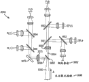
图33示出了根据本发明的示例性实施例的声学流式细胞计量系统的示意图。系统3000具有包含通过毛细管3006泵送的样本3004(可能包括一个或多个罕见事件颗粒的颗粒)的样本管3002。邻近毛细管3006布置的压电元件3008可以在处理器3022的控制下由控制电路3009操作,并且可以基于例如包括尺寸和密度的颗粒性质施加声能以声学聚焦和/或分离颗粒。经声学聚焦的颗粒随后可以进入探询区3010,在那里它们穿过探询源3012的光束(例如被高度聚焦的激光束或者两个或更多个激光束),并且它们中的一些或全部可以在废物部3014被收集。由探询源3012与颗粒的相互作用导致的散射光可以由收集透镜和光学收集块3016收集,并且可以使用与数据采集模块3012和处理器3022互连的光电倍增管阵列3018进行分析。FIG. 33 shows a schematic diagram of an acoustic flow cytometry system according to an exemplary embodiment of the present invention. The system 3000 has a
图34示出了根据本发明的示例性实施例的声学流式细胞计数器中的声学聚焦毛细管的示意图以显示压电元件对颗粒的影响。样本3004中的颗粒3001a和3001b在声学聚焦毛细管3006中的背景或载体流体3003中移动。可以是压电陶瓷元件的压电元件3008将颗粒3001a声学聚焦到内部同轴流3030中并且将颗粒3001b聚焦到外部同轴流3032中。FIG. 34 shows a schematic diagram of an acoustic focusing capillary in an acoustic flow cytometer according to an exemplary embodiment of the present invention to show the influence of piezoelectric elements on particles.
图35示出了根据本发明的示例性实施例的声学流式细胞计数器中的光学收集块的一部分。光学收集块3016包括具有第一激光器3040(例如405nm紫光激光器)和第二激光器3042(例如488nm蓝光激光器)的探询区3010。由激光器3040和3042发射的光束进入束成形光学器件3044的布置中,所述束成形光学器件将它们紧密地聚焦到颗粒3001a的经声学聚焦的同轴流上(图34)。当颗粒3001a穿过激光束时,散射光由聚光透镜3046收集并且在传到检测器阵列3018(图33)之前进入光学收集块3048(图36)。当与长通过时间结合时具有或不具有脉冲的405nm波长可以是很有用的,并且尤其有用于可用于多色测定的量子点的激发。也可以使用其它波长,例如包括640nm。相同颗粒可以由两个不同的激光器分析。较强的激光器可以用于分析较暗的颗粒,而不同的、较弱的激光器可以用于较亮的颗粒。单一的较弱的激光器也可以与增加的通过时间一起用于信号积分,并且这类激光器还可以通过在不同时间施加更强和更弱的脉冲而在脉冲调制系统中使用。FIG. 35 shows a portion of an optical collection block in an acoustic flow cytometer according to an exemplary embodiment of the present invention. The
两个激光器的使用有助于改善自发荧光和背景变化问题并且通过减小信号和背景的变化增加信噪比。例如,第一激光器可以在激发激光器的波长之上激发自发荧光,并且在该波长之上检测到的信号可以用于估计预期用于主检测激光器的自发荧光贡献。这可以用具有紫光激光器和蓝光激光器或仅仅具有紫光激光器或具有激发一个以上颜色的紫光激光器(如果存在有监测自发荧光的独立色带)的系统完成。可以仅监测蓝色荧光通道,并且然后可以减去其它通道中的预期贡献。也可以使用红光激光器。对于具有长寿命探针的脉冲或调制系统,可以测量与长寿命探针的初始输出组合的自发荧光的短寿命贡献。也可以测量并且反算自发荧光衰减之后的长寿命探针的荧光以确定所有通道中的自发荧光贡献。The use of two lasers helps to improve autofluorescence and background variation problems and increases the signal-to-noise ratio by reducing signal and background variation. For example, the first laser may excite autofluorescence above the wavelength of the excitation laser, and the signal detected above this wavelength may be used to estimate the autofluorescence contribution expected for the primary detection laser. This can be done with a system with a violet laser and a blue laser or just a violet laser or a violet laser that excites more than one color if there is a separate color band to monitor autofluorescence. Only the blue fluorescence channel can be monitored, and expected contributions in other channels can then be subtracted. Red lasers can also be used. For pulsed or modulated systems with long-lived probes, the short-lived contribution of autofluorescence combined with the initial output of the long-lived probe can be measured. Fluorescence of long-lived probes after autofluorescence decay can also be measured and back-calculated to determine the autofluorescence contribution in all channels.
根据本发明的示例性实施例,可以使用


图36示出了根据本发明的示例性实施例的声学流式细胞计数器中的光学数据收集块的示意图。来自激光器3040和3042的散射光3050由聚光透镜3046收集,以箭头A的方向进入收集块3048,并且行进到一对空间滤光针孔3052,其中的一个由反射镜(未显示)背衬,由此将光束3050分离为初级光束3054和次级光束3056。初级光束3054进入准直透镜3060并且横越分束器BS1、BS2和BS3和关联的聚焦透镜以进入检测器阵列3018(图33)中的侧向散射通道3062以及荧光通道FL4、FL5和FL6。次级光束3056进入准直透镜3070并且横越反射镜3072和分束器BS5和BS4和关联的聚焦透镜以进入检测器阵列3018中的荧光通道FL1、FL2和FL3。36 shows a schematic diagram of an optical data collection block in an acoustic flow cytometer according to an exemplary embodiment of the present invention. Scattered light 3050 from
图37示出了根据本发明的示例性实施例的声学流式细胞计数器中的射流系统的示意图。在系统3100中,泵部分3702中的样本泵3102将来自样本管3002的样本流体3004(图34)泵送到样本部分3705中的歧管3104并且在经过用于检测样本线中的空气的存在的气泡传感器3008之后到达隔膜泵3106。因此,可能不需要做预定容积样本抽取,并且样本可被抽取至样本结束状态,原因是在样本抽取阶段内检测到空气将标志样本的结束。气泡传感器3008可以基于各种模态,包括可以确定空气相对于流体样本的存在的超声、阻抗、电容、光学或任何其它类型的传感器模态。歧管3014中的样本流体然后可以被泵送到下歧管部分3703中的下歧管3110以进入声学聚焦毛细管3006。鞘液泵3124将保持在容器部分3701中的鞘容器3120中的鞘液3205(图38)泵送到鞘缓冲罐3122中并在随后被泵送到在毛细管3006的上歧管部分3704中的上歧管3130中。水和冲洗流体可以保持在水容器3140和冲洗流体容器3150中,并在必要时被泵送到系统中,同时在废物容器3160中进行废物收集。FIG. 37 shows a schematic diagram of a fluidic system in an acoustic flow cytometer according to an exemplary embodiment of the present invention. In
图38示出了根据本发明的示例性实施例的具有上游声学聚焦并跟随有下游流体动力学聚焦的单换能器声学聚焦毛细管的示意图。包括颗粒3204(可能包括一个或多个罕见事件颗粒)的样本流体流动通过毛细管3206。单换能器3208然后可以将颗粒3204(作为3201)沿着毛细管3206的大致中心轴线声学聚焦到第一区域中。这可以在任何流体动力学聚焦之前进行。鞘液3205然后可以用于围绕经声学聚焦的样本流体和颗粒流动并且进一步将流体和颗粒3201流体动力学地聚焦到第一区域的下游的第二区域中。使用用于上游声学聚焦的一个以上换能器是可行的,但是优选单换能器。同样地,在第一、上游声学聚焦相之后使用经声学和流体动力学两者聚焦颗粒3201的第二、下游双聚焦相也是可行的。然而优选地,颗粒首先被声学聚焦并于随后在声学聚焦位置下游的第二位置被流体动力学聚焦而不需要进一步声学聚焦,原因是这一组合提供相当令人惊叹的罕见事件检测能力。38 shows a schematic diagram of a single transducer acoustic focusing capillary with upstream acoustic focusing followed by downstream hydrodynamic focusing, according to an exemplary embodiment of the invention. A sample fluid including particles 3204 (possibly including one or more rare event particles) flows through
尽管可以代替流体动力学聚焦单独使用具有明显优点的声学聚焦,但是组合使用声学聚焦和流体动力学聚焦的某些配置是特别有用的。例如,组合声学/流体动力学聚焦可以抵抗外力以进一步稳定颗粒流的绝对位置;可以进一步固定经聚焦的颗粒流的焦点(这在样本被稀释或“粘性”细胞必须保持在较低浓度以防止聚集的情况下是特别有用的);并且可以有助于保证样本不会接触壁(这在一些应用中可能是重要的)。最后,意外证明在上游单独使用声学聚焦并且接着沿着相同通道在下游使用流体动力学聚焦产生优良性质,允许在相对较短时间内检测某些罕见事件,如以上示例性实施例中的一些所述。While acoustic focusing alone can be used instead of hydrodynamic focusing with clear advantages, certain configurations using acoustic and hydrodynamic focusing in combination are particularly useful. For example, combined acoustic/hydrodynamic focusing can resist external forces to further stabilize the absolute position of the particle stream; it can further fix the focal point of the focused particle stream (this is necessary when samples are diluted or "sticky" cells must be kept at low concentrations to prevent are particularly useful in cases of aggregation); and can help ensure that the sample does not touch the walls (which may be important in some applications). Finally, it was surprisingly demonstrated that the use of acoustic focusing alone upstream followed by hydrodynamic focusing downstream along the same channel yields superior properties, allowing the detection of certain rare events in relatively short periods of time, as demonstrated in some of the above exemplary examples. stated.
根据本发明的示例性实施例,样本泵3102和鞘液泵3124可以由处理器控制以调节毛细管3006中鞘液与样本流体的容积比,从而保持探询区中大致恒定的总颗粒速度。例如,鞘液与样本流体的容积比可以保持在从大约1:10至大约100:1。在保持紧密聚焦颗粒流的同时调节样本输入速率的能力允许调节通过探询激光器的速度(和因此花费的时间)。更长的探询时间通过允许随着时间收集更多光子而允许更高灵敏度的测量。当颗粒分析系统只能控制样本流动时,对于指定样本浓度,可调节流速限制增加或减小颗粒分析速率的能力,原因是增加或减小样本输入速率必然增加或减小通过时间。通过包括响应样本输入调节的鞘流动使得总流体流动保持恒定,可以允许宽范围的样本输入速率而不改变总流体速度。然后,通过改变总流体速度,能够利用更长探询时间的优点。通过不加速颗粒和同轴鞘流动,通过声学流式细胞计数器的激光器探询区域的颗粒通过时间可以为常规流体动力学聚焦系统中的大约20-100倍。优选地,它们可以为至少20μs、至少25μs、至少30μs、至少35μs、至少40μs、至少60μs、至少80μs或至少100μs。这可以允许更高灵敏度光学测量,同时保持类似的颗粒分析速率。According to an exemplary embodiment of the invention,
图39示出了根据本发明的示例性实施例的可以调节声学流式细胞计数器中的前向散射孔径的阻挡杆装置的示意图。在前向散射光进入聚光透镜3048之前阻挡杆3300可以用于改变前向散射光的孔径。由激光器3040和3042发出的光束3041与颗粒的经声学聚焦流(流出图中纸面)相互作用。阻挡杆3300可以安装在圆柱形销柱3302的下侧和离轴位置使得自旋销柱3302改变它相对于散射激光束3050的传播的位置。一旦激光器与颗粒流对准,这允许操作者使阻挡杆3300与激光器3040和3042对准。可以通过改变阻挡杆3300的形状而改变前向散射器3050的孔径,这可以通过将轴环加入杆上而完成。销柱3302可以旋转以定位阻挡杆3300,从而提供在大约15°至大约23°之间或大约17°至21°之间或大约19°的前向散射器3050的孔径α。39 shows a schematic diagram of a barrier rod arrangement that can adjust the forward scatter aperture in an acoustic flow cytometer according to an exemplary embodiment of the present invention. The blocking rod 3300 can be used to change the aperture of the forward scattered light before it enters the
图40A-40F示出了根据本发明的示例性实施例的将0.050%和0.045%CD34阳性细胞的罕见事件群体检测作为活CD45阳性细胞的亚群。其中,周围血液被染色并且以500μl/min(图40A-40C)和1000μl/min(图40D-40F)的流速以设在500,000个总细胞的停止门限使用具有上游声学聚焦和下游流体动力学聚焦的声学聚焦细胞计数器运行。图40A和40D显示了用AADvancedTM死细胞染色剂染色的总细胞并且显示了活细胞门限。图40B和40E显示了在活细胞上选通的细胞。图40C和40F显示了在活CD45阳性细胞上选通的细胞。在500μl/min的流速下,采集时间为大约6分26秒;检测到白血球的0.050%CD34阳性细胞;并且CD34阳性细胞的直接测量值为0.07细胞/μl。在1000μl/min的流速下,采集时间为大约4分28秒;检测到白血球的0.045%CD34阳性细胞;并且CD34阳性细胞的直接测量值为0.05细胞/μl。40A-40F illustrate the detection of rare event populations of 0.050% and 0.045% CD34 positive cells as subpopulations of live CD45 positive cells, according to an exemplary embodiment of the invention. where peripheral blood was stained and used with upstream acoustic focusing and downstream hydrodynamic focusing at flow rates of 500 μl/min (Figures 40A-40C) and 1000 μl/min (Figures 40D-40F) with a stop threshold set at 500,000 total cells The acoustic focusing cell counter is run. Figures 40A and 40D show the use of Total cells stained with AADvanced ™ Dead Cell Stain and live cell threshold shown. Figures 40B and 40E show cells gated on live cells. Figures 40C and 40F show cells gated on live CD45 positive cells. At a flow rate of 500 μl/min, the acquisition time was approximately 6 minutes and 26 seconds; 0.050% CD34 positive cells of leukocytes were detected; and the direct measurement of CD34 positive cells was 0.07 cells/μl. At a flow rate of 1000 μl/min, the acquisition time was approximately 4 minutes and 28 seconds; 0.045% CD34 positive cells of leukocytes were detected; and the direct measurement of CD34 positive cells was 0.05 cells/μl.
图41A-41D示出了在非声学流式细胞计数器上和声学聚焦细胞计数器上运行的细胞检测的比较输出绘图。来自正常供体的周围血液掺加有CD34阳性细胞和50μl的CountBrightTM绝对计数小珠并且用于计算CD34计数。图41A和41B显示了在活细胞(图41A)和CD45阳性细胞(图41B)上选通的数据,其在仅仅流体动力学聚焦流式细胞计数器上运行。图41C和41D显示了在活细胞(图41C)和CD45阳性细胞(图41D)上选通的数据,其在根据本发明的示例性实施例的具有上游声学聚焦和下游流体动力学聚焦的声学聚焦细胞计数器上运行。使用仅仅流体动力学聚焦流式细胞计数器,采集时间为13分49秒;使用小珠导出的CD34阳性细胞计数为8.01细胞/μl;并且不产生直接测量值。使用声学聚焦流式细胞计数器,采集时间为1分17秒;使用小珠导出的CD34阳性细胞计数为7.91细胞/μl;并且CD34阳性细胞的直接测量值为8细胞/μl。Figures 41A-41D show comparative output plots for cell assays run on a non-acoustic flow cytometer and an acoustic focusing cytometer. Peripheral blood from normal donors was spiked with CD34 positive cells and 50 μl of CountBright ™ Absolute Counting Beads and used to calculate CD34 counts. Figures 41A and 41B show data gated on live cells (Figure 41A) and CD45 positive cells (Figure 41B), run on a hydrodynamic focusing only flow cytometer. 41C and 41D show data gated on live cells (FIG. 41C) and CD45-positive cells (FIG. 41D) in acoustic cells with upstream acoustic focusing and downstream hydrodynamic focusing according to an exemplary embodiment of the invention. Run on a focusing cell counter. Using a hydrodynamic focusing flow cytometer only, the acquisition time was 13 minutes and 49 seconds; the bead-derived CD34 positive cell count was 8.01 cells/[mu]l; and no direct measurements were produced. Using an acoustic focusing flow cytometer, the acquisition time was 1 minute 17 seconds; the bead-derived CD34 positive cell count was 7.91 cells/μl; and the direct measurement of CD34 positive cells was 8 cells/μl.
图42示出了根据本发明的示例性实施例的声学聚焦细胞计数器的各部件的示意图。示例性细胞计数器包括用于鞘液的第一流体路径4205和用于样本流体的第二流体路径4209,第一流体路径包括鞘液4208、鞘液过滤器4207和鞘液容器4206,第二流体路径包括样本流体4213、气泡传感器4212、样本回路4211和毛细管组件4210。鞘液和样本流体可以在流动室4204中流动并且第一激光器4202和第二激光器4203可以探询可能在样本流体中的颗粒。最后,一些鞘液和样本流体可以被送入废物容器4201。FIG. 42 shows a schematic diagram of components of an acoustic focusing cytometer according to an exemplary embodiment of the present invention. The exemplary cytometer includes a
本发明的实施例可以更快地分析罕见细胞事件;在更少的时间内运行更多的细胞而没有灵敏度的损失;检测细胞中抗原的弱表达;并且以更小的含糊性更清楚地分辨细胞群体。本发明的实施例可以提供对样本浓度、流速、可检测光子的数量、实验时间长度和样本吞吐量的有力控制。声学聚焦细胞计量术可以改变许多目前的细胞测定的执行方式,并且提供用于创建新细胞测定的机会。它可以使用例如2MHz以上的超声波以在没有高速或高容积鞘液的情况下将细胞定位到沿着流动通道中心轴线的单聚焦线中,并且可以与容积无关地集中细胞。声学聚焦可以利用细胞或颗粒相对于背景介质的物理差异,允许细胞保持紧密聚焦。声学聚焦可以用声能将细胞集中在流体的中心,这创建被分析样本浓度的相当大灵活性。更重要地,声学聚焦可以使细胞的对准与颗粒流速分离,因此可以在不破坏毛细管中细胞的聚焦的情况下增加或减小细胞的流速。该可调节流速的精确性可以有助于研究者确定被分析细胞的数量和细胞在经聚焦激光束中花费的时间量。声学流式细胞计量术的附加特征和优点可以在Ward等人的“Fundamentals of Acoustic Cytometry”,Current Protocols inCytometry,Supplement 49,1.11.1-1.22.12(2009)中找到,上述文献的完整公开通过引用被合并于本文中。Embodiments of the invention can analyze rare cellular events more quickly; run more cells in less time without loss of sensitivity; detect weak expression of antigens in cells; and resolve more clearly with less ambiguity cell population. Embodiments of the present invention can provide powerful control over sample concentration, flow rate, number of detectable photons, length of experiment, and sample throughput. Acoustic focusing cytometry can change the way many current cellular assays are performed and provide opportunities for creating new cellular assays. It can use ultrasound, eg, above 2 MHz, to localize cells into a single focal line along the central axis of the flow channel without high speed or high volume of sheath fluid, and can focus cells independent of volume. Acoustic focusing can take advantage of the physical differences of cells or particles relative to the background medium, allowing cells to remain tightly focused. Acoustic focusing can use acoustic energy to focus cells in the center of the fluid, which creates considerable flexibility in the concentration of the sample being analyzed. More importantly, acoustic focusing can decouple the alignment of the cells from the particle flow rate, so the flow rate of the cells can be increased or decreased without disrupting the focusing of the cells in the capillary. The precision of this adjustable flow rate can help the researcher determine the number of cells analyzed and the amount of time the cells spend in the focused laser beam. Additional features and advantages of acoustic flow cytometry can be found in Ward et al., "Fundamentals of Acoustic Cytometry", Current Protocols in Cytometry, Supplement 49, 1.11.1-1.22.12 (2009), the full disclosure of which is via References are incorporated herein.
在仅使用流体动力学聚焦的系统中,样本核心由快速流动鞘液“夹持”,并且鞘液的体积典型地是样本流体的100至1000倍。这样的大比率导致低样本输入速率,这通常阻碍分辨率。然而根据本发明的示例性实施例,先前经声学聚焦的样本可以在声学聚焦的下游进一步地被流体动力学地聚焦,鞘液和样本流体之间的容积比可被显著地减小。例如,该容积比可以减小到例如大约50比1、40比1、30比1、20比1、10比1、9比1、8比1、7比1、6比1、5比1、4比1、3比1、或2比1。该容积比也可以为大约1比1、1比2、1比3、1比4、1比5、1比6、1比7、1比8、1比9、和1比10。这些数字是示例性的并且也可以使用它们之间的其它分数比。优选地,鞘液和样本流体之间的容积比可以在大约10比1至1比10之间或大约5比1至1比5之间。该系统可以使具有颗粒的流体样本以大约200μl/min至大约1000μl/min的样本流体输入速率在毛细管中的样本通道中流动并且使鞘液以大约2200μl/min至大约1400μl/min的鞘液输入速率在鞘流动通道中流动,同时保持样本流体和鞘液的总输入速率恒定,由此保证通过一个或多个探询激光器的颗粒的探询时间保持恒定,与样本流体输入速率无关。该系统也可以使样本流体以大约25μl/min至大约1000μl/min之间的样本流速流动并使鞘液以大约2375μl/min至大约1400μl/min的鞘流速流动。该系统也可以使样本流体以至少200μl/min的样本流速流动并且使鞘液以至多2200μl/min的鞘液流速流动,同时将鞘液与样本流体的容积比调节到大约11比1至大约1.4比1之间的比率。该系统也可以使样本流体以至少500μl/min的样本流速流动并且使鞘液以至多1900μl/min的鞘液流速流动,同时将鞘液与样本流体的容积比调节到大约3.8比1至大约1.4比1之间的比率。In systems using only hydrodynamic focusing, the sample core is "clamped" by a fast-flowing sheath fluid, and the volume of the sheath fluid is typically 100 to 1000 times that of the sample fluid. Such a large ratio results in a low sample input rate, which often hinders resolution. However, according to an exemplary embodiment of the present invention, the previously acoustically focused sample can be further hydrodynamically focused downstream of the acoustic focusing, and the volume ratio between the sheath fluid and the sample fluid can be significantly reduced. For example, the volume ratio can be reduced to, for example, about 50 to 1, 40 to 1, 30 to 1, 20 to 1, 10 to 1, 9 to 1, 8 to 1, 7 to 1, 6 to 1, 5 to 1 , 4 to 1, 3 to 1, or 2 to 1. The volume ratio may also be about 1 to 1, 1 to 2, 1 to 3, 1 to 4, 1 to 5, 1 to 6, 1 to 7, 1 to 8, 1 to 9, and 1 to 10. These numbers are exemplary and other fractional ratios between them can also be used. Preferably, the volume ratio between the sheath fluid and the sample fluid may be between about 10:1 and 1:10 or between about 5:1 and 1:5. The system can flow a fluid sample with particles in a sample channel in a capillary at a sample fluid input rate of about 200 μl/min to about 1000 μl/min and sheath fluid at a sheath input rate of about 2200 μl/min to about 1400 μl/min The rate flows in the sheath flow channel while maintaining the total input rate of sample fluid and sheath fluid constant, thereby ensuring that the interrogation time of particles passing through one or more interrogation lasers remains constant independent of the sample fluid input rate. The system can also flow the sample fluid at a sample flow rate of between about 25 μl/min to about 1000 μl/min and the sheath fluid at a sheath flow rate of about 2375 μl/min to about 1400 μl/min. The system can also flow the sample fluid at a sample flow rate of at least 200 μl/min and the sheath fluid at a sheath fluid flow rate of at most 2200 μl/min while adjusting the sheath fluid to sample fluid volume ratio from about 11 to 1 to about 1.4 A ratio of 1 to 1. The system can also flow the sample fluid at a sample flow rate of at least 500 μl/min and the sheath fluid at a sheath fluid flow rate of at most 1900 μl/min while adjusting the sheath fluid to sample fluid volume ratio from about 3.8 to 1 to about 1.4 A ratio of 1 to 1.
根据本发明的实施例,提供了一种流式细胞计数器,包括:(1)包括样本通道的毛细管;(2)耦合至所述毛细管的至少一个振动产生换能器,所述至少一个振动产生换能器被配置成产生声信号,所述声信号引起所述样本通道内的声辐射压力以声学集中在所述样本通道中的流体样本流内流动的颗粒;以及(3)包括紫光激光器和蓝光激光器的探询源,所述紫光激光器和所述蓝光激光器被配置成与经声学集中的颗粒的至少一些相互作用以产生输出信号。According to an embodiment of the present invention, there is provided a flow cytometer, comprising: (1) a capillary tube including a sample channel; (2) at least one vibration generating transducer coupled to the capillary tube, the at least one vibration generating transducer a transducer configured to generate an acoustic signal that induces acoustic radiation pressure within the sample channel to acoustically concentrate particles flowing within a fluid sample stream in the sample channel; and (3) comprising a violet laser and An interrogation source of a blue laser, the violet laser and the blue laser configured to interact with at least some of the acoustically concentrated particles to generate an output signal.
在这样的流式细胞计数器中,所述至少一个振动产生换能器可以包括压电装置,所述紫光激光器可以具有大约405纳米的波长,并且所述蓝光激光器可以具有大约488纳米的波长。此外,所述毛细管还可以包括鞘流动通道,所述鞘流动通道被配置成在由所述声辐射压力引起的所述颗粒的声学聚焦的下游使鞘液围绕所述流体样本流流动以流体动力学地集中所述流体样本流内的经声学集中的颗粒。此外,这样的流式细胞计数器可以包括第一泵和第二泵,所述第一泵被配置成使包括颗粒的流体样本以大约200微升每分钟至大约1000微升每分钟的样本流体输入速率在所述毛细管中的所述样本通道内流动,所述第二泵被配置成使鞘液以大约2200微升每分钟至大约1400微升每分钟的鞘液输入速率在所述鞘流动通道中流动,并且所述第一泵和所述第二泵被配置成保持在所述毛细管中流动的样本流体和鞘液的总输入速率恒定,由此保证通过所述紫光激光器和所述蓝光激光器的经声学集中的颗粒的至少一些的探询时间保持恒定,而与所述样本流体输入速率无关。In such a flow cytometer, the at least one vibration generating transducer may comprise a piezoelectric device, the violet laser may have a wavelength of about 405 nanometers, and the blue laser may have a wavelength of about 488 nanometers. In addition, the capillary may further include a sheath flow channel configured to flow a sheath fluid around the fluid sample flow downstream of the acoustic focusing of the particles caused by the acoustic radiation pressure to hydrodynamically The acoustically concentrated particles within the fluid sample stream are concentrated acoustically. Additionally, such a flow cytometer may include a first pump and a second pump, the first pump configured to input a fluid sample including particles at a sample fluid input rate of about 200 microliters per minute to about 1000 microliters per minute flow rate in the sample channel in the capillary, and the second pump is configured to cause sheath fluid to flow through the sheath flow channel at a sheath fluid input rate of about 2200 microliters per minute to about 1400 microliters per minute. flow in the capillary, and the first pump and the second pump are configured to keep the total input rate of the sample fluid and the sheath fluid flowing in the capillary constant, thereby ensuring The interrogation time of at least some of the acoustically concentrated particles remains constant regardless of the sample fluid input rate.
这类流式细胞计数器还可以包括:光学模块,其收集来自所述探询源的输出信号;检测器模块,其检测所述光学模块的输出信号;以及数据采集模块,其处理所述检测器模块的输出,并且这类流式细胞计数器还可以包括处理器,所述处理器被配置成控制所述至少一个振动产生换能器、所述检测器模块和所述数据采集模块中的至少一个。此外,这类流式细胞计数器可以包括在所述毛细管和所述光学模块之间的阻挡杆,所述阻挡杆可以附连到大致圆柱形的销柱,所述销柱可旋转以定位所述阻挡杆并且调节所述探询源的输出信号的输出孔径,并且所述探询源的输出信号的输出孔径在大约17度至大约21度之间。此外,所述光学模块可以包括收集来自所述探询源的输出信号的收集透镜,并且所述收集透镜的输出用空间滤光针孔装置分成两个光束,其中第一光束是来自所述紫光激光器的输出并且第二光束是来自所述蓝光激光器的输出。并且,所述检测器模块可以包括检测器,所述检测器检测来自由所述紫光激光器输出的所述第一光束的前向散射信号和侧向散射信号。Such flow cytometers may also include: an optical module that collects the output signal from the interrogation source; a detector module that detects the output signal of the optical module; and a data acquisition module that processes the detector module and such flow cytometers may further include a processor configured to control at least one of the at least one vibration-generating transducer, the detector module, and the data acquisition module. Additionally, such flow cytometers may include a barrier rod between the capillary and the optical module, which may be attached to a generally cylindrical pin that is rotatable to position the blocking the rod and adjusting the output aperture of the output signal of the interrogation source, and the output aperture of the output signal of the interrogation source is between about 17 degrees and about 21 degrees. Additionally, the optical module may include a collection lens that collects the output signal from the interrogation source, and the output of the collection lens is split into two beams with a spatially filtered pinhole arrangement, wherein the first beam is from the violet laser and the second beam is the output from the blue laser. Also, the detector module may include a detector that detects a forward scatter signal and a side scatter signal from the first light beam output by the violet laser.
根据本发明的另一个实施例,提供了一种流式细胞计数器,包括:(1)毛细管,其被配置成允许包括颗粒的样本流体在其中流动;(2)第一聚焦机构,其被配置成将所述样本流体内的所述颗粒的至少一些声学聚焦在所述毛细管内的第一区域中;(3)第二聚焦机构,其被配置成将包括所述至少一些经声学聚焦的颗粒的样本流体流体动力学地聚焦在所述第一区域的下游的所述毛细管内的第二区域中;(4)在所述毛细管中或下游的探询区,经声学和流体动力学聚焦的颗粒的至少一些能够流动通过所述探询区;以及(5)至少一个检测器,其被配置成检测在所述探询区获得的关于经声学和流体动力学聚焦的颗粒的至少一些的至少一个信号。According to another embodiment of the present invention, there is provided a flow cytometer, comprising: (1) a capillary configured to allow a sample fluid including particles to flow therein; (2) a first focusing mechanism configured to to acoustically focus at least some of the particles within the sample fluid in a first region within the capillary; (3) a second focusing mechanism configured to include the at least some of the acoustically focused particles (4) In the interrogation zone in or downstream of the capillary, the acoustically and hydrodynamically focused particles at least some of the interrogation zone are capable of flowing through the interrogation zone; and (5) at least one detector configured to detect at least one signal obtained in the interrogation zone for at least some of the acoustically and hydrodynamically focused particles.
这类流式细胞计数器还可以包括样本流体泵和鞘液泵,所述样本流体泵被配置成使样本流体以大约25微升每分钟至大约1000微升每分钟之间的样本流速流入所述毛细管,所述鞘液泵被配置成使鞘液以大约2375微升每分钟至大约1400微升每分钟之间的鞘流速流入所述毛细管。此外,所述第一聚焦机构可以被配置成将所述第一区域中的经声学聚焦的颗粒的至少一些聚焦到从所述第一区域流动到所述第二区域的单行线,并且所述样本流体泵和所述鞘液泵可以被配置成保持在所述毛细管中流动的样本流体和鞘液的总速率恒定,由此保证通过所述探询区的经声学和流体动力学聚焦颗粒的至少一些的探询时间保持恒定,而与所述样本流速无关。这类流式细胞计数器还可以包括样本流体泵和鞘液泵,所述样本流体泵被配置成使样本流体以大约200微升每分钟至大约1000微升每分钟之间的样本流速流入所述毛细管,所述鞘液泵被配置成使鞘液以大约2200微升每分钟至大约1400微升每分钟之间的鞘流速流入所述毛细管。Such flow cytometers may also include a sample fluid pump and a sheath fluid pump configured to flow sample fluid into said a capillary, the sheath fluid pump configured to flow sheath fluid into the capillary at a sheath flow rate between about 2375 microliters per minute and about 1400 microliters per minute. Furthermore, the first focusing mechanism may be configured to focus at least some of the acoustically focused particles in the first region into a single file line flowing from the first region to the second region, and the sample The fluid pump and the sheath fluid pump may be configured to maintain a constant total rate of sample fluid and sheath fluid flowing in the capillary, thereby ensuring that at least some of the acoustically and hydrodynamically focused particles pass through the interrogation zone. The interrogation time remains constant regardless of the sample flow rate. Such flow cytometers may also include a sample fluid pump and a sheath fluid pump configured to flow sample fluid into said a capillary, the sheath fluid pump configured to flow sheath fluid into the capillary at a sheath flow rate between about 2200 microliters per minute and about 1400 microliters per minute.
根据本发明的另一个实施例,提供了一种使用流式细胞计数器检测罕见事件的方法,包括:(1)使包括颗粒的样本流体流入通道;(2)通过将声辐射压力施加到包含在所述通道内的第一区域而使得所述样本流体中的所述颗粒的至少一些声学聚焦在所述第一区域中;(3)通过使鞘液围绕所述样本流体流动将包括所述至少一些经声学聚焦的颗粒的样本流体流体动力学地聚焦在所述第一区域的下游的第二区域中;(4)调节所述鞘液与所述样本流体的容积比以保持所述第二区域中或下游的探询区中的大致恒定的总颗粒速度;(5)分析所述探询区中的经声学和流体动力学聚焦的颗粒的至少一些;以及(6)基于在所述探询区检测到的至少一个信号检测一个或多个罕见事件,所述一个或多个罕见事件选自下列:一个或多个罕见荧光事件、一个或多个罕见细胞类型以及一个或多个死亡细胞。According to another embodiment of the present invention, there is provided a method for detecting rare events using a flow cytometer, comprising: (1) flowing a sample fluid including particles into a channel; (2) applying acoustic radiation pressure to the a first region within the channel such that at least some of the particles in the sample fluid are acoustically focused in the first region; (3) flowing sheath fluid around the sample fluid will include the at least A sample fluid of some acoustically focused particles is hydrodynamically focused in a second region downstream of the first region; (4) adjusting the volume ratio of the sheath fluid to the sample fluid to maintain the second an approximately constant total particle velocity in the interrogation zone in or downstream of the region; (5) analyzing at least some of the acoustically and hydrodynamically focused particles in the interrogation zone; and (6) based on detection in the interrogation zone The at least one detected signal detects one or more rare events selected from the group consisting of: one or more rare fluorescent events, one or more rare cell types, and one or more dead cells.
这类方法还可以包括使所述样本流体以至少200微升每分钟的样本流速流动并且使所述鞘液以至多2200微升每分钟的鞘液流速流动,并且调节所述鞘液与所述样本流体的容积比可以包括将所述鞘液与所述样本流体的容积比调节到大约11比1至大约1.4比1之间的比率。此外,这类方法可以包括使所述样本流体以至少500微升每分钟的样本流速流动并且使所述鞘液以至多1900微升每分钟的鞘液流速流动,并且调节所述鞘液与所述样本流体的容积比可以包括将所述鞘液与所述样本流体的容积比调节到大约3.8比1至大约1.4比1之间的比率。此外,所述方法可以包括保证所述经声学和流体动力学聚焦的颗粒通过所述探询区的通过时间超过大约20微秒,或保证所述经声学和流体动力学聚焦的颗粒通过所述探询区的通过时间超过大约40微秒。Such methods may also include flowing the sample fluid at a sample flow rate of at least 200 microliters per minute and flowing the sheath fluid at a sheath fluid flow rate of at most 2200 microliters per minute, and adjusting the sheath fluid and the The volume ratio of the sample fluid may include adjusting the volume ratio of the sheath fluid to the sample fluid to a ratio between about 11 to 1 to about 1.4 to 1. Additionally, such methods may include flowing the sample fluid at a sample flow rate of at least 500 microliters per minute and flowing the sheath fluid at a sheath fluid flow rate of at most 1900 microliters per minute, and adjusting the sheath fluid to the The volume ratio of the sample fluid may include adjusting the volume ratio of the sheath fluid to the sample fluid to a ratio between about 3.8 to 1 to about 1.4 to 1. Additionally, the method may include ensuring that the transit time of the acoustically and hydrodynamically focused particles through the interrogation zone exceeds about 20 microseconds, or ensuring that the acoustically and hydrodynamically focused particles pass through the interrogation zone The transit time of the zone exceeds about 40 microseconds.
根据本发明的另一个示例性实施例,提供了一种包括计算机可读指令的计算机可读介质,当由声学流式细胞计量装置中的或与其通信的计算机执行时所述计算机可读指令控制所述装置以:(1)使包括颗粒的样本流体流入通道;(2)通过将声辐射压力施加到包含在所述通道内的第一区域将所述样本流体中的所述颗粒的至少一些声学聚焦在所述第一区域中;(3)通过使鞘液围绕所述样本流体流动将包括所述至少一些经声学聚焦的颗粒的样本流体流体动力学地聚焦在所述第一区域的下游的第二区域中;(4)调节所述鞘液与所述样本流体的容积比以保持所述第二区域中或下游的探询区中的大致恒定的总颗粒速度;(5)分析所述探询区中的经声学和流体动力学聚焦颗粒的至少一些;以及(6)基于在所述探询区检测到的至少一个信号检测一个或多个罕见事件,所述一个或多个罕见事件选自下列:一个或多个罕见荧光事件、一个或多个罕见细胞类型以及一个或多个死亡细胞。According to another exemplary embodiment of the present invention, there is provided a computer readable medium comprising computer readable instructions which when executed by a computer in or in communication with an acoustic flow cytometry device controls The device is configured to: (1) cause a sample fluid including particles to flow into a channel; (2) displace at least some of the particles in the sample fluid by applying acoustic radiation pressure to a first region contained within the channel. Acoustically focusing in the first region; (3) hydrodynamically focusing a sample fluid comprising the at least some acoustically focused particles downstream of the first region by flowing a sheath fluid around the sample fluid (4) adjust the volume ratio of the sheath fluid to the sample fluid to maintain a substantially constant total particle velocity in the second region or in an interrogation zone downstream; (5) analyze the at least some of the acoustically and hydrodynamically focused particles in the interrogation zone; and (6) detecting one or more rare events based on at least one signal detected in the interrogation zone, the one or more rare events selected from The following: one or more rare fluorescent events, one or more rare cell types, and one or more dead cells.
这类计算机可读介质还可以控制所述装置以使所述样本流体在至少200微升每分钟的样本流速下流动并且使所述鞘液在至多2200微升每分钟的鞘液流速下流动,并且通过将所述鞘液与所述样本流体的容积比调节到大约11比1至大约1.4比1之间的比率调节所述鞘液与所述样本流体的容积比。此外,这类计算机可读介质还可以控制所述装置以使所述样本流体在至少500微升每分钟的样本流速下流动并且使所述鞘液在至多1900微升每分钟的鞘液流速下流动,并且通过将所述鞘液与所述样本流体的容积比调节到大约3.8比1至大约1.4比1之间的比率调节所述鞘液与所述样本流体的容积比。此外,这类计算机可读介质还可以控制所述装置以保证所述经声学和流体动力学聚焦的颗粒通过所述探询区的通过时间超过大约20微秒,或保证所述经声学和流体动力学聚焦的颗粒通过所述探询区的通过时间超过大约40微秒。Such computer readable medium can also control the device to flow the sample fluid at a sample flow rate of at least 200 microliters per minute and flow the sheath fluid at a sheath fluid flow rate of at most 2200 microliters per minute, And the volume ratio of the sheath fluid to the sample fluid is adjusted by adjusting the volume ratio of the sheath fluid to the sample fluid to a ratio between about 11:1 to about 1.4:1. In addition, such computer readable media can control the device such that the sample fluid flows at a sample flow rate of at least 500 microliters per minute and the sheath fluid at a sheath fluid flow rate of at most 1900 microliters per minute. and adjusting the volume ratio of the sheath fluid to the sample fluid by adjusting the volume ratio of the sheath fluid to the sample fluid to a ratio between about 3.8 to 1 to about 1.4 to 1. In addition, such computer readable media can control the device to ensure that the transit time of the acoustically and hydrodynamically focused particles through the interrogation zone exceeds about 20 microseconds, or to ensure that the acoustically and hydrodynamically focused The transit time of the optically focused particles through the interrogation zone exceeds about 40 microseconds.
根据本发明的实施例,提供了一种装置,其包括:(1)包括通道的毛细管;(2)耦合至所述毛细管的至少一个振动源,所述至少一个振动源被配置成将振动施加于所述通道;以及(3)包括405nm激光器的探询源,所述探询源被配置成具有输出,所述输出与在所述毛细管中流动的一个或多个颗粒相互作用。所述探询源还可以包括488nm激光器。所述振动源可以包括压电材料。所述振动源可以被配置成产生声信号,所述声信号引起所述通道内的声辐射压力,所述声辐射压力可以集中所述通道中的流体样本流内的多个选定颗粒,并且所述毛细管可以包括鞘流动通道以流体动力学地集中所述流体样本流内的选定颗粒。According to an embodiment of the present invention, there is provided an apparatus comprising: (1) a capillary including a channel; (2) at least one vibration source coupled to the capillary, the at least one vibration source being configured to apply vibration to and (3) an interrogation source comprising a 405 nm laser configured to have an output that interacts with one or more particles flowing in the capillary. The interrogation source may also include a 488nm laser. The vibration source may include a piezoelectric material. the vibration source can be configured to generate an acoustic signal that induces an acoustic radiation pressure within the channel that can concentrate a plurality of selected particles within the flow of fluid sample in the channel, and The capillary can include a sheath flow channel to hydrodynamically focus selected particles within the fluid sample stream.
根据本发明的另一个实施例,提供了一种系统,其包括:(1)具有通道的毛细管;(2)耦合至所述毛细管的至少一个振动产生换能器,所述至少一个振动产生换能器被配置成产生声信号,所述声信号引起所述通道内的声辐射压力,其中所述声辐射压力集中所述通道中的流体样本流内的多个选定颗粒;以及(3)包括405nm激光器的探询源,所述探询源被配置成具有输出,所述输出与所述选定颗粒中的至少一些相互作用以产生输出信号;(4)光学模块,其收集来自所述探询源的输出信号;(5)检测器模块,其检测所述光学模块的输出信号;以及(6)数据采集模块,其处理所述检测器模块的输出。所述振动产生换能器可以包括压电装置。所述探询源还可以包括488nm激光器,并且所述405nm激光器和所述488nm激光器都可以探询所述选定颗粒中的至少一些。所述毛细管可以包括至少一个鞘流动通道,并且所述鞘流动通道可以包括鞘液以流体动力学地集中所述流体样本流内的所述选定颗粒。所述系统可以包括处理器,所述处理器被配置成控制所述振动产生换能器、所述检测器模块和所述数据采集模块中的至少一个。它还可以包括在所述毛细管和所述光学模块之间的阻挡杆,并且所述阻挡杆可以附连到大致圆柱形的销柱,所述销柱可以是可旋转的,以定位所述阻挡杆并且调节所述探询源的输出信号的输出孔径。所述探询源的输出信号的输出孔径可以为大约19°。所述光学模块可以包括收集来自所述探询源的输出信号的收集透镜,并且所述收集透镜的输出用空间滤光针孔装置分成两个光束,其中第一光束是来自所述405nm激光器的输出并且第二光束是来自所述488nm激光器的输出。所述检测器模块可以包括检测器,所述检测器检测来自由所述405nm激光器输出的所述第一光束的前向散射信号和侧向散射信号。所述系统可以包括泵,所述泵将样本流体从容器沿着样本流动路径移动到所述毛细管,所述样本流动路径可以包括气泡传感器,并且所述泵可以被配置成在大约200μl每分钟至大约1000μl每分钟的样本输入速率下将所述样本流体输入到所述毛细管中。所述系统还可以包括用于成像所述流体样本流中的颗粒的成像器。According to another embodiment of the present invention, there is provided a system comprising: (1) a capillary having a channel; (2) at least one vibration generating transducer coupled to the capillary, the at least one vibration generating transducer The transducer is configured to generate an acoustic signal that induces an acoustic radiation pressure within the channel, wherein the acoustic radiation pressure concentrates a plurality of selected particles within the fluid sample flow in the channel; and (3) an interrogation source comprising a 405 nm laser configured to have an output that interacts with at least some of the selected particles to generate an output signal; (4) an optical module that collects data from the interrogation source (5) a detector module that detects the output signal of the optical module; and (6) a data acquisition module that processes the output of the detector module. The vibration generating transducer may comprise a piezoelectric device. The interrogation source may also include a 488nm laser, and both the 405nm laser and the 488nm laser may interrogate at least some of the selected particles. The capillary can include at least one sheath flow channel, and the sheath flow channel can include a sheath fluid to hydrodynamically concentrate the selected particles within the fluid sample stream. The system may include a processor configured to control at least one of the vibration generating transducer, the detector module, and the data acquisition module. It may also include a blocking rod between the capillary and the optical module, and the blocking rod may be attached to a generally cylindrical pin which may be rotatable to position the blocking rod. rod and adjusts the output aperture of the interrogation source's output signal. The output aperture of the output signal of the interrogation source may be approximately 19°. The optical module may include a collection lens that collects the output signal from the interrogation source, and the output of the collection lens is split into two beams with a spatial filtering pinhole arrangement, wherein the first beam is the output from the 405nm laser And the second beam is the output from the 488nm laser. The detector module may include a detector that detects a forward scatter signal and a side scatter signal from the first light beam output by the 405 nm laser. The system may include a pump that moves sample fluid from a container to the capillary along a sample flow path, which may include an air bubble sensor, and the pump may be configured to operate at about 200 μl per minute to The sample fluid is injected into the capillary at a sample input rate of approximately 1000 μl per minute. The system may also include an imager for imaging particles in the fluid sample stream.
根据本发明的另一个实施例,提供了一种流式细胞计量系统,包括:(1)第一泵,其被配置成使包括颗粒的样本流体在毛细管中的第一通道中流动;(2)压电装置,其被配置成产生在平面方向上的声辐射压力以声学聚焦所述第一通道中的所述颗粒;(3)第二泵,其被配置成使鞘液在所述毛细管中的第二通道中在第二平面方向上流动以在所述第二平面方向上流体动力学地聚焦所述颗粒并且进一步聚焦所述颗粒;(4)探询源,其中来自所述探询源的输出会输出来自405nm激光器的第一光束和来自488nm激光器的第二光束,并且其中所述第一光束和所述第二光束与在所述毛细管中流动的所述颗粒的至少一些相互作用以产生输出信号;(5)光学模块,其收集来自所述探询源的输出信号;(6)检测器模块,其检测所述光学模块的输出信号;以及(7)数据采集模块,其处理所述检测器模块的输出。According to another embodiment of the present invention, a flow cytometry system is provided, including: (1) a first pump configured to make a sample fluid including particles flow in a first channel in a capillary; (2 ) a piezoelectric device configured to generate acoustic radiation pressure in a planar direction to acoustically focus the particles in the first channel; (3) a second pump configured to cause the sheath fluid to flow in the capillary Flow in a second channel in a second planar direction to hydrodynamically focus the particles in the second planar direction and further focus the particles; (4) an interrogation source, wherein the interrogation source from the interrogation source outputting a first beam from a 405 nm laser and a second beam from a 488 nm laser, and wherein the first beam and the second beam interact with at least some of the particles flowing in the capillary to produce output signal; (5) an optical module that collects the output signal from the interrogation source; (6) a detector module that detects the output signal of the optical module; and (7) a data acquisition module that processes the detected the output of the converter module.
根据本发明的另一个实施例,提供了一种使用流式细胞计数器检测罕见事件的方法,其包括:(1)使包括颗粒的样本以25μl每分钟至大约1000μl每分钟之间的流速在流动通道中流动;(2)将所述样本中的所述颗粒的至少一些声学聚焦在包含在所述流动通道内的第一区域中;(3)将包括所述至少一些经声学聚焦的颗粒的样本流体动力学地聚焦在所述第一区域的下游的第二区域中;以及(4)基于在探询区检测到的至少一个信号检测罕见事件,允许经声学和流体动力学聚焦的颗粒的至少一些流动通过所述探询区。所述方法可以包括使所述样本以至少200μl每分钟的流速或至少500μl每分钟的流速流动。它还可以包括保证经声学和流体动力学聚焦的颗粒通过所述探询区的通过时间超过大约20微秒,或超过大约40微秒。并且它可以包括检测罕见荧光事件、检测罕见细胞类型的一个或多个细胞和/或检测一个或多个死亡细胞。According to another embodiment of the present invention, there is provided a method for detecting rare events using a flow cytometer, comprising: (1) flowing a sample including particles at a flow rate between 25 μl per minute and about 1000 μl per minute (2) acoustically focus at least some of the particles in the sample in a first region contained within the flow channel; (3) include the at least some of the acoustically focused particles the sample is hydrodynamically focused in a second region downstream of the first region; and (4) detecting rare events based on at least one signal detected in the interrogation zone, allowing at least Some flows through the interrogation zone. The method may comprise flowing the sample at a flow rate of at least 200 μl per minute or at a flow rate of at least 500 μl per minute. It may also include ensuring that the transit time of the acoustically and hydrodynamically focused particles through the interrogation zone exceeds about 20 microseconds, or exceeds about 40 microseconds. And it can include detecting a rare fluorescent event, detecting one or more cells of a rare cell type, and/or detecting one or more dead cells.
根据本发明的另一个示例性实施例,提供了一种包括计算机可读指令的计算机可读介质,当由声学流式细胞计量装置中的或与其通信的计算机执行时所述计算机可读指令控制所述装置以:(1)使包括颗粒的样本以25μl每分钟至大约1000μl每分钟之间的流速在流动通道中流动;(2)将所述样本中的所述颗粒的至少一些声学聚焦在包含在所述流动通道内的第一区域中;(3)将包括所述至少一些经声学聚焦的颗粒的样本流体动力学地聚焦在所述第一区域的下游的第二区域中;以及(4)基于在探询区检测到的至少一个信号检测罕见事件,允许经声学和流体动力学聚焦的颗粒的至少一些得以流动通过所述探询区。所述计算机可读介质还可以控制所述装置以使所述样本在至少200μl每分钟的流速下或在至少500μl每分钟的流速下流动。它还可以控制所述装置以保证经声学和流体动力学聚焦的颗粒通过所述探询区的通过时间超过大约20微秒,或超过大约40微秒。并且它可以控制所述装置以检测罕见荧光事件、检测罕见细胞类型的一个或多个细胞和/或检测一个或多个死亡细胞。According to another exemplary embodiment of the present invention, there is provided a computer readable medium comprising computer readable instructions which when executed by a computer in or in communication with an acoustic flow cytometry device controls The device is configured to: (1) flow a sample comprising particles in a flow channel at a flow rate between 25 μl per minute and about 1000 μl per minute; (2) acoustically focus at least some of the particles in the sample on contained in a first region within the flow channel; (3) hydrodynamically focusing a sample including the at least some acoustically focused particles in a second region downstream of the first region; and ( 4) Detecting rare events based on at least one signal detected in an interrogation zone, allowing at least some of the acoustically and hydrodynamically focused particles to flow through the interrogation zone. The computer readable medium can also control the device to cause the sample to flow at a flow rate of at least 200 μl per minute or at a flow rate of at least 500 μl per minute. It may also control the device to ensure that the transit time of the acoustically and hydrodynamically focused particles through the interrogation zone exceeds about 20 microseconds, or exceeds about 40 microseconds. And it can control the device to detect rare fluorescent events, detect one or more cells of a rare cell type, and/or detect one or more dead cells.
根据本发明的另一个实施例,提供了一种用于流式细胞计量术的方法,包括:(1)使包括颗粒的样本流体流入流体通道;(2)将所述颗粒声学聚焦在所述流体通道的第一区域中;(3)将鞘液流动到在所述第一区域的下游的所述流体通道的第二区域中以进一步流体动力学地聚焦所述声学聚焦颗粒;(4)调节样本流体与鞘液的容积比以在所述第二区域中保持总颗粒速度大致恒定;以及(5)分析所述第二区域中的所述颗粒。According to another embodiment of the present invention, there is provided a method for flow cytometry, comprising: (1) flowing a sample fluid including particles into a fluid channel; (2) acoustically focusing the particles on the (3) flowing sheath fluid into a second region of the fluid channel downstream of the first region to further hydrodynamically focus the acoustically focusing particles; (4) adjusting the volume ratio of sample fluid to sheath fluid to maintain a substantially constant total particle velocity in the second region; and (5) analyzing the particles in the second region.
根据本发明的另一个示例性实施例,提供了一种包括计算机可读指令的计算机可读介质,当由声学流式细胞计量装置中的或与其通信的计算机执行时所述计算机可读指令控制所述装置以:(1)使包括颗粒的样本流体流入流体通道;(2)将所述颗粒声学聚焦在所述流体通道的第一区域中;(3)将鞘液流动到在所述第一区域的下游的所述流体通道的第二区域中以进一步流体动力学地聚焦所述声学聚焦颗粒;(4)调节样本流体与鞘液的容积比以在所述第二区域中保持总颗粒速度大致恒定;以及(5)分析所述第二区域中的所述颗粒。According to another exemplary embodiment of the present invention, there is provided a computer readable medium comprising computer readable instructions which when executed by a computer in or in communication with an acoustic flow cytometry device controls The device is configured to: (1) flow a sample fluid including particles into a fluid channel; (2) acoustically focus the particles in a first region of the fluid channel; in a second region of the fluid channel downstream of the first region to further hydrodynamically focus the acoustically focused particles; (4) adjust the volume ratio of sample fluid to sheath fluid to maintain total particle the velocity is substantially constant; and (5) analyzing the particles in the second region.
可以在本发明的以上示例性实施例中的一个或多个中存在或检测或使用的罕见事件或罕见事件颗粒的例子包括干细胞(任何类型)、微小残留病细胞、四聚物、NKT细胞、胎母输血综合症细胞、死亡细胞、具有罕见荧光标记的细胞等,并且更一般地可以包括具有某些识别特性的任何识别颗粒或细胞或者颗粒或细胞的群体,将预料到所述识别特性仅仅存在于样本中总体颗粒或细胞的一小部分内。对于给定问题或应用可适用部分将当然取决于特定细胞或颗粒。例如,罕见识别群体可以表示具有颗粒或细胞的总数量的大约5%、或大约2.5%、或大约1%、或大约0.1%、或大约0.05%、或大约0.01%的颗粒或细胞。这些值仅仅是示例性的并且在它们中的任何两个之间的其它值也是可能的,也可以是更小的值。Examples of rare events or rare event particles that may be present or detected or used in one or more of the above exemplary embodiments of the invention include stem cells (of any type), minimal residual disease cells, tetramers, NKT cells, Fetomaternal transfusion syndrome cells, dead cells, cells with rare fluorescent labels, etc., and more generally may include any recognized particle or cell or population of particles or cells with certain recognition properties that would be expected to be only Present in a small fraction of the total particles or cells in a sample. Applicable moieties for a given problem or application will of course depend on the particular cell or particle. For example, a rare recognition population can represent particles or cells having about 5%, or about 2.5%, or about 1%, or about 0.1%, or about 0.05%, or about 0.01% of the total number of particles or cells. These values are exemplary only and other values between any two of them are possible, as well as smaller values.
适合用在或用于本发明的以上示例性实施例中的一个或多个的测定的例子包括抗原或配体密度测量、凋亡分析、细胞周期研究、细胞增殖测定、细胞分选、染色体分析、DNA/RNA内容分析、药物吸收和外排测定、酶活性测定、荧光蛋白测定、基因表达或转染测定、免疫表型、膜电势分析、代谢研究、多重小珠分析、核染色检测、网状细胞和血小板分析、干细胞分析以及生存性和细胞毒性测定。Examples of assays suitable for use in or for one or more of the above exemplary embodiments of the invention include antigen or ligand density measurements, apoptosis assays, cell cycle studies, cell proliferation assays, cell sorting, chromosome analysis , DNA/RNA content analysis, drug uptake and efflux assays, enzyme activity assays, fluorescent protein assays, gene expression or transfection assays, immunophenotyping, membrane potential analysis, metabolism studies, multiplex bead analysis, nuclear staining assays, network Stem cell and platelet assays, stem cell assays, and viability and cytotoxicity assays.
适合用在或用于本发明的以上示例性实施例中的一个或多个的介质制剂的例子包括泛影酸(amidotrizoate);具有非离子表面活性剂的氯化铯,例如

适合用在或用于本发明的以上示例性实施例中的一个或多个的探针的例子包括包含BFP的染料、生物发光和/或化学发光物质、C点、Ca2+/发光蛋白质、加载染料的纳米球、藻红蛋白和荧光素、与纯荧光素结合使用的荧光素/铽配合物、荧光蛋白、消光系数小于25,000cm-1M-1(例如Alexa


适合用在或用于本发明的以上示例性实施例中的一个或多个的二级试剂的例子包括使用配体(例如生物素、蛋白A和G)的次级试剂,二次抗体、抗生蛋白链菌素、轭合到抗体或其它配体的紫外激发染料(包括紫光激发二次轭合物,例如轭合到抗生蛋白链菌素/生物素或蛋白A/G的Pacific BlueTM或Pacific OrangeTM)以及在二次编排中使用的
上述本发明的各种示例性实施例中的一个或多个可以与许多类型的环境和工业样本一起使用(尤其当感兴趣颗粒是罕见的并且通常需要相当大的浓度时)。例如,它们可以用于从城市用水、特定核酸探针和其它微生物探针、包括饮料、牛奶、啤酒、嗽口水等的各种食品的类似微生物试验处理或分析微生物;从而使环境和工业分析物从诸如染色探针的试剂分离;从而分析在某些工业过程(例如用于复印机和打印机的油墨生产和巧克力制造中的质量控制)中重要的颗粒的形状和粒度;从而从废物流或馈送流集中和/或去除颗粒;从而延长某些过滤器的寿命;从而从加工流体去除金属、陶瓷或其它微粒或从废油(例如机油和食用油等)去除微粒。One or more of the various exemplary embodiments of the invention described above may be used with many types of environmental and industrial samples (especially when the particles of interest are rare and typically require substantial concentrations). For example, they can be used to process or analyze microorganisms from urban water, specific nucleic acid probes and other microbial probes, similar microbial tests of various foods including beverages, milk, beer, mouthwash, etc.; thus making environmental and industrial analytes Separation from reagents such as dyed probes; thereby analyzing the shape and size of particles important in certain industrial processes such as ink production for copiers and printers and quality control in chocolate manufacturing; Concentrates and/or removes particulates; thereby extending the life of certain filters; thereby removing metal, ceramic or other particulates from process fluids or from waste oils such as motor and cooking oils, etc.
以上方法中的任何一个可以用处理器和数据库自动化。包含指令的计算机可读介质可以导致数据处理介质(例如计算机系统)中的程序执行以上示例性实施例中所述的任何一个或多个步骤。Any of the above methods can be automated with a processor and database. A computer-readable medium containing instructions may cause a program in a data processing medium (eg, a computer system) to perform any one or more steps described in the exemplary embodiments above.
可以通过加入或替换上面在前述示例性实施例中描述的一般或具体描述的部件和/或物质和/或步骤和/或操作条件同样成功地重复前述的示例性实施例。尽管特别地参考以上示例性实施例详细地描述了本发明,但是其它实施例也是可能的并且在本发明的范围内。本领域的技术人员通过考虑说明书和附图以及在说明书和附图中描述的本发明的实施将显而易见本发明的变型和修改。The preceding exemplary embodiments can be repeated with equal success by adding or substituting the generically or specifically described components and/or substances and/or steps and/or operating conditions described above in the preceding exemplary embodiments. Although the invention has been described in detail with particular reference to the above exemplary embodiments, other embodiments are possible and within the scope of the invention. Variations and modifications of the invention will become apparent to those skilled in the art from consideration of the specification and drawings and from practice of the invention described in the specification and drawings.
Claims (20)
Priority Applications (1)
| Application Number | Priority Date | Filing Date | Title |
|---|---|---|---|
| CN201410492244.6A CN104330346B (en) | 2009-12-04 | 2010-11-29 | Method FOR ACOUSTIC FLOW CYTOMETRY |
Applications Claiming Priority (7)
| Application Number | Priority Date | Filing Date | Title |
|---|---|---|---|
| US26690709P | 2009-12-04 | 2009-12-04 | |
| US61/266,907 | 2009-12-04 | ||
| US30393810P | 2010-02-12 | 2010-02-12 | |
| US61/303,938 | 2010-02-12 | ||
| US35931010P | 2010-06-28 | 2010-06-28 | |
| US61/359,310 | 2010-06-28 | ||
| PCT/US2010/058242 WO2011068764A2 (en) | 2009-12-04 | 2010-11-29 | Apparatuses, systems, methods, and computer readable media for acoustic flow cytometry |
Related Child Applications (1)
| Application Number | Title | Priority Date | Filing Date |
|---|---|---|---|
| CN201410492244.6A Division CN104330346B (en) | 2009-12-04 | 2010-11-29 | Method FOR ACOUSTIC FLOW CYTOMETRY |
Publications (2)
| Publication Number | Publication Date |
|---|---|
| CN102762990A true CN102762990A (en) | 2012-10-31 |
| CN102762990B CN102762990B (en) | 2014-10-29 |
Family
ID=44081725
Family Applications (2)
| Application Number | Title | Priority Date | Filing Date |
|---|---|---|---|
| CN201080061774.5A Active CN102762990B (en) | 2009-12-04 | 2010-11-29 | Devices, systems, methods and computer readable media for acoustic flow cytometry |
| CN201410492244.6A Active CN104330346B (en) | 2009-12-04 | 2010-11-29 | Method FOR ACOUSTIC FLOW CYTOMETRY |
Family Applications After (1)
| Application Number | Title | Priority Date | Filing Date |
|---|---|---|---|
| CN201410492244.6A Active CN104330346B (en) | 2009-12-04 | 2010-11-29 | Method FOR ACOUSTIC FLOW CYTOMETRY |
Country Status (6)
| Country | Link |
|---|---|
| US (2) | US20110134426A1 (en) |
| EP (1) | EP2507639B1 (en) |
| JP (2) | JP5887275B2 (en) |
| CN (2) | CN102762990B (en) |
| AU (1) | AU2010326180B2 (en) |
| WO (1) | WO2011068764A2 (en) |
Cited By (13)
| Publication number | Priority date | Publication date | Assignee | Title |
|---|---|---|---|---|
| CN105829238A (en) * | 2013-09-05 | 2016-08-03 | 伯乐生命医学产品有限公司 | Multidimensional hydrodynamic focusing chamber |
| CN106556561A (en) * | 2016-11-28 | 2017-04-05 | 中国科学院苏州生物医学工程技术研究所 | A kind of fluid particles motion control device |
| CN106796169A (en) * | 2014-10-01 | 2017-05-31 | 水光科技私人有限公司 | The sensor of particle in detection fluid |
| CN106872339A (en) * | 2017-01-10 | 2017-06-20 | 中国科学院苏州生物医学工程技术研究所 | A kind of particle sort method and its device and purposes |
| CN107202773A (en) * | 2017-06-01 | 2017-09-26 | 重庆大学 | A kind of cell cycle scattered light intensity method for establishing model of utilization polystyrene spheres |
| CN107430050A (en) * | 2015-01-12 | 2017-12-01 | 仪器实验室公司 | Spatial Separation of Particles in Particle-Containing Solutions for Biomedical Sensing and Detection |
| CN108444897A (en) * | 2018-03-06 | 2018-08-24 | 山东大学 | Label-free micro-current controlled cell instrument and method based on mating plate illumination and sheath Flow Technique |
| CN109540771A (en) * | 2018-12-18 | 2019-03-29 | 武汉大学 | A kind of the acousto-optic micro-fluidic chip and its method for separating of accurate sorting leukocyte sub-type |
| WO2019119368A1 (en) * | 2017-12-21 | 2019-06-27 | 深圳先进技术研究院 | Acoustically driven flow-type cell detection apparatus |
| CN109946217A (en) * | 2017-12-21 | 2019-06-28 | 深圳先进技术研究院 | A kind of sound-driving flow cytometry detection device |
| CN111480053A (en) * | 2017-11-08 | 2020-07-31 | 恩德莱斯和豪瑟尔欧洲两合公司 | System and method for spatially resolved determination of at least one physical or chemical process variable |
| CN113692528A (en) * | 2019-04-18 | 2021-11-23 | 光子科学研究所基金会 | Fluidic device for optical interrogation of individual microorganisms |
| CN113840640A (en) * | 2019-05-15 | 2021-12-24 | 弗洛设计声能学公司 | Acoustic edge effect |
Families Citing this family (87)
| Publication number | Priority date | Publication date | Assignee | Title |
|---|---|---|---|---|
| US7340957B2 (en) * | 2004-07-29 | 2008-03-11 | Los Alamos National Security, Llc | Ultrasonic analyte concentration and application in flow cytometry |
| US7835000B2 (en) | 2006-11-03 | 2010-11-16 | Los Alamos National Security, Llc | System and method for measuring particles in a sample stream of a flow cytometer or the like |
| ATE538377T1 (en) | 2007-04-02 | 2012-01-15 | Acoustic Cytometry Systems Inc | METHOD FOR IMPROVED ANALYSIS OF CELLS AND PARTICLES FOCUSED IN AN ACOUSTIC FIELD |
| US8083068B2 (en) | 2007-04-09 | 2011-12-27 | Los Alamos National Security, Llc | Apparatus for separating particles utilizing engineered acoustic contrast capture particles |
| US7837040B2 (en) | 2007-04-09 | 2010-11-23 | Los Alamos National Security, Llc | Acoustic concentration of particles in fluid flow |
| US8528406B2 (en) * | 2007-10-24 | 2013-09-10 | Los Alamos National Security, LLP | Method for non-contact particle manipulation and control of particle spacing along an axis |
| US8263407B2 (en) * | 2007-10-24 | 2012-09-11 | Los Alamos National Security, Llc | Method for non-contact particle manipulation and control of particle spacing along an axis |
| US9144383B2 (en) | 2007-12-13 | 2015-09-29 | The Board Of Trustees Of The University Of Arkansas | Device and method for in vivo noninvasive magnetic manipulation of circulating objects in bioflows |
| US20090156932A1 (en) | 2007-12-13 | 2009-06-18 | Board Of Trustees Of The University Of Arkansas | Device and method for in vivo flow cytometry using the detection of photoacoustic waves |
| US9451884B2 (en) | 2007-12-13 | 2016-09-27 | Board Of Trustees Of The University Of Arkansas | Device and method for in vivo detection of clots within circulatory vessels |
| US8266950B2 (en) * | 2007-12-19 | 2012-09-18 | Los Alamos National Security, LLP | Particle analysis in an acoustic cytometer |
| US8714014B2 (en) * | 2008-01-16 | 2014-05-06 | Life Technologies Corporation | System and method for acoustic focusing hardware and implementations |
| US8658734B2 (en) * | 2009-05-11 | 2014-02-25 | Stc.Unm | Synthesis of stable elastomeric negative acoustic contrast particles |
| JP5887275B2 (en) * | 2009-12-04 | 2016-03-16 | ライフ テクノロジーズ コーポレーション | Apparatus, system, method and computer readable medium for acoustic flow cytometry |
| JP5437148B2 (en) * | 2010-04-23 | 2014-03-12 | ベイバイオサイエンス株式会社 | Flow cytometer and cell sorter |
| RU2587328C2 (en) * | 2010-05-05 | 2016-06-20 | БЕКМАН КАУЛТЕР БАЙОМЕДИКАЛ, ЭлЭлСи | Diagnostic system and components |
| WO2012098287A1 (en) * | 2011-01-19 | 2012-07-26 | Teknologian Tutkimuskeskus Vtt | Method and system for determining particle size information |
| USD698038S1 (en) | 2011-03-03 | 2014-01-21 | Life Technologies Corporation | Plate loader |
| JP5973471B2 (en) | 2011-03-03 | 2016-08-23 | ライフ テクノロジーズ コーポレーション | Sampling probe, system, apparatus and method |
| WO2013003498A2 (en) * | 2011-06-27 | 2013-01-03 | Life Technologies Corporation | Acoustic cytometry methods and protocols |
| EP2594334A1 (en) | 2011-11-21 | 2013-05-22 | Drive O2 | Sample vial for digital holographic analysis of a liquid cell sample |
| CA2842377C (en) | 2011-07-19 | 2019-08-27 | Ovizio Imaging Systems N.V. | A method and system for detecting and/or classifying cancerous cells in a cell sample |
| US9656263B2 (en) | 2011-09-28 | 2017-05-23 | Acousort Ab | System and method to separate cells and/or particles |
| EP2626686A1 (en) * | 2012-02-13 | 2013-08-14 | Ovizio Imaging Systems NV/SA | Flow cytometer with digital holographic microscope |
| US10704021B2 (en) | 2012-03-15 | 2020-07-07 | Flodesign Sonics, Inc. | Acoustic perfusion devices |
| US9950282B2 (en) | 2012-03-15 | 2018-04-24 | Flodesign Sonics, Inc. | Electronic configuration and control for acoustic standing wave generation |
| US10967298B2 (en) | 2012-03-15 | 2021-04-06 | Flodesign Sonics, Inc. | Driver and control for variable impedence load |
| US9458450B2 (en) | 2012-03-15 | 2016-10-04 | Flodesign Sonics, Inc. | Acoustophoretic separation technology using multi-dimensional standing waves |
| JP6110484B2 (en) * | 2012-06-21 | 2017-04-05 | エスティーシー. ユーエヌエムStc.Unm | Spatial correlation focusing from multiple sample streams excited by a line focusing light source |
| EP2898310B1 (en) | 2012-09-20 | 2019-05-01 | Ovizio Imaging Systems NV/SA | Digital holographic microscope with fluid systems |
| WO2014052449A1 (en) | 2012-09-25 | 2014-04-03 | The Board Of Trustees Of The University Of Arkansas | Device and method for in vivo photoacoustic diagnosis and photothermal purging of infected blood |
| CN105008895B (en) * | 2012-10-15 | 2019-02-15 | 纳诺赛莱克特生物医药股份有限公司 | System, apparatus and method for particle sorting |
| US9757726B2 (en) | 2013-03-14 | 2017-09-12 | Inguran, Llc | System for high throughput sperm sorting |
| US10371622B2 (en) | 2013-03-14 | 2019-08-06 | Inguran, Llc | Device for high throughput sperm sorting |
| US10662408B2 (en) | 2013-03-14 | 2020-05-26 | Inguran, Llc | Methods for high throughput sperm sorting |
| CA3005826C (en) | 2013-04-15 | 2021-11-23 | Becton, Dickinson And Company | Biological fluid collection device and biological fluid separation and testing system |
| EP2986383B1 (en) * | 2013-04-15 | 2020-08-26 | Becton, Dickinson and Company | Biological fluid separation device and biological fluid separation and testing system |
| US9645080B2 (en) * | 2013-04-16 | 2017-05-09 | University Of Washington | Systems, devices, and methods for separating, concentrating, and/or differentiating between cells from a cell sample |
| WO2014191003A1 (en) | 2013-05-28 | 2014-12-04 | Chemometec A/S | Image forming cytometer |
| US10126220B2 (en) * | 2013-07-22 | 2018-11-13 | National Oilwell Varco, L.P. | Systems and methods for determining specific gravity and minerological properties of a particle |
| US9920313B2 (en) * | 2013-10-21 | 2018-03-20 | Biomet Biologics, Llc | Cell washing device using a wave |
| WO2015105955A1 (en) | 2014-01-08 | 2015-07-16 | Flodesign Sonics, Inc. | Acoustophoresis device with dual acoustophoretic chamber |
| US9377400B2 (en) | 2014-03-31 | 2016-06-28 | Redshift Systems Corporation | Motion modulation fluidic analyzer system |
| JP6104475B2 (en) | 2014-08-28 | 2017-04-05 | シスメックス株式会社 | Particle imaging apparatus and particle imaging method |
| EP3209402B1 (en) | 2014-10-24 | 2019-11-20 | Life Technologies Corporation | Acoustically settled liquid-liquid sample purification system |
| JP6646298B2 (en) * | 2014-12-16 | 2020-02-14 | 国立大学法人 東京大学 | Method of manufacturing rope-like structure |
| WO2016100954A1 (en) * | 2014-12-19 | 2016-06-23 | Captl Llc | Flow cytometry using hydrodynamically planar flow |
| EP3240472B1 (en) | 2014-12-31 | 2023-07-26 | Bioventures, LLC. | Devices and methods for fractionated photoacoustic flow cytometry |
| US11708572B2 (en) | 2015-04-29 | 2023-07-25 | Flodesign Sonics, Inc. | Acoustic cell separation techniques and processes |
| US11021699B2 (en) | 2015-04-29 | 2021-06-01 | FioDesign Sonics, Inc. | Separation using angled acoustic waves |
| US11377651B2 (en) | 2016-10-19 | 2022-07-05 | Flodesign Sonics, Inc. | Cell therapy processes utilizing acoustophoresis |
| KR102341153B1 (en) * | 2015-05-13 | 2021-12-17 | 가부시키가이샤 에아렉크스 | How to control particles |
| JP6713730B2 (en) * | 2015-05-20 | 2020-06-24 | シスメックス株式会社 | Cell detection device and cell detection method |
| US10036698B2 (en) | 2015-06-19 | 2018-07-31 | Captl Llc | Time-sequential cytometry |
| US11459540B2 (en) | 2015-07-28 | 2022-10-04 | Flodesign Sonics, Inc. | Expanded bed affinity selection |
| US11474085B2 (en) | 2015-07-28 | 2022-10-18 | Flodesign Sonics, Inc. | Expanded bed affinity selection |
| WO2017054839A1 (en) * | 2015-09-28 | 2017-04-06 | Siemens Healthcare Gmbh | A technique for aligning a non-spherical biological entity in a flow cell |
| US10648900B2 (en) * | 2015-12-23 | 2020-05-12 | Becton, Dickinson And Company | Multi-color flow cytometric analysis of samples with low cell numbers |
| HUE061715T2 (en) * | 2015-12-23 | 2023-08-28 | Siemens Healthcare Gmbh | Flow cell for analyzing particles in a liquid to be examined, method and use |
| EP3196631A1 (en) | 2016-01-19 | 2017-07-26 | Ovizio Imaging Systems NV/SA | Digital holographic microscope with electro-fluidic system, said electro-fluidic system and methods of use |
| US11085035B2 (en) | 2016-05-03 | 2021-08-10 | Flodesign Sonics, Inc. | Therapeutic cell washing, concentration, and separation utilizing acoustophoresis |
| US11214789B2 (en) | 2016-05-03 | 2022-01-04 | Flodesign Sonics, Inc. | Concentration and washing of particles with acoustics |
| FR3051556A1 (en) * | 2016-05-18 | 2017-11-24 | Biocytex | USE OF FLUORESCENT MARKERS FOR THE DETECTION OF BIOLOGICAL TARGET IN A SAMPLE |
| SG11201811220UA (en) * | 2016-07-01 | 2019-01-30 | Life Technologies Corp | Methods, systems and devices for concentration of particles |
| US11207683B2 (en) * | 2016-08-02 | 2021-12-28 | Imec Vzw | Method and arrangement for focusing objects in a flow |
| US10994281B2 (en) * | 2016-09-30 | 2021-05-04 | Arkray, Inc. | Method for separating particles, separation apparatus, and separation system |
| CA3041517A1 (en) | 2016-10-19 | 2018-04-26 | Flodesign Sonics, Inc. | Affinity cell extraction by acoustics |
| EP3541536B1 (en) | 2016-11-19 | 2025-10-22 | Cytek Biosciences, Inc. | Flow cytometry systems with stepper flow control valve |
| US10436697B2 (en) | 2016-11-19 | 2019-10-08 | Cytek Biosciences, Inc. | Flow cytometery system with fluidics control system |
| US11525765B2 (en) * | 2017-02-10 | 2022-12-13 | Tosoh Corporation | Particle detection device and particle detection method |
| EP3602003B1 (en) | 2017-03-31 | 2026-04-22 | Life Technologies Corporation | Apparatuses, systems and methods for imaging flow cytometry |
| EP4292520A3 (en) | 2017-10-16 | 2024-07-10 | Massachusetts Institute of Technology | Systems, devices and methods for non-invasive hematological measurements |
| WO2019118921A1 (en) | 2017-12-14 | 2019-06-20 | Flodesign Sonics, Inc. | Acoustic transducer drive and controller |
| CN108120665A (en) * | 2017-12-28 | 2018-06-05 | 无锡奥芬光电科技有限公司 | The method and apparatus of monochromatic structure optical test molecule |
| US11698364B2 (en) | 2018-06-27 | 2023-07-11 | University Of Washington | Real-time cell-surface marker detection |
| EP3874254B1 (en) * | 2018-10-30 | 2025-04-30 | Becton, Dickinson and Company | Particle sorting module with alignment window, systems and methods of use thereof |
| US11788965B2 (en) * | 2019-02-21 | 2023-10-17 | Arizona Board Of Regents On Behalf Of Arizona State University | Folic acid functionalized copper sulfide nanoparticles for the detection of ovarian cancer cells in flow |
| EP4529839A3 (en) | 2019-07-24 | 2025-04-16 | Massachusetts Institute of Technology | Finger inserts for a nailfold imaging device |
| EP3842149A1 (en) * | 2019-12-29 | 2021-06-30 | Applied Cells Inc. | Biological entity separation device |
| EP4157088A4 (en) | 2020-05-28 | 2024-04-24 | Leuko Labs, Inc. | METHOD FOR DETECTING WHITE BLOOD CELLS AND/OR WHITE BLOOD CELL SUBTYPES FROM NON-INVASIVE CAPILLARY VIDEOS |
| WO2021247918A1 (en) | 2020-06-03 | 2021-12-09 | Kinetic River Corp. | Configurable particle analyzer apparatuses and methods |
| JP7042301B2 (en) * | 2020-06-04 | 2022-03-25 | シスメックス株式会社 | Cell detection method |
| JP2023553838A (en) * | 2020-11-25 | 2023-12-26 | シリス,インコーポレイテッド | Organic microspheres used in personalized medicine and drug development |
| EP4086607B1 (en) * | 2021-05-07 | 2024-12-04 | Deutsches Elektronen-Synchrotron DESY | Device and system for bunching of sample particles |
| US12584902B2 (en) | 2023-12-10 | 2026-03-24 | Instrumentation Laboratory Company | Acoustic separation of a test sample |
| WO2025264676A1 (en) * | 2024-06-17 | 2025-12-26 | Lumacyte, Inc. | Apparatus, systems, and methods of analyzing biological particles |
| CN119101655A (en) * | 2024-10-09 | 2024-12-10 | 上海交通大学 | A method for capturing cells in three-dimensional space based on single-beam focused acoustic field |
Citations (3)
| Publication number | Priority date | Publication date | Assignee | Title |
|---|---|---|---|---|
| WO2008122051A1 (en) * | 2007-04-02 | 2008-10-09 | Acoustic Cytometry Systems, Inc. | Methods and devices for enhanced analysis of field focused cells and particles |
| US20090158823A1 (en) * | 2007-12-19 | 2009-06-25 | Gregory Kaduchak | Particle analysis in an acoustic cytometer |
| US20090178716A1 (en) * | 2008-01-16 | 2009-07-16 | Acoustic Cytometry Systems, Inc. | System and Method for Acoustic Focusing Hardware and Implementations |
Family Cites Families (33)
| Publication number | Priority date | Publication date | Assignee | Title |
|---|---|---|---|---|
| US4284412A (en) * | 1979-07-13 | 1981-08-18 | Ortho Diagnostics, Inc. | Method and apparatus for automated identification and enumeration of specified blood cell subclasses |
| US4298836A (en) * | 1979-11-23 | 1981-11-03 | Coulter Electronics, Inc. | Particle shape determination |
| JPS61153546A (en) * | 1984-12-26 | 1986-07-12 | Canon Inc | Particle analysis device |
| JPS61159135A (en) * | 1984-12-31 | 1986-07-18 | Canon Inc | Particle analysis device |
| JPS62207933A (en) * | 1986-03-08 | 1987-09-12 | Canon Inc | Particle analysis device |
| US4786165A (en) * | 1986-07-10 | 1988-11-22 | Toa Medical Electronics Co., Ltd. | Flow cytometry and apparatus therefor |
| JPH0449573Y2 (en) * | 1987-08-28 | 1992-11-20 | ||
| JPH0277636A (en) * | 1988-09-13 | 1990-03-16 | Canon Inc | Particle measuring device |
| JP3052665B2 (en) * | 1993-01-26 | 2000-06-19 | 株式会社日立製作所 | Flow cell device |
| JPH07270301A (en) * | 1994-03-31 | 1995-10-20 | Hitachi Ltd | Flow-type particle image analysis method and apparatus |
| JP3375203B2 (en) * | 1994-08-08 | 2003-02-10 | シスメックス株式会社 | Cell analyzer |
| JPH08297121A (en) * | 1995-04-26 | 1996-11-12 | Hitachi Ltd | Particle analyzer |
| JPH09126989A (en) * | 1995-11-07 | 1997-05-16 | Hitachi Ltd | Flow-type particle image analyzer |
| US5895869A (en) * | 1995-11-17 | 1999-04-20 | Mwi, Inc. | Method and apparatus for analyzing particulate matter |
| GB9707096D0 (en) * | 1997-04-08 | 1997-05-28 | Smithkline Beecham Plc | Novel device |
| US6074879A (en) * | 1997-06-23 | 2000-06-13 | Bayer Corporation | Synthetic polymer particles for use as standards and calibrators in flow cytometry |
| US5880474A (en) * | 1997-08-29 | 1999-03-09 | Becton Dickinson And Company | Multi-illumination-source flow particle analyzer with inter-location emissions crosstalk cancelation |
| US5895764A (en) * | 1997-11-24 | 1999-04-20 | University Of New Mexico | Controlled sheath flow injection cytometry |
| US6067157A (en) * | 1998-10-09 | 2000-05-23 | University Of Washington | Dual large angle light scattering detection |
| US6473171B1 (en) * | 1999-01-15 | 2002-10-29 | Coors Brewing Company | Biocompatible apparatus for ultrasensitive and rapid detection of contaminants in liquids |
| US6249341B1 (en) * | 1999-01-25 | 2001-06-19 | Amnis Corporation | Imaging and analyzing parameters of small moving objects such as cells |
| US6890487B1 (en) * | 1999-09-30 | 2005-05-10 | Science & Technology Corporation ©UNM | Flow cytometry for high throughput screening |
| US6263745B1 (en) * | 1999-12-03 | 2001-07-24 | Xy, Inc. | Flow cytometer nozzle and flow cytometer sample handling methods |
| WO2002061112A2 (en) * | 2001-01-29 | 2002-08-08 | The University Of Tennessee Research Corporation | Methods for detection of incorporation of a nucleotide onto a nucleic acid primer |
| US6467350B1 (en) * | 2001-03-15 | 2002-10-22 | The Regents Of The University Of California | Cylindrical acoustic levitator/concentrator |
| JP2004535572A (en) * | 2001-06-13 | 2004-11-25 | ケニス エフ. ウーヘンハイマー, | Automated fluid handling systems and methods |
| US20030110840A1 (en) * | 2001-07-24 | 2003-06-19 | Arriaga Edgar A. | Systems and methods for detecting a particle |
| US7201875B2 (en) * | 2002-09-27 | 2007-04-10 | Becton Dickinson And Company | Fixed mounted sorting cuvette with user replaceable nozzle |
| US7340957B2 (en) * | 2004-07-29 | 2008-03-11 | Los Alamos National Security, Llc | Ultrasonic analyte concentration and application in flow cytometry |
| US8083068B2 (en) * | 2007-04-09 | 2011-12-27 | Los Alamos National Security, Llc | Apparatus for separating particles utilizing engineered acoustic contrast capture particles |
| US7837040B2 (en) * | 2007-04-09 | 2010-11-23 | Los Alamos National Security, Llc | Acoustic concentration of particles in fluid flow |
| US20100009333A1 (en) * | 2008-07-08 | 2010-01-14 | Beckman Coulter, Inc. | Methods for Acoustic Particle Focusing in Biological Sample Analyzers |
| JP5887275B2 (en) * | 2009-12-04 | 2016-03-16 | ライフ テクノロジーズ コーポレーション | Apparatus, system, method and computer readable medium for acoustic flow cytometry |
-
2010
- 2010-11-29 JP JP2012542118A patent/JP5887275B2/en active Active
- 2010-11-29 AU AU2010326180A patent/AU2010326180B2/en not_active Ceased
- 2010-11-29 CN CN201080061774.5A patent/CN102762990B/en active Active
- 2010-11-29 US US12/955,282 patent/US20110134426A1/en not_active Abandoned
- 2010-11-29 WO PCT/US2010/058242 patent/WO2011068764A2/en not_active Ceased
- 2010-11-29 CN CN201410492244.6A patent/CN104330346B/en active Active
- 2010-11-29 EP EP10834985.3A patent/EP2507639B1/en active Active
-
2016
- 2016-02-15 JP JP2016025611A patent/JP6336496B2/en active Active
-
2017
- 2017-07-06 US US15/643,019 patent/US20180067044A1/en not_active Abandoned
Patent Citations (4)
| Publication number | Priority date | Publication date | Assignee | Title |
|---|---|---|---|---|
| WO2008122051A1 (en) * | 2007-04-02 | 2008-10-09 | Acoustic Cytometry Systems, Inc. | Methods and devices for enhanced analysis of field focused cells and particles |
| US20090048805A1 (en) * | 2007-04-02 | 2009-02-19 | Gregory Kaduchak | Particle Imaging Systems and Methods Using Acoustic Radiation Pressure |
| US20090158823A1 (en) * | 2007-12-19 | 2009-06-25 | Gregory Kaduchak | Particle analysis in an acoustic cytometer |
| US20090178716A1 (en) * | 2008-01-16 | 2009-07-16 | Acoustic Cytometry Systems, Inc. | System and Method for Acoustic Focusing Hardware and Implementations |
Cited By (19)
| Publication number | Priority date | Publication date | Assignee | Title |
|---|---|---|---|---|
| CN105829238B (en) * | 2013-09-05 | 2017-07-07 | 伯乐生命医学产品有限公司 | The fluid dynamic concentration chamber of various dimensions |
| CN105829238A (en) * | 2013-09-05 | 2016-08-03 | 伯乐生命医学产品有限公司 | Multidimensional hydrodynamic focusing chamber |
| US10670514B2 (en) | 2014-10-01 | 2020-06-02 | Water Optics Technology Pte. Ltd. | Sensor for particle detection in a fluid |
| CN106796169A (en) * | 2014-10-01 | 2017-05-31 | 水光科技私人有限公司 | The sensor of particle in detection fluid |
| CN107430050A (en) * | 2015-01-12 | 2017-12-01 | 仪器实验室公司 | Spatial Separation of Particles in Particle-Containing Solutions for Biomedical Sensing and Detection |
| CN106556561A (en) * | 2016-11-28 | 2017-04-05 | 中国科学院苏州生物医学工程技术研究所 | A kind of fluid particles motion control device |
| CN106872339A (en) * | 2017-01-10 | 2017-06-20 | 中国科学院苏州生物医学工程技术研究所 | A kind of particle sort method and its device and purposes |
| CN107202773A (en) * | 2017-06-01 | 2017-09-26 | 重庆大学 | A kind of cell cycle scattered light intensity method for establishing model of utilization polystyrene spheres |
| CN107202773B (en) * | 2017-06-01 | 2019-07-05 | 重庆大学 | A kind of cell cycle scattered light intensity method for establishing model using polystyrene spheres |
| CN111480053B (en) * | 2017-11-08 | 2022-05-03 | 恩德莱斯和豪瑟尔欧洲两合公司 | Method for spatially resolved determination of at least one physical or chemical process variable |
| US11359944B2 (en) | 2017-11-08 | 2022-06-14 | Endress+Hauser SE+Co. KG | System and method for spatially resolved determination of at least one physical or chemical process variable |
| CN111480053A (en) * | 2017-11-08 | 2020-07-31 | 恩德莱斯和豪瑟尔欧洲两合公司 | System and method for spatially resolved determination of at least one physical or chemical process variable |
| WO2019119368A1 (en) * | 2017-12-21 | 2019-06-27 | 深圳先进技术研究院 | Acoustically driven flow-type cell detection apparatus |
| CN109946217A (en) * | 2017-12-21 | 2019-06-28 | 深圳先进技术研究院 | A kind of sound-driving flow cytometry detection device |
| CN109946217B (en) * | 2017-12-21 | 2021-08-24 | 深圳先进技术研究院 | An acoustically driven flow cytometry device |
| CN108444897A (en) * | 2018-03-06 | 2018-08-24 | 山东大学 | Label-free micro-current controlled cell instrument and method based on mating plate illumination and sheath Flow Technique |
| CN109540771A (en) * | 2018-12-18 | 2019-03-29 | 武汉大学 | A kind of the acousto-optic micro-fluidic chip and its method for separating of accurate sorting leukocyte sub-type |
| CN113692528A (en) * | 2019-04-18 | 2021-11-23 | 光子科学研究所基金会 | Fluidic device for optical interrogation of individual microorganisms |
| CN113840640A (en) * | 2019-05-15 | 2021-12-24 | 弗洛设计声能学公司 | Acoustic edge effect |
Also Published As
| Publication number | Publication date |
|---|---|
| JP5887275B2 (en) | 2016-03-16 |
| EP2507639A4 (en) | 2017-12-27 |
| US20180067044A1 (en) | 2018-03-08 |
| WO2011068764A3 (en) | 2011-10-13 |
| JP2013513109A (en) | 2013-04-18 |
| WO2011068764A2 (en) | 2011-06-09 |
| EP2507639A2 (en) | 2012-10-10 |
| AU2010326180A1 (en) | 2012-07-12 |
| CN104330346B (en) | 2017-04-12 |
| AU2010326180B2 (en) | 2015-06-18 |
| JP6336496B2 (en) | 2018-06-06 |
| CN104330346A (en) | 2015-02-04 |
| CN102762990B (en) | 2014-10-29 |
| US20110134426A1 (en) | 2011-06-09 |
| JP2016128832A (en) | 2016-07-14 |
| EP2507639B1 (en) | 2020-04-01 |
Similar Documents
| Publication | Publication Date | Title |
|---|---|---|
| CN104330346B (en) | Method FOR ACOUSTIC FLOW CYTOMETRY | |
| US12188856B2 (en) | Particle analyzing systems and methods using acoustic radiation pressure | |
| JP2013513109A5 (en) | ||
| JP4782844B2 (en) | Method and application for distinguishing at least two cell populations | |
| AU2015224429B2 (en) | Apparatuses, systems, methods, and computer readable media for acoustic flow cytometry |
Legal Events
| Date | Code | Title | Description |
|---|---|---|---|
| C06 | Publication | ||
| PB01 | Publication | ||
| C10 | Entry into substantive examination | ||
| SE01 | Entry into force of request for substantive examination | ||
| C14 | Grant of patent or utility model | ||
| GR01 | Patent grant |