CN101379387A - Differential white blood count on a disposable card - Google Patents
Differential white blood count on a disposable card Download PDFInfo
- Publication number
- CN101379387A CN101379387A CNA2006800531502A CN200680053150A CN101379387A CN 101379387 A CN101379387 A CN 101379387A CN A2006800531502 A CNA2006800531502 A CN A2006800531502A CN 200680053150 A CN200680053150 A CN 200680053150A CN 101379387 A CN101379387 A CN 101379387A
- Authority
- CN
- China
- Prior art keywords
- differentiation
- fraction
- leukocytes
- blood cells
- white blood
- Prior art date
- Legal status (The legal status is an assumption and is not a legal conclusion. Google has not performed a legal analysis and makes no representation as to the accuracy of the status listed.)
- Granted
Links
Images


Classifications
-
- B—PERFORMING OPERATIONS; TRANSPORTING
- B01—PHYSICAL OR CHEMICAL PROCESSES OR APPARATUS IN GENERAL
- B01L—CHEMICAL OR PHYSICAL LABORATORY APPARATUS FOR GENERAL USE
- B01L3/00—Containers or dishes for laboratory use, e.g. laboratory glassware; Droppers
- B01L3/50—Containers for the purpose of retaining a material to be analysed, e.g. test tubes
- B01L3/502—Containers for the purpose of retaining a material to be analysed, e.g. test tubes with fluid transport, e.g. in multi-compartment structures
- B01L3/5027—Containers for the purpose of retaining a material to be analysed, e.g. test tubes with fluid transport, e.g. in multi-compartment structures by integrated microfluidic structures, i.e. dimensions of channels and chambers are such that surface tension forces are important, e.g. lab-on-a-chip
- B01L3/502715—Containers for the purpose of retaining a material to be analysed, e.g. test tubes with fluid transport, e.g. in multi-compartment structures by integrated microfluidic structures, i.e. dimensions of channels and chambers are such that surface tension forces are important, e.g. lab-on-a-chip characterised by interfacing components, e.g. fluidic, electrical, optical or mechanical interfaces
-
- B—PERFORMING OPERATIONS; TRANSPORTING
- B01—PHYSICAL OR CHEMICAL PROCESSES OR APPARATUS IN GENERAL
- B01L—CHEMICAL OR PHYSICAL LABORATORY APPARATUS FOR GENERAL USE
- B01L3/00—Containers or dishes for laboratory use, e.g. laboratory glassware; Droppers
- B01L3/50—Containers for the purpose of retaining a material to be analysed, e.g. test tubes
- B01L3/502—Containers for the purpose of retaining a material to be analysed, e.g. test tubes with fluid transport, e.g. in multi-compartment structures
- B01L3/5027—Containers for the purpose of retaining a material to be analysed, e.g. test tubes with fluid transport, e.g. in multi-compartment structures by integrated microfluidic structures, i.e. dimensions of channels and chambers are such that surface tension forces are important, e.g. lab-on-a-chip
- B01L3/502769—Containers for the purpose of retaining a material to be analysed, e.g. test tubes with fluid transport, e.g. in multi-compartment structures by integrated microfluidic structures, i.e. dimensions of channels and chambers are such that surface tension forces are important, e.g. lab-on-a-chip characterised by multiphase flow arrangements
- B01L3/502776—Containers for the purpose of retaining a material to be analysed, e.g. test tubes with fluid transport, e.g. in multi-compartment structures by integrated microfluidic structures, i.e. dimensions of channels and chambers are such that surface tension forces are important, e.g. lab-on-a-chip characterised by multiphase flow arrangements specially adapted for focusing or laminating flows
-
- G—PHYSICS
- G01—MEASURING; TESTING
- G01N—INVESTIGATING OR ANALYSING MATERIALS BY DETERMINING THEIR CHEMICAL OR PHYSICAL PROPERTIES
- G01N33/00—Investigating or analysing materials by specific methods not covered by groups G01N1/00 - G01N31/00
- G01N33/48—Biological material, e.g. blood, urine; Haemocytometers
- G01N33/50—Chemical analysis of biological material, e.g. blood, urine; Testing involving biospecific ligand binding methods; Immunological testing
- G01N33/5002—Partitioning blood components
-
- B—PERFORMING OPERATIONS; TRANSPORTING
- B01—PHYSICAL OR CHEMICAL PROCESSES OR APPARATUS IN GENERAL
- B01L—CHEMICAL OR PHYSICAL LABORATORY APPARATUS FOR GENERAL USE
- B01L2200/00—Solutions for specific problems relating to chemical or physical laboratory apparatus
- B01L2200/02—Adapting objects or devices to another
- B01L2200/026—Fluid interfacing between devices or objects, e.g. connectors, inlet details
- B01L2200/027—Fluid interfacing between devices or objects, e.g. connectors, inlet details for microfluidic devices
-
- B—PERFORMING OPERATIONS; TRANSPORTING
- B01—PHYSICAL OR CHEMICAL PROCESSES OR APPARATUS IN GENERAL
- B01L—CHEMICAL OR PHYSICAL LABORATORY APPARATUS FOR GENERAL USE
- B01L2200/00—Solutions for specific problems relating to chemical or physical laboratory apparatus
- B01L2200/06—Fluid handling related problems
- B01L2200/0636—Focussing flows, e.g. to laminate flows
-
- B—PERFORMING OPERATIONS; TRANSPORTING
- B01—PHYSICAL OR CHEMICAL PROCESSES OR APPARATUS IN GENERAL
- B01L—CHEMICAL OR PHYSICAL LABORATORY APPARATUS FOR GENERAL USE
- B01L2200/00—Solutions for specific problems relating to chemical or physical laboratory apparatus
- B01L2200/10—Integrating sample preparation and analysis in single entity, e.g. lab-on-a-chip concept
-
- B—PERFORMING OPERATIONS; TRANSPORTING
- B01—PHYSICAL OR CHEMICAL PROCESSES OR APPARATUS IN GENERAL
- B01L—CHEMICAL OR PHYSICAL LABORATORY APPARATUS FOR GENERAL USE
- B01L2200/00—Solutions for specific problems relating to chemical or physical laboratory apparatus
- B01L2200/14—Process control and prevention of errors
- B01L2200/143—Quality control, feedback systems
-
- B—PERFORMING OPERATIONS; TRANSPORTING
- B01—PHYSICAL OR CHEMICAL PROCESSES OR APPARATUS IN GENERAL
- B01L—CHEMICAL OR PHYSICAL LABORATORY APPARATUS FOR GENERAL USE
- B01L2300/00—Additional constructional details
- B01L2300/06—Auxiliary integrated devices, integrated components
- B01L2300/0672—Integrated piercing tool
-
- B—PERFORMING OPERATIONS; TRANSPORTING
- B01—PHYSICAL OR CHEMICAL PROCESSES OR APPARATUS IN GENERAL
- B01L—CHEMICAL OR PHYSICAL LABORATORY APPARATUS FOR GENERAL USE
- B01L2300/00—Additional constructional details
- B01L2300/08—Geometry, shape and general structure
- B01L2300/0809—Geometry, shape and general structure rectangular shaped
- B01L2300/0816—Cards, e.g. flat sample carriers usually with flow in two horizontal directions
-
- B—PERFORMING OPERATIONS; TRANSPORTING
- B01—PHYSICAL OR CHEMICAL PROCESSES OR APPARATUS IN GENERAL
- B01L—CHEMICAL OR PHYSICAL LABORATORY APPARATUS FOR GENERAL USE
- B01L2300/00—Additional constructional details
- B01L2300/08—Geometry, shape and general structure
- B01L2300/0861—Configuration of multiple channels and/or chambers in a single devices
- B01L2300/087—Multiple sequential chambers
-
- B—PERFORMING OPERATIONS; TRANSPORTING
- B01—PHYSICAL OR CHEMICAL PROCESSES OR APPARATUS IN GENERAL
- B01L—CHEMICAL OR PHYSICAL LABORATORY APPARATUS FOR GENERAL USE
- B01L2400/00—Moving or stopping fluids
- B01L2400/04—Moving fluids with specific forces or mechanical means
- B01L2400/0475—Moving fluids with specific forces or mechanical means specific mechanical means and fluid pressure
- B01L2400/0481—Moving fluids with specific forces or mechanical means specific mechanical means and fluid pressure squeezing of channels or chambers
-
- B—PERFORMING OPERATIONS; TRANSPORTING
- B01—PHYSICAL OR CHEMICAL PROCESSES OR APPARATUS IN GENERAL
- B01L—CHEMICAL OR PHYSICAL LABORATORY APPARATUS FOR GENERAL USE
- B01L2400/00—Moving or stopping fluids
- B01L2400/04—Moving fluids with specific forces or mechanical means
- B01L2400/0475—Moving fluids with specific forces or mechanical means specific mechanical means and fluid pressure
- B01L2400/0487—Moving fluids with specific forces or mechanical means specific mechanical means and fluid pressure fluid pressure, pneumatics
-
- B—PERFORMING OPERATIONS; TRANSPORTING
- B01—PHYSICAL OR CHEMICAL PROCESSES OR APPARATUS IN GENERAL
- B01L—CHEMICAL OR PHYSICAL LABORATORY APPARATUS FOR GENERAL USE
- B01L2400/00—Moving or stopping fluids
- B01L2400/06—Valves, specific forms thereof
- B01L2400/0605—Valves, specific forms thereof check valves
-
- B—PERFORMING OPERATIONS; TRANSPORTING
- B01—PHYSICAL OR CHEMICAL PROCESSES OR APPARATUS IN GENERAL
- B01L—CHEMICAL OR PHYSICAL LABORATORY APPARATUS FOR GENERAL USE
- B01L9/00—Supporting devices; Holding devices
- B01L9/52—Supports specially adapted for flat sample carriers, e.g. for plates, slides, chips
- B01L9/527—Supports specially adapted for flat sample carriers, e.g. for plates, slides, chips for microfluidic devices, e.g. used for lab-on-a-chip
-
- G—PHYSICS
- G01—MEASURING; TESTING
- G01N—INVESTIGATING OR ANALYSING MATERIALS BY DETERMINING THEIR CHEMICAL OR PHYSICAL PROPERTIES
- G01N15/00—Investigating characteristics of particles; Investigating permeability, pore-volume or surface-area of porous materials
- G01N15/01—Investigating characteristics of particles; Investigating permeability, pore-volume or surface-area of porous materials specially adapted for biological cells, e.g. blood cells
- G01N2015/016—White blood cells
-
- G—PHYSICS
- G01—MEASURING; TESTING
- G01N—INVESTIGATING OR ANALYSING MATERIALS BY DETERMINING THEIR CHEMICAL OR PHYSICAL PROPERTIES
- G01N15/00—Investigating characteristics of particles; Investigating permeability, pore-volume or surface-area of porous materials
- G01N15/10—Investigating individual particles
- G01N15/14—Optical investigation techniques, e.g. flow cytometry
- G01N2015/1486—Counting the particles
Landscapes
- Chemical & Material Sciences (AREA)
- Health & Medical Sciences (AREA)
- Life Sciences & Earth Sciences (AREA)
- Hematology (AREA)
- Engineering & Computer Science (AREA)
- Analytical Chemistry (AREA)
- General Health & Medical Sciences (AREA)
- Biomedical Technology (AREA)
- Dispersion Chemistry (AREA)
- Molecular Biology (AREA)
- Urology & Nephrology (AREA)
- Immunology (AREA)
- Clinical Laboratory Science (AREA)
- Chemical Kinetics & Catalysis (AREA)
- Food Science & Technology (AREA)
- Physics & Mathematics (AREA)
- Biochemistry (AREA)
- Medicinal Chemistry (AREA)
- General Physics & Mathematics (AREA)
- Pathology (AREA)
- Microbiology (AREA)
- Cell Biology (AREA)
- Biotechnology (AREA)
- Investigating Or Analysing Biological Materials (AREA)
- Investigating Or Analysing Materials By Optical Means (AREA)
- Investigating, Analyzing Materials By Fluorescence Or Luminescence (AREA)
- Automatic Analysis And Handling Materials Therefor (AREA)
Abstract
一种用于在卡片或柱体中进行白细胞的多部分区分,例如,三、四和五部分区分的设备和方法。它可以是在多种顺序的和平行的运行中几种类型的裂解和/或染色/标记的组合,来实现对细胞的区分。还可以进行各种细胞计数。

An apparatus and method for multipart differentiation of leukocytes, such as three-, four-, and five-part differentiation, in cards or columns. It can achieve cell differentiation through a combination of several types of lysis and/or staining/labeling in multiple sequential and parallel runs. Various cell counting techniques can also be performed.
Description
[段落1]本专利申请是2005年5月12日提交的美国专利申请号10/908,460的部分延续,其要求了2004年5月14日提交的临时专利申请号60/571,235的优先权。[Paragraph 1] This patent application is a continuation-in-part of US Patent Application Serial No. 10/908,460, filed May 12, 2005, which claims priority to Provisional Patent Application Serial No. 60/571,235, filed May 14, 2004.
[段落2]并且,本专利申请是2005年4月25日提交的美国专利申请号10/908,014的部分延续,其是2002年11月26日提交的美国专利申请号10/304,773的部分延续,其是2000年8月2日提交的美国专利申请号09/630,924、现在的美国专利号6,597,438的部分延续。[Paragraph 2] And, that this patent application is a continuation-in-part of U.S. Patent Application Serial No. 10/908,014, filed April 25, 2005, which is a continuation-in-part of U.S. Patent Application Serial No. 10/304,773, filed November 26, 2002, It is a continuation-in-part of US Patent Application No. 09/630,924, filed August 2, 2000, now US Patent No. 6,597,438.
[段落3]并且,本专利申请是2005年4月25日提交的美国专利申请号10/908,014的部分延续,其是2004年11月3日提交的美国专利申请号10/980,685的部分延续,其是2002年6月19日提交的美国专利申请号10/174,851、现在的美国专利号6,837,476的分案。[Paragraph 3] And, that this patent application is a continuation-in-part of U.S. Patent Application Serial No. 10/908,014, filed April 25, 2005, which is a continuation-in-part of U.S. Patent Application Serial No. 10/980,685, filed November 3, 2004, It is a division of US Patent Application No. 10/174,851, filed June 19, 2002, now US Patent No. 6,837,476.
[段落4]并且,本专利申请也是2005年4月25日提交的美国专利申请号10/908,014的部分延续,其是2003年1月10日提交的美国专利申请号10/340,231、现在的美国专利号6,889,567的部分延续,其是2000年6月2日提交的美国专利申请号09/586,093、现在的美国专利号6,568,286的分案。[Paragraph 4] Also, this patent application is also a continuation-in-part of U.S. Patent Application Serial No. 10/908,014, filed April 25, 2005, which is U.S. Patent Application Serial No. 10/340,231, filed January 10, 2003, now U.S. A continuation-in-part of Patent No. 6,889,567, which is a division of US Patent Application No. 09/586,093, filed June 2, 2000, now US Patent No. 6,568,286.
[段落5]并且,本申请是2004年9月27日提交的美国专利申请号10/950,898的部分延续。[Paragraph 5] Also, this application is a continuation-in-part of US Patent Application Serial No. 10/950,898 filed September 27,2004.
[段落6]并且,本专利申请是2004年9月9日提交的美国专利申请号10/938,265的部分延续,其是2002年11月26日提交的美国专利申请号10/304,773的部分延续。[Paragraph 6] Also, this patent application is a continuation-in-part of US Patent Application Serial No. 10/938,265, filed September 9, 2004, which is a continuation-in-part of US Patent Application Serial No. 10/304,773, filed November 26, 2002.
[段落7]并且,本专利申请是2004年9月9日提交的美国专利申请号10/938,265的部分延续,其是2002年8月21日提交的美国专利申请号10/225,325、现在的美国专利号6,970,245的部分延续。[Paragraph 7] Also, this patent application is a continuation-in-part of U.S. Patent Application Serial No. 10/938,265, filed September 9, 2004, which is U.S. Patent Application Serial No. 10/225,325, filed August 21, 2002, now U.S. Continuation-in-Part of Patent No. 6,970,245.
[段落8]并且,本申请是2005年4月11日提交的美国专利申请号10/932,662的部分延续。[Paragraph 8] Also, this application is a continuation-in-part of US Patent Application Serial No. 10/932,662, filed April 11,2005.
[段落9]并且,本申请是2004年7月27日提交的美国专利申请号10/899,607的部分延续。[Paragraph 9] Also, this application is a continuation-in-part of US Patent Application Serial No. 10/899,607, filed July 27,2004.
[段落10]并且,本专利申请是2004年9月9日提交的美国专利申请号10/938,245的部分延续,其是2004年4月14日提交的美国专利申请号10/824,859的延续,其是2002年8月21日提交的美国专利申请号10/225,325、现在的美国专利号6,970,245的部分延续,其是2000年8月2日提交的美国专利申请号09/630,927、现在的美国专利号6,549,275的部分延续。[Paragraph 10] Also, this patent application is a continuation-in-part of U.S. Patent Application Serial No. 10/938,245, filed September 9, 2004, which is a continuation-in-part of U.S. Patent Application Serial No. 10/824,859, filed April 14, 2004, which A continuation-in-part of U.S. Patent Application No. 10/225,325, filed August 21, 2002, now U.S. Patent No. 6,970,245, which is U.S. Patent Application No. 09/630,927, filed August 2, 2000, now U.S. Patent No. Partial continuation of 6,549,275.
[段落11]并且,本专利申请是2004年1月16日提交的美国专利申请号10/759,875的部分延续,其是2001年6月29日提交的美国专利申请号09/896,230、现在的美国专利号6,700,130的部分延续。[Paragraph 11] Also, this patent application is a continuation-in-part of U.S. Patent Application No. 10/759,875, filed January 16, 2004, which is U.S. Patent Application No. 09/896,230, filed June 29, 2001, now U.S. Continuation-in-Part of Patent No. 6,700,130.
[段落12]并且,本专利申请是2004年1月16日提交的美国专利申请号10/759,875的部分延续,其是2002年11月26日提交的美国专利申请号10/304,773的部分延续。[Paragraph 12] Also, this patent application is a continuation-in-part of US Patent Application Serial No. 10/759,875, filed January 16, 2004, which is a continuation-in-part of US Patent Application Serial No. 10/304,773, filed November 26, 2002.
[段落13]并且,本专利申请是2002年11月26日提交的美国专利申请号10/304,773的部分延续,其是2000年8月2日提交的美国专利申请号09/630,924、现在的美国专利号6,597,438的部分延续。[Paragraph 13] Also, this patent application is a continuation-in-part of U.S. Patent Application Serial No. 10/304,773, filed November 26, 2002, which is U.S. Patent Application Serial No. 09/630,924, filed August 2, 2000, now U.S. Continuation-in-Part of Patent No. 6,597,438.
[段落14]并且,本专利申请是2005年4月25日提交的美国专利申请号10/908,014的部分延续,其是2004年9月28日提交的美国专利申请号10/953,197的部分延续。[Paragraph 14] Also, this patent application is a continuation-in-part of US Patent Application Serial No. 10/908,014, filed April 25, 2005, which is a continuation-in-part of US Patent Application Serial No. 10/953,197, filed September 28, 2004.
[段落15]并且,本专利申请是2004年12月30日提交的美国专利申请号11/027,134的部分延续,其是2002年11月26日提交的美国专利申请号10/304,773的部分延续,其是2000年8月2日提交的美国专利申请号09/630,924、现在的美国专利号6,597,438的部分延续。[Paragraph 15] And, this patent application is a continuation-in-part of U.S. Patent Application Serial No. 11/027,134, filed December 30, 2004, which is a continuation-in-part of U.S. Patent Application Serial No. 10/304,773, filed November 26, 2002, It is a continuation-in-part of US Patent Application No. 09/630,924, filed August 2, 2000, now US Patent No. 6,597,438.
[段落16]并且,本申请是2005年12月27日提交的美国专利申请号11/306,402的部分延续。[Paragraph 16] Also, this application is a continuation-in-part of US Patent Application Serial No. 11/306,402 filed December 27,2005.
[段落17]2004年5月14日提交的临时专利申请号60/571,235通过引用合并在此。2000年6月2日提交的美国专利申请号09/586,093、现在的美国专利号6,568,286通过引用合并在此。2000年8月2日提交的美国专利申请号09/630,924、现在的美国专利号6,597,438通过引用合并在此。2000年8月2日提交的美国专利申请号09/630,927、现在的美国专利号6,549,275通过引用合并在此。2001年6月29日提交的美国专利申请号09/896,230、现在的美国专利号6,700,130通过引用合并在此。2002年6月19日提交的美国专利申请号10/174,851、现在的美国专利号6,837,476通过引用合并在此。2002年8月21日提交的美国专利申请号10/225,325、现在的美国专利号6,970,245通过引用合并在此。2002年11月26日提交的美国专利申请号10/304,773通过引用合并在此。2003年1月10日提交的美国专利申请号10/340,231,现在的美国专利6,889,567通过引用合并在此。2004年1月16日提交的美国专利申请号10/759,875通过引用合并在此。2004年4月14日提交的美国专利申请号10/824,859通过引用合并在此。2004年7月27日提交的美国专利申请号10/899,607通过引用合并在此。2005年4月25日提交的美国专利申请号10/908,014通过引用合并在此。2005年5月12日提交的美国专利申请号10/908,460通过引用合并在此。2005年4月11日提交的美国专利申请号10/932,662通过引用合并在此。2004年9月9日提交的美国专利申请号10/938,245通过引用合并在此。2004年9月9日提交的美国专利申请号10/938,265通过引用合并在此。2004年9月27日提交的美国专利申请号10/950,898通过引用合并在此。2004年9月28日提交的美国专利申请号10/953,197通过引用合并在此。2004年11月3日提交的美国专利申请号10/980,685通过引用合并在此。2004年12月30日提交的美国专利申请号11/027,134通过引用合并在此。2005年12月27日提交的美国专利申请号11/306,402通过引用合并在此。[Paragraph 17] Provisional Patent Application No. 60/571,235, filed May 14, 2004, is hereby incorporated by reference. US Patent Application No. 09/586,093, filed June 2, 2000, now US Patent No. 6,568,286, is hereby incorporated by reference. US Patent Application No. 09/630,924, filed August 2, 2000, now US Patent No. 6,597,438, is hereby incorporated by reference. US Patent Application No. 09/630,927, filed August 2, 2000, now US Patent No. 6,549,275, is hereby incorporated by reference. US Patent Application No. 09/896,230, filed June 29, 2001, now US Patent No. 6,700,130, is hereby incorporated by reference. US Patent Application No. 10/174,851, filed June 19, 2002, now US Patent No. 6,837,476, is hereby incorporated by reference. US Patent Application No. 10/225,325, filed August 21, 2002, now US Patent No. 6,970,245, is hereby incorporated by reference. US Patent Application No. 10/304,773, filed November 26, 2002, is hereby incorporated by reference. US Patent Application No. 10/340,231, filed January 10, 2003, now US Patent 6,889,567, is hereby incorporated by reference. US Patent Application No. 10/759,875, filed January 16, 2004, is hereby incorporated by reference. US Patent Application No. 10/824,859, filed April 14, 2004, is hereby incorporated by reference. US Patent Application No. 10/899,607, filed July 27, 2004, is hereby incorporated by reference. US Patent Application No. 10/908,014, filed April 25, 2005, is hereby incorporated by reference. US Patent Application No. 10/908,460, filed May 12, 2005, is hereby incorporated by reference. US Patent Application No. 10/932,662, filed April 11, 2005, is hereby incorporated by reference. US Patent Application No. 10/938,245, filed September 9, 2004, is hereby incorporated by reference. US Patent Application No. 10/938,265, filed September 9, 2004, is hereby incorporated by reference. US Patent Application No. 10/950,898, filed September 27, 2004, is hereby incorporated by reference. US Patent Application No. 10/953,197, filed September 28, 2004, is hereby incorporated by reference. US Patent Application No. 10/980,685, filed November 3, 2004, is hereby incorporated by reference. US Patent Application No. 11/027,134, filed December 30, 2004, is hereby incorporated by reference. US Patent Application No. 11/306,402, filed December 27, 2005, is hereby incorporated by reference.
背景background
[段落18]本发明一般地涉及细胞计数,特别地涉及便携的细胞计数。更特别地,本发明涉及血液分析。[Paragraph 18] The present invention relates generally to cell counting, and in particular to portable cell counting. More particularly, the invention relates to blood analysis.
[段落19]本发明与Cabuz等人2005年1月28日提交的、标题为“Mesovalve调节物”的美国专利申请系列号号10/905,995相关,通过引用合并在此。并且,本发明与Cabuz等人2004年12月21日提交的、标题为“介质分离的静电驱动的阀”的美国专利申请系列号号11/018,799相关,通过引用合并在此。这些专利申请属于拥有本发明的同一机构。[Paragraph 19] This invention is related to US Patent Application Serial No. 10/905,995, filed January 28, 2005, by Cabuz et al., entitled "Mesovalve Modulators," which is incorporated herein by reference. Also, this disclosure is related to US Patent Application Serial No. 11/018,799, filed December 21, 2004, entitled "Electrostatically Actuated Valve for Media Separation," by Cabuz et al., which is incorporated herein by reference. These patent applications belong to the same institution that owns this invention.
[段落20]本发明还相关于Cabuz等人的、标题为“流动细胞计的流体驱动系统”、2002年5月7日授权的美国专利号6,382,228 B1;Cabuz等人的、标题为“具有弹性恢复力的静电驱动的泵”、2004年5月4日授权的美国专利号6,729,856 B2;Cabuz等的、标题为“具有宏观的力和替换的聚合物宏观驱动阵列”、2001年7月3日授权的美国专利号6,255,758 B1;Ohnstein等人的、标题为“用于均衡压力或流动控制的可寻址的阀阵列”、2001年6月5日授权的美国专利号6,240,944 B1;Herb等人的、标题为“双隔板、单室的中央泵”、2001年1月30日授权的美国专利6,179,586 B1;和Cabuz的、标题为“具有多个基本栅格的静电驱动的中央泵”、1998年11月17日授权的美国专利号5,836,750;所有这些通过引用合并在此。这些专利申请属于拥有本发明的同一机构。[Paragraph 20] This invention is also related to U.S. Patent No. 6,382,228 B1, issued May 7, 2002, to Cabuz et al., entitled "Fluid Drive System for a Flow Cytometer"; Electrostatically Actuated Pumps for Restorative Force," U.S. Patent No. 6,729,856 B2, issued May 4, 2004; Cabuz et al., entitled "Polymer Macroscopically Actuated Arrays with Macroscopic Force and Displacement," July 3, 2001 Granted U.S. Patent No. 6,255,758 B1; U.S. Patent No. 6,240,944 B1, issued June 5, 2001, to Ohnstein et al., entitled "Addressable Valve Arrays for Equalizing Pressure or Flow Control"; Herb et al. , U.S. Patent 6,179,586 B1, issued January 30, 2001, entitled "Double-baffle, single-chamber central pump," and Cabuz, entitled "Electrostatically driven central pump with multiple elementary grids," 1998 US Patent No. 5,836,750, issued November 17; all of which are incorporated herein by reference. These patent applications belong to the same institution that owns this invention.
概述overview
[段落21]本发明可以包括用于实现白细胞的多部分区分的流体分析卡片。[Paragraph 21] The present invention may include a fluid analysis card for achieving multi-part differentiation of white blood cells.
附图的简要说明Brief description of the drawings
[段落22]图1是举例说明的样品分析器和柱体的透视图。[Paragraph 22] Figure 1 is a perspective view of an illustrative sample analyzer and cartridge.
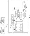
[段落23]图2是图1的举例说明的样品分析器和柱体的示意图。[Paragraph 23] Figure 2 is a schematic diagram of the illustrated sample analyzer and cartridge of Figure 1 .
[段落24]图3是显示图2的样品分析器和柱体的流动控制的更详细的示意图。[paragraph 24] FIG. 3 is a more detailed schematic diagram showing flow control of the sample analyzer and cartridge of FIG. 2 .
[段落25]图4a是具有各种分析电路的举例说明的柱体的图;[Paragraph 25] Figure 4a is a diagram of an illustrative cylinder with various analysis circuits;
[段落26]图4b显示了包括图4a的电路的举例说明的柱体的平面图;[paragraph 26] Figure 4b shows a plan view of an illustrative cylinder comprising the circuit of Figure 4a;
[段落27]图5是可以包括到柱体中的多个举例说明的储存池的示意图;[paragraph 27] FIG. 5 is a schematic diagram of a plurality of illustrative storage cells that may be incorporated into a cartridge;
[段落28]图6是显示分析血液样品的举例说明的方法的示意流程图;[paragraph 28] Figure 6 is a schematic flow diagram showing an illustrative method of analyzing a blood sample;
[段落29]图7是显示分析血液样品的另一个举例说明的方法的示意流程图;[paragraph 29] Figure 7 is a schematic flow diagram showing another illustrative method of analyzing a blood sample;
[段落30]图8是多部分测量方法的各个组件的示意图;[paragraph 30] Figure 8 is a schematic diagram of the various components of the multi-part measurement method;
[段落31]图9a是具有各种分析电路的举例说明的柱体的图;[Paragraph 31] Figure 9a is a diagram of an illustrative cylinder with various analysis circuits;
[段落32]图9b显示了包括图9a的电路的举例说明的柱体的平面图;[paragraph 32] Figure 9b shows a plan view of an illustrative cylinder comprising the circuit of Figure 9a;
[段落33]图9c显示了包括图9a的电路的举例说明的柱体的平面图;[paragraph 33] Figure 9c shows a plan view of an illustrative cylinder comprising the circuit of Figure 9a;
[段落34]图10a是具有各种分析电路的另一个举例说明的柱体的图;[Paragraph 34] Figure 10a is a diagram of another illustrated cartridge with various analysis circuits;
[段落35]图10b显示了包括图10a的电路的举例说明的柱体的平面图;[paragraph 35] Figure 10b shows a plan view of an illustrative cylinder comprising the circuit of Figure 10a;
[段落36]图10c显示了包括图10a的电路的举例说明的柱体的平面图;[paragraph 36] Figure 10c shows a plan view of an illustrative cylinder comprising the circuit of Figure 10a;
[段落37]图11a是具有各种分析电路的另一个举例说明的柱体的图;[Paragraph 37] Figure 11a is a diagram of another illustrated cartridge with various analysis circuits;
[段落38]图11b显示了包括图11a的电路的举例说明的柱体的平面图;[Paragraph 38] Figure 11b shows a plan view of an illustrative cylinder comprising the circuit of Figure 11a;
[段落39]图12a是具有各种分析电路的另一个举例说明的柱体的图;[Paragraph 39] Figure 12a is a diagram of another illustrated cartridge with various analysis circuits;
[段落40]图12b显示了包括图12a的电路的举例说明的柱体的平面图;[paragraph 40] Figure 12b shows a plan view of an illustrative cylinder comprising the circuit of Figure 12a;
[段落41]图12c显示了包括图12a的电路的举例说明的柱体的平面图;[paragraph 41] Figure 12c shows a plan view of an illustrative cylinder comprising the circuit of Figure 12a;
[段落42]图13a是具有各种分析电路的另一个举例说明的柱体的图;[Paragraph 42] Figure 13a is a diagram of another illustrated cartridge with various analysis circuits;
[段落43]图13b显示了包括图13a的电路的举例说明的柱体的平面图;[paragraph 43] Figure 13b shows a plan view of an illustrative cylinder comprising the circuit of Figure 13a;
[段落44]图13c显示了包括图13a的电路的举例说明的柱体的平面图;[paragraph 44] Figure 13c shows a plan view of an illustrative cylinder comprising the circuit of Figure 13a;
[段落45]图14a是具有各种分析电路的另一个举例说明的柱体的图;[paragraph 45] FIG. 14a is a diagram of another illustrated cartridge with various analysis circuits;
[段落46]图14b显示了包括图14a的电路的举例说明的柱体的平面图;和[paragraph 46] Figure 14b shows a plan view of an illustrative cylinder comprising the circuit of Figure 14a; and
[段落47]图15a和15b显示了对血细胞的四部分的区分的数据和图表。[paragraph 47] Figures 15a and 15b show data and graphs for the differentiation of the four fractions of blood cells.
说明illustrate
[段落48]本发明一般涉及样品分析器,更特别地,涉及具有可更换的和/或一次性的柱体的样品分析器,用于在照料患者的地点,例如医师办公室、在家、或在实地的其他地方使用。通过提供具有所有所需试剂和/或液体的可更换的和/或一次性的柱体,所述样品分析器可以很少或没有专业训练时在实验室环境之外可靠地使用。例如,这可以帮助流水线样品的分析处理、降低成分和对医务或其他人员的负担、为许多患者提高样品分析的便利性,包括需要相对频繁的血液监视/分析的那些患者。[Paragraph 48] The present invention relates generally to sample analyzers, and more particularly, to sample analyzers having replaceable and/or disposable cartridges for use in a patient care setting, such as a physician's office, at home, or in a used elsewhere in the field. By providing replaceable and/or disposable cartridges with all required reagents and/or liquids, the sample analyzer can be reliably used outside of a laboratory setting with little or no specialized training. For example, this can help streamline analytical processing of samples, reduce composition and burden on medical or other personnel, improve convenience of sample analysis for many patients, including those requiring relatively frequent blood monitoring/analysis.
[段落49]容许在颗粒悬浮样品中进行快速和有效的颗粒辨别的方法是流式细胞计。在这种方法中,颗粒的悬浮液,一般是血液样品中的细胞,被传送通过流动通道,其中用一个或多个聚焦的光束照射样品中的单个颗粒。通过一个或多个光检测器检测光束与流动通过所述流动通道的单个颗粒的相互作用。通常,检测器被设计以测量特定的波束或发射波长下的光吸收或荧光发射,和/或在特定的散射角的光散射。因而,关于与颗粒的吸收、荧光、光散射或其他光学或电学性质相关的一个或多个特征,通过流动通道的每个颗粒可以被表征。由所述检测器测量的性质可以容许每个颗粒被映射到特征空间中,特征空间的轴是光强度或检测器测量的其他性质。理想地,样品中的不同颗粒被映射到特征空间的不同的和不重叠的区域,容许每个颗粒根据它在特征空间中的映射被分析。这种分析包括颗粒的计数、鉴定、定量(对于一个或多个物理性质)和/或分选。[Paragraph 49] A method that allows rapid and efficient particle discrimination in particle suspension samples is flow cytometry. In this method, a suspension of particles, typically cells in a blood sample, is transported through a flow channel where one or more focused beams of light illuminate individual particles in the sample. Interaction of the light beam with individual particles flowing through the flow channel is detected by one or more photodetectors. Typically, detectors are designed to measure light absorption or fluorescence emission at specific beam or emission wavelengths, and/or light scattering at specific scattering angles. Thus, each particle passing through a flow channel can be characterized with respect to one or more characteristics related to the particle's absorption, fluorescence, light scattering, or other optical or electrical properties. The properties measured by the detector may allow each particle to be mapped into a characteristic space whose axis is light intensity or other properties measured by the detector. Ideally, different particles in the sample are mapped to distinct and non-overlapping regions of feature space, allowing each particle to be analyzed according to its mapping in feature space. Such analysis includes enumeration, identification, quantification (for one or more physical properties) and/or sorting of particles.
[段落50]一个举例说明的实例可以是一种样品分析器,其具有接受采集的样品,例如采集的全血样品的可移动柱体,一旦所述可移动柱体被安装,分析器被激活,所述分析器和柱体自动地处理样品,分析器提供足够的信息为用户做出临床判断。在某些实例中,所述分析器显示或打印出定量结果(例如,在预定范围之内和/或之外),从而不需要用户进一步计算或解释。[Paragraph 50] An illustrative example may be a sample analyzer having a movable cartridge that receives a collected sample, such as a collected whole blood sample, once the movable cartridge is installed, the analyzer is activated , the analyzer and cartridge process the sample automatically, and the analyzer provides sufficient information for the user to make a clinical judgment. In some instances, the analyzer displays or prints out quantitative results (eg, within and/or outside predetermined ranges) so that no further calculations or interpretations by the user are required.
[段落51]样品分析器可以用于,例如,测定血液样品中白细胞的数量和/或种类。在一个举例说明的实例中,所述分析器包括腔室和可更换的流体柱体,其中所述腔室适合于接受所述可更换的流体性柱体。在某些情况下,所述可更换的流体柱体是一次性的柱体。在一个举例说明的实例中,所述可更换的流体柱体可以包括一种或多种试剂(例如,裂解试剂、染料、稀释剂,等等)、一个或多个分析通道、一个或多个流量传感器、一个或多个阀门、和/或适合于处理(例如,裂解、染色、混合,等等)样品并将处理的样品递送到柱体的合适分析通道上的流路。为了支持所述卡片,所述腔体可以包括,例如,压力源、一个或多个光源、一个或多个光检测器、处理器和电源。压力源可以向所述可更换的流体柱体端口提供合适的压力来驱动液体按需要通过所述流路。分析器的一个或多个光源可以用于询问(interrogate)所述可移动柱体的至少选定的分析通道中准备的样品,所述分析器的一个或多个光探测器可以检测穿过样品、被样品吸收和/或散射的光线。所述处理器可以与至少一些所述光源和检测器连接,并且可以测定样品的一个或多个参数。在某些实例中,所述可更换的流体柱体上的一个或多个分析通道可以包括一个或多个流式细胞计通道。[Paragraph 51] The sample analyzer can be used, for example, to determine the number and/or type of white blood cells in a blood sample. In one illustrated example, the analyzer includes a chamber and a replaceable fluidic cartridge, wherein the chamber is adapted to receive the replaceable fluidic cartridge. In some cases, the replaceable fluid cartridge is a disposable cartridge. In one illustrated example, the replaceable fluid cartridge can include one or more reagents (eg, lysing reagents, dyes, diluents, etc.), one or more analytical channels, one or more A flow sensor, one or more valves, and/or a flow path suitable for processing (eg, lysing, staining, mixing, etc.) the sample and delivering the processed sample to a suitable analytical channel of the cartridge. To support the card, the cavity may include, for example, a pressure source, one or more light sources, one or more light detectors, a processor and a power supply. A pressure source may provide suitable pressure to the replaceable fluid cartridge port to drive fluid through the flow path as desired. One or more light sources of the analyzer can be used to interrogate (interrogate) the sample prepared in at least selected analysis channels of the movable cylinder, and the one or more light detectors of the analyzer can detect , the light absorbed and/or scattered by the sample. The processor can be coupled to at least some of the light sources and detectors and can determine one or more parameters of the sample. In some examples, the one or more analytical channels on the replaceable fluid cartridge can include one or more flow cytometer channels.
[段落52]在某些举例说明的实例中,可以向所述可更换的流体柱体提供全血样品,所述可移动柱体可以适合于进行血液分析。[Paragraph 52] In certain illustrative examples, a whole blood sample can be provided to the replaceable fluid cartridge, which can be adapted for blood analysis.
[段落53]为了计数和分类白细胞,至少一部分所述全血样品可以被提供给所述可移动柱体中的白细胞测量通道。向所述白细胞测量通道提供的血液样品可以,例如,如果希望的话,被稀释,红细胞可以被动态(on the fly)裂解,产生的样品可以被水动力学地聚焦用于核形成,并最终向第二细胞计数通道提供。细胞计数通道也可以沿着可移动柱体的透明的流动束流窗口或位于其下方,从而流动束流中的细胞可以被相应的光源和检测器光学地询问。在某些情况下,流量传感器可以在可移动柱体上提供,来提供通过第二细胞计数通道的流速的测量。[Paragraph 53] For counting and classifying white blood cells, at least a portion of said whole blood sample may be provided to a white blood cell measurement channel in said movable cartridge. The blood sample provided to the leukocyte measurement channel can, for example, be diluted if desired, the red blood cells can be lysed on the fly, and the resulting sample can be hydrodynamically focused for nucleation and ultimately sent to A second cell counting channel is provided. The cytometry channel can also be located along or below the transparent flow beam window of the movable cylinder so that cells in the flow beam can be optically interrogated by corresponding light sources and detectors. In some cases, a flow sensor may be provided on the movable cartridge to provide a measurement of the flow rate through the second cytometry channel.
[段落54]在某些情况下白细胞测量通道的举例说明的测量的参数可以包括,例如,对二(2)、三(3)、四(4)或五(5)部分白细胞的区分、总白细胞计数和/或轴上白细胞体积。对于白细胞区分,所述第一、第二、第三、第四和第五部分、类型或种类的白细胞,可以分别指淋巴细胞、单核细胞、嗜中性细胞、嗜曙红细胞和嗜碱性细胞。白细胞也可以被分类成三个组,其包括淋巴细胞、单核细胞和粒细胞(LMG)。粒细胞可以包括嗜中性细胞、嗜曙红细胞和嗜碱性细胞。[Paragraph 54] Illustrative measured parameters of the leukocyte measurement channel in some cases may include, for example, differentiation of two (2), three (3), four (4) or five (5) fractions of leukocytes, total White blood cell count and/or on-axis white blood cell volume. For leukocyte differentiation, said first, second, third, fourth and fifth parts, types or classes of leukocytes may refer to lymphocytes, monocytes, neutrophils, eosinophils and basophils, respectively cell. White blood cells can also be classified into three groups which include lymphocytes, monocytes and granulocytes (LMG). Granulocytes may include neutrophils, eosinophils and basophils.
[段落55]取决于希望的应用,可以测量或计算五种其他参数。在某些情况下,在向细胞计数通道提供样品之前,染料和/或荧光标签可以被添加到样品中,其在某些情况下可以帮助细胞区分。[Paragraph 55] Depending on the desired application, five other parameters can be measured or calculated. In some cases, dyes and/or fluorescent tags can be added to the sample prior to providing the sample to the cytometry channel, which in some cases can aid in cell differentiation.
[段落56]几种一般类型的光散射测量可以在流式细胞计中进行。在小角度(相对于入射光束约1-25度)进行的光强测量,通常称为正向或小角度散射,给出了细胞大小的信息。正向散射还强烈取决于细胞和细胞外介质之间的折射的差异,从而可以辨别例如具有损坏的膜的细胞。在入射光的约65度-115度的角度下进行的光强度测量,通常称为直角或大角度散射,可以提供关于颗粒结构的大小和程度的信息。[Paragraph 56] Several general types of light scattering measurements can be performed in a flow cytometer. Light intensity measurements at small angles (approximately 1-25 degrees relative to the incident beam), often referred to as forward or small-angle scatter, give information on cell size. Forward scatter is also strongly dependent on the difference in refraction between the cell and the extracellular medium, so that eg cells with damaged membranes can be discerned. Measurements of light intensity at angles of about 65°-115° to incident light, commonly referred to as right-angle or large-angle scattering, can provide information about the size and extent of grain structure.
[段落57]在不同角的同时光散射测量,或与吸收或荧光测量组合,可以在流式细胞计方法中使用。例如,与光散射组合的光吸收可以在流式细胞计中被使用来区分各种类型的白细胞。这种方法有时使用了细胞的染色。[Paragraph 57] Simultaneous light scattering measurements at different angles, or in combination with absorption or fluorescence measurements, can be used in flow cytometric methods. For example, light absorption combined with light scattering can be used in flow cytometry to distinguish various types of leukocytes. This method sometimes uses staining of cells.
[段落58]典型地,这种颗粒辨别方法至少部分地使用在此总称为样品分析器的设备的一个或多个部分来实现。许多样品分析器优于在实验室环境中由受训的人员使用的大型设备。为了使用许多样品分析器,在向样品分析器提供准备的样品之前,采集的样品必需首先被处理,例如,通过将样品稀释到期望的水平、添加合适的试剂、将样品离心来提供期望的分离等等。为了得到精确的结果,这种样品处理一般必需由受训的人员进行,这会提高进行样品分析所需的成本和时间。[Paragraph 58] Typically, such particle discrimination methods are implemented at least in part using one or more portions of an apparatus collectively referred to herein as a sample analyzer. Many sample analyzers are superior to larger devices used by trained personnel in a laboratory setting. In order to use many sample analyzers, before providing the prepared sample to the sample analyzer, the collected sample must first be processed, for example, by diluting the sample to the desired level, adding appropriate reagents, centrifuging the sample to provide the desired separation etc. To obtain accurate results, such sample handling typically must be performed by trained personnel, which increases the cost and time required to perform sample analysis.
[段落59]许多样品分析器还需要在分析阶段期间的操作员介入,例如需要另外的信息输入或另外的样品处理。这会进一步提高进行期望的样品分析所需的成本和时间。并且,许多样品分析器仅仅提供了未加工的分析数据作为输出,进一步的计算和/或解释通常必需由受训的人员进行,来做出合适的临床判断。[Paragraph 59] Many sample analyzers also require operator intervention during the analysis phase, eg requiring additional information entry or additional sample processing. This further increases the cost and time required to perform the desired sample analysis. Also, many sample analyzers provide only raw analytical data as output, and further calculations and/or interpretations often must be performed by trained personnel to make appropriate clinical judgments.
[段落60]图1是举例说明的样品分析器和柱体的透视图。举例说明的样品分析器一般地以10显示,包括腔室12和可更换的或一次性的柱体14。举例说明的腔室12包括基部16、盖子18和铰链20,所述铰链将基部16连接到盖子18,但这不是必需的。在举例说明的范例中,基部16包括第一光源22a、第二光源22b和第三光源22c以及相关的光学器件和操作样品分析器的必需电学器件。每个光源可以是单一光源或多光源,取决于应用。在某些情况下,腔室的总尺寸可以小于1立方英尺、小于二分之一立方英尺、小于四分之一立方英尺或更小,如所希望的。同样地,腔室的总重量可以小于10磅、小于5磅、小于1磅,或更小,如所希望的。[Paragraph 60] Figure 1 is a perspective view of an illustrative sample analyzer and cartridge. The illustrated sample analyzer, shown generally at 10 , includes a
[段落61]举例说明的盖子12包括压力源(例如,具有控制微阀的压力室)、第一光检测器24a、第二光检测器22b和第三光检测器22c,每个与光学器件和电学器件相连。每个光检测器也可以是单光检测器或多光检测器,取决于应用。如果希望,还可以提供偏光器和/或滤光器,取决于应用。[Paragraph 61] The illustrated
[段落62]举例说明的可移动柱体14适合于经由样品采集器端口接收样品液体,其在举例说明的实例中,包括刺血针32。在某些实例中,刺血针32可以是可收缩的和/或弹簧加载的。当可移动柱体14不使用时,帽子38可以用于保护样品收集器端口和/或刺血针32。[Paragraph 62] The illustrated
[段落63]在举例说明的实例中,可移动柱体14对全血样品进行血液分析。刺血针32可以被用于刺伤用户的手指来产生血液样品,通过毛细管作用血液样品可以被抽吸到可移动柱体14中抗凝剂包被的毛细管中。可以以类似于可从Micronics Technologies获得的流路的方式构建可移动柱体14,其中一些使用具有酸蚀通道的层状结构来制造。然而,期待的是,可以按任何适合的方式来构建可移动柱体14,包括通过注射模塑或其他任何适合的制造过程或方法,如所希望的。[Paragraph 63] In the illustrated example, the
[段落64]在使用期间,在血液样品白被抽吸到可移动柱体14中之后,当盖子18处于打开位置时可移动柱体14可以被插入所述腔体中。在某些情况下,可移动柱体14可以包括孔洞26a和26b,用于接受基部16中的插脚28a和28b,其可以帮助提供仪器的不同部分之间的对准和连接。可移动柱体14也可以包括第一透明流动束流窗口30a、第二透明流动束流窗口30b和第三透明流动束流窗口30c,其分别对准第一、第二和第三光源22a、22b和22c以及第一、第二和第三光检测器24a、24b和24c。[paragraph 64] During use, after a blood sample has been drawn into the
[段落65]当盖子移动到闭合位置时,系统被密封,盖子18可以经由压力提供端口36a、36b、36c和36d提供受控的压力来分别压迫在举例说明的可移动柱体14中的接收端口34a、34b、34d和34d。期待的是,可以使用更多或更少的压力提供和压力接收端口,取决于应用。作为选择,或另外,一个或多个微泵,例如静电驱动的中央泵,可以在可移动柱体14之上或之中被提供,来提供必需的压力来操作可移动柱体14上的流路。在例如美国专利号5,836,750、6,106,245、6179,586、6,729,856和6,767,190中描述了一些举例说明的静电驱动的中央泵,都被转让给本发明的受让人,全部通过引用合并在此。[Paragraph 65] When the lid is moved to the closed position, the system is sealed, and the
[段落66]一旦被密封,举例说明的仪器可以对收集的血液样品进行血液分析。在某些情况下,所述血液分析可以包括白细胞计数(WBC)。[Paragraph 66] Once sealed, the illustrated instrument can perform blood analysis on the collected blood sample. In some instances, the blood analysis may include a white blood cell count (WBC).
[段落67]为了计数和分类白细胞,全血样品可以被提供给可移动柱体14中的白血测量通道。然后,如不希望,血液样品可以被稀释,红细胞可以被即时裂解,产生的样品可以被以水动力学方式聚焦用于核形成,并最终向第二细胞计数通道提供。细胞计数通道可以沿着可移动柱体14的第二透明流动束流窗口30b放置,从而流动束流中的细胞可以被第二光源22b和第二光检测器24b以光学方式询问。流量传感器可以在可移动柱体14上提供,来提供对通过所述细胞计数通道的流速的测量。在某些情况下,测量的白细胞参数可以包括,例如,对三(3)或(5)部分白细胞的区分,总白细胞计数和/或轴上白细胞体积。取决于应用,也可以测量或计算其他参数。[Paragraph 67] A whole blood sample may be provided to a white blood measurement channel in the
[段落68]即便图1显示了一个举例说明的样品分析器和柱体组合,期待的是可以使用其他样品分析器结构。例如,样品分析器10和可移动柱体可以类似于Schwichtenberg等人的美国专利申请2004/0211077中描述的,通过引用将其合并在此。[Paragraph 68] Even though Figure 1 shows an illustrative sample analyzer and cartridge combination, it is contemplated that other sample analyzer configurations may be used. For example, the
[段落69]在某些情况下,样品分析器10可以适合于在照料患者的地点使用,例如在医生办公室、在家里、或实地的其他地方。提供很少或不需专业训练、可以在实验室环境之外可靠地使用的样品分析器10的能力,可以帮助流水线样品的分析处理、降低成本和对医务人员的负担、以及提高许多患者的样品分析的便利性,包括需要相对频繁的血液监视/分析的那些患者。[Paragraph 69] In some cases,
[段落70]在操作期间,样品分析器10可以接收采集的样品,例如采集的全血样品,一旦分析器被激活,样品分析器10可以自动地处理样品并向用户提供信息来进行临床判断。在某些实例中,样品分析器10可以显示或打印出定量结果(例如,在预定范围之内和/或之外),从而不需要用户进一步计算或解释。[Paragraph 70] During operation,
[段落71]图2是图1的举例说明的样品分析器和柱体的示意图。如以上详述的,在举例说明的实例中,基部16可以包括多个光源22,相关的光学器件和用于操作所述分析器的必需的控制和处理电学器件40。基部16还可以包括电池42、变压器或其他电源。显示了盖子12具有压力源/流动控制模块44和具有相关的光学器件的多个光检测器24。[paragraph 71] Figure 2 is a schematic diagram of the illustrated sample analyzer and cartridge of Figure 1 . As detailed above, in the illustrated example, the
[段落72]可移动柱体14可以经由样品采集器端口或刺血针32接收样品液体。当通过压力源/流动控制模块44加压时,可移动柱体14可以对接收的血液样品进行血液分析。在某些实施例中,以及如上所述的,可移动柱体14可以包括多个试剂49以及用于混合所述试剂与血液样品来制备用于分析的血液样品的流路。并且,可移动柱体14可以包括多个流量传感器来帮助控制和/或证实流路的正确操作。[paragraph 72] The
[段落73]在某些情况下,制备血液样品(例如,裂解的、染色的、稀释的和/或以其他方式处理的),然后在一个或多个板载的细胞计数通道,例如细胞计数通道50中以水动力学方式聚焦用于核形成。在举例说明的实施例中,细胞计数通道50可以经过可移动柱体14中的透明流动束流窗口,例如第一透明流动束流窗口30a。基部16中的光源22的阵列和相关的光学器件可以经由流动束流窗口30a提供穿过核束流的光线。光检测器24的阵列和相关的光学器件可以也经由所述流动束流窗口30a接收来自所述核的散射和非散射光。控制器或处理器40可以从所述检测器24的阵列接收输出信号,并且区分和/或计数存在于所述核束流中的选定细胞。[Paragraph 73] In some cases, blood samples are prepared (e.g., lysed, stained, diluted, and/or otherwise processed) and then processed in one or more on-board cytometry channels, e.g. Hydrodynamically focused in
[段落74]考虑到可移动柱体14可以包括流体控制模块48,用于帮助控制可移动柱体14上至少一些流体的速度。在举例说明的实施例中,所述流体控制模块48可以包括流量传感器,用于感应各种流体的速度并将所述速度报告给控制器或处理器40。控制器或处理器40然后可以调整一个或多个控制信号,后者被提供给压力源/流动控制模块44,来实现期望的压力和因而期望的流体速度用于分析器的正确操作。[paragraph 74] It is contemplated that the
[段落75]由于血液和其他生物废物可能传播疾病,可移动柱体14可以在所述举例说明细胞计数通道50下游包括废物储存池52。废物储存池52可以接收和保存可移动柱体14中流动束流的流体。当测试完成时,可移动柱体14可以被从分析器上移出并且抛弃,优选抛弃到与生物废物相容的容器中。[Paragraph 75] Since blood and other biological waste may spread disease, the
[段落76]图3是显示图2的样品分析器和柱体的流动控制的更详细的示意图。在举例说明的实施例中,盖子18中的压力源/流量控制器44提供五种受控的压力,包括推动样品(P)的压力36a、裂解(L)压力36b、染料(ST)压力36c和鞘(SH)压力36d。这些仅仅是举例说明的,期待的是更多、更少或不同的压力(例如,向稀释剂储存池的稀释剂压力)可以由压力源/流量控制器44提供,取决于应用。并且,期待的是盖子18可以包括压力源/流量控制器44。相反,可移动柱体14可以包括板载的压力源,例如压缩空气储存器、一个或多个微泵,例如如上所述的静电驱动的中央泵,或任何其他适合的压力源,如所希望的。光源和检测器的阵列没有在图3中示出。[paragraph 76] FIG. 3 is a more detailed schematic diagram showing the flow control of the sample analyzer and cartridge of FIG. 2 . In the illustrated embodiment, a pressure source/
[段落77]在举例说明的实施例中,压力源36a经由推进流体65向血液样品储存池62提供压力,压力源36b向裂解储存池64提供压力,压力源36c向染料储存池66提供压力,压力源36d向鞘储存池68提供压力。[Paragraph 77] In the illustrated embodiment,
[段落78]在一个举例说明的实施例中,每个压力源可以包括用于接收输入压力的第一压力室和用于向可移动柱体提供受控的压力的第二压力室。可以在第一压力室和第二压力室之间提供第一阀门,用于可控地将第一压力室中的压力释放到第二压力室中。在流体通路与第二压力室中的第二阀门可以可控地将第二压力室中的压力释放到大气中。这可以容许压力源/流量控制器44向可移动柱体14上的每个压力接收端口提供受控的压力。每个阀门可以是分别可寻址和可控制的静电驱动的微阀,如在例如标题“用于均衡压力或流动控制的可寻址的阀阵列”共同待决美国专利申请系列号09/404,560中描述的,通过引用合并在此。作为选择,每个阀门可以是静电驱动的微阀的阵列,其是采用可控制的工作周期脉冲调节的,来实现受控的“有效”流动或泄漏速率。如果希望,也可以使用其他阀门。[Paragraph 78] In an illustrative embodiment, each pressure source may include a first pressure chamber for receiving an input pressure and a second pressure chamber for providing a controlled pressure to the movable column. A first valve may be provided between the first pressure chamber and the second pressure chamber for controllably releasing pressure in the first pressure chamber into the second pressure chamber. A second valve in the fluid passage and the second pressure chamber can controllably release the pressure in the second pressure chamber to atmosphere. This may allow the pressure source/
[段落79]举例说明的可移动柱体14包括五个压力接收端口34a、34b、34c和34d,每一个用于从压力源/流量控制器44接收相应的受控压力。在举例说明的实施例中,压力接收端口34a、34b、34c和34d可以分别将受控压力指向血液储存池62、裂解储存池64、染料储存池66和鞘储存池68。裂解储存池64、染料储存池66和鞘储存池68可以在可移动柱体14装运使用之前被填充,而血液储存池62可以在本地经由样品采集器端口或刺血针32被填充。[Paragraph 79] The illustrated
[段落80]如所示的,可以串联地为流量传感器提供每种或选定的流体。每个流量传感器80a、80b、80c和80d可以测量相应的流体的速度。流量传感器80a、80b、80c和80d优选地是热风速型流量传感器,更优选地是微桥型流量传感器。在例如美国专利号4,478,076、美国专利号4,478,077、美国专利号4,501,144、美国专利号4,651,564、美国专利号4,683,159和美国专利号5,050429中描述了微桥流量传感器,所有这些通过引用被合并在此。来自每个流量传感器80a-80d的输出信号可以被提供给控制器或处理器40。如所示,控制器或处理器40可以向压力源/控制器44提供控制信号。例如,当血液样品的速度降至低于第一预定值时,为了控制向血液样品提供的压力,控制器或处理器40可以打开压力源/控制器44中的第一压力室和第二压力室之间的第一阀门,用于可控地释放第一压力室中的压力到所述第二压力室中。同样地,当血液样品的速度提高到高于第二预定值时,控制器或处理器40可以打开第二阀门,其泄出第二压力室中的压力。控制器或处理器40可以按类似的方式控制裂解试剂、染料和鞘流体的速度。[Paragraph 80] As indicated, each or selected fluids may be provided to the flow sensor in series. Each
[段落81]在某些情况下,控制器或处理器40可以检测通过流动通道的流动速度方面的一种或多种改变。流动速度方面的改变可以相应于,例如,流动通道中的一个或多个气泡、起因于例如血液样品的凝结、流动通道中不希望的或外来的物体的流动通道的闭塞或部分闭塞,以及流动通道的其他不希望的特征。控制器或处理器40可以被编程来检测来自流动速度的这些特征,在某些情况下,发出警告和/或关闭样品分析器。[paragraph 81] In some cases, the controller or
[段落82]热风速仪型流量传感器一般包括加热元件,其当被激发时在液体中产生一个或多个热脉冲,并进一步包括位于加热元件的上游和/或下游的一个或多个热传感器来检测一个或多个热脉冲。可以将通过流动通道的流体的速度与热脉冲从加热元件移动到间隔的热传感器之一花费的时间相关联。[paragraph 82] Thermal anemometer-type flow sensors generally include a heating element that, when energized, generates one or more heat pulses in the liquid, and further include one or more thermal sensors located upstream and/or downstream of the heating element to detect one or more heat pulses. The velocity of the fluid through the flow channel can be correlated to the time it takes for a heat pulse to travel from the heating element to one of the spaced thermal sensors.
[段落83]在某些情况下,热风速仪型流量传感器可以用于检测热传导率和/或流体的比热。流体的热传导率和/或比热方面的改变可以相应于流体特性方面的改变,例如,流体的状态的变化(血液样品的凝结)、流体中的气泡、流体中不希望的或外来的物体,等等。因而,在某些实施例中,期待的是控制器或处理器40可以通过监视经过热风速仪型流量传感器的流体热传导率和/或比热来检测流体的特征。[Paragraph 83] In some cases, thermal anemometer-type flow sensors may be used to detect thermal conductivity and/or specific heat of a fluid. Changes in the thermal conductivity and/or specific heat of the fluid may correspond to changes in the properties of the fluid, for example, changes in the state of the fluid (coagulation of the blood sample), air bubbles in the fluid, unwanted or foreign objects in the fluid, etc. Thus, in certain embodiments, it is contemplated that the controller or
[段落84]在某些情况下,可以以与流体通道具有流体连通的方式提供阻抗传感器。控制器或处理器40可以与阻抗传感器耦合。流体的阻抗方面的改变可以表明流体特性方面的改变,例如,流体的状态的变化(血液样品的凝结)、流体中的气泡、流体中不希望的或外来的物体,等等。因而,在某些实施例中,期待的是控制器或处理器40可以通过监视经过阻抗传感器的流体的阻抗来检测流体的特征。[paragraph 84] In some cases, an impedance sensor may be provided in fluid communication with the fluid channel. A controller or
[段落85]还可以提供在111中一般地显示的下游阀门。控制器或处理器40可以按需要地打开/闭合下游阀111。例如,下游阀门111可以保持关闭直到系统被完全地加压。这可以帮助防止血液、裂解试剂、球化试剂、鞘流体和稀释剂在系统完全加压之前流入流路86。并且,下游阀门111可以被控制来帮助进行某些测试,例如零流动测试,等等。在另一个实施例中,下游阀门111可以通过机械作用被打开,例如,当盖子被闭合时。[Paragraph 85] A downstream valve shown generally at 111 may also be provided. The controller or
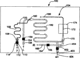
[段落86]图4a是结构430的可移动卡片或柱体的各个组件的图或示意图,所述结构430可以在一个运行中提供对白细胞的四部分的区分。一滴全血可以从来源431被提供给样品采集器432。血液可以被提供给即时裂解注射器机构或模块434。血液向模块434的流动速度可以通过流速控制机构或模块433控制。裂解试剂可以以流速控制机构433控制的流速从裂解试剂储存池435提供给即时裂解注射器434。裂解的血液可以从即时裂解机构434提供给水动力学聚焦室437。来自储存池436的鞘试剂可以进入聚焦室437来在血细胞进入光通道438时提供围绕血细胞的鞘。鞘试剂的流动可以由控制块433控制。血细胞可以通过光学和处理系统439来区分。系统439的散射和分析可以提供对细胞的四部分的区分。细胞和相关的流体可以从光通道进入废物储存池441。裂解和鞘试剂可以是相同的或不同的试剂。裂解和鞘试剂的来源可以分别是所述卡片或柱体之上或之外的储存池435和436。废物贮存池441可以在所述卡片或柱体之上或之外。流速控制机构433可以在所述卡片之上或之外,或者它可以部分地在所述卡片或柱体上。[paragraph 86] FIG. 4a is a diagram or schematic diagram of various components of a removable card or cartridge of a structure 430 that can provide four-part differentiation of white blood cells in one run. A drop of whole blood may be provided from
[段落87]图4B显示了举例说明的可移动卡片或柱体中图4a的结构430的示意图的实施方式,其被设计为一次性的。柱体一般地显示在这个图的100处,其可以类似于以上参考图1、2和3显示和描述的可移动柱体14。要理解的是,可移动柱体100仅仅是举例说明的,本发明可以应用于许多微流柱体,不考虑形式、功能或结构。例如,本发明可以应用于适合流式细胞计、血液学、临床化学、血液化学分析、尿分析、血气分析、病毒分析、细菌分析、电解质测量等等的的可移动柱体。还期待的是,本发明的可移动柱体,例如可移动柱体100可以从任何适合的材料或材料系统制造,包括,例如,玻璃、塑料、硅、一种或多种聚合物、杂合体材料、或任何其他适合的材料或材料系统,或材料或材料系统的组合。[Paragraph 87] Figure 4B shows an embodiment of a schematic diagram of the structure 430 of Figure 4a in an illustrative removable card or cylinder, which is designed to be disposable. A column is shown generally at 100 in this figure, which may be similar to the
[段落88]举例说明的可移动柱体100包括测量通道104,然而可以使用更多或更少的测量通道,如所希望的。测量通道104是白细胞测量通道。全血样品由可移动柱体100经由血液接收端口106接收,所述血液接收端口106通过毛细管作用,吸出已知数量的血液进入抗凝剂包被的血液样品贮存毛细管108。推动样品(P)的压力,例如图3的推动样品(P)的压力36a被提供给推动样品的流体储存池,例如图3的推动样品的流体储存池65。当施加压力时,推动样品的流体被从推动样品流体储存池压到推动血液样品的通道110中。[Paragraph 88] The illustrated
[段落89]在某些举例说明的实例中,可以以与推动血液样品的通道110串联的方式提供阀门112和流量传感器114。当希望推动血液样品通过流路时,阀门112可以被控制来打开。流量传感器114可以测量推动血液样品的流体的流动速度和因而通过抗凝剂包被的毛细管108的血液样品的流动速度。流量传感器114提供的流动速度可以用于帮助控制提供给可移动柱体100的推动样品(P)的压力。[Paragraph 89] In certain illustrative examples,
[段落90]在举例说明的实例中,全血样品经由毛细管108被提供给白细胞测量通道104。提供阀门120来控制血液样品从毛细血管108流动进入白细胞测量通道104。[Paragraph 90] In the illustrated example, a whole blood sample is provided to white blood
[段落91]对于白细胞测量通道104,白细胞裂解试剂(L)压力,例如图3的裂解压力(PIN(L))36b,被提供给裂解试剂储存池,例如图3的裂解储存池64。当施加压力时,裂解储存池64中的裂解试剂被压迫到裂解试剂管或通道154中。通道在某些环境中是指管道、毛细管、蛇形流动路径、传送带等等;然而,术语“通道”在此以一般的含义使用。[Paragraph 91] For the
[段落92]在某些举例说明的实施例中,可以以与裂解试剂通道154串联的方式提供阀门156和流量传感器158。当希望推动裂解试剂进入流路时,阀门156可以被控制来打开。流量传感器158可以测量裂解试剂的流动速度,并提供通过裂解试剂通道154的裂解试剂流动速度的测量。流量传感器158提供的流动速度可以用于帮助控制裂解压力,所述裂解压力通过压力源/控制器44被提供给可更换的或一次性的柱体100。[Paragraph 92] In certain illustrative embodiments,
[段落93]在举例说明的可移动柱体100的正常功能运行期间,裂解试剂以裂解试剂流动速度被提供给组合、连接或交叉区域160,血液样品以血液样品流动速度被提供给交叉区域160。血液样品流动速度和裂解试剂流动速度可以由压力源/控制器,例如图3的压力源/控制器44来控制。[Paragraph 93] During normal functional operation of the illustrated
[段落94]交叉区域160可以被配置,使得当两种流体流过交叉区域160时,裂解试剂围绕血液样品四周流动。虽然接点或组合、联结或交叉区域160或170可以用对其目的更为描述性的术语(例如,水动力性聚焦室)来称呼,术语“交叉区域”在此按一般含义使用。在某些情况下,裂解试剂流动速度可以高于血液样品的流动速度,其可以帮助改善动态裂解通道162(其可以具有蛇形路径))中的流动特征,以及在某些情况下,来帮助形成完全地和均匀地被裂解试剂围绕的薄的血液带。这种带流动可以在它们穿过即时裂解通道162时帮助裂解试剂均匀地裂解红细胞。此外,即时裂解通道162的长度以及裂解试剂和血液样品的流动速度可以被设置,使得血液样品暴露于裂解试剂持续合适的时间量。[paragraph 94]
[段落95]鞘流体(SH)压力,例如图3的鞘(SH)压力36d可以被提供给鞘流体储存池,例如图3的鞘流体储存池68。当施加压力时,鞘流体被从鞘流体储存池68被压到鞘通道164中。在某些举例说明的实例中,可以以与鞘通道164串联的方式提供阀门166和流量传感器168。当希望推动鞘流体进入流路时,阀门166可以被控制来打开。流量传感器168可以测量鞘流体的流动速度,并且可以提供通过鞘通道164的鞘流体流动速度的测量。由流量传感器168提供的流动速度可以用于帮助控制提供给可移动柱体100的鞘压力(SH)。[Paragraph 95] A sheath fluid (SH) pressure, such as sheath (SH)
[段落96]在举例说明的实施例中,鞘流体被以鞘流体流动速度提供给交叉区域170,裂解的血液样品被以裂解的血液样品流动速度提供给交叉区域170。虽然这个区域170可以被称为鞘形成、水动力学聚焦或其他目的效果等等的区域,它在此被一般地称为“交叉区域”。裂解的血液样品流动速度和鞘流动速度可以由压力源/控制器,例如图3的压力源/控制器44来控制。[Paragraph 96] In the illustrated embodiment, the sheath fluid is provided to the
[段落97]交叉区域170可以被配置,使得当两种流体流过交叉区域170时,鞘流体围绕裂解的血液样品四周流动。在某些情况下,鞘流动速度显著地高于裂解的血液样品的流动速度,其可以帮助改善下游的流动光学细胞计数通道172中的核形成。例如,在某些流式细胞计应用中,所述交叉区域170可以被配制为水动力学地聚焦和排列裂解血液样品中的白细胞成单纵队的核,当它们穿过可移动柱体100中的光学窗口区域174时,每个白细胞可以被分析器分别地光学询问。在某些情况下,穿过细胞计数通道172的流体通过管道或通道186向图3的板载的废物储存池52提供。[Paragraph 97]
[段落98]图5是可以被包括到可移动柱体中的多个举例说明的储存池的示意图。在举例说明的实例中,可移动柱体例如图4的可移动柱体100可以包括,例如,裂解试剂储存池64、推进流体储存池65、染料储存池66、鞘流体储存池68和废物储存池52。这些仅是举例说明的,预期的是,更多、更少或不同的储存池可以在可移动柱体之上或之中提供。[paragraph 98] FIG. 5 is a schematic diagram of a plurality of illustrative storage tanks that may be incorporated into a movable column. In the illustrated example, a movable cartridge such as
[段落99]每个储存池可以调整大小,并且包括合适的数量的流体和/或试剂来支持可移动柱体的期望的操作。同样地,在某些实例中,储存池例如染料储存池66可能是希望的,来添加染料到白细胞通道中来支持对白细胞的区分。期待的是,保存在储存池中的试剂和/或流体起初可以是处于液体或冻干形式,取决于应用。[Paragraph 99] Each reservoir can be sized and include an appropriate amount of fluid and/or reagents to support the desired operation of the movable cartridge. Likewise, in some instances, a reservoir, such as
[段落100]图6是显示使用可移动柱体分析血液样品的举例说明的方法的示意流程图。在举例说明的方法中,首先在步骤200获得血液样品。然后,血液样品可以被提供给可移动柱体中的抗凝剂包被的毛细管202。然后,血液样品可以被提供给白细胞(WBC)测量通道206。[Paragraph 100] Figure 6 is a schematic flow diagram showing an illustrative method of analyzing a blood sample using a movable cartridge. In the illustrated method, a blood sample is first obtained at
[段落101]在举例说明的WBC测量通道206中,全血230的红细胞可以如232所示首先被裂解,如233所示染色或标记,然后以水动力学方式聚焦并提供单纵队流下可移动柱体中的WBC细胞计数通道234。光源236,例如垂直空腔表面发射激光器(VCSEL)可以在单个的细胞通过WBC细胞计数通道234的分析区域时将光线照射到单个的细胞上。在某些情况下,可以提供VCSEL设备的阵列,当单个的细胞通过WBC细胞计数通道234的分析区域时仅有与单个的细胞对准的VCSEL被激活。VCSEL提供的一些入射光被散射,检测器238检测所述散射光。可以使用其他类型的光源。在某些情况下,检测器238可以检测正向角度散射光线(FALS)、小角度散射光线(SALS)和大角度散射光线(LASL)。检测器239可以检测来自某些细胞的荧光。在某些情况下,如在240中所示,许多参数可以在分析期间测量,包括,例如,轴上细胞体积、总WBC计数和WBC五(5)部分区分。[Paragraph 101] In the illustrated
[段落102]图7是显示分析血液样品的另一个举例说明方法的示意流程图。在这个举例说明的方法中,可以获得血液样品,提供给血液样品储存池,如步骤300所示。然后,血液样品可以被提供给可移动柱体中的抗凝剂包被的毛细管302,并稀释。血液样品可以被提供给白细胞(WBC)测量通道340。[Paragraph 102] Figure 7 is a schematic flow diagram showing another illustrative method of analyzing a blood sample. In this illustrated method, a blood sample may be obtained and provided to a blood sample storage reservoir, as shown in
[段落103]在举例说明的WBC测量通道340中,红细胞可以被裂解,视情况注射染料,如342所示。然后细胞可以被以水动力学方式聚焦并提供单纵队流下可移动柱体中的WBC细胞计数数通道344。光源346,例如垂直空腔表面发射激光器(VCSEL)可以在单个的细胞通过WBC细胞计数通道344的分析区域时将光线照射到单个的细胞上。在某些情况下,可以提供VCSEL设备的阵列,当单个的细胞通过WBC细胞计数通道344的分析区域时仅有与单个的细胞对准的VCSEL被激活。[Paragraph 103] In the illustrated
[段落104]在单个的细胞/颗粒穿过聚焦的入射光束时,一些光线被阻挡、散射或遮挡,其可以通过检测器来检测(未显示)。当两个或更多光源沿着WBC细胞计数通道344聚焦于不同间隙的点时,可以检测每个细胞的前和/或后边缘。通过测量细胞横穿一个聚焦点到下一个的距离花费的时间,可以测定流动速度和因而测定细胞速度。使用测定的细胞速度,如348所示,可以将细胞阻挡、散射或遮挡光束的持续时间与细胞大小和/或细胞体积相关联。[Paragraph 104] As individual cells/particles pass through the focused incident beam, some light is blocked, scattered or blocked, which can be detected by a detector (not shown). When two or more light sources are focused at differently spaced points along the
[段落105]在某些实施例中,可以提供光源350和相关的光学器件和/或偏光器。光源346和350可以被组合成一个光源(甚至组合成一束用于期望的测量),其中在同时和在同一个细胞上进行所有的测量。相关的光源350的光学器件可以瞄准光线、测量轴下散射,例如SALS、FALS和LALS散射,如354所示。如上,SALS、FALS和LALS散射可以用于测量,例如计数的白细胞(NWBC)的数目352,以及帮助白细胞区分,如356所示。在某些情况下,提供一个或多个偏光器来偏振化光源提供的光线,在检测器检测的偏振消光/旋转的水平可以用于帮助进行白细胞区分,但在所有实施例中这不是必需的。同时,来自某些细胞(即,染色的、标记的或标签化的)的荧光可以被检测,如355所示。[paragraph 105] In some embodiments, a
[段落106]图8是白细胞五部分区分测量计划500的提纲。为了启动,可以是通过散射的三部分区分。第四部分区分502可以是通过散射,例如与三部分方法一起。因此,如果散射的第四部分的回答是“是”,然后可以进入第五部分模块503。从模块503,测定可以是箭头504的方向,其中第五部分测定与另一个四部分散射测量平行地进行。第五部分的平行方法可以包括用荧光选择性染色CD45和散射。从模块503的测定反而可以是沿箭头505方向的,其中第五部分测定与另一个四部分散射测量按顺序地进行。这种第五部分测量可以是选择性的裂解或发荧光。[paragraph 106] FIG. 8 is an outline of a white blood cell five-part differential measurement plan 500 . For priming, there can be a three-part distinction by scatter. The fourth part differentiation 502 may be by scatter, for example with the three-part method. Thus, if the answer to the fourth part of the scatter is yes, then the fifth part module 503 can be entered. From block 503, a measurement may be in the direction of arrow 504, where a fifth part measurement is performed in parallel with another four part scatter measurement. Parallel approaches to the fifth part can include selective staining of CD45 and scatter with fluorescence. Measurements from module 503 may instead be in the direction of arrow 505, wherein a fifth part measurement is performed sequentially with another four part scatter measurement. This fifth part measurement can be selective lysis or fluorescence.
[段落107]如果对第四部分是否通过散射,例如,与第三部分散射一起的模块502问题的答案是“否”,可以跟随箭头到模块506,表明还需要进行第四和第五部分区分。在箭头507的方向上,与三部分的散射测量平行地,第四和第五部分的区分可以使用选择性染料例如中性红来进行,和/或使用可以涉及合适染料的使用的荧光来实现。另一方面,第四和第五部分区分可以沿箭头508的方向,相对于其他三部分散射测量以连续方式进行。连续的区分可以用选择性裂解和/或荧光来进行。[Paragraph 107] If the answer to the question of whether the fourth part passes scatter, e.g., block 502 along with the third part scatter is "No", the arrow can be followed to block 506, indicating that a fourth and fifth part distinction also needs to be made . In the direction of arrow 507, in parallel to the scatterometry of the three parts, the differentiation of the fourth and fifth parts can be done using a selective dye such as neutral red, and/or using fluorescence which can involve the use of a suitable dye . On the other hand, the fourth and fifth fractional distinctions can be made in a sequential manner, in the direction of arrow 508, relative to the other three fractional scatter measurements. Serial differentiation can be performed using selective lysis and/or fluorescence.
[段落108]图9a显示了类似于图4a的结构430的结构440的可移动卡片或柱体的版本。结构440可以提供对流动通过光通道438的细胞的四部分的区分。它涉及来自采集器432的样品的选择性裂解。用来提供这种选择性裂解的是选择性裂解试剂储存池442,其可以向即时裂解注射器434提供选择性裂解试剂。来自储存池442的选择性裂解试剂的流动速度可以由流速控制机构433控制。在一个血液样品上可以有两个连续的运行。裂解的第一次运行可以产生对三部分的区分。使用选择性裂解对相同样品的第二次运行可以提供对移动通过光通道438的血细胞的第四部分区分。[Paragraph 108] Figure 9a shows a removable card or cylinder version of a
[段落109]图9b类似于图4b,分别相对于图9a和4a中结构440和430的它们的实施方式。图9b中的实施方式的其他特征可以被解释。也就是说,选择性裂解试剂414可以被提供给通道451,其被连接到用于将第一裂解试剂传送到交叉区域160的通道154。通道451上的阀门413可以控制选择性裂解试剂414什么时候可以流向区域160。流量传感器412可以监视选择性裂解试剂的流动,并向流动控制机构提供指示流动的信号。[paragraph 109] Figure 9b is similar to Figure 4b, with respect to their implementations of
[段落110]图9c类似于图9b,在于不存在流量传感器412,用于传送选择性裂解试剂414的通道451被连接到流量传感器158上游的裂解试剂室管道154。流量传感器158可以用于裂解运行,以及用于选择性裂解运行,其可以在不同的或独立的时间发生。[paragraph 110] FIG. 9c is similar to FIG. 9b in that there is no
[段落111]图10a是结构450的图表或示意图,其可以以对一个样品的三个顺序运行来提供对白细胞的五部分的区分。使用来自裂解试剂储存池435的裂解的第一次运行可以在光通道438中产生对血细胞的三部分的区分。使用来自试剂储存池442的第一次选择性裂解的第二次运行可以在通道438中产生对血细胞的第四部分的区分。使用来自另一个选择性裂解试剂储存池443的第二选择性裂解的第三次运行可以在通道438中产生对血细胞的第五部分的区分。[paragraph 111] Figure 10a is a diagram or schematic diagram of a
[段落112]图10b类似于图9b,分别相对于图10a和9a中结构450和440的它们的实施方式。图10b的其他特征是被提供给通道452的第二选择性裂解试剂406,所述通道452通过通道154将裂解试剂传送到交叉区域160。通道452上的阀门407可以控制第二选择性裂解试剂406什么时候要流向区域160。流量传感器411可以监视第二选择性裂解试剂406经过通道452的流动,并向流动控制机构提供指示流动的信号。[paragraph 112] Figure 10b is similar to Figure 9b, with respect to their implementations of
[段落113]图10c类似于图10b,在于不存在流量传感器412和411,并且分别用于传送第一选择性裂解试剂414和第二选择性裂解试剂406的管道或通道451和452被连接到流量传感器158上游的裂解试剂通道154。流量传感器158可以用于裂解运行、第一选择性裂解运行和第二选择性裂解运行,其可以在不同的时间发生。[paragraph 113] FIG. 10c is similar to FIG. 10b in that there are no
[段落114]图11a是结构460的图表或示意图,其可以采用对一个样品的一个运行提供对白细胞的四部分的区分。可以有储存池444,其含有与裂解试剂混合的染色/荧光试剂,其可以被提供给动态裂解注射器434。除了储存池444的这种混合物和不存在储存池435之外,结构460类似于图4a的结构430。三部分散射和第四部分染色/荧光测量可以平行地进行。[paragraph 114] Figure 11a is a diagram or schematic diagram of a
[段落115]图11b显示了图11a的结构460的实施方式。图11b类似于图4b,只是不同于裂解试剂,相反地有裂解试剂和染色试剂的混合物401输入到通道154中。[Paragraph 115] Figure 11b shows an embodiment of the
[段落116]图12a是结构470的图表或示意图,其可以采用对一个样品的两个连续运行提供对白细胞的五部分的区分。裂解试剂和染色试剂混合物储存池444可以从图11a中保留。添加选择性裂解试剂储存池442(如图9a中)。储存池442和444都可以提供选择性裂解试剂以及裂解试剂和染色试剂的混合物,其可以经由相应的管道或通道,但不必在同一运行中,被提供给即时裂解注射器434。使用染色试剂和裂解试剂的混合物的第一运行还可以在光通道438中提供使用荧光/散射检测的对第四部分的区分,除了使用血细胞的散射检测提供三部分区分之外。使用选择性裂解试剂的第二次运行可以提供对细胞的第五部分的区分。[paragraph 116] Figure 12a is a diagram or schematic diagram of a
[段落117]图12b显示了图12a的结构470的实施方式。图12b可以类似于图11b,在于它具有裂解试剂和染色试剂的混合物401经由阀门156进入通道154。还存在流量传感器158,用于使用发给流速控制机构的信号来监测混合物401的流动。类似于图9b,还存在经由阀门413进入通道451的选择性裂解试剂414.试剂414可以经由通道451通过流量传感器412进入通道154。传感器412可以使用发给流速控制机构的信号来监测试剂414的流动。[Paragraph 117] Figure 12b shows an embodiment of the
[段落118]图12c类似于图12b中的实施方式,只是除去了用于监视选择性裂解试剂414的流量传感器412。并且,用于传送试剂的通道451被连接到流量传感器158上游的通道154,用于裂解试剂和染色试剂的混合物401。对于与裂解试剂401和选择性裂解试剂414混合的染料可以使用同一流量传感器,因为它们可以在不同的时间作为独立的运行发生。[Paragraph 118] Figure 12c is similar to the embodiment in Figure 12b, except that the
[段落119]图13a是结构480的图表或示意图,其可以在光通道438中提供对细胞的四部分的区分。这个结构480可以类似于图4a的结构430,只是结构480另外具有染色试剂储存池445,其具有通向动态裂解注射器434的出口。并且,所述储存池445被连接到流速控制机构433,用于控制染色试剂向注射器机构434的流动。结构480可以具有使用来自储存池435的裂解试剂的第一次运行,用于在光通道438中对血细胞的三部分的区分。使用来自储存池445的染色试剂的血细胞染色的第二次运行和光通道438中细胞的荧光/散射观察结果可以提供对细胞的四部分的区分。[paragraph 119] FIG. 13a is a diagram or schematic diagram of a
[段落120]图13b显示了图13a的结构480的实施方式。根据结构透视,卡片100的布局看起来类似于图9b的布局,除了染色试剂3经由通道453输入通道154,而不是选择性裂解试剂414之外。染色试剂417可以经由管道154和交叉区域160上的阀门416和流量传感器415进入通道453。[Paragraph 120] Figure 13b shows an embodiment of the
[段落121]图13c可以类似于图13b,除了缺少用于监测染色试剂流动的流量传感器415和用于传送染色试剂417的通道453被连接到流量传感器158上游的通道154之外。裂解试剂和染色试剂可以利用同一个流量传感器158,因为裂解试剂和试剂417可以在不同的时间在两个独立的运行中被使用。[paragraph 121] FIG. 13c may be similar to FIG. 13b except that
[段落122]图14a是结构490的图表或示意图,其可以在光通道438中提供对血细胞的四部分的区分。结构490可以类似于结构480,除了染色试剂储存池445的出口不连接到即时裂解注射器机构434的入口,而是它连接到注射器机构434出口和水动力学聚焦室437的入口之间的通道之外。[paragraph 122] FIG. 14a is a diagram or schematic diagram of a
[段落123]图14b显示了图14a的结构390的实施方式。图14b中的实施方式可以类似于图4b中的实施方式,只是通道454在交叉区域170的紧邻上游的地方被连接到通道162。这个通道454可以被连接到染色试剂储存池并经由阀门403和流量传感器402将染色试剂404传送到通道162。传感器402可以向流速控制机构提供关于试剂404流动的信号。[Paragraph 123] Figure 14b shows an embodiment of the structure 390 of Figure 14a. The embodiment in FIG. 14b may be similar to the embodiment in FIG. 4b except that
[段落124]图15a和15b显示了关于四部分血细胞区分的数据和图表,利用了与在此讨论的类似的结构。[Paragraph 124] Figures 15a and 15b show data and graphs for the differentiation of four-part blood cells, using a structure similar to that discussed here.
[段落125]注意到的是,图4a、4b、9a、9b、9c、10a、10b、10c、11a、11b、12a、12b、12c、13a、13b、13c、14a和14b的结构是用于血细胞的多部分区分的各种方法的代表。其他结构,包括在此公开的结构的各种排列和其他方案可以用于对血细胞的多部分的区分。[Paragraph 125] Note that the structures of Figures 4a, 4b, 9a, 9b, 9c, 10a, 10b, 10c, 11a, 11b, 12a, 12b, 12c, 13a, 13b, 13c, 14a, and 14b are for Representative of various methods for multipart differentiation of blood cells. Other structures, including various arrangements of the structures disclosed herein, and other schemes can be used to differentiate blood cell fractions.
[段落126]在本说明书中,虽然以另一种方式或时态声明了,但是一些物质可以具有推定的或预测的性质。[Paragraph 126] In this specification, although stated in another manner or tense, some substances may have putative or predicted properties.
[段落127]虽然已经根据至少一个举例说明的实例描述了本发明,在阅读本说明书时多种改变和修饰对于本领域技术人员变得明显。因而,鉴于先有技术,附随的权利要求被尽可能宽泛地解释的意图,包括了所有这些改变和修饰。[Paragraph 127] While the invention has been described in terms of at least one illustrative example, various changes and modifications will become apparent to those skilled in the art upon reading this specification. It is therefore intended that the appended claims be interpreted as broadly as possible in light of the prior art to embrace all such changes and modifications.
Claims (49)
Applications Claiming Priority (3)
| Application Number | Priority Date | Filing Date | Title |
|---|---|---|---|
| US11/306,508 | 2005-12-30 | ||
| US11/306,508 US20060263888A1 (en) | 2000-06-02 | 2005-12-30 | Differential white blood count on a disposable card |
| PCT/US2006/047792 WO2007084232A1 (en) | 2005-12-30 | 2006-12-14 | Differential white blood count on a disposable card |
Publications (2)
| Publication Number | Publication Date |
|---|---|
| CN101379387A true CN101379387A (en) | 2009-03-04 |
| CN101379387B CN101379387B (en) | 2013-02-06 |
Family
ID=38293337
Family Applications (1)
| Application Number | Title | Priority Date | Filing Date |
|---|---|---|---|
| CN2006800531502A Expired - Fee Related CN101379387B (en) | 2005-12-30 | 2006-12-14 | Differential white blood count on a disposable card |
Country Status (5)
| Country | Link |
|---|---|
| US (1) | US20060263888A1 (en) |
| EP (1) | EP1966587B1 (en) |
| JP (1) | JP2009522546A (en) |
| CN (1) | CN101379387B (en) |
| WO (1) | WO2007084232A1 (en) |
Cited By (6)
| Publication number | Priority date | Publication date | Assignee | Title |
|---|---|---|---|---|
| CN103170377A (en) * | 2011-12-20 | 2013-06-26 | 中国科学院深圳先进技术研究院 | Hemocyte analysis chip and system for using chip thereof |
| CN103403547A (en) * | 2010-11-09 | 2013-11-20 | 综合医院公司 | Counting particles using an electrical differential counter |
| WO2016173547A1 (en) * | 2015-04-30 | 2016-11-03 | Winnoz Technology, Inc | System and method for detecting biomarker |
| CN111157433A (en) * | 2020-01-06 | 2020-05-15 | 中国科学院生态环境研究中心 | Kit for marking single cell of red blood cell and detection method thereof |
| CN115752304A (en) * | 2022-11-29 | 2023-03-07 | 安徽瑞控智能科技有限公司 | Motormeter dish tucking detection device |
| US12332265B2 (en) | 2019-12-27 | 2025-06-17 | Beckman Coulter, Inc. | Sample preparation instrument |
Families Citing this family (123)
| Publication number | Priority date | Publication date | Assignee | Title |
|---|---|---|---|---|
| JP2006507921A (en) | 2002-06-28 | 2006-03-09 | プレジデント・アンド・フェロウズ・オブ・ハーバード・カレッジ | Method and apparatus for fluid dispersion |
| GB0307428D0 (en) | 2003-03-31 | 2003-05-07 | Medical Res Council | Compartmentalised combinatorial chemistry |
| US20060078893A1 (en) | 2004-10-12 | 2006-04-13 | Medical Research Council | Compartmentalised combinatorial chemistry by microfluidic control |
| GB0307403D0 (en) | 2003-03-31 | 2003-05-07 | Medical Res Council | Selection by compartmentalised screening |
| EP2127736A1 (en) | 2003-04-10 | 2009-12-02 | The President and Fellows of Harvard College | Formation and control of fluidic species |
| CN104069784B (en) | 2003-08-27 | 2017-01-11 | 哈佛大学 | electronic control of fluidic species |
| US20050221339A1 (en) | 2004-03-31 | 2005-10-06 | Medical Research Council Harvard University | Compartmentalised screening by microfluidic control |
| ES2548567T3 (en) | 2004-04-07 | 2015-10-19 | Levine, Robert Aaron | Disposable chamber to analyze biological fluids |
| US9477233B2 (en) | 2004-07-02 | 2016-10-25 | The University Of Chicago | Microfluidic system with a plurality of sequential T-junctions for performing reactions in microdroplets |
| US7655470B2 (en) | 2004-10-29 | 2010-02-02 | University Of Chicago | Method for manipulating a plurality of plugs and performing reactions therein in microfluidic systems |
| US7968287B2 (en) | 2004-10-08 | 2011-06-28 | Medical Research Council Harvard University | In vitro evolution in microfluidic systems |
| EP2248578B1 (en) | 2005-03-04 | 2012-06-06 | President and Fellows of Harvard College | Method for forming multiple emulsions |
| US20070054119A1 (en) * | 2005-03-04 | 2007-03-08 | Piotr Garstecki | Systems and methods of forming particles |
| US7731901B2 (en) | 2005-10-19 | 2010-06-08 | Abbott Laboratories | Apparatus and method for performing counts within a biologic fluid sample |
| JP2009536313A (en) | 2006-01-11 | 2009-10-08 | レインダンス テクノロジーズ, インコーポレイテッド | Microfluidic devices and methods for use in nanoreactor formation and control |
| US9976192B2 (en) | 2006-03-10 | 2018-05-22 | Ldip, Llc | Waveguide-based detection system with scanning light source |
| US9528939B2 (en) | 2006-03-10 | 2016-12-27 | Indx Lifecare, Inc. | Waveguide-based optical scanning systems |
| US9562837B2 (en) | 2006-05-11 | 2017-02-07 | Raindance Technologies, Inc. | Systems for handling microfludic droplets |
| ATE540750T1 (en) | 2006-05-11 | 2012-01-15 | Raindance Technologies Inc | MICROFLUIDIC DEVICE AND METHOD |
| EP3536396B1 (en) | 2006-08-07 | 2022-03-30 | The President and Fellows of Harvard College | Fluorocarbon emulsion stabilizing surfactants |
| US8772046B2 (en) | 2007-02-06 | 2014-07-08 | Brandeis University | Manipulation of fluids and reactions in microfluidic systems |
| US7776927B2 (en) | 2007-03-28 | 2010-08-17 | President And Fellows Of Harvard College | Emulsions and techniques for formation |
| US8592221B2 (en) | 2007-04-19 | 2013-11-26 | Brandeis University | Manipulation of fluids, fluid components and reactions in microfluidic systems |
| US12038438B2 (en) | 2008-07-18 | 2024-07-16 | Bio-Rad Laboratories, Inc. | Enzyme quantification |
| EP2315629B1 (en) | 2008-07-18 | 2021-12-15 | Bio-Rad Laboratories, Inc. | Droplet libraries |
| EP2813837B1 (en) * | 2008-10-02 | 2020-05-27 | Pixcell Medical Technologies Ltd. | Optical imaging based on viscoelastic focusing |
| EP3415235B1 (en) | 2009-03-23 | 2025-11-12 | Bio-Rad Laboratories, Inc. | Manipulation of microfluidic droplets |
| WO2010110740A1 (en) * | 2009-03-25 | 2010-09-30 | Haiqing Gong | A fluidic apparatus and/or method for differentiating viable cells |
| US20120211084A1 (en) | 2009-09-02 | 2012-08-23 | President And Fellows Of Harvard College | Multiple emulsions created using jetting and other techniques |
| WO2011042564A1 (en) | 2009-10-09 | 2011-04-14 | Universite De Strasbourg | Labelled silica-based nanomaterial with enhanced properties and uses thereof |
| CN102713720B (en) * | 2009-10-28 | 2016-05-11 | 阿兰蒂克微科学股份有限公司 | Microscopic imaging device and microscopic imaging method |
| US20140152801A1 (en) | 2009-10-28 | 2014-06-05 | Alentic Microscience Inc. | Detecting and Using Light Representative of a Sample |
| US9075225B2 (en) | 2009-10-28 | 2015-07-07 | Alentic Microscience Inc. | Microscopy imaging |
| EP4459241A3 (en) | 2009-12-07 | 2025-01-01 | Meso Scale Technologies, LLC. | Assay cartridge reader |
| JP5709894B2 (en) | 2009-12-18 | 2015-04-30 | アボット ポイント オブ ケア インコーポレイテッド | Biological fluid analysis cartridge |
| EP2517025B1 (en) | 2009-12-23 | 2019-11-27 | Bio-Rad Laboratories, Inc. | Methods for reducing the exchange of molecules between droplets |
| US10351905B2 (en) | 2010-02-12 | 2019-07-16 | Bio-Rad Laboratories, Inc. | Digital analyte analysis |
| US9399797B2 (en) | 2010-02-12 | 2016-07-26 | Raindance Technologies, Inc. | Digital analyte analysis |
| US9366632B2 (en) | 2010-02-12 | 2016-06-14 | Raindance Technologies, Inc. | Digital analyte analysis |
| CA2789425C (en) | 2010-02-12 | 2020-04-28 | Raindance Technologies, Inc. | Digital analyte analysis with polymerase error correction |
| CN105424665B (en) * | 2010-04-15 | 2019-03-26 | 洛伊克德克斯有限公司 | For quickly determining the equipment, system and method for medical conditions |
| BR112012026366B1 (en) * | 2010-04-16 | 2020-05-05 | Opko Diagnostics, Llc | microfluidic sample analyzer and method for analyzing a microfluidic sample |
| WO2012045012A2 (en) | 2010-09-30 | 2012-04-05 | Raindance Technologies, Inc. | Sandwich assays in droplets |
| EP2658653B1 (en) | 2010-12-30 | 2015-03-04 | Abbott Point Of Care, Inc. | Biologic fluid analysis cartridge with sample handling portion and analysis chamber portion |
| EP3859011A1 (en) | 2011-02-11 | 2021-08-04 | Bio-Rad Laboratories, Inc. | Methods for forming mixed droplets |
| EP3736281A1 (en) | 2011-02-18 | 2020-11-11 | Bio-Rad Laboratories, Inc. | Compositions and methods for molecular labeling |
| EP2714254B1 (en) | 2011-05-23 | 2017-09-06 | President and Fellows of Harvard College | Control of emulsions, including multiple emulsions |
| US8841071B2 (en) | 2011-06-02 | 2014-09-23 | Raindance Technologies, Inc. | Sample multiplexing |
| EP3709018A1 (en) | 2011-06-02 | 2020-09-16 | Bio-Rad Laboratories, Inc. | Microfluidic apparatus for identifying components of a chemical reaction |
| BR112014000141A2 (en) | 2011-07-06 | 2017-06-13 | Harvard College | Multiple emulsions and techniques for forming multiple emulsions |
| US8658430B2 (en) | 2011-07-20 | 2014-02-25 | Raindance Technologies, Inc. | Manipulating droplet size |
| CN105817276B (en) | 2011-08-24 | 2018-02-06 | 艾博特健康公司 | Biologicfluid sample analyzes box |
| US8741233B2 (en) | 2011-12-27 | 2014-06-03 | Honeywell International Inc. | Disposable cartridge for fluid analysis |
| US8741234B2 (en) | 2011-12-27 | 2014-06-03 | Honeywell International Inc. | Disposable cartridge for fluid analysis |
| US8741235B2 (en) | 2011-12-27 | 2014-06-03 | Honeywell International Inc. | Two step sample loading of a fluid analysis cartridge |
| US8663583B2 (en) | 2011-12-27 | 2014-03-04 | Honeywell International Inc. | Disposable cartridge for fluid analysis |
| US10752949B2 (en) | 2012-08-14 | 2020-08-25 | 10X Genomics, Inc. | Methods and systems for processing polynucleotides |
| US10323279B2 (en) | 2012-08-14 | 2019-06-18 | 10X Genomics, Inc. | Methods and systems for processing polynucleotides |
| US9701998B2 (en) | 2012-12-14 | 2017-07-11 | 10X Genomics, Inc. | Methods and systems for processing polynucleotides |
| US9951386B2 (en) | 2014-06-26 | 2018-04-24 | 10X Genomics, Inc. | Methods and systems for processing polynucleotides |
| US11591637B2 (en) | 2012-08-14 | 2023-02-28 | 10X Genomics, Inc. | Compositions and methods for sample processing |
| US10221442B2 (en) | 2012-08-14 | 2019-03-05 | 10X Genomics, Inc. | Compositions and methods for sample processing |
| CA3216609C (en) | 2012-08-14 | 2024-05-14 | 10X Genomics, Inc. | Microcapsule compositions and methods |
| US10273541B2 (en) | 2012-08-14 | 2019-04-30 | 10X Genomics, Inc. | Methods and systems for processing polynucleotides |
| US10584381B2 (en) | 2012-08-14 | 2020-03-10 | 10X Genomics, Inc. | Methods and systems for processing polynucleotides |
| WO2014093676A1 (en) | 2012-12-14 | 2014-06-19 | 10X Technologies, Inc. | Methods and systems for processing polynucleotides |
| US10533221B2 (en) | 2012-12-14 | 2020-01-14 | 10X Genomics, Inc. | Methods and systems for processing polynucleotides |
| US10610861B2 (en) | 2012-12-17 | 2020-04-07 | Accellix Ltd. | Systems, compositions and methods for detecting a biological condition |
| US20140170678A1 (en) | 2012-12-17 | 2014-06-19 | Leukodx Ltd. | Kits, compositions and methods for detecting a biological condition |
| EP2932268A4 (en) | 2012-12-17 | 2016-10-19 | Leukodx Ltd | SYSTEMS AND METHODS FOR DETECTING A BIOLOGICAL STATE |
| US10502666B2 (en) | 2013-02-06 | 2019-12-10 | Alentic Microscience Inc. | Sample processing improvements for quantitative microscopy |
| AU2014214682B2 (en) | 2013-02-08 | 2018-07-26 | 10X Genomics, Inc. | Polynucleotide barcode generation |
| US20140235480A1 (en) * | 2013-02-11 | 2014-08-21 | Craig David Shimasaki | Ultra-sensitive Detection of Analytes |
| CA3005826C (en) * | 2013-04-15 | 2021-11-23 | Becton, Dickinson And Company | Biological fluid collection device and biological fluid separation and testing system |
| JP2016531282A (en) | 2013-06-26 | 2016-10-06 | アレンティック マイクロサイエンス インコーポレイテッド | Improved sample handling for microscopy |
| US10395758B2 (en) | 2013-08-30 | 2019-08-27 | 10X Genomics, Inc. | Sequencing methods |
| US11901041B2 (en) | 2013-10-04 | 2024-02-13 | Bio-Rad Laboratories, Inc. | Digital analysis of nucleic acid modification |
| US10018640B2 (en) * | 2013-11-13 | 2018-07-10 | Becton, Dickinson And Company | Optical imaging system and methods for using the same |
| US9944977B2 (en) | 2013-12-12 | 2018-04-17 | Raindance Technologies, Inc. | Distinguishing rare variations in a nucleic acid sequence from a sample |
| US9824068B2 (en) | 2013-12-16 | 2017-11-21 | 10X Genomics, Inc. | Methods and apparatus for sorting data |
| US11193176B2 (en) | 2013-12-31 | 2021-12-07 | Bio-Rad Laboratories, Inc. | Method for detecting and quantifying latent retroviral RNA species |
| WO2015157567A1 (en) | 2014-04-10 | 2015-10-15 | 10X Genomics, Inc. | Fluidic devices, systems, and methods for encapsulating and partitioning reagents, and applications of same |
| CN113249435B (en) | 2014-06-26 | 2024-09-03 | 10X基因组学有限公司 | Method for analyzing nucleic acid from single cell or cell population |
| JP2017526046A (en) | 2014-06-26 | 2017-09-07 | 10エックス ゲノミクス,インコーポレイテッド | Nucleic acid sequence assembly process and system |
| MX2016016904A (en) | 2014-06-26 | 2017-03-27 | 10X Genomics Inc | NUCLEIC ACID SEQUENCE ANALYSIS. |
| US12312640B2 (en) | 2014-06-26 | 2025-05-27 | 10X Genomics, Inc. | Analysis of nucleic acid sequences |
| CN106659483A (en) * | 2014-06-27 | 2017-05-10 | 脉冲健康有限责任公司 | Analytical cartridges and methods of use thereof |
| CN107003234B (en) * | 2014-07-29 | 2020-01-21 | Ldip有限责任公司 | Partially packaged waveguide-based sensing chip, system and method of use |
| MX2017005267A (en) | 2014-10-29 | 2017-07-26 | 10X Genomics Inc | Methods and compositions for targeted nucleic acid sequencing. |
| US9975122B2 (en) | 2014-11-05 | 2018-05-22 | 10X Genomics, Inc. | Instrument systems for integrated sample processing |
| SG11201705615UA (en) | 2015-01-12 | 2017-08-30 | 10X Genomics Inc | Processes and systems for preparing nucleic acid sequencing libraries and libraries prepared using same |
| EP3245605B1 (en) | 2015-01-13 | 2022-04-20 | 10X Genomics, Inc. | Systems and methods for visualizing structural variation and phasing information |
| WO2016130578A1 (en) | 2015-02-09 | 2016-08-18 | 10X Genomics, Inc. | Systems and methods for determining structural variation and phasing using variant call data |
| BR112017018054A2 (en) | 2015-02-24 | 2018-07-24 | 10X Genomics Inc | Methods for Covering Targeted Nucleic Acid Sequences |
| EP4286516A3 (en) | 2015-02-24 | 2024-03-06 | 10X Genomics, Inc. | Partition processing methods and systems |
| WO2016138427A1 (en) | 2015-02-27 | 2016-09-01 | Indx Lifecare, Inc. | Waveguide-based detection system with scanning light source |
| US11071982B2 (en) | 2015-08-27 | 2021-07-27 | Ativa Medical Corporation | Fluid holding and dispensing micro-feature |
| US20170059590A1 (en) | 2015-08-27 | 2017-03-02 | Ativa Medical Corporation | Fluid holding and dispensing micro-feature |
| US9366606B1 (en) | 2015-08-27 | 2016-06-14 | Ativa Medical Corporation | Fluid processing micro-feature devices and methods |
| US10647981B1 (en) | 2015-09-08 | 2020-05-12 | Bio-Rad Laboratories, Inc. | Nucleic acid library generation methods and compositions |
| SG11201804086VA (en) | 2015-12-04 | 2018-06-28 | 10X Genomics Inc | Methods and compositions for nucleic acid analysis |
| JP6735348B2 (en) | 2016-02-11 | 2020-08-05 | 10エックス ジェノミクス, インコーポレイテッド | Systems, methods and media for de novo assembly of whole genome sequence data |
| WO2017197343A2 (en) | 2016-05-12 | 2017-11-16 | 10X Genomics, Inc. | Microfluidic on-chip filters |
| WO2017197338A1 (en) | 2016-05-13 | 2017-11-16 | 10X Genomics, Inc. | Microfluidic systems and methods of use |
| CN109983133B (en) | 2016-06-28 | 2023-09-08 | 慕尼黑工业大学 | Combination of reversible and irreversible cell labels for analysis of receptor-ligand Koff rates |
| US10815525B2 (en) | 2016-12-22 | 2020-10-27 | 10X Genomics, Inc. | Methods and systems for processing polynucleotides |
| US10011872B1 (en) | 2016-12-22 | 2018-07-03 | 10X Genomics, Inc. | Methods and systems for processing polynucleotides |
| US10550429B2 (en) | 2016-12-22 | 2020-02-04 | 10X Genomics, Inc. | Methods and systems for processing polynucleotides |
| US12264411B2 (en) | 2017-01-30 | 2025-04-01 | 10X Genomics, Inc. | Methods and systems for analysis |
| CN110214186B (en) | 2017-01-30 | 2023-11-24 | 10X基因组学有限公司 | Methods and systems for droplet-based single cell barcoding |
| EP3625715A4 (en) | 2017-05-19 | 2021-03-17 | 10X Genomics, Inc. | SYSTEMS AND PROCEDURES FOR THE ANALYSIS OF DATA SETS |
| US10844372B2 (en) | 2017-05-26 | 2020-11-24 | 10X Genomics, Inc. | Single cell analysis of transposase accessible chromatin |
| CN109526228B (en) | 2017-05-26 | 2022-11-25 | 10X基因组学有限公司 | Single cell analysis of transposase accessible chromatin |
| WO2019035082A1 (en) | 2017-08-17 | 2019-02-21 | Abbott Point Of Care Inc. | Techniques for performing optical and electrochemical assays with universal circuitry |
| US11067526B2 (en) | 2017-08-17 | 2021-07-20 | Abbott Point Of Care Inc. | Devices, systems, and methods for performing optical and electrochemical assays |
| EP3669178B1 (en) | 2017-08-17 | 2023-07-19 | Abbott Point Of Care Inc | Systems for performing optical and electrochemical assays |
| EP3954782A1 (en) | 2017-11-15 | 2022-02-16 | 10X Genomics, Inc. | Functionalized gel beads |
| US10829815B2 (en) | 2017-11-17 | 2020-11-10 | 10X Genomics, Inc. | Methods and systems for associating physical and genetic properties of biological particles |
| WO2019169287A1 (en) * | 2018-03-02 | 2019-09-06 | Teleflex Medical Incorporated | Infection detection systems and methods |
| CN112262218B (en) | 2018-04-06 | 2024-11-08 | 10X基因组学有限公司 | Systems and methods for quality control in single cell processing |
| US11679388B2 (en) * | 2019-04-08 | 2023-06-20 | Sqz Biotechnologies Company | Cartridge for use in a system for delivery of a payload into a cell |
| CN115428088A (en) | 2020-02-13 | 2022-12-02 | 10X基因组学有限公司 | Systems and methods for joint interactive visualization of gene expression and DNA chromatin accessibility |
| US20240296589A1 (en) * | 2023-03-01 | 2024-09-05 | Honeywell International Inc. | Imaging chamber for an imaging system |
Family Cites Families (103)
| Publication number | Priority date | Publication date | Assignee | Title |
|---|---|---|---|---|
| US3822095A (en) * | 1972-08-14 | 1974-07-02 | Block Engineering | System for differentiating particles |
| US3928094A (en) * | 1975-01-16 | 1975-12-23 | Fairchild Camera Instr Co | Method of aligning a wafer beneath a mask and system therefor and wafer having a unique alignment pattern |
| US3976862A (en) * | 1975-03-18 | 1976-08-24 | Block Engineering, Inc. | Flow stream processor |
| US4284412A (en) * | 1979-07-13 | 1981-08-18 | Ortho Diagnostics, Inc. | Method and apparatus for automated identification and enumeration of specified blood cell subclasses |
| JPS58143206A (en) * | 1982-02-19 | 1983-08-25 | Canon Inc | Signal processing unit for detection of position |
| US4478077A (en) * | 1982-09-30 | 1984-10-23 | Honeywell Inc. | Flow sensor |
| US4683159A (en) * | 1982-09-30 | 1987-07-28 | Honeywell Inc. | Semiconductor device structure and processing |
| US4651564A (en) * | 1982-09-30 | 1987-03-24 | Honeywell Inc. | Semiconductor device |
| US4501144A (en) * | 1982-09-30 | 1985-02-26 | Honeywell Inc. | Flow sensor |
| US4478076A (en) * | 1982-09-30 | 1984-10-23 | Honeywell Inc. | Flow sensor |
| US4695034A (en) * | 1984-11-27 | 1987-09-22 | Stec Inc. | Fluid control device |
| US4745279A (en) * | 1986-01-02 | 1988-05-17 | American Hospital Supply Corporation | Hematocrit measuring apparatus |
| US4704033A (en) * | 1986-03-06 | 1987-11-03 | Micronix Corporation | Multiple wavelength linear zone plate alignment apparatus and method |
| NL8601000A (en) * | 1986-04-21 | 1987-11-16 | Jan Greve T H Twente Afdeling | THE USE OF POLARIZED LIGHT IN FLOW CYTOMETRY. |
| JPH079423B2 (en) * | 1986-11-27 | 1995-02-01 | 東亜医用電子株式会社 | Leukocyte classification reagent by flow cytometry |
| KR970007077B1 (en) * | 1987-03-13 | 1997-05-02 | 코울터 일렉트로닉스 인커퍼레이티드 | Multi-Partial Identification Method Using Light Scattering Technique |
| US4818263A (en) * | 1987-06-11 | 1989-04-04 | Tektronix, Inc. | Method and apparatus for precisely positioning microlenses on optical fibers |
| US4874949A (en) * | 1987-09-14 | 1989-10-17 | Vanderbilt University | Method of measuring lung vascular function and transcapillary transport by the use of nonradioactive markers |
| JPS6453965U (en) * | 1987-09-30 | 1989-04-03 | ||
| US4911616A (en) * | 1988-01-19 | 1990-03-27 | Laumann Jr Carl W | Micro miniature implantable pump |
| US5760900A (en) * | 1989-03-18 | 1998-06-02 | Canon Kabushiki Kaisha | Method and apparatus for optically measuring specimen |
| US4932989A (en) * | 1989-04-05 | 1990-06-12 | At&T Bell Laboratories | Method and apparatus for fabricating microlenses on optical fibers |
| CH679555A5 (en) * | 1989-04-11 | 1992-03-13 | Westonbridge Int Ltd | |
| CA2016699C (en) * | 1989-05-15 | 2003-11-18 | Paul N. Marshall | Lytic agents and uses thereof |
| US5224843A (en) * | 1989-06-14 | 1993-07-06 | Westonbridge International Ltd. | Two valve micropump with improved outlet |
| DE3926066A1 (en) * | 1989-08-07 | 1991-02-14 | Ibm Deutschland | MICROMECHANICAL COMPRESSOR CASCADE AND METHOD FOR INCREASING PRINTER AT EXTREMELY LOW WORKING PRESSURE |
| CH681168A5 (en) * | 1989-11-10 | 1993-01-29 | Westonbridge Int Ltd | Micro-pump for medicinal dosing |
| US5244537A (en) * | 1989-12-27 | 1993-09-14 | Honeywell, Inc. | Fabrication of an electronic microvalve apparatus |
| US5082242A (en) * | 1989-12-27 | 1992-01-21 | Ulrich Bonne | Electronic microvalve apparatus and fabrication |
| KR910012538A (en) * | 1989-12-27 | 1991-08-08 | 야마무라 가쯔미 | Micro pump and its manufacturing method |
| US5050429A (en) * | 1990-02-22 | 1991-09-24 | Yamatake-Honeywell Co., Ltd. | Microbridge flow sensor |
| US5096388A (en) * | 1990-03-22 | 1992-03-17 | The Charles Stark Draper Laboratory, Inc. | Microfabricated pump |
| EP0483469B1 (en) * | 1990-10-30 | 1994-10-12 | Hewlett-Packard Company | Micropump |
| DE69129260T2 (en) * | 1990-11-03 | 1998-11-19 | Horiba Ltd | Device for measuring the particle size distribution |
| US5108623A (en) * | 1990-11-19 | 1992-04-28 | Gould Inc. | Moving web filter assembly |
| US5194909A (en) * | 1990-12-04 | 1993-03-16 | Tycko Daniel H | Apparatus and method for measuring volume and hemoglobin concentration of red blood cells |
| DE4119955C2 (en) * | 1991-06-18 | 2000-05-31 | Danfoss As | Miniature actuator |
| US5176358A (en) * | 1991-08-08 | 1993-01-05 | Honeywell Inc. | Microstructure gas valve control |
| JPH05322882A (en) * | 1992-05-21 | 1993-12-07 | Hitachi Ltd | Blood analyzer |
| CA2138671C (en) * | 1992-07-02 | 2003-11-25 | Benzion Landa | Concentration detector for colored toner |
| JP3215175B2 (en) * | 1992-08-10 | 2001-10-02 | シスメックス株式会社 | Particle analyzer |
| US5441597A (en) * | 1992-12-01 | 1995-08-15 | Honeywell Inc. | Microstructure gas valve control forming method |
| EP0698211B1 (en) * | 1993-05-14 | 2003-04-02 | Coulter International Corporation | Reticulocyte analyzing method and apparatus utilizing light scatter techniques |
| JP3324050B2 (en) * | 1994-10-31 | 2002-09-17 | 日本光電工業株式会社 | Leukocyte classification reagent and leukocyte classification method |
| US5601080A (en) * | 1994-12-28 | 1997-02-11 | Coretech Medical Technologies Corporation | Spectrophotometric blood analysis |
| US5793485A (en) * | 1995-03-20 | 1998-08-11 | Sandia Corporation | Resonant-cavity apparatus for cytometry or particle analysis |
| US5528045A (en) * | 1995-04-06 | 1996-06-18 | Becton Dickinson And Company | Particle analyzer with spatially split wavelength filter |
| US5716852A (en) * | 1996-03-29 | 1998-02-10 | University Of Washington | Microfabricated diffusion-based chemical sensor |
| EP0871539B1 (en) * | 1995-06-16 | 2002-02-20 | University of Washington | Tangential flow planar microfabricated fluid filter |
| US5932100A (en) * | 1995-06-16 | 1999-08-03 | University Of Washington | Microfabricated differential extraction device and method |
| US5717631A (en) * | 1995-07-21 | 1998-02-10 | Carnegie Mellon University | Microelectromechanical structure and process of making same |
| US5633724A (en) * | 1995-08-29 | 1997-05-27 | Hewlett-Packard Company | Evanescent scanning of biochemical array |
| US5726751A (en) * | 1995-09-27 | 1998-03-10 | University Of Washington | Silicon microchannel optical flow cytometer |
| IT1280475B1 (en) * | 1995-11-09 | 1998-01-20 | Fiat Ricerche | COLOR AND IMAGE SELECTIVE MICROFILTER DEVICES. |
| DE19546570C1 (en) * | 1995-12-13 | 1997-03-27 | Inst Mikro Und Informationstec | Fluid micropump incorporated in silicon chip |
| JP3308441B2 (en) * | 1995-12-19 | 2002-07-29 | シスメックス株式会社 | Urine particle analyzer |
| US5863502A (en) * | 1996-01-24 | 1999-01-26 | Sarnoff Corporation | Parallel reaction cassette and associated devices |
| US5948684A (en) * | 1997-03-31 | 1999-09-07 | University Of Washington | Simultaneous analyte determination and reference balancing in reference T-sensor devices |
| EP0910474B1 (en) * | 1996-06-14 | 2004-03-24 | University of Washington | Absorption-enhanced differential extraction method |
| US5764674A (en) * | 1996-06-28 | 1998-06-09 | Honeywell Inc. | Current confinement for a vertical cavity surface emitting laser |
| US5799030A (en) * | 1996-07-26 | 1998-08-25 | Honeywell Inc. | Semiconductor device with a laser and a photodetector in a common container |
| EP0929746B1 (en) * | 1996-10-03 | 2003-06-11 | Westonbridge International Limited | Micro-machined device for fluids and method of manufacture |
| US6124663A (en) * | 1996-12-16 | 2000-09-26 | The Boeing Company | Fiber optic connector having a microelectromechanical positioning apparatus and an associated fabrication method |
| US5683159A (en) * | 1997-01-03 | 1997-11-04 | Johnson; Greg P. | Hardware mounting rail |
| US6097859A (en) * | 1998-02-12 | 2000-08-01 | The Regents Of The University Of California | Multi-wavelength cross-connect optical switch |
| US5974867A (en) * | 1997-06-13 | 1999-11-02 | University Of Washington | Method for determining concentration of a laminar sample stream |
| AU8148898A (en) * | 1997-06-23 | 1999-01-04 | Luminex Corporation | Interlaced lasers for multiple fluorescence measurement |
| US6082185A (en) * | 1997-07-25 | 2000-07-04 | Research International, Inc. | Disposable fluidic circuit cards |
| US5989402A (en) * | 1997-08-29 | 1999-11-23 | Caliper Technologies Corp. | Controller/detector interfaces for microfluidic systems |
| US5880474A (en) * | 1997-08-29 | 1999-03-09 | Becton Dickinson And Company | Multi-illumination-source flow particle analyzer with inter-location emissions crosstalk cancelation |
| US6007775A (en) * | 1997-09-26 | 1999-12-28 | University Of Washington | Multiple analyte diffusion based chemical sensor |
| US5836750A (en) * | 1997-10-09 | 1998-11-17 | Honeywell Inc. | Electrostatically actuated mesopump having a plurality of elementary cells |
| US5822170A (en) * | 1997-10-09 | 1998-10-13 | Honeywell Inc. | Hydrophobic coating for reducing humidity effect in electrostatic actuators |
| US5901939A (en) * | 1997-10-09 | 1999-05-11 | Honeywell Inc. | Buckled actuator with enhanced restoring force |
| US6106245A (en) * | 1997-10-09 | 2000-08-22 | Honeywell | Low cost, high pumping rate electrostatically actuated mesopump |
| US6054335A (en) * | 1997-12-12 | 2000-04-25 | Xerox Corporation | Fabrication of scanning III-V compound light emitters integrated with Si-based actuators |
| US6116756A (en) * | 1997-12-12 | 2000-09-12 | Xerox Corporation | Monolithic scanning light emitting devices |
| US6350613B1 (en) * | 1998-03-07 | 2002-02-26 | Belton Dickinson & Co. | Determination of white blood cell differential and reticulocyte counts |
| CA2320296A1 (en) * | 1998-05-18 | 1999-11-25 | University Of Washington | Liquid analysis cartridge |
| US6091197A (en) * | 1998-06-12 | 2000-07-18 | Xerox Corporation | Full color tunable resonant cavity organic light emitting diode |
| JP4001436B2 (en) * | 1998-07-23 | 2007-10-31 | 三菱電機株式会社 | Optical switch and optical path switching device using optical switch |
| US6032689A (en) * | 1998-10-30 | 2000-03-07 | Industrial Technology Research Institute | Integrated flow controller module |
| US6091537A (en) * | 1998-12-11 | 2000-07-18 | Xerox Corporation | Electro-actuated microlens assemblies |
| US6184607B1 (en) * | 1998-12-29 | 2001-02-06 | Honeywell International Inc. | Driving strategy for non-parallel arrays of electrostatic actuators sharing a common electrode |
| US6184608B1 (en) * | 1998-12-29 | 2001-02-06 | Honeywell International Inc. | Polymer microactuator array with macroscopic force and displacement |
| US6215221B1 (en) * | 1998-12-29 | 2001-04-10 | Honeywell International Inc. | Electrostatic/pneumatic actuators for active surfaces |
| US6249341B1 (en) * | 1999-01-25 | 2001-06-19 | Amnis Corporation | Imaging and analyzing parameters of small moving objects such as cells |
| US6097485A (en) * | 1999-03-08 | 2000-08-01 | Integrated Waveguides, Inc. | Microchip optical transport technology for use in a personal flow cytometer |
| US6214625B1 (en) * | 1999-04-28 | 2001-04-10 | Coulter International Corp. | Composition and method for differentiation of basophils and eosinophils in blood |
| US6179586B1 (en) * | 1999-09-15 | 2001-01-30 | Honeywell International Inc. | Dual diaphragm, single chamber mesopump |
| US6240944B1 (en) * | 1999-09-23 | 2001-06-05 | Honeywell International Inc. | Addressable valve arrays for proportional pressure or flow control |
| US6632676B1 (en) * | 1999-09-24 | 2003-10-14 | Clinical Diagnostic Solutions | Multi-purpose reagent system and method for enumeration of red blood cells, white blood cells and thrombocytes and differential determination of white blood cells |
| US6281975B1 (en) * | 2000-03-07 | 2001-08-28 | Eldex Laboratories, Inc. | Capillary flow cell with bulbous ends |
| US7553453B2 (en) * | 2000-06-02 | 2009-06-30 | Honeywell International Inc. | Assay implementation in a microfluidic format |
| US8071051B2 (en) * | 2004-05-14 | 2011-12-06 | Honeywell International Inc. | Portable sample analyzer cartridge |
| US6549275B1 (en) * | 2000-08-02 | 2003-04-15 | Honeywell International Inc. | Optical detection system for flow cytometry |
| US7630063B2 (en) * | 2000-08-02 | 2009-12-08 | Honeywell International Inc. | Miniaturized cytometer for detecting multiple species in a sample |
| US7978329B2 (en) * | 2000-08-02 | 2011-07-12 | Honeywell International Inc. | Portable scattering and fluorescence cytometer |
| US6597438B1 (en) * | 2000-08-02 | 2003-07-22 | Honeywell International Inc. | Portable flow cytometry |
| US6382228B1 (en) * | 2000-08-02 | 2002-05-07 | Honeywell International Inc. | Fluid driving system for flow cytometry |
| US6729856B2 (en) * | 2001-10-09 | 2004-05-04 | Honeywell International Inc. | Electrostatically actuated pump with elastic restoring forces |
| US7168675B2 (en) * | 2004-12-21 | 2007-01-30 | Honeywell International Inc. | Media isolated electrostatically actuated valve |
| US7445017B2 (en) * | 2005-01-28 | 2008-11-04 | Honeywell International Inc. | Mesovalve modulator |
-
2005
- 2005-12-30 US US11/306,508 patent/US20060263888A1/en not_active Abandoned
-
2006
- 2006-12-14 JP JP2008548573A patent/JP2009522546A/en active Pending
- 2006-12-14 CN CN2006800531502A patent/CN101379387B/en not_active Expired - Fee Related
- 2006-12-14 EP EP06847664.7A patent/EP1966587B1/en not_active Ceased
- 2006-12-14 WO PCT/US2006/047792 patent/WO2007084232A1/en not_active Ceased
Cited By (12)
| Publication number | Priority date | Publication date | Assignee | Title |
|---|---|---|---|---|
| CN103403547A (en) * | 2010-11-09 | 2013-11-20 | 综合医院公司 | Counting particles using an electrical differential counter |
| US9976973B2 (en) | 2010-11-09 | 2018-05-22 | The General Hospital Corporation | Counting particles using an electrical differential counter |
| CN103403547B (en) * | 2010-11-09 | 2019-07-16 | 综合医院公司 | Counting Particles Using an Electrical Differential Counter |
| US10527568B2 (en) | 2010-11-09 | 2020-01-07 | The General Hospital Corporation | Counting particles using an electrical differential counter |
| CN103170377A (en) * | 2011-12-20 | 2013-06-26 | 中国科学院深圳先进技术研究院 | Hemocyte analysis chip and system for using chip thereof |
| CN103170377B (en) * | 2011-12-20 | 2015-02-18 | 中国科学院深圳先进技术研究院 | Hemocyte analysis chip and system for using chip thereof |
| WO2016173547A1 (en) * | 2015-04-30 | 2016-11-03 | Winnoz Technology, Inc | System and method for detecting biomarker |
| US9884320B2 (en) | 2015-04-30 | 2018-02-06 | Winnoz Technology, Inc. | System and method for detecting biomarker |
| CN107709990A (en) * | 2015-04-30 | 2018-02-16 | 伊勒伯科技股份有限公司 | Systems and methods for detecting biomarkers |
| US12332265B2 (en) | 2019-12-27 | 2025-06-17 | Beckman Coulter, Inc. | Sample preparation instrument |
| CN111157433A (en) * | 2020-01-06 | 2020-05-15 | 中国科学院生态环境研究中心 | Kit for marking single cell of red blood cell and detection method thereof |
| CN115752304A (en) * | 2022-11-29 | 2023-03-07 | 安徽瑞控智能科技有限公司 | Motormeter dish tucking detection device |
Also Published As
| Publication number | Publication date |
|---|---|
| JP2009522546A (en) | 2009-06-11 |
| US20060263888A1 (en) | 2006-11-23 |
| WO2007084232A1 (en) | 2007-07-26 |
| EP1966587B1 (en) | 2018-07-25 |
| CN101379387B (en) | 2013-02-06 |
| EP1966587A1 (en) | 2008-09-10 |
Similar Documents
| Publication | Publication Date | Title |
|---|---|---|
| CN101379387B (en) | Differential white blood count on a disposable card | |
| JP4621249B2 (en) | Sample analysis system | |
| EP1745275B1 (en) | Portable sample analyzer cartridge | |
| Huh et al. | Microfluidics for flow cytometric analysis of cells and particles | |
| CN1985168B (en) | Portable sample analyzer with removable cartridge | |
| US12025550B2 (en) | Fluidic cartridge for cytometry and additional analysis | |
| CN101262950B (en) | A flow metered analyzer | |
| Chung et al. | Recent advances in miniaturized microfluidic flow cytometry for clinical use | |
| EP3121601A1 (en) | Portable sample analyzer system | |
| WO2007075919A2 (en) | Portable sample analyzer system | |
| CN101389947A (en) | Assay Tools in Microfluidic Format | |
| WO2007005974A2 (en) | A flow metered analyzer | |
| WO2022187608A1 (en) | Systems and methods for concentrating sorted cell populations | |
| Suthanthiraraj et al. | Essential fluidics for a flow cytometer | |
| EP3535056B1 (en) | Fluidic cartridge for cytometry and additional analysis | |
| 신세현 et al. | Microfluidic Applications for Flow Cytometeric Analysis of Cells and Particles | |
| WO2018085678A1 (en) | Fluidic cartridge for cytometry and additional analysis |
Legal Events
| Date | Code | Title | Description |
|---|---|---|---|
| C06 | Publication | ||
| PB01 | Publication | ||
| C10 | Entry into substantive examination | ||
| SE01 | Entry into force of request for substantive examination | ||
| C14 | Grant of patent or utility model | ||
| GR01 | Patent grant | ||
| CF01 | Termination of patent right due to non-payment of annual fee |
Granted publication date: 20130206 |
|
| CF01 | Termination of patent right due to non-payment of annual fee |